Embed Size (px)
Citation preview
REVIEW Open Access
Hierarchies in eukaryotic genome organization:Insights from polymer theory and simulationsBalaji VS Iyer, Martin Kenward and Gaurav Arya*
Abstract
Eukaryotic genomes possess an elaborate and dynamic higher-order structure within the limiting confines of thecell nucleus. Knowledge of the physical principles and the molecular machinery that govern the 3D organization ofthis structure and its regulation are key to understanding the relationship between genome structure and function.Elegant microscopy and chromosome conformation capture techniques supported by analysis based on polymermodels are important steps in this direction. Here, we review results from these efforts and provide someadditional insights that elucidate the relationship between structure and function at different hierarchical levels ofgenome organization.
IntroductionThe term “genome” refers to the complete linear DNAsequence containing all of the hereditary material pos-sessed by an organism. One of the goals of the humangenome project was to determine the sequence of the 3billion base pairs that constitute human DNA. Despitethe wealth of information this tour-de-force of scientificenterprise has generated, it is increasingly becoming clearthat the cellular function of the human genome is notmerely determined by the linear ordering of its DNAbase pairs. In fact, many of the functional aspects of thegenome are governed by its three dimensional (3D)structure, which involves meters long DNA packagedinto the limiting space of a micrometer sized cell nucleus.The DNA, thus packaged, occupies a significant por-
tion of the nucleus volume while cellular factors thatread, copy, modify, and maintain the genome, occupythe remaining. Ultimately, sophisticated patterns in cel-lular function arise due to a coupling between the acces-sibility of genetic information in the packaged DNA, andthe organization and activity of cellular factors withinthe cell nucleus. For instance, nuclear processes liketranscription, translation, repair and recombination donot occur ubiquitously in the nucleus, but are spatiallycompartmentalized in transcription, replication andrecombination factories [1-3]. Clearly, how the 3D orga-nization of the genome modulates these nuclear
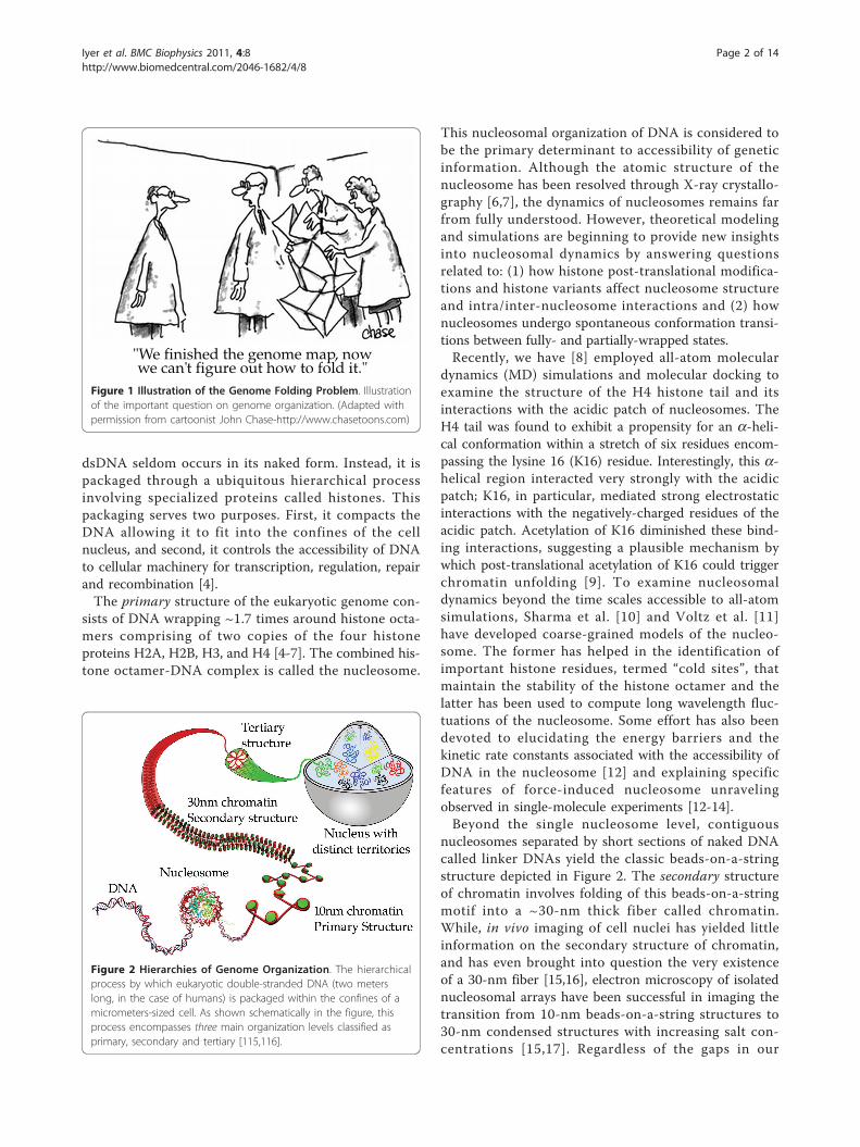
processes and how the nuclear processes in turn modifygenome structure are important questions in moderncell biology. A critical step in addressing these questionsrequires a fundamental understanding of the genome3D structure and the physical principles governing itsorganization, as articulated concisely yet powerfully inthe cartoon of Figure 1.Increasingly, ideas from polymer theory and simula-
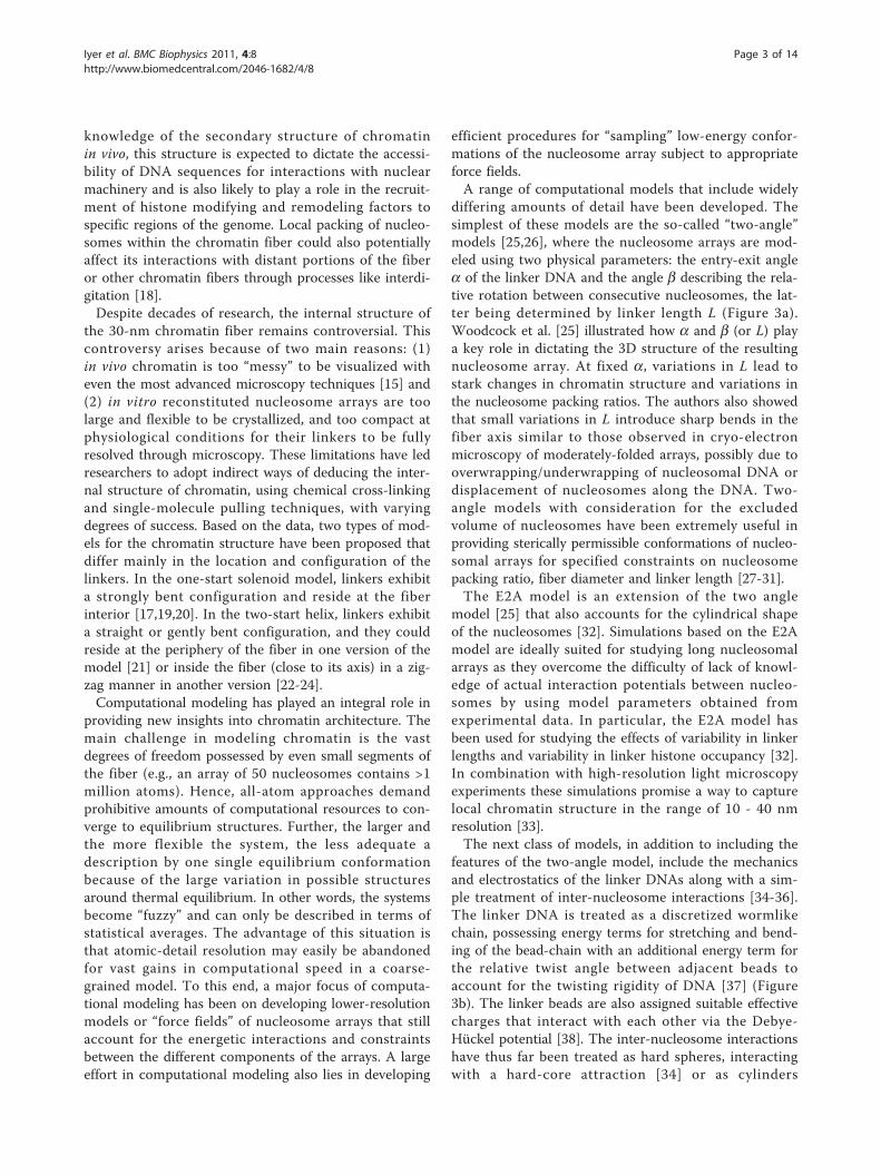
tions coupled with state-of-the-art microscopy and chro-mosome conformation capture techniques are beingused to determine the 3D structure of the genome andthe physical principles governing its folding. In this arti-cle, we present an overview of the key aspects andinsights gained from these studies at the different hier-archical levels of organization shown in Figure 2. Webegin with a discussion of mesoscale models and simu-lation methods used to decipher the secondary structureof the genome, the folded chromatin fiber, on the scaleof 1-10 kbp. Next, we discuss coarse-grained modelsand simulations of the genome tertiary structure. At thetertiary structure level genome compaction varies all theway from ~50 to 100,000 fold, therefore, we have cho-sen to divide it into two sub-structures: tertiary-a struc-ture at the gene locus level (10-2000 kbp) and tertiary-bstructure at the chromosomal level (1-200 Mbp).
Primary and Secondary Structure: ChromatinOrganizationThe classic image of double stranded DNA (dsDNA) isthat of a naked double helix. However, in eukaryotes,
* Correspondence: [email protected] of NanoEngineering, University of California San Diego, 9500Gilman Drive, La Jolla, CA 92093-0448, USA
Iyer et al. BMC Biophysics 2011, 4:8http://www.biomedcentral.com/2046-1682/4/8
© 2011 Iyer et al; licensee BioMed Central Ltd. This is an Open Access article distributed under the terms of the Creative CommonsAttribution License (http://creativecommons.org/licenses/by/2.0), which permits unrestricted use, distribution, and reproduction inany medium, provided the original work is properly cited.
dsDNA seldom occurs in its naked form. Instead, it ispackaged through a ubiquitous hierarchical processinvolving specialized proteins called histones. Thispackaging serves two purposes. First, it compacts theDNA allowing it to fit into the confines of the cellnucleus, and second, it controls the accessibility of DNAto cellular machinery for transcription, regulation, repairand recombination [4].The primary structure of the eukaryotic genome con-
sists of DNA wrapping ~1.7 times around histone octa-mers comprising of two copies of the four histoneproteins H2A, H2B, H3, and H4 [4-7]. The combined his-tone octamer-DNA complex is called the nucleosome.
This nucleosomal organization of DNA is considered tobe the primary determinant to accessibility of geneticinformation. Although the atomic structure of thenucleosome has been resolved through X-ray crystallo-graphy [6,7], the dynamics of nucleosomes remains farfrom fully understood. However, theoretical modelingand simulations are beginning to provide new insightsinto nucleosomal dynamics by answering questionsrelated to: (1) how histone post-translational modifica-tions and histone variants affect nucleosome structureand intra/inter-nucleosome interactions and (2) hownucleosomes undergo spontaneous conformation transi-tions between fully- and partially-wrapped states.Recently, we have [8] employed all-atom molecular
dynamics (MD) simulations and molecular docking toexamine the structure of the H4 histone tail and itsinteractions with the acidic patch of nucleosomes. TheH4 tail was found to exhibit a propensity for an a-heli-cal conformation within a stretch of six residues encom-passing the lysine 16 (K16) residue. Interestingly, this a-helical region interacted very strongly with the acidicpatch; K16, in particular, mediated strong electrostaticinteractions with the negatively-charged residues of theacidic patch. Acetylation of K16 diminished these bind-ing interactions, suggesting a plausible mechanism bywhich post-translational acetylation of K16 could triggerchromatin unfolding [9]. To examine nucleosomaldynamics beyond the time scales accessible to all-atomsimulations, Sharma et al. [10] and Voltz et al. [11]have developed coarse-grained models of the nucleo-some. The former has helped in the identification ofimportant histone residues, termed “cold sites”, thatmaintain the stability of the histone octamer and thelatter has been used to compute long wavelength fluc-tuations of the nucleosome. Some effort has also beendevoted to elucidating the energy barriers and thekinetic rate constants associated with the accessibility ofDNA in the nucleosome [12] and explaining specificfeatures of force-induced nucleosome unravelingobserved in single-molecule experiments [12-14].Beyond the single nucleosome level, contiguous
nucleosomes separated by short sections of naked DNAcalled linker DNAs yield the classic beads-on-a-stringstructure depicted in Figure 2. The secondary structureof chromatin involves folding of this beads-on-a-stringmotif into a ~30-nm thick fiber called chromatin.While, in vivo imaging of cell nuclei has yielded littleinformation on the secondary structure of chromatin,and has even brought into question the very existenceof a 30-nm fiber [15,16], electron microscopy of isolatednucleosomal arrays have been successful in imaging thetransition from 10-nm beads-on-a-string structures to30-nm condensed structures with increasing salt con-centrations [15,17]. Regardless of the gaps in our
Figure 1 Illustration of the Genome Folding Problem. Illustrationof the important question on genome organization. (Adapted withpermission from cartoonist John Chase-http://www.chasetoons.com)
Figure 2 Hierarchies of Genome Organization. The hierarchicalprocess by which eukaryotic double-stranded DNA (two meterslong, in the case of humans) is packaged within the confines of amicrometers-sized cell. As shown schematically in the figure, thisprocess encompasses three main organization levels classified asprimary, secondary and tertiary [115,116].
Iyer et al. BMC Biophysics 2011, 4:8http://www.biomedcentral.com/2046-1682/4/8
Page 2 of 14
knowledge of the secondary structure of chromatinin vivo, this structure is expected to dictate the accessi-bility of DNA sequences for interactions with nuclearmachinery and is also likely to play a role in the recruit-ment of histone modifying and remodeling factors tospecific regions of the genome. Local packing of nucleo-somes within the chromatin fiber could also potentiallyaffect its interactions with distant portions of the fiberor other chromatin fibers through processes like interdi-gitation [18].Despite decades of research, the internal structure of
the 30-nm chromatin fiber remains controversial. Thiscontroversy arises because of two main reasons: (1)in vivo chromatin is too “messy” to be visualized witheven the most advanced microscopy techniques [15] and(2) in vitro reconstituted nucleosome arrays are toolarge and flexible to be crystallized, and too compact atphysiological conditions for their linkers to be fullyresolved through microscopy. These limitations have ledresearchers to adopt indirect ways of deducing the inter-nal structure of chromatin, using chemical cross-linkingand single-molecule pulling techniques, with varyingdegrees of success. Based on the data, two types of mod-els for the chromatin structure have been proposed thatdiffer mainly in the location and configuration of thelinkers. In the one-start solenoid model, linkers exhibita strongly bent configuration and reside at the fiberinterior [17,19,20]. In the two-start helix, linkers exhibita straight or gently bent configuration, and they couldreside at the periphery of the fiber in one version of themodel [21] or inside the fiber (close to its axis) in a zig-zag manner in another version [22-24].Computational modeling has played an integral role in
providing new insights into chromatin architecture. Themain challenge in modeling chromatin is the vastdegrees of freedom possessed by even small segments ofthe fiber (e.g., an array of 50 nucleosomes contains >1million atoms). Hence, all-atom approaches demandprohibitive amounts of computational resources to con-verge to equilibrium structures. Further, the larger andthe more flexible the system, the less adequate adescription by one single equilibrium conformationbecause of the large variation in possible structuresaround thermal equilibrium. In other words, the systemsbecome “fuzzy” and can only be described in terms ofstatistical averages. The advantage of this situation isthat atomic-detail resolution may easily be abandonedfor vast gains in computational speed in a coarse-grained model. To this end, a major focus of computa-tional modeling has been on developing lower-resolutionmodels or “force fields” of nucleosome arrays that stillaccount for the energetic interactions and constraintsbetween the different components of the arrays. A largeeffort in computational modeling also lies in developing
efficient procedures for “sampling” low-energy confor-mations of the nucleosome array subject to appropriateforce fields.A range of computational models that include widely
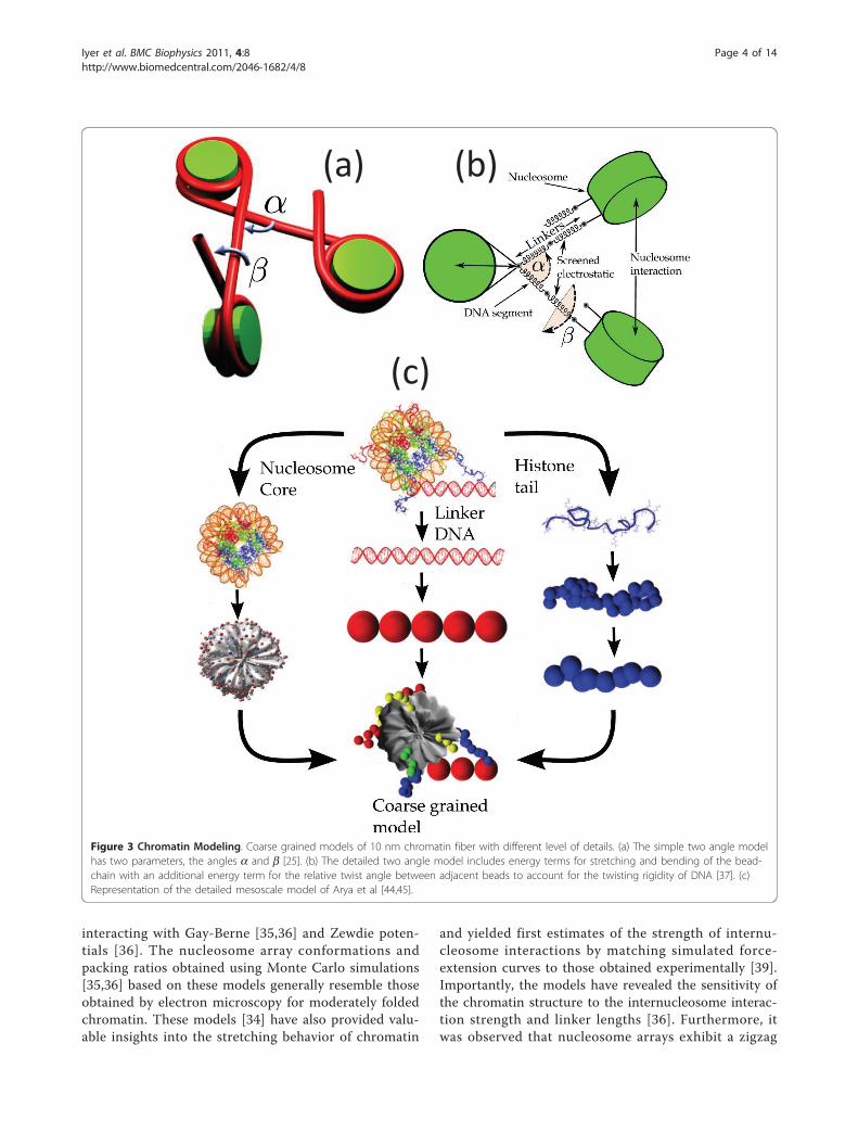
differing amounts of detail have been developed. Thesimplest of these models are the so-called “two-angle”models [25,26], where the nucleosome arrays are mod-eled using two physical parameters: the entry-exit anglea of the linker DNA and the angle b describing the rela-tive rotation between consecutive nucleosomes, the lat-ter being determined by linker length L (Figure 3a).Woodcock et al. [25] illustrated how a and b (or L) playa key role in dictating the 3D structure of the resultingnucleosome array. At fixed a, variations in L lead tostark changes in chromatin structure and variations inthe nucleosome packing ratios. The authors also showedthat small variations in L introduce sharp bends in thefiber axis similar to those observed in cryo-electronmicroscopy of moderately-folded arrays, possibly due tooverwrapping/underwrapping of nucleosomal DNA ordisplacement of nucleosomes along the DNA. Two-angle models with consideration for the excludedvolume of nucleosomes have been extremely useful inproviding sterically permissible conformations of nucleo-somal arrays for specified constraints on nucleosomepacking ratio, fiber diameter and linker length [27-31].The E2A model is an extension of the two angle
model [25] that also accounts for the cylindrical shapeof the nucleosomes [32]. Simulations based on the E2Amodel are ideally suited for studying long nucleosomalarrays as they overcome the difficulty of lack of knowl-edge of actual interaction potentials between nucleo-somes by using model parameters obtained fromexperimental data. In particular, the E2A model hasbeen used for studying the effects of variability in linkerlengths and variability in linker histone occupancy [32].In combination with high-resolution light microscopyexperiments these simulations promise a way to capturelocal chromatin structure in the range of 10 - 40 nmresolution [33].The next class of models, in addition to including the
features of the two-angle model, include the mechanicsand electrostatics of the linker DNAs along with a sim-ple treatment of inter-nucleosome interactions [34-36].The linker DNA is treated as a discretized wormlikechain, possessing energy terms for stretching and bend-ing of the bead-chain with an additional energy term forthe relative twist angle between adjacent beads toaccount for the twisting rigidity of DNA [37] (Figure3b). The linker beads are also assigned suitable effectivecharges that interact with each other via the Debye-Hückel potential [38]. The inter-nucleosome interactionshave thus far been treated as hard spheres, interactingwith a hard-core attraction [34] or as cylinders
Iyer et al. BMC Biophysics 2011, 4:8http://www.biomedcentral.com/2046-1682/4/8
Page 3 of 14
interacting with Gay-Berne [35,36] and Zewdie poten-tials [36]. The nucleosome array conformations andpacking ratios obtained using Monte Carlo simulations[35,36] based on these models generally resemble thoseobtained by electron microscopy for moderately foldedchromatin. These models [34] have also provided valu-able insights into the stretching behavior of chromatin
and yielded first estimates of the strength of internu-cleosome interactions by matching simulated force-extension curves to those obtained experimentally [39].Importantly, the models have revealed the sensitivity ofthe chromatin structure to the internucleosome interac-tion strength and linker lengths [36]. Furthermore, itwas observed that nucleosome arrays exhibit a zigzag
(a) (b)
(c)
Figure 3 Chromatin Modeling. Coarse grained models of 10 nm chromatin fiber with different level of details. (a) The simple two angle modelhas two parameters, the angles a and b [25]. (b) The detailed two angle model includes energy terms for stretching and bending of the bead-chain with an additional energy term for the relative twist angle between adjacent beads to account for the twisting rigidity of DNA [37]. (c)Representation of the detailed mesoscale model of Arya et al [44,45].
Iyer et al. BMC Biophysics 2011, 4:8http://www.biomedcentral.com/2046-1682/4/8
Page 4 of 14

structure without the linker histone and a more solenoi-dal structure for linker histone-bound arrays [40].Very detailed models of local interactions within oligo-
nucleosomes have been developed by Schlick and cow-orkers [41-49] (Figure 3c). The earliest model [41,42]treated the nucleosome as a rigid cylinder (along with asmall protrusion to represent the H3 histone tail) withhundreds of “pseudo” charges scattered uniformly onthe surface. The magnitude of the charges were opti-mized to reproduce as closely as possible the electricfield in the vicinity of the nucleosome. This approachallowed one to account for the salt-dependence in theinter-nucleosome interactions. The model was laterrefined by employing an irregular-shaped representationof the nucleosome [43] that was based on a more recentnucleosome crystal structure with all histone tails fullyresolved [50]. The model reproduced the experimentallyobserved [15,17] compaction of the arrays with increas-ing salt concentration and indicated that the arraysmaintain a zigzag morphology under monovalent saltconditions. Further, the simulations demonstrated thatreduced electrostatic repulsion between the linkers isthe main mechanism responsible for the folding ofarrays at high salt. These models are also being used tostudy the dynamics of chromatin arrays, especiallyunder different kinds of forces including torsional stres-ses [51].Recently, this model was further improved by account-
ing for histone tail flexibility [44], linker histone binding[47], and effects of divalent ions [47]. The tails weretreated as coarse-grained bead-chains, where each beadrepresented five amino acid residues. The stretching,bending, and the electrostatic terms in the bead-chainwere parametrized using an iterative procedure. The lin-ker histone was coarse-grained as three charged beadsrigidly bound at the nucleosome dyad with the magni-tude of the charges optimized to reproduce the electricfield of the atomic linker histone. Divalent ions weretreated phenomenologically in terms of their effect onflexibility and electrostatic screening of the linkerDNAs. A configurational-bias Monte Carlo approachwas used to sample the tail configurations and transla-tion, rotation and pivot moves were used to sample theglobal array configurations [46]. This model helped elu-cidate the role of each histone tail, the linker histoneand physiological salt condition in chromatin folding[45,49]. Specifically, the H4 tails were found to mediatethe strongest internucleosome interactions, the H3 tailsmediated strong internucleosome interactions andscreened electrostatic repulsion between the entering/exiting linkers, and the H2A and H2B tails mediatedinter-fiber interactions [45]. The linker histones con-stricted the linker entry/exit angle to bring alternatenucleosomes together. Divalent ions were also found to
facilitate tight packing of nucleosomes by allowing afraction of the linkers to bend and by strongly screeningthe linker repulsion at the fiber axis. Moreover, themodel, in conjunction with sophisticated cross-linkingexperiments, confirmed the existence of a hetero-morphic fiber containing both zigzag and solenoid con-formations in the presence of additional divalent cationsand linker histones [49]. This model has also recentlybeen used to reproduce the linker length dependence inthe observed chromatin structures [48].In summary, mesoscale models at varying levels of
sophistication such as those discussed above are provingto be valuable tools for examining chromatin structure.The choice of which model to use is dictated by theamount of detail required and the amount of computa-tional resources available. For instance, the detailedmodels accounting for histone tail flexibility and nucleo-some geometry may provide the most accurate repre-sentation of short nucleosome arrays. For long arrayscontaining hundreds of nucleosomes, these modelsrapidly become computationally intractable and theintermediate-resolution models like the E2A model [32]become more suitable.
Tertiary-a Structure: Gene Locus OrganizationIn the previous section, we discussed the secondarystructure of chromatin from a static perspective. In rea-lity, the chromatin fiber within the cell nucleus is pre-sent in a dynamic state [52]-it is flexible over lengthsmuch larger than the fiber diameter [53] and it is con-stantly being subjected to various kinds of remodelingactivities, including histone modifications [54-56], slid-ing and depletion of nucleosomes [57-64], and incor-poration of histone variants [65]. There is strongevidence from light microscopy studies indicating thatat the gene locus level the chromatin fiber is organizedinto loops [66]. Studies on several multigene clustersconceive such loops as instrumental in bringing togetherdistant enhancer and promoter regions crucial for geneactivation, regulation and recombination [67-69].Although the detailed mechanism of looping is not fullyunderstood, there is no question that the “intrinsic”bending rigidity of the chromatin fiber dictates to a con-siderable degree the loop size-dependent statistical prob-ability of two distant regions of the fiber coming intoclose proximity to form a loop.Emerging evidence suggests that the relationship
between flexibility and looping probability may be uti-lized by cells for gene regulation. Specifically, the ideathat modulation of flexibility through remodeling pro-cesses like acetylation of histones alters the probabilityof interactions between a remote enhancer and cognategene by means of looping has been successfully appliedto explain gene regulation of the Hoxd gene cluster and
Iyer et al. BMC Biophysics 2011, 4:8http://www.biomedcentral.com/2046-1682/4/8
Page 5 of 14
the b-globin locus [70]. Strong support for this idea alsocomes from recent experiments in the Murre lab thatshow large-scale conformational changes in the IgH-locus during B-Cell development accompanying gen-ome-wide and locus-specific histone modifications andnucleosome depletion events [66,71,72]. Thus, it seemsthat chromatin remodeling events, apart from modulat-ing the local structure of chromatin and DNA accessi-bility, could lead to changes in the higher-order foldingof chromatin through its effects on macroscopic proper-ties of the fiber such as its flexibility.So far, little effort has been devoted to investigating
the intrinsic flexibility of chromatin and the associatedlooping probability as a function of loop size; especiallyhow the flexibility correlates with external conditionslike monovalent/divalent salt concentration and systemparameters like nucleosome repeat length, DNA wrap-ping angle, histone variants, histone modifications andpresence/absence of linker histones. Chromatin flexibil-ity as characterized by its persistence length, Lp, seemsto exhibit large variations, depending on the experimen-tal method used for its determination and the conditionsand type of chromatin investigated. For example, single-molecule pulling of nucleosome arrays at low salt [39]and in vivo looping driven cross-linking/recombinationassays [53,73] measure Lp as low as 30-50 nm while ana-lysis of fluorescent markers in the genome of erythro-cytes measure Lp of 100-200 nm [74,75]. However, it isimportant to review these results in light of a recentfundamental study [76] indicating that standard defini-tions of persistence length as used in these studies maynot describe the local intrinsic flexibility of chromatin.Aumann et al. [77] recently examined Lp of nucleo-
some arrays using Monte Carlo simulations of a mesos-cale model of chromatin. The persistence length wasobtained from the decay in the correlation of the tan-gent vector representing the local fiber axis. It wasfound that Lp decreased strongly with increasing nucleo-some repeat length and increasing entry/exit angle, andthat binding of the linker histone led to an increase inLp, consistent with experimental observations of linkerhistone deficient and inclusive chromatin. A comparisonbetween the magnitude of the bending and elastic rigid-ity suggested that chromatin is much easier to bendthan stretch, leading to the interesting hypothesis that itmay be easier for the cells to pack chromatin via tightloops rather than by linear compression of the fiber.As discussed earlier, chromatin exists in a highly
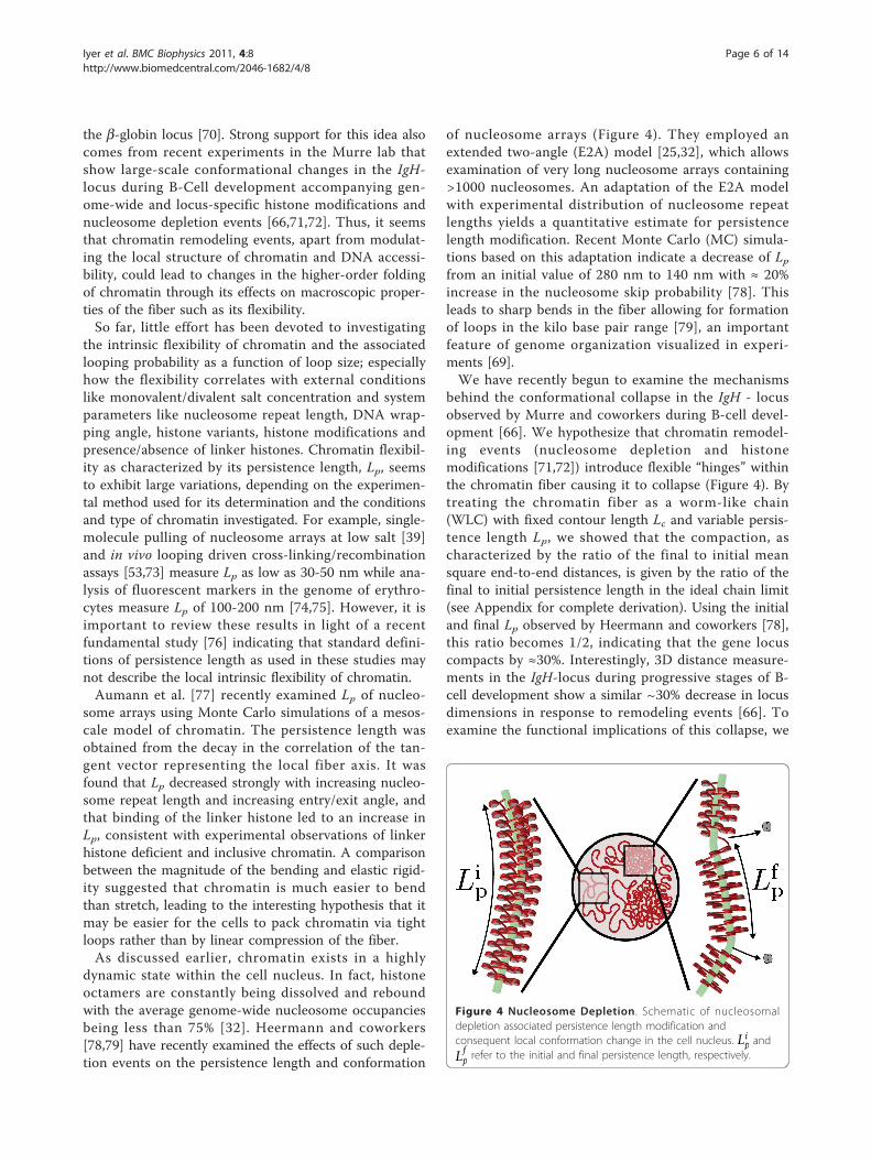
dynamic state within the cell nucleus. In fact, histoneoctamers are constantly being dissolved and reboundwith the average genome-wide nucleosome occupanciesbeing less than 75% [32]. Heermann and coworkers[78,79] have recently examined the effects of such deple-tion events on the persistence length and conformation
of nucleosome arrays (Figure 4). They employed anextended two-angle (E2A) model [25,32], which allowsexamination of very long nucleosome arrays containing>1000 nucleosomes. An adaptation of the E2A modelwith experimental distribution of nucleosome repeatlengths yields a quantitative estimate for persistencelength modification. Recent Monte Carlo (MC) simula-tions based on this adaptation indicate a decrease of Lpfrom an initial value of 280 nm to 140 nm with ≈ 20%increase in the nucleosome skip probability [78]. Thisleads to sharp bends in the fiber allowing for formationof loops in the kilo base pair range [79], an importantfeature of genome organization visualized in experi-ments [69].We have recently begun to examine the mechanisms
behind the conformational collapse in the IgH - locusobserved by Murre and coworkers during B-cell devel-opment [66]. We hypothesize that chromatin remodel-ing events (nucleosome depletion and histonemodifications [71,72]) introduce flexible “hinges” withinthe chromatin fiber causing it to collapse (Figure 4). Bytreating the chromatin fiber as a worm-like chain(WLC) with fixed contour length Lc and variable persis-tence length Lp, we showed that the compaction, ascharacterized by the ratio of the final to initial meansquare end-to-end distances, is given by the ratio of thefinal to initial persistence length in the ideal chain limit(see Appendix for complete derivation). Using the initialand final Lp observed by Heermann and coworkers [78],this ratio becomes 1/2, indicating that the gene locuscompacts by ≈30%. Interestingly, 3D distance measure-ments in the IgH-locus during progressive stages of B-cell development show a similar ~30% decrease in locusdimensions in response to remodeling events [66]. Toexamine the functional implications of this collapse, we
Figure 4 Nucleosome Depletion . Schematic of nucleosomaldepletion associated persistence length modification andconsequent local conformation change in the cell nucleus. Li
p and
Lfp refer to the initial and final persistence length, respectively.
Iyer et al. BMC Biophysics 2011, 4:8http://www.biomedcentral.com/2046-1682/4/8
Page 6 of 14
used a simple Flory type argument to show that the col-lapse leads to an 8-fold increase in the number of binaryinteractions nb within the gene locus. Assuming that thefrequency of promoter-enhancer (P-E) interactions isproportional to nb, we conclude that the conformationalcollapse facilitates transcriptional regulation and/orrecombination by allowing for higher probability of P-Einteractions. Thus, the chromatin remodeling inducedchanges in the persistence length offers one possiblemechanism for governing the conformational state ofthe gene locus and consequent modification of the func-tional state of the cell (Figure 4).
Tertiary-b Structure: Chromosomal OrganizationInterestingly, the condensed higher-order structures ofchromatin, namely chromosomes, were observed as dis-tinct entities during mitosis as early as the 19th century,much before the primary and secondary structures ofchromatin were known to exist. However, disappearanceof this condensed structure during interphase and theunderlying chromatin organization remained a mysterytill the late 20th century [80]. In the beginning two dif-ferent models were proposed for the interphase chroma-tin organization: (1) random organization akin to a bowlof spaghetti without any apparent structure and (2)organization in territories that later condense duringmitosis to form distinct chromosomes [81]. The earliestclues to deciding between the two models came fromexperiments shining a laser light onto a specific volumeof the cell nucleus and observing the effects of the con-sequent damage on replication [81]. It was found thatonly a few chromosomes were affected by the laserlight, indicating existence of distinct chromosome terri-tories within the cell nucleus. This conjectural evidencehas now been confirmed beyond doubt by advances inimaging techniques like fluorescent in situ hybridization(FISH) [82-84] and chromosome painting [82,83].While there is concrete evidence for the existence of
territories, the internal architecture of chromatin withinchromosome territories, the interaction between terri-tories, and the organizational principles governing theirformation remain poorly understood [85]. Results fromMC simulations of confined polymer chains suggest thatterritory formation could arise from simple, non-specificentropic forces and from segregation of long chainsattempting to conserve their topological state whileundergoing confined Brownian motion [86,87]. Increas-ingly, insights from such coarse-grained polymer modelscoupled with experiments are being used to examine theinternal architecture of chromosome territories.Two predominant themes recur during discussions of
higher-order structures of interphase chromosomes: (1)formation of loop structures and (2) confined fractalorganization. Interestingly, each of these themes derive
their support from different kind of experiments. Theloop structure theme is predominantly supported bylight microscopy experiments [75,88-90] and the fractaltheme by chromosome conformation capture [91], smallangle neutron scattering [92] and tracer diffusion [93]experiments. Although the two themes are not mutuallyexclusive, it is often possible to classify the polymermodels based on them. Here, we follow this classifica-tion in discussing the polymer models and their centralfeatures.The earliest polymer model [94] considered the orga-
nization as a classical fractal where the recurring motifis that of the confined Gaussian chain. The confinementidea was introduced to explain the observation that thegeometric separation between two points in the struc-ture follows a Gaussian polymer model between the 0.1-1.5 Mbp genomic separation scale [88] while beyondthis scale the geometric separation tends to becomeindependent of genomic separation. However, an alter-native proposition explaining this leveling off of geo-metric separation through incorporation of loops wasfound to be consistent with the observation of loopstructures in different light microscopy experiments[95]. The loop proposition consequently led to the for-mulation of the random walk giant loop (RW-GL)model [75] followed by increasingly refined models likethe multiloop subcompartment (MLS) [96] and the ran-dom loop (RL) [97] (Figure 5).The basic feature of the RW-GL model is the exis-
tence of 1-3 Mbp size loops along a randomly orientedbackbone [75]. This analytical model explained theobserved leveling off of 2D mean geometric distance inthe 10-200 Mbp genomic distance range fairly well.However, it had limitations in capturing the compart-mentalized chromosome territory structure and spatialchromatin distribution observed in experiments [96].These limitations were attributed to the use of phantomchains in the model [96]. The MLS model based simula-tions, with excluded-volume interactions and subcom-partments consisting of 120 kbp loops that can beopened up to accommodate giant loops, overcame thelimitations of the RW-GL model and correctly predictedthe 2D mean geometric distances [96]. The RW-GL andthe MLS models demonstrated beyond doubt the useful-ness of simulations based on polymer models for studyof higher-order structures of the genome. However,these models were unable to explain the drastic levelingoff of 3D geometric distances above the 10 Mbp rangethat were observed by recent 3D-FISH experiments [90].The random loop model that proposed existence ofloops at all scales >150 kbp could successfully explainthis leveling off beyond the 10 Mbp scale (Figure 6). Akey feature of this model is that it accounts for observedexperimental features of existence of loops at several
Iyer et al. BMC Biophysics 2011, 4:8http://www.biomedcentral.com/2046-1682/4/8
Page 7 of 14

scales along with a means to account for cell-to-cell var-iation of measurements through averaging over differentconfigurations of loops [97]. Further, the presence ofrandom loops is expected to lead to formation of segre-gated chromosome territories as they repel each othermore strongly than the linear structures [98].The proposed loop models show a power-law behavior
for geometric distances versus contour length at scales<10 Mbp, with scaling exponents ranging from ν = 1/3-1/2. This suggests two different internal structures at work:(1) equilibrium/globular state at short scales and (2) ran-dom looped structure at large scales. The transitionbetween these structures is not clearly understood. Incontrast, the confined fractals do not encounter these dif-ficulties as they propose a single size exponent at allscales. Recent advances in chromosome conformationcapture [91] have enabled genome-wide measurementsof probability of formation of loops as a function of thesize of the loops in the human genome (Figure 6). Com-parison of the probability measurements with confinedfreely jointed chain (FJC) simulations indicate that the
Figure 5 Loop Models. Illustration of the different loop models; (a)Random Loop model indicating loops at all scales > 150 kbp [90],(b) multi-loop subcompartment model with 120 kbp rosettestructures [96] and (c) random-walk-giant-loop model with giantloops organized along a random backbone [75].
Figure 6 Random Loop and Fractal Globule. (a) Predictions of the random loop model for mean-square displacement (adapted from Mateos-Langerak et al [90]). (b) Predictions of the fractal globule model for probability of contact (adapted from the article of Lieberman-Aiden et alwith permission from AAAS [91]).
Iyer et al. BMC Biophysics 2011, 4:8http://www.biomedcentral.com/2046-1682/4/8
Page 8 of 14
human genome may be organized as a fractal globulewith size scaling exponent ν = 1/3 [91]. However, wenote that the agreement of the probability measurementsis observed only in the 1-10 Mbp range (Figure 6).The advantage of this simplified picture of confined
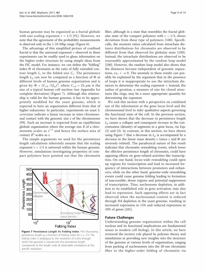
fractal is that the universal exponent ν observed in theseexperiments can be readily used to glean information onthe higher-order structure by using simple ideas fromthe FJC model. For instance, we can define the “folding”index F of chromatin as the ratio of fully extended con-tour length Lc to the folded size Cs. The persistencelength Lp can now be computed as a function of F atdifferent levels of human genome organization and isgiven by: F = (Cs,0 /2Lp)
2, where Cs,0 ≈ 25 μm is thesize of a typical human cell nucleus (see Appendix forcomplete derivation) (Figure 7). Although this relation-ship is valid for the human genome, it has to be appro-priately modified for the yeast genome, which isexpected to have an organization different from that ofhigher eukaryotes. In particular, experiments on yeast S.cerevisiae indicate a linear increase in inter-chromoso-mal contact with the genomic size s of the chromosome[99]. Such an increase is expected from an equilibriumglobule organization where the average size R of a chro-mosome scales as s1/2 and hence the surface area ofcontact R2 scales as s.The simple arguments we used for the persistence
length calculations inherently assume that the scalingexponent ν = 1/3 is universal within the human genome.However, simulations investigating properties of com-pact polymers have pointed out that the chromatin
fiber, although in a state that resembles the fractal glob-ular state of the compact polymers with ν = 1/3, showsdeviations from these type of polymers [100]. Specifi-cally, the moment ratios calculated from intrachain dis-tance distributions for chromatin are observed to bedifferent from that observed for globular state [100].Instead, the intrachain distributions are observed to bereasonably approximated by the random loop model[100]. However, the random loop model also shows thatthe distances become independent of genomic separa-tions, i.e., ν ® 0. The anomaly in these results can pos-sibly be explained by the argument that in the presenceof loops it is inappropriate to use the intrachain dis-tances to determine the scaling exponent ν. Instead, theradius of gyration, a measure of size for closed struc-tures like rings, may be a more appropriate quantity fordetermining the exponent.We end this section with a perspective on combined
use of the information at the gene locus level and thechromosomal level to infer qualitative aspects governingthe functional state of the cell. In the previous section,we have shown that the decrease in persistence lengthLp causes a collapse and consequent increase in the con-centration (density) of segments in a gene locus, via Eqs.(2) and (3). In contrast, in this section, we have shownusing Figure 7 that a decrease in Lp is accompanied by adecrease in the linear mass density c (since c and F areinversely related). The paradoxical nature of this resultindicates that chromatin remodeling events, which lowerthe effective persistence length of chromatin, could haveopposing effects on gene-related activities like transcrip-tion. On one hand, locus-wide remodeling could openup regions for transcription and lead to increased fre-quency of interactions between promoters and enhan-cers; while on the other hand, genome-wide remodelingevents could cause genome folding leading to formationof inaccessible, dense regions and potential suppressionof transcription. Thus, nucleosome depletion, in addi-tion to its established role in gene activation, may alsolead to repression. Such opposing effects are in factobserved when the nucleosomal content is reducedthrough H4 depletion in the yeast genome, resulting inincreased expression in 15% and reduced expression in10% of genes [101].
Future ChallengesUnderstanding genome organization within the cellnucleus and its functional implications are fundamentalissues in modern cell biology. In this article, we havereviewed the incisive role played by polymer theory andsimulations in providing new insights into the structureof the genome at various levels of organization, rangingfrom packing of nucleosomes into the 30-nm chromatinfiber to the higher-order folding of chromatin via
Figure 7 Persistence Length Vs Folding Index. Plot illustratingpersistence length as a function of folding index for ν = 1/3. Thefolding index is analogous to the resolution of a lens throughwhich the genome is viewed and the persistence lengthcorresponds to the length scale of observable correlations at thisspecific resolution.
Iyer et al. BMC Biophysics 2011, 4:8http://www.biomedcentral.com/2046-1682/4/8
Page 9 of 14
looping into chromosomes. Despite enormous progressin both experimental and computational fronts, a com-prehensive model of genome organization at the chro-matin and chromosome level is still lacking. Below welist four main challenges that computational modelswould have to overcome to tackle this highly intriguingand important problem.First, genome organization is heterogeneous. At the
chromatin level, this heterogeneity arises from variationsin the nucleosome repeat length, nucleosome composi-tion (histone modifications, histone variants, and linkerhistones), and potential entrapment of chromatin inmetastable states. Most models of chromatin, includingthose developed in our group, tend to assume uniformnucleosome positioning and composition. Only recentlyhave studies begun to examine the effects of chromatinheterogeneity; preliminary work in this direction hasalready yielded some very interesting results. At thechromosome level, the size, shape and location of chro-matin loops is not fixed. Thus, a key aspect towardsexamining heterogeneity in chromosome models wouldbe through more realistic modeling of the interactionsacross chromatin fibers, e.g., allowing loops to dynami-cally form and break according to the associated ener-getics of loop formation in chromosome models, asopposed to “fixing” looping points and loop sizes.Second, the chromatin fiber and chromosomes are
highly dynamic. Nucleosomes are constantly being dis-placed, modified, dissolved and reformed through var-ious mechanisms. At the higher scale, chromatin loopsare continuously being broken and reformed. In addi-tion, a range of nuclear proteins including transcriptionfactors and architectural proteins dynamically bind anddissociate from chromatin [2]. Such binding/dissociationevents could affect chromatin structure and in turn maybe affected by chromatin structure. Most models cur-rently look at the genome from a static perspective.Incorporation of the above mentioned kinetic featuresinto polymer models coupled with structural heteroge-neity would be a crucial step in building more compre-hensive models of the genome.Third, development of accurate, coarse-grained mod-
els of chromosomes poses a grand challenge. Onepotential route is via “multi-scale” approaches, whereeach level represents coarse-graining of the previous,higher-resolution level. Such an approach generallyinvolves the use of potential of mean forces (PMFs) fortreating the interactions between coarse-grained subu-nits. The PMFs account for effects of the degrees offreedom that are “averaged out” during each coarse-graining step. The challenge is to maintain self-consis-tency from one level to another, as the PMFs are validonly when there is a clear separation of length and
time scales across the levels [102]. When there is noclear separation of time/length scales, one might needto resort to “multi-resolution” approaches. In thisapproach, different portions of the system are treatedat different resolutions, depending on their relativeimportance to the phenomena being investigated (inthe same spirit as the QM/MM method [103]employed in enzyme catalysis). The challenge here isto identify the portions of the system (or degrees offreedom) important enough to be treated at higherresolution as opposed to those regions that can betreated at lower resolution. Another challenge is devel-oping suitable potentials for linking low- and high-resolution regions.Fourth, little modeling effort has been invested in
determining structure-function relationships in genomeorganization. A definitive connection between structureand function comes from the observation that gene den-sity is high in largely decondensed euchromatin and lowin highly condensed heterochromatin [104]. Further,gene-rich and gene-poor regions are found to be physi-cally separated from each other [105,106]. Specifically,in human lymphocytes gene-rich regions are positionedin the nuclear interior while the gene-poor regions arepositioned towards its periphery [107]. Apart from genedensity patterns, the molecular species responsible fornuclear processes and gene expression are connectedthrough a complex and extensively coupled network giv-ing rise to functional coupling between macromolecularstructures and compartments [108,109]. Though manyaspects of these studies are been driven by the experi-ments, polymer theory and simulations could contributeto better understanding of such structure-functionrelationships.
AppendixEffect of persistence length modifications on gene locusconformationTo study the generic effects of changes in the persis-tence length of the chromatin fiber on the conformationof the gene locus, we have treated the chromatin fiberas a worm-like chain (WLC) with fixed contour lengthLc. The WLC is the standard model used to describesemi-flexible polymer chains, and it has been success-fully used to explain the conformational properties ofdouble-stranded DNA and other biopolymers [110].Though this model is too idealized to represent in vivochromatin (as it neglects fiber-fiber interactions, exis-tence of stable loops and heterogeneous flexibility), itcan nonetheless provide qualitative insights into themagnitude of the conformational changes.We begin by writing down the mean squared end-to-
end distance of a WLC with an “effective” persistence
Iyer et al. BMC Biophysics 2011, 4:8http://www.biomedcentral.com/2046-1682/4/8
Page 10 of 14
length Lp of the chromatin fiber that embodies alleffects of the remodeling [111]:
〈R2e 〉 = 2L2
p
[Lc
Lp− 1 + exp
(−Lc
Lp
)]. (1)
We consider Lp as a variable that changes from an
initial value Lip to a final value Lf
p due to chromatin
remodeling. Because we anticipate a weak effect ofremodeling on the overall contour length at the locuslevel, we hold Lc constant. In the ideal chain limit of theWLC, Lc ≫ Lp, the ratio of the final to initial meansquare end-to-end distances is given by
〈R2e,f 〉/〈R2
e,i〉 ≈ Lfp/Li
p. (2)
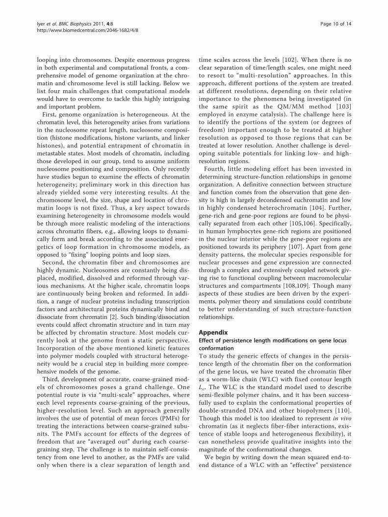
Plugging in values of initial and final Lp observed byHeermann and coworkers [78], the ratio becomes 140/280 = 1/2. We have further confirmed this conforma-tional collapse by using simple lattice MC simulations ofphantom linear chains of different lengths and regionsof flexibility. The simulations indicate that introductionof regions of flexibility in an overall stiff chain, differingby a nominal bending energy of 2kBT per segment,strongly decreases the mean squared radius of gyrationof the chains (Table 1).The implications to this modification in size can be
explored through the Flory approximation. According tothe Flory approximation, the number of binary interac-tions in a polymer chain nb ~ c2, where c is the concen-tration of segments [112]. Since c ~ 1/V (where V is thevolume of the chain) and V ∼ 〈R2
e 〉3/2 assuming sphericalsymmetry, the ratio of the final to the initial number ofbinary interactions is given by:
nb,f /nb,i ≈ (〈R2e,i〉/〈R2
e,f 〉)3 ≈ (Lip/Lf
p)3. (3)
In the case of the IgH locus, this translates to an 8-fold increase in nb.
Persistence length as a function of folding indexWe treat the chromatin fiber as a FJC at the genome-wide level with a contour length given by:
Lc = l × Nr , (4)
where Nr is the number of repeat units, each of lengthl = 2Lp. The characteristic size of a FJC is given by[113]:
Cs ≈ l × (Nr)ν , (5)
where ν is a scaling exponent that connects the size ofthe chain to its number of repeat units. Note that thefractal dimension, df , is the exponent that connects themass of the chain M to its size via M ∼ (Cs)df and thatM is also proportional to the number of repeat units M~ Nr ~ (Cs)
1/ν (from Eq. 5). Hence, ν is the inverse ofthe fractal dimension ν = 1/df and determines the orga-nization of the FJC at all length scales.We can define the “folding” index F of FJC as the
ratio of fully extended contour length Lc to the foldedsize Cs, which can be simplified further using Eqs. (4)and (5):
� ≡ Lc/Cs ≈ N1−νr = (Lc/2Lp)1−ν . (6)
Since the contour length Lc = d/c, where d is thegenomic distance in bp and c is the linear mass densityin bp/nm, F increases with decreasing linear massdensity.The contour length within the FJC framework can be
partitioned arbitrarily, i.e., if the contour length of astructure is 100 μm then it can be partitioned as, say,100 repeat units each of 1 μm length or 1000 repeatunits of length 0.1 μm. The flexibility of the contourlength partitioning allows for choice of either number ofrepeat units Nr or the persistence length Lp as a variablein Eq. (6). However, the contour length Lc according toEq. (6) is determined by the product of the foldingindex F and size Cs. This constraint has to be satisfiedwhen Lp is chosen as the variable and the contourlength in the framework changes with the change in thefolding index. Thus F can be used like a lens of variableresolution to view genome organization at differentlength scales.The persistence length Lp can now be computed as a
function of F at different levels of human genome orga-nization. We begin with the organization at the level ofdsDNA, whose persistence and contour lengths areknown: Lc ≈ 2m for dsDNA with 6 billion bp at a linearmass density c = 2.94 bp/nm [114]. Given that Lp = 50nm [114] and ν = 1/3, the number of repeat units Nr ≈2 × 10⁷ from Eq. (4), the folding index F ≈ 8 × 10⁴
from Eq. (6), and Cs ≈ 25 μm from Eq. (5). Interestingly,the FJC size Cs is consistent with typical dimensions of
Table 1 Ratio of mean-square radius of gyration ofchains
Nr Fa R(0)b R(1) R(2) R(3)
16 5.15 2.38 1.46 1.30 1.19
32 10.11 2.73 1.77 1.622 1.49
64 20.16 2.91 1.91 1.83 1.76
128 40.20 3.02 2.02 1.95 1.91
256 80.46 3.07 2.06 2.00 1.98
512 160.78 3.10 2.11 2.04 2.03
1024 321.84 3.11 2.10 2.05 2.05
Ratio of mean-square radius of gyration of chains of different lengths (Nr) withdifferent regions of flexibility to that of a fully flexible chain (F).a Absolute values of mean square radius of gyration.b In R(x), x denotes the number of flexible regions sandwiched between stiffregions that are of the same length as the stiff regions.
Iyer et al. BMC Biophysics 2011, 4:8http://www.biomedcentral.com/2046-1682/4/8
Page 11 of 14
human cell nuclei. Since, this size is invariant across allorganization levels, we denote it as Cs,0. With this con-straint, Eq. (6) reduces to
� = (Cs,0/2Lp)(1−ν)/ν |ν=1/3 = (Cs,0/2Lp)2. (7)
Equation (7) thus provides the relationship betweenthe persistence length Lp and folding index F for allorganization levels, which is plotted in Figure 7.
AcknowledgementsWe thank Prof. Cornelis Murre of the Division of Biological Sciences at UCSDfor many delightful and insightful discussions on organization ofchromosomes.
Authors’ contributionsBVSI and GA organized the article, contributed to the writing of all sectionsof the article, and developed the theoretical analysis presented in theAppendices. MK designed some of the figures and contributed to thewriting of the section on secondary structure. All the authors have read andapproved the final manuscript.
Received: 7 January 2011 Accepted: 15 April 2011Published: 15 April 2011
References1. Cook PR: The Organization of Replication and Transcription. Science 1999,
284(5421):1790-1795.2. Misteli T: Beyond the Sequence: Cellular Organization of Genome
Function. Cell 2007, 128:787-800.3. Lucas JS, Bossen C, Murre C: Transcription and recombination factories:
common features? Curr Opin Cell Biol 2010, 23:1-7.4. Kornberg RD, Lorch Y: Twenty-five years of the nucleosome, fundamental
particle of the eukaryote chromosome. Cell 1999, 98:285-294.5. Kornberg RD: Chromatin Structure: A Repeating Unit of Histones and
DNA. Science 1974, 184:868-871.6. Luger K, Mäder AW, Richmond RK, Sargent DF, Richmond TJ: Crystal
structure of the nucleosome core particel at 2.8 Å resolution. Nature1997, 389:251-260.
7. Richmond TJ, Davey CA: The structure of DNA in the nucleosome core.Nature 2003, 423:145-150.
8. Yang D, Arya G: Structure and binding of the H4 histone tail and theeffects of lysine 16 acetylation. Phys Chem Chem Phys 2011, 13:2911-2921.
9. Shogren-Knaak M, Ishil H, Sun JM, Pazin MJ, Davie JR, Peterson CL: HistoneH4-K16 acetylation controls chromatin structure and proteininteractions. Science 2006, 311(5762):844-847.
10. Sharma S, Ding F, Dokholyan NV: Multiscale modeling of nucleosomedynamics. Biophys J 2007, 92(5):1457-1470.
11. Voltz K, Trylska J, Tozzini V, Kurkal-Siebert V, Langowski J, Smith J: Coarse-Grained Force Field for the Nucleosome from Self-ConsistentMultiscaling. J Comp Chem 2008, 29(9):2006-2008.
12. Wocjan T, Klenin K, Langowski J: Brownian Dynamics Simulation of DNAUnrolling from the Nucleosome. J Phys Chem B 2009, 113(9):2639-2646.
13. Kulic IM, Schiessel H: DNA spools under tension. Phys Rev Lett 2004,92(22):228101.
14. Sudhanshu B, Mihardja S, Koslover EF, Mehraeen S, Bustamante C,Spakowitz AJ: Tension-dependent structural deformation alters single-molecule transition kinetics. Proc Natl Acad Sci USA 2011,108(5):1885-1890.
15. Woodcock CL, Ghosh RP: Chromatin Higher-order Structure andDynamics. Cold Spring Harb Perspect Biol 2010, 2(5), a000596-1:22.
16. Fussner E, Ching RW, Bazett-Jones DP: Living without 30 nm chromatinfibers. Trends Biochem Sci 2011, 36:1-6.
17. Thoma F, Koller T, Klug A: Involvement of histone H1 in the organizationof the nucleosome and of the salt-dependent superstructures ofchromatin. J Cell Biol 1979, 83:403-427.
18. Grigoryev SA: Keeping fingers crossed: heterochromatin spreadingthrough interdigitation of nucleosome arrays. FEBS Lett 2004, 564:4-8.
19. Finch JT, Klug A: Solenoidal model for superstructure in chromatin. ProcNatl Acad Sci USA 1976, 73(6):1897-1901.
20. Widom J, Klug A: Structure of the 300 Å chromatin filament: X-raydiffraction from oriented samples. Cell 1985, 43:207-213.
21. Woodcock CLF, Frado LLY, Rattner JB: The higher-order structure ofchromatin: evidence for a helical ribbon arrangement. J Cell Biol 1984,99:42-52.
22. Williams SP, Athey BD, Muglia LJ, Schappe RS, Gough AH, Langmore JP:Chromatin fibers are left-handed double helices with diameter and massper unit length that depend on linker length. Biophys J 1986, 49:233-248.
23. Dorigo B, Schalch T, Kulangara A, Duda S, Schroeder RR, Richmond TJ:Nucleosome arrays reveal the two-start organization of the chromatinfiber. Science 2004, 306:1571-1573.
24. Schalch T, Duda S, Sargent D, Richmond TJ: X-ray structure of atetranucleosome and its implications for the chromatin fibre. Nature2005, 436:138-141.
25. Woodcock CL, Grigoryev SA, Horowitz RA, Whitaker N: A chromatin foldingmodel that incorporates linker variability generates fibers resemblingthe native structures. Proc Natl Acad Sci USA 1993, 90(19):9021-9025.
26. van Holde K, Zlatanova J: Chromatin Higher Order Structure: Chasing aMirage? J Biol Chem 1995, 270(15):8373-8376.
27. Schiessel H, Gelbart WM, Bruinsma R: DNA folding: Structural andmechanical properties of the two-angle model for chromatin. Biophys J2001, 80:1940-1956.
28. Mergell B, Everaers R, Schiessel H: Nucleosome interactions in chromatin:Fiber stiffening and hairpin formation. Phys Rev E 2004, 70, 011915-1-9).
29. Diesinger PM, Heermann DW: Two-angle model and phase diagram forchromatin. Phys Rev E 2006, 74:031904.
30. Wong H, Victor J, Mozziconacci J: An all-atom model of the chromatinfiber containing linker histones reveals a versatile structure tuned by thenucleosomal repeat length. PloS One 2007, 2(9), e877-1:8.
31. Elena K, Fuller CJ, Straight AF, Spakowitz AJ: Local geometry and elasticityin compact chromatin structure. Biophys J 2010, 99:3941-3950.
32. Diesinger PM, Heerman DW: The Influence of the Cylindrical Shape of theNucleosomes and H1 Defects on Properties of Chromatin. Biophys J 2008,94:4165-4172.
33. Diesinger PM, Heermann DW: Monte Carlo Simulations indicate thatChromatin Nanostructure is accessible by Light Microscopy. PMC Biophys2010, 3(11):1-20.
34. Katritch KV, Bustamante C, Olson WK: Pulling chromatin fibers: computersimulations of direct physical micromanipulations. J Mol Biol 2000,295:29-40.
35. Wedemann G, Langowski J: Computer simulation of the 30-nanometerchromatin fiber. Biophys J 2002, 82(6):2847-2859.
36. Stehr R, Kepper N, Rippe K, Wedemann G: The Effect of InternucleosomalInteraction on Folding of the Chromatin Fiber. Biophys J 2008, 95.
37. Allison SA, Austin R, Hogan M: Bending and twisting dynamics of shortlinear DNAs: analysis of the triplet anistropy decay of a 209 base pairfragment by Brownian simulation. J Chem Phys 1989, 90:3843-3854.
38. Stigter D: Interactions of highly charged colloidal cylinders withapplications to double-stranded DNA. Biopolymers 1977,16:1435-1448.
39. Cui Y, Bustamante C: Pulling a single chromatin fiber reveals the forcesthat maintain its higher-order structure. Proc Natl Acad Sci USA 2000,97:127-132.
40. Kepper N, Foethke D, Stehr R, Wedemann G, Rippe K: NucleosomeGeometry and Internucleosomal Interactions Control the ChromatinFiber Conformation. Biophys J 2008, 95:3692-3705.
41. Beard D, Schlick T: Modeling salt-mediated electrostatics ofmacromolecules: The discrete surface charge optimization algorithmand its application to the nucleosome. Biopolymers 2001, 58:106-115.
42. Beard D, Schlick T: Computational modeling predicts the structure anddynamics of chromatin fiber. Structure 2001, 9:105-114.
43. Sun J, Zhang Q, Schlick T: Electrostatic mechanism of nucleosomal arrayfolding revealed by computer simulation. Proc Natl Acad Sci USA 2005,102:8180-8185.
44. Arya G, Schlick T: Role of histone tails in chromatin folding revealed by amesoscopic oligonucleosome model. Proc Natl Acad Sci USA 2006,103(44):16236-16241.
45. Arya G, Zhang Q, Schlick T: Flexible histone tails in a new mesoscopicoligonucleosome model. Biophys J 2006, 91:133-150.
Iyer et al. BMC Biophysics 2011, 4:8http://www.biomedcentral.com/2046-1682/4/8
Page 12 of 14
46. Arya G, Schlick T: Efficient global biopolymer sampling with end-transferconfigurational bias Monte Carlo. J Chem Phys 2007, 126:044107.
47. Arya G, Schlick T: A tale of tails: how histone tails mediate chromatincompaction in different salt and linker histone environments. J PhysChem A 2009, 113:4045-4059.
48. Schlick T, Perisić O: Mesoscale simulations of two nucleosome-repeatlength oligonucleosomes. Phys Chem Chem Phys 2009, 11(45):10729-10737.
49. Grigoryev SA, Arya G, Correll S, Woodcock CL, Schlick T: Evidence forheteromorphic chromatin fibers from analysis of nucleosomeinteractions. Proc Natl Acad Sci USA 2009, 106(32):13317-13322.
50. Davey CA, Sargent DF, Luger K, Maeder AW, Richmond TJ: Solvent-mediated interactions in the structure of the nucleosome core particleat 1.9 Åresolution. J Mol Biol 2002, 319:1097-1113.
51. Dobrovolskaia IV, Kenward M, Arya G: Twist propagation in dinucleosomearrays. Biophys J 2010, 99:3355-3364.
52. Deal R, Henikoff S: Capturing the dynamic epigenome. Genome Biol 2010,11(10):218.
53. Ringrose L, Chabanis S, Angrand PO, Woodroofe C, Stewart AF:Quantitative comparison of DNA looping in vitro and in vivo: chromatinincreases effective DNA flexibility at short distances. EMBO J 1999,18:6630-6641.
54. Bruno M, Flaus A, Stockdale C, Rencurel C, Helder TFerreira, Owen Hughes:Histone H2A/H2B dimer exchange by ATP-dependent chromatinremodeling activities. Mol Cell 2003, 12:1599-1606.
55. Kouzarides T: Chromatin Modifications and Their Function. Cell 2007,128:693-705.
56. Yang X, Zaurin R, Beato M, Peterson CL: Swi3p controls SWI/SNF assemblyand ATP-dependent H2A-H2B displacement. Nat Struct Mol Biol 2007,14:540-547.
57. Lorch Y, Zhang M, Kornberg RD: Histone Octamer Transfer by aChromatin-Remodeling Complex. Cell 1999, 96:389-392.
58. Whitehouse I, Flaus A, Cairns BR, White MF, Workman JL, Owen-Hughes T:Nucleosome mobilization catalysed by the yeast SWI/SNF complex.Nature 1999, 400:784-787.
59. Hamiche A, Sandaltzopoulos R, Gdula DA, Wu C: ATP-Dependent HistoneOctamer Sliding Mediated by the Chromatin Remodeling ComplexNURF. Cell 1999, 97:833-842.
60. Längst G, Bonte EJ, Corona DFV, Becker PB: Nucleosome Movement byCHRAC and ISWI without disruption or trans-displacement oftheHhistone Octamer. Cell 1999, 97:843-852.
61. Boeger H, Griesenbeck J, Strattan J, Kornberg RD: Nucleosomes unfoldcompletely at a transcriptionally active promoter. Mol Cell 2003, 11:1587-1598.
62. Reinke H, Hörz W: Histones are first hyperacetylated and then losecontact with the activated PHO5 promoter. Mol Cell 2003, 11:1599-1607.
63. Boeger H, Griesenbeck J, Strattan J, Kornberg R: Removal of promoternucleosomes by disassembly rather than sliding in vivo. Mol Cell 2004,14:667-673.
64. Cairns BR: Chromatin remodeling: insights and intrigue from single-molecule studies. Nat Struct Mol Biol 2007, 14(11):989-996.
65. Mizuguchi G, Shen X, Landry J, Wu WH, Sen S, Wu C: ATP-driven exchangeof histone H2AZ variant catalyzed by SWR1 chromatin remodelingcomplex. Science 2004, 303:343-348.
66. Jhunjhunwala S, van Zelm MC, Peak MM, Cutchin S, Roy R, vanDongen JJM, Grosveld FG, Knoch TA, Murre C: The 3D Structure of theImmunoglobulin Heavy-Chain Locus: Implications for Long-RangeGenomic Interactions. Cell 2008, 133:265-279.
67. Carter D, Chakalova L, Osborne CS, Dai Y, Fraser P: Long-range chromatinregulatory interactions in vivo. Nat Genet 2002, 32(4):623-626.
68. Tolhuis B, Palstra R, Splinter E, Grosveld F, de Laat W: Looping andinteraction between hypersensitive sites in the active beta-globin locus.Mol Cell 2002, 10:1453-1465.
69. Sayegh C, Jhunjhunwala S, Riblet R, Murre C: Visualization of loopinginvolving the immunoglobulin heavy-chain locus in developing B cells.Gene Dev 2005, 19:322-327.
70. Li Q, Barkess G, Qian H: Chromatin looping and the probability oftranscription. Trends Genet 2006, 22(4):197-202.
71. Heinz S, Benner C, Spann N, Bertolino E, Lin YC, Laslo P, Cheng JX, Murre C,Singh H, Glass CK: Simple Combinations of Lineage-Determining FactorsPrime cis-Regulatory Elements Required for Macrophage and B-CellIdentities. Mol Cell 2010, 38:576-589.
72. Lin YC, Jhunjhunwala S, Benner C, Heinz S, Welinder E, Mansson R,Sigvardsson M, Hagman J, Espinoza CA, Dutkowski J, Ideker T, Glass CK,Murre C: A global network of transcription factors, involving E2A, EBF1and Foxo1, that orchestrates B cell fate. Nat Immunol 2010, 11(7):635-643.
73. Dekker J, Rippe K, Dekker M, Kleckner N: Capturing ChromosomeConformation. Science 2002, 295:1306-1311.
74. Bystricky K, Heun P, Gehlen L, Langowski J, Gasser SM: Long-rangecompaction and flexibility of interphase chromatin in budding yeastanalyzed by high-resolution imaging techniques. Proc Natl Acad Sci USA2004, 101:16495-6500.
75. Sachs RK, van den Engh G, Trask B, Yokota H, Hearst JE: A random-walk/giant-loop model for inter-phase chromosomes. Proc Natl Acad Sci USA1995, 92:2710-2714.
76. Hsu HP, Paul W, Binder K: Standard Definitions of Persistence Length DoNot Describe the Local “Intrinsic” Stiffness of Real Polymer Chains.Macromolecules 2010, 43(6):3094-3102.
77. Aumann F, Lankas F, Caudron M, Langowski J: Monte Carlo simulation ofchromatin stretching. Phys Rev E 2006, 73:041927.
78. Diesinger PM, Heermann DW: Depletion Effects Massively ChangeChromatin Properties and Influence Genome Folding. Biophys J 2009,97:2146-2153.
79. Diesinger PM, Kunkel S, Langowski J, Heermann DW: Histone depletionfacilitates chromatin loops on the kilobasepair scale. Biophys J 2010,99(9):2995-3001.
80. Razin SV, Petrov A, Hair A, Vassetzky YS: Chromatin Domains andTerritories: Flexibly Rigid. Crit Rev Eukar Gene 2004, 14(1-2):79-88.
81. Cremer T, Cremer C, Baumann H, Luedtke EK, Sperling K, Teuber V, Zorn C:Rabl’s Model of the Inter-phase Chromosome Arrangement Tested inChinese Hamster Cells by Premature Chromosome Condensation andLaser-UV-Microbeam Experiments. Hum Genet 1982, 60:46-56.
82. van der Ploeg M: Cytochemical nucleic acid research during thetwentieth century. Eur J Histochem 2000, 44:7-42.
83. Cremer T, Cremer C: Chromosome territories, nuclear architecture andgene regulation in mammalian cells. Nat Rev Genet 2001, 2:292-301.
84. Parada LA, Misteli T: Chromosome positioning in the interphase nucleus.Trends Cell Biol 2002, 12(9):425-432.
85. Cremer T, Cremer M, Dietzel S, Müller S, Solovei I, Fakan S: Chromosometerritories - a functional nuclear landscape. Curr Opin Cell Biol 2006,18:307-316.
86. Rosa A, Everaers R: Structure and Dynamics of Interphase Chromosomes.PLOS Comput Biol 2008, 4(8):e1000153-1-10.
87. Cook PR, Marenduzzo D: Entropic organization of interphasechromosomes. J Cell Biol 2009, 186(6):825-834.
88. van den Engh G, Sachs RK, Trask BJ: Estimating genomic distance fromDNA sequence location in cell nuclei by a random walk model. Science1992, 257:1410-1412.
89. Münkel C, Eils R, Dietzel S, Zink D, Mehring C, Wedemann G, Cremer T,Langowski J: Compartmentalization of interphase chromosomesobserved in simulation and experiment. J Mol Biol 1999, 285:1053-1065.
90. Mateos-Langerak J, Bohn M, de Leeuw W, Giromus O, Manders EMM,Verschure PJ, Indemans MH, Gierman HJ, Heermann DW, van Driel R,Goetze S: Spatially confined folding of chromatin in the interphasenucleus. Proc Natl Acad Sci USA 2009, 106(10):3812-3817.
91. Lieberman-Aiden E, van Berkum NL, Williams L, Imakaev M, Ragoczy T,Telling A, Amit I, Lajoie BR, Sabo PJ, Dorschner MO, Sandstrom R,Bernstein B, Bender MA, Groudine M, Gnirke A, Stamatoyannopoulos J,Mirny LA, Lander ES, Dekker J: Comprehensive Mapping of Long-RangeInteractions Reveals Folding Principles of the Human Genome. Science2009, 326(5950):289-293.
92. Lebedev DV, Filatov MV, Kuklin AI, Islamov AK, Kentzinger E, Pantina R,Toperverg BP, Isaev-Ivanov VV: Fractal nature of chromatin organization ininterphase chicken erythrocyte nuclei: DNA structure exhibits biphasicfractal properties. FEBS Lett 2005, 579:1465-1468.
93. Bancaud A, Huet S, Daigle N, Mozziconacci J, Ellenberg J: Molecularcrowding affects diffusion and binding of nuclear proteins inheterochromatin and reveals. EMBO J 2009, 28(24):3785-3798.
94. Hahnfeldt P, Hearst JE, Brenner DJ, Sachs RK, Hlatky LR: Polymer models forinterphase chromosomes. Proc Natl Acad Sci USA 1993, 90(16):7854-7858.
95. Ostashevsky J, Lange C: The 30 nm chromatin fiber as a felxible polymer.J Biomol Struct Dyn 1994, 11:813-820.
Iyer et al. BMC Biophysics 2011, 4:8http://www.biomedcentral.com/2046-1682/4/8
Page 13 of 14

96. Münkel C, Langowski J: Chromosome structure predicted by a polymermodel. Phys Rev E 1998, 57(5):5888-5896.
97. Bohn M, Heermann DW, van Driel R: A Random Loop Model for LongPolymers. Phys Rev E 2007, 76:051805-1-051805-8.
98. Bohn M, Heermann DW: Topological interactions between ring polymers:Implications for chromatin loops. J Chem Phys 2010, 132(4), 044904-1:6.
99. Rodley CDM, Bertels F, Jones B, O’Sullivan JM: Global identification ofyeast chromosome interactions using Genome conformation capture.Fungal Genet Biol 2009, 46(11):879-886.
100. Bohn M, Heermann DW: Conformational properties of compact polymers.J Chem Phys 2009, 130:174901-1-174901-11.
101. Wyrick JJ, Holstege FCP, Jennings EG, Causton HC, Shore D, Grunstein M,Lander ES, Young RA: Chromosomal landscape of nucleosome-dependentgene expression and silencing in yeast. Nature 1999, 402:418-421.
102. Müller-Plathe F: Coarse-Graining in Polymer Simulation: From theAtomistic to the Mesoscopic Scale and Back. Chemphyschem 2002,3(9):755-769.
103. Warshel A, Levitt M: Theoretical studies of enzymic reactions: Dielectric,electrostatic and steric stabilization of the carbonium ion in the reactionof lysozyme. J Mol Biol 1976, 103:227-249.
104. Gilbert N, Boyle S, Fiegler H, Woodfine K, Carter NP, Bickmore WA:Chromatin architecture of the human genome: gene-rich domains areenriched in open chromatin fibers. Cell 2004, 118(5):555-566.
105. Boutanaev AM, Mikhaylova LM, Nurminsky DI: The Pattern of ChromosomeFolding in Interphase Is Outlined by the Linear Gene Density Profile. MolCell Biol 2005, 25(18):8379-8386.
106. Shopland LS, Lynch CR, Peterson KA, Thornton K, Kepper N, von Hase J,Stein S, Vincent S, Molloy KR, Kreth G, Cremer C, Bult CJ, O’Brien TP: Foldingand organization of a contiguous chromosome region according to thegene distribution pattern in primary genomic sequence. J Cell Biol 2006,174:27-38.
107. Boyle S, Gilchrist S, Bridger JM, Mahy NL, Ellis JA, Bickmore WA: The spatialorganization of human chromosomes within the nuclei of normal andemerin-mutant cells. Hum Mol Genet 2001, 10(3):211-219.
108. Maniatis T, Reed R: An extensive network of coupling among geneexpression machines. Nature 2002, 416:499-506.
109. O’Brien TP, Bult CJ, Cremer C, Grunze M, Knowles BB, Langowski J,McNally J, Pederson T, Politz JC, Pombo A, Schmahl G, Spatz JP, van Driel R:Genome Function and Nuclear Architecture: From Gene Expression toNanoscience. Genome Res 2003, 13:1029-1041.
110. Lu Y, Weers B, Stellwagen NC: DNA persistence length revisited.Biopolymers 2002, 61:261-275.
111. Langowski J, Heermann DW: Computational modeling of the chromatinfiber. Semin Cell Dev Biol 2007, 18(5):659-667.
112. de Gennes PG: Scaling Concepts in Polymer Physics Ithaca and London:Cornell University Press; 1985.
113. Rubinstein M, Colby RH: Polymer Physics New York: Oxford University Press;2003.
114. Langowski J: Polymer chain models of DNA and chromatin. Eur Phys J ESoft Matter 2006, 19:241-249.
115. Woodcock CL, Dimitrov S: Higher-order structure of chromatin andchromosomes. Curr Opin Genet Dev 2001, 11(2):130-135.
116. Woodcock CL: Chromatin architecture. Curr Opin Struc Biol 2006,16(2):213-220.
doi:10.1186/2046-1682-4-8Cite this article as: Iyer et al.: Hierarchies in eukaryotic genomeorganization: Insights from polymer theory and simulations. BMCBiophysics 2011 4:8. Submit your next manuscript to BioMed Central
and take full advantage of:
• Convenient online submission
• Thorough peer review
• No space constraints or color figure charges
• Immediate publication on acceptance
• Inclusion in PubMed, CAS, Scopus and Google Scholar
• Research which is freely available for redistribution
Submit your manuscript at www.biomedcentral.com/submit
Iyer et al. BMC Biophysics 2011, 4:8http://www.biomedcentral.com/2046-1682/4/8
Page 14 of 14