Embed Size (px)
Citation preview
Hierarchically Structured ZnO/Petal Hybrid Composites with TunedOptoelectronic and Mechanical PropertiesCheolmin Park,†,‡ Hye-Mi So,‡ Hyeon Jun Jeong,§ Mun Seok Jeong,§ Eckhard Pippel,∥
Won Seok Chang,*,†,‡ and Seung-Mo Lee*,†,‡
†Nano Mechatronics, Korea University of Science and Technology (UST), 217 Gajeong-ro, Yuseong-gu, Daejeon 305-333, SouthKorea‡Department of Nanomechanics, Nano-Convergence Mechanical Systems Research Division, Korea Institute of Machinery &Materials (KIMM), 156 Gajungbuk-ro, Yuseong-gu, Daejeon 305-343, South Korea§Department of Energy Science, Center for Integrated Nanostructure Physics, Institute for Basic Science, Sungkyunkwan University,2066 Seobu-ro, Suwon 440-746, South Korea∥Max Planck Institute of Microstructure Physics, Weinberg 2, D-06120 Halle (Saale), Germany
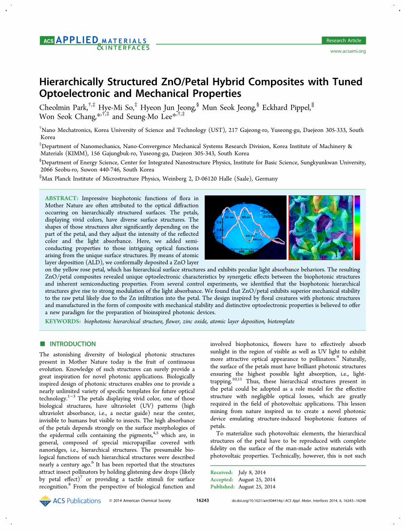
ABSTRACT: Impressive biophotonic functions of flora inMother Nature are often attributed to the optical diffractionoccurring on hierarchically structured surfaces. The petals,displaying vivid colors, have diverse surface structures. Theshapes of those structures alter significantly depending on thepart of the petal, and they adjust the intensity of the reflectedcolor and the light absorbance. Here, we added semi-conducting properties to those intriguing optical functionsarising from the unique surface structures. By means of atomiclayer deposition (ALD), we conformally deposited a ZnO layeron the yellow rose petal, which has hierarchical surface structures and exhibits peculiar light absorbance behaviors. The resultingZnO/petal composites revealed unique optoelectronic characteristics by synergetic effects between the biophotonic structuresand inherent semiconducting properties. From several control experiments, we identified that the biophotonic hierarchicalstructures give rise to strong modulation of the light absorbance. We found that ZnO/petal exhibits superior mechanical stabilityto the raw petal likely due to the Zn infiltration into the petal. The design inspired by floral creatures with photonic structuresand manufactured in the form of composite with mechanical stability and distinctive optoelectronic properties is believed to offera new paradigm for the preparation of bioinspired photonic devices.
KEYWORDS: biophotonic hierarchical structure, flower, zinc oxide, atomic layer deposition, biotemplate
■ INTRODUCTION
The astonishing diversity of biological photonic structurespresent in Mother Nature today is the fruit of continuousevolution. Knowledge of such structures can surely provide agreat inspiration for novel photonic applications. Biologicallyinspired design of photonic structures enables one to provide anearly unlimited variety of specific templates for future opticaltechnology.1−3 The petals displaying vivid color, one of thosebiological structures, have ultraviolet (UV) patterns (highultraviolet absorbance, i.e., a nectar guide) near the center,invisible to humans but visible to insects. The high absorbanceof the petals depends strongly on the surface morphologies ofthe epidermal cells containing the pigments,4,5 which are, ingeneral, composed of special micropapillae covered withnanoridges, i.e., hierarchical structures. The presumable bio-logical functions of such hierarchical structures were describednearly a century ago.6 It has been reported that the structuresattract insect pollinators by holding glistening dew drops (likelyby petal effect)7 or providing a tactile stimuli for surfacerecognition.8 From the perspective of biological function and
involved biophotonics, flowers have to effectively absorbsunlight in the region of visible as well as UV light to exhibitmore attractive optical appearance to pollinators.9 Naturally,the surface of the petals must have brilliant photonic structuresensuring the highest possible light absorption, i.e., light-trapping.10,11 Thus, these hierarchical structures present inthe petal could be adopted as a role model for the effectivestructure with negligible optical losses, which are greatlyrequired in the field of photovoltaic applications. This lessonmining from nature inspired us to create a novel photonicdevice emulating structure-induced biophotonic features ofpetals.To materialize such photovoltaic elements, the hierarchical
structures of the petal have to be reproduced with completefidelity on the surface of the man-made active materials withphotovoltaic properties. Technically, however, this is not such
Received: July 8, 2014Accepted: August 25, 2014Published: August 25, 2014
Research Article
www.acsami.org
© 2014 American Chemical Society 16243 dx.doi.org/10.1021/am504414q | ACS Appl. Mater. Interfaces 2014, 6, 16243−16248
an easy undertaking. Provided that conformality can beguaranteed, the replication approach could be the best possiblesolution. The resulting replica may also introduce somesuperlative properties induced by synergetic effects betweenthe used biotemplates and the coated man-made materials (inparticular, oxides),12−14 so long as the conformal replicationcan be done and the as-coated film can have high crystallinity.In general, oxides require high processing temperatures to becrystallized. This implies that the resulting oxide/petalcomposites may be mechanically too fragile to be used forthe intended applications. However, this issue can be readilyresolved, if one uses the composites just as is without removingthe petal, or if the employed oxide can be crystallized by low-temperature processing. Unlike other oxides requiring highprocessing temperatures to crystallize, ZnO with goodcrystallinity and extreme conformality can be coated on diversebiological templates by means of low-temperature atomic layerdeposition (ALD) technique.12−14 Here, through ZnO ALD,ZnO/petal composites with good mechanical stability wereprepared. Interestingly, the native petals revealed peculiarlocation-dependent absorption characteristics due to thevariations of hierarchical structures present on the petal. Ascompared to flat ZnO without any structural feature, ZnOcoated on the petal, excluding any pigment effect of the petal,showed noticeable optical absorption enhancements in the UVas well as visible range in concert with the optical properties ofthe deposited ZnO. In addition, the photocurrent generated byZnO exhibited distinct structure- and location-dependenttrends as well. The design inspired by floral creatures andmanufactured in the form of composite with mechanicaldurability and distinctive optoelectrical characteristics couldherald a new paradigm for the preparation of bioinspiredphotonic devices.
■ EXPERIMENTAL SECTIONPreparation of a Dried Yellow Rose Petal. Individual rose
petals were pulled out from living yellow roses that were purchasedfrom a local flower garden in Daejeon, Korea. After being cleanedcarefully with deionized water, a petal was sandwiched between two 5× 5 cm2 slide glasses and kept at room conditions for drying.
ZnO ALD. The rose petal was placed in an ALD reactor (Savannah100, Cambridge Nanotech, Inc.) and dried at 70 °C for 30 min invacuum (1 × 10⁻² Torr) with a steady N2 gas stream (20 sccm). Forthe ZnO deposition, DEZn (diethylzinc, Zn(C2H5)2, Sigma-Aldrich)and deionized H2O were used as zinc and oxygen sources, respectively.During the deposition process, the petals were alternately exposed to/purged from Zn(C2H5)2 and H2O vapor for 250 cycles. The pulse/exposure/purge time of Zn(C2H5)2 and H2O was 0.1/30/40 s and0.1/30/40 s, respectively.
Characterizations. The absorbance of each sample was measuredby UV−vis−NIR spectrophotometer (UV-3600, Shimadzu) in thespectral range of 350−800 nm with 0.5 nm resolution. Thebackground noises of the sample were corrected by measuring thereference. The macrophotoluminescence (macro-PL) spectra of ZnOdeposited samples were measured using a spectrofluorometer(FluoroLog-3) in the spectral range of 350−700 nm with 0.35 nmresolution. Its excitation light is 295 nm wavelengths, and the samplewas positioned at 45 deg between the excitation light and the detectorwith a spot size of 1.5 × 1.5 cm2. Spatially resolved micro-PL mappingwas performed by NT-MDT, NTEGRA-SPECTRA, with a 355 nmwavelength of excitation. The scan area of 75 × 75 μm2 was measuredby a 40× objective lens with ∼800 nm spatial resolution. After osmiumcoating of 5 nm thickness on the samples, the morphology wascarefully measured by field emission scanning electron microscope(FE-SEM, S-4800, Hitachi) of cold type with 1 kV acceleration voltageand 8.6 pA emissions current. The crystallinity of the deposited ZnOwas measured by an X-ray diffractometer (XRD, Bruker D8 Advance)with Cu Kα radiation (wavelength λ = 1.5418 Å). The 2 theta rangewas from 20−80 deg with a scan speed of 0.2 deg/min. Thedetermination of the chemical nature of the deposited ZnO on thepetal was performed by an X-ray photoelectron spectrometer (XPS,KRATOS, AXIS NOVA). For the preparation of the cross-sectionalsamples of the ZnO/petal, a focused ion beam was used. Transmission
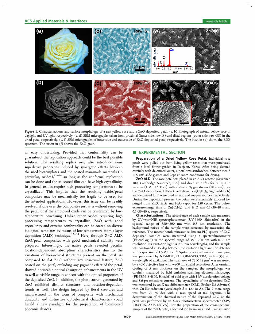
Figure 1. Characterizations and surface morphology of a raw yellow rose and a ZnO deposited petal. (a, b) Photograph of natural yellow rose indaylight and UV light, respectively. (c, d) SEM micrographs taken from proximal (inner side, raw IS) and distal regions (outer side, raw OS) in thedried petal, respectively. (e, f) SEM micrographs of inner side and outer side of ZnO deposited petal, respectively. The inset in (e) shows the EDXspectrum. The insert in (f) shows the ZnO grain.
ACS Applied Materials & Interfaces Research Article
dx.doi.org/10.1021/am504414q | ACS Appl. Mater. Interfaces 2014, 6, 16243−1624816244
electron microscope (TEM) and energy-dispersive X-ray (EDX)investigations (imaging and point analyses) were performed with a FEITITAN 80−300 microscope (300 kV) in scanning transmission mode.For photocurrent measurement, the Ti/Au electrode of 30/500 nmthickness was deposited on the surface of the ZnO/petal compositesand ZnO/quartz by thermal evaporation method (KOREAVAC, KVE-T2000) with a shadow mask (gap = 50 μm). During the thermalevaporation process, the sample holder was water-cooled to avoidthermal damage to the ZnO/petal. In a dark room, the photogeneratedcurrents of ZnO/petal and ZnO/quartz were measured using a high-speed source/monitor unit (E5262A, Agilent Technologies) withillumination of 355 nm laser (CNI, DOI-UV-F-355-CW, 11.5 mW/cm2).Tensile Test. All samples (2 mm × ∼3 cm) were carefully cut with
a knife (BAYHA, Blades, No. 24) under the optical microscope (LeitzAristomet); subsequently those were mounted in a thick paper jig(∼500 μm thick) having 20 mm punched holes. The paper jigs wereused to enable alignment and clamping of the specimens during thetensile test. Furthermore, the jigs allowed the specimens to be cuteasily through the cutting line so that specimen and paper jigs werenot loaded together during the actual test. Pattex Blitz Kleber (Henkel,Germany) was used as glue to fix the sample to the edge of the jigs. AZWICK 1445 tensile test machine with 500 g HBM load cell with 0.1mN resolution and 0.5% uncertainty was used. The upper part of thejig was fixed to the load cell by a screw-type clamping system, andsubsequently the jig was cut through the middle line with scissors.After controlling the vertical alignment of the specimen attached to thejig, the lower part of the jig was also fixed in the same way as the upperpart. The extension rate was 10% of the initial length per minute (2mm/min). The sample was extended until failure occurred. SEM andoptical microscope were used to measure and confirm the cross-sectional area of the specimens. Because the thickness of the samplewas not perfectly uniform, along the horizontal direction of eachsample, at many points the thicknesses were measured and averaged.In the case of the width of each sample, a similar procedure wasapplied. On the basis of the measured cross-sectional area, the rawforce (mN)−strain (%) data for each specimen exported from thesoftware of the machine were rescaled into the engineering stress (σ)−strain (ε). To obtain a tensile testing curve of one sample, more than15 samples were prepared identically and measured at identicalconditions. All graphic works including data rescaling were performedwith ORIGIN 8.0.
■ RESULTS AND DISCUSSION
In fauna, structural color, originating from physical interactionsof light with photonic micro/nanostructures, has been reportedin a range of animal species, while in flora, examples ofphotonic structures are quite rare. In addition, those have beenvery recently demonstrated.9,15 Particularly, the color of thepetal is believed to be changed with the chemical nature of thepigments. However, it is known that the intensity of thereflected color depends strongly on the shape and dimension ofepidermal cells.4 For instance, a yellow petal (Figure 1a)contains pigments that absorb green and blue light, allowing theyellow light to pass through and out of the petal.16 Althoughthe petal appears yellow to us, it can be figured out that thereexists a nectar guide with strong absorption of UV light (Figure1b). The strong UV absorption was believed to be induced bythe differential distribution of flavonoids in some plants.17
However, in the case of the yellow rose petal used in thisresearch, the microstructure investigation by scanning electronmicroscope (SEM) indicated that the morphology anddistribution of the hierarchical structures (nanoridges onmicropapillae) on the petal vary with the location. Namely,though micropapillaes covered with nanoridges are widelyspread in common in both the proximal region (inner side) andthe distal region (outer side) of the petal, the sizes of individual
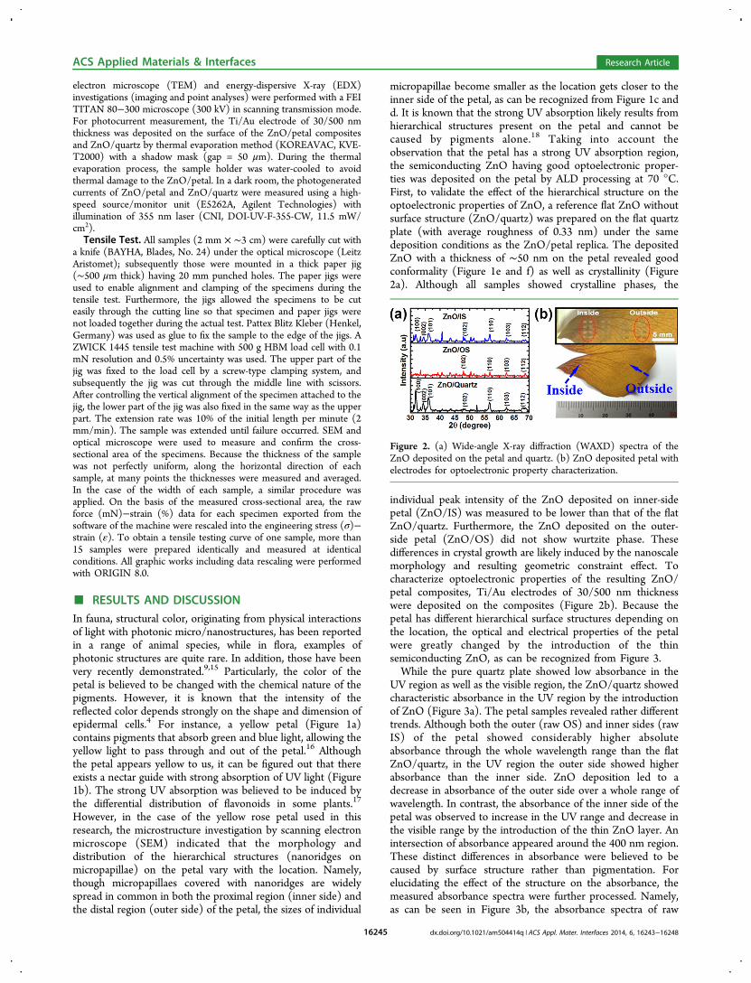
micropapillae become smaller as the location gets closer to theinner side of the petal, as can be recognized from Figure 1c andd. It is known that the strong UV absorption likely results fromhierarchical structures present on the petal and cannot becaused by pigments alone.18 Taking into account theobservation that the petal has a strong UV absorption region,the semiconducting ZnO having good optoelectronic proper-ties was deposited on the petal by ALD processing at 70 °C.First, to validate the effect of the hierarchical structure on theoptoelectronic properties of ZnO, a reference flat ZnO withoutsurface structure (ZnO/quartz) was prepared on the flat quartzplate (with average roughness of 0.33 nm) under the samedeposition conditions as the ZnO/petal replica. The depositedZnO with a thickness of ∼50 nm on the petal revealed goodconformality (Figure 1e and f) as well as crystallinity (Figure2a). Although all samples showed crystalline phases, the
individual peak intensity of the ZnO deposited on inner-sidepetal (ZnO/IS) was measured to be lower than that of the flatZnO/quartz. Furthermore, the ZnO deposited on the outer-side petal (ZnO/OS) did not show wurtzite phase. Thesedifferences in crystal growth are likely induced by the nanoscalemorphology and resulting geometric constraint effect. Tocharacterize optoelectronic properties of the resulting ZnO/petal composites, Ti/Au electrodes of 30/500 nm thicknesswere deposited on the composites (Figure 2b). Because thepetal has different hierarchical surface structures depending onthe location, the optical and electrical properties of the petalwere greatly changed by the introduction of the thinsemiconducting ZnO, as can be recognized from Figure 3.While the pure quartz plate showed low absorbance in the
UV region as well as the visible region, the ZnO/quartz showedcharacteristic absorbance in the UV region by the introductionof ZnO (Figure 3a). The petal samples revealed rather differenttrends. Although both the outer (raw OS) and inner sides (rawIS) of the petal showed considerably higher absoluteabsorbance through the whole wavelength range than the flatZnO/quartz, in the UV region the outer side showed higherabsorbance than the inner side. ZnO deposition led to adecrease in absorbance of the outer side over a whole range ofwavelength. In contrast, the absorbance of the inner side of thepetal was observed to increase in the UV range and decrease inthe visible range by the introduction of the thin ZnO layer. Anintersection of absorbance appeared around the 400 nm region.These distinct differences in absorbance were believed to becaused by surface structure rather than pigmentation. Forelucidating the effect of the structure on the absorbance, themeasured absorbance spectra were further processed. Namely,as can be seen in Figure 3b, the absorbance spectra of raw
Figure 2. (a) Wide-angle X-ray diffraction (WAXD) spectra of theZnO deposited on the petal and quartz. (b) ZnO deposited petal withelectrodes for optoelectronic property characterization.
ACS Applied Materials & Interfaces Research Article
dx.doi.org/10.1021/am504414q | ACS Appl. Mater. Interfaces 2014, 6, 16243−1624816245
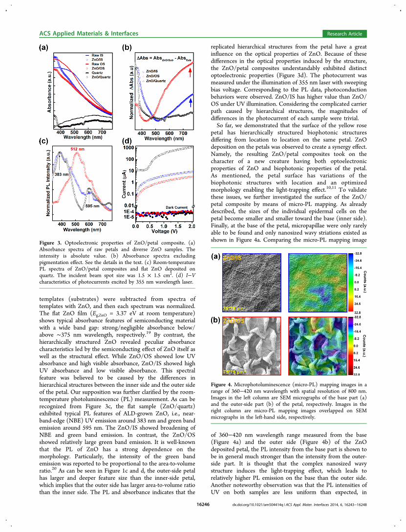
templates (substrates) were subtracted from spectra oftemplates with ZnO, and then each spectrum was normalized.The flat ZnO film (Eg,ZnO = 3.37 eV at room temperature)shows typical absorbance features of semiconducting materialwith a wide band gap: strong/negligible absorbance below/above ∼375 nm wavelength, respectively.19 By contrast, thehierarchically structured ZnO revealed peculiar absorbancecharacteristics led by the semiconducting effect of ZnO itself aswell as the structural effect. While ZnO/OS showed low UVabsorbance and high visible absorbance, ZnO/IS showed highUV absorbance and low visible absorbance. This spectralfeature was believed to be caused by the differences inhierarchical structures between the inner side and the outer sideof the petal. Our supposition was further clarified by the room-temperature photoluminescence (PL) measurement. As can berecognized from Figure 3c, the flat sample (ZnO/quartz)exhibited typical PL features of ALD-grown ZnO, i.e., near-band-edge (NBE) UV emission around 383 nm and green bandemission around 595 nm. The ZnO/IS showed broadening ofNBE and green band emission. In contrast, the ZnO/OSshowed relatively large green band emission. It is well-knownthat the PL of ZnO has a strong dependence on themorphology. Particularly, the intensity of the green bandemission was reported to be proportional to the area-to-volumeratio.20 As can be seen in Figure 1c and d, the outer-side petalhas larger and deeper feature size than the inner-side petal,which implies that the outer side has larger area-to-volume ratiothan the inner side. The PL and absorbance indicates that the
replicated hierarchical structures from the petal have a greatinfluence on the optical properties of ZnO. Because of thesedifferences in the optical properties induced by the structure,the ZnO/petal composites understandably exhibited distinctoptoelectronic properties (Figure 3d). The photocurrent wasmeasured under the illumination of 355 nm laser with sweepingbias voltage. Corresponding to the PL data, photoconductionbehaviors were observed. ZnO/IS has higher value than ZnO/OS under UV illumination. Considering the complicated carrierpath caused by hierarchical structures, the magnitudes ofdifferences in the photocurrent of each sample were trivial.So far, we demonstrated that the surface of the yellow rose
petal has hierarchically structured biophotonic structuresdiffering from location to location on the same petal. ZnOdeposition on the petals was observed to create a synergy effect.Namely, the resulting ZnO/petal composites took on thecharacter of a new creature having both optoelectronicproperties of ZnO and biophotonic properties of the petal.As mentioned, the petal surface has variations of thebiophotonic structures with location and an optimizedmorphology enabling the light-trapping effect.10,11 To validatethese issues, we further investigated the surface of the ZnO/petal composite by means of micro-PL mapping. As alreadydescribed, the sizes of the individual epidermal cells on thepetal become smaller and smaller toward the base (inner side).Finally, at the base of the petal, micropapillae were only rarelyable to be found and only nanosized wavy striations existed asshown in Figure 4a. Comparing the micro-PL mapping image
of 360−420 nm wavelength range measured from the base(Figure 4a) and the outer side (Figure 4b) of the ZnOdeposited petal, the PL intensity from the base part is shown tobe in general much stronger than the intensity from the outer-side part. It is thought that the complex nanosized wavystructure induces the light-trapping effect, which leads torelatively higher PL emission on the base than the outer side.Another noteworthy observation was that the PL intensities ofUV on both samples are less uniform than expected, in
Figure 3. Optoelectronic properties of ZnO/petal composite. (a)Absorbance spectra of raw petals and diverse ZnO samples. Theintensity is absolute value. (b) Absorbance spectra excludingpigmentation effect. See the details in the text. (c) Room-temperaturePL spectra of ZnO/petal composites and flat ZnO deposited onquartz. The incident beam spot size was 1.5 × 1.5 cm2. (d) I−Vcharacteristics of photocurrents excited by 355 nm wavelength laser.
Figure 4. Microphotoluminescence (micro-PL) mapping images in arange of 360−420 nm wavelength with spatial resolution of 800 nm.Images in the left column are SEM micrographs of the base part (a)and the outer-side part (b) of the petal, respectively. Images in theright column are micro-PL mapping images overlapped on SEMmicrographs in the left-hand side, respectively.
ACS Applied Materials & Interfaces Research Article
dx.doi.org/10.1021/am504414q | ACS Appl. Mater. Interfaces 2014, 6, 16243−1624816246
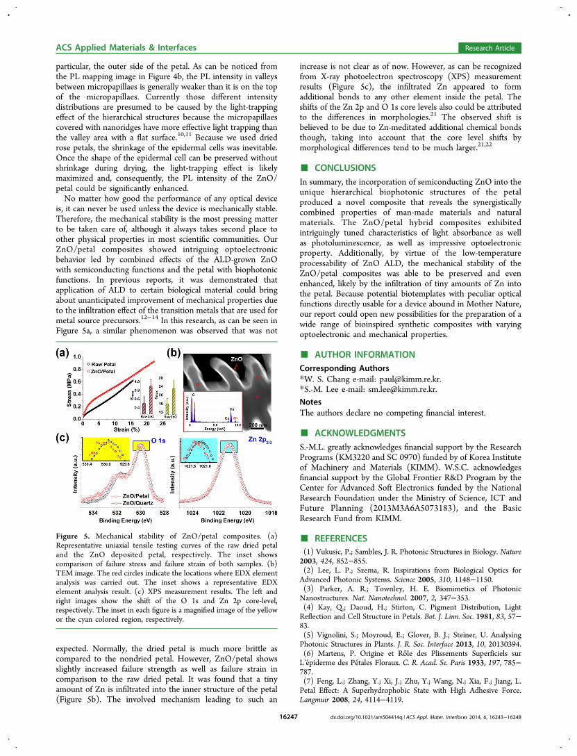
particular, the outer side of the petal. As can be noticed fromthe PL mapping image in Figure 4b, the PL intensity in valleysbetween micropapillaes is generally weaker than it is on the topof the micropapillaes. Currently those different intensitydistributions are presumed to be caused by the light-trappingeffect of the hierarchical structures because the micropapillaescovered with nanoridges have more effective light trapping thanthe valley area with a flat surface.10,11 Because we used driedrose petals, the shrinkage of the epidermal cells was inevitable.Once the shape of the epidermal cell can be preserved withoutshrinkage during drying, the light-trapping effect is likelymaximized and, consequently, the PL intensity of the ZnO/petal could be significantly enhanced.No matter how good the performance of any optical device
is, it can never be used unless the device is mechanically stable.Therefore, the mechanical stability is the most pressing matterto be taken care of, although it always takes second place toother physical properties in most scientific communities. OurZnO/petal composites showed intriguing optoelectronicbehavior led by combined effects of the ALD-grown ZnOwith semiconducting functions and the petal with biophotonicfunctions. In previous reports, it was demonstrated thatapplication of ALD to certain biological material could bringabout unanticipated improvement of mechanical properties dueto the infiltration effect of the transition metals that are used formetal source precursors.12−14 In this research, as can be seen inFigure 5a, a similar phenomenon was observed that was not
expected. Normally, the dried petal is much more brittle ascompared to the nondried petal. However, ZnO/petal showsslightly increased failure strength as well as failure strain incomparison to the raw dried petal. It was found that a tinyamount of Zn is infiltrated into the inner structure of the petal(Figure 5b). The involved mechanism leading to such an
increase is not clear as of now. However, as can be recognizedfrom X-ray photoelectron spectroscopy (XPS) measurementresults (Figure 5c), the infiltrated Zn appeared to formadditional bonds to any other element inside the petal. Theshifts of the Zn 2p and O 1s core levels also could be attributedto the differences in morphologies.21 The observed shift isbelieved to be due to Zn-meditated additional chemical bondsthough, taking into account that the core level shifts bymorphological differences tend to be much larger.21,22
■ CONCLUSIONSIn summary, the incorporation of semiconducting ZnO into theunique hierarchical biophotonic structures of the petalproduced a novel composite that reveals the synergisticallycombined properties of man-made materials and naturalmaterials. The ZnO/petal hybrid composites exhibitedintriguingly tuned characteristics of light absorbance as wellas photoluminescence, as well as impressive optoelectronicproperty. Additionally, by virtue of the low-temperatureprocessability of ZnO ALD, the mechanical stability of theZnO/petal composites was able to be preserved and evenenhanced, likely by the infiltration of tiny amounts of Zn intothe petal. Because potential biotemplates with peculiar opticalfunctions directly usable for a device abound in Mother Nature,our report could open new possibilities for the preparation of awide range of bioinspired synthetic composites with varyingoptoelectronic and mechanical properties.
■ AUTHOR INFORMATIONCorresponding Authors*W. S. Chang e-mail: [email protected].*S.-M. Lee e-mail: [email protected].
NotesThe authors declare no competing financial interest.
■ ACKNOWLEDGMENTSS.-M.L. greatly acknowledges financial support by the ResearchPrograms (KM3220 and SC 0970) funded by of Korea Instituteof Machinery and Materials (KIMM). W.S.C. acknowledgesfinancial support by the Global Frontier R&D Program by theCenter for Advanced Soft Electronics funded by the NationalResearch Foundation under the Ministry of Science, ICT andFuture Planning (2013M3A6A5073183), and the BasicResearch Fund from KIMM.
■ REFERENCES(1) Vukusic, P.; Sambles, J. R. Photonic Structures in Biology. Nature2003, 424, 852−855.(2) Lee, L. P.; Szema, R. Inspirations from Biological Optics forAdvanced Photonic Systems. Science 2005, 310, 1148−1150.(3) Parker, A. R.; Townley, H. E. Biomimetics of PhotonicNanostructures. Nat. Nanotechnol. 2007, 2, 347−353.(4) Kay, Q.; Daoud, H.; Stirton, C. Pigment Distribution, LightReflection and Cell Structure in Petals. Bot. J. Linn. Soc. 1981, 83, 57−83.(5) Vignolini, S.; Moyroud, E.; Glover, B. J.; Steiner, U. AnalysingPhotonic Structures in Plants. J. R. Soc. Interface 2013, 10, 20130394.(6) Martens, P. Origine et Role des Plissements Superficiels surL’epiderme des Petales Floraux. C. R. Acad. Se. Paris 1933, 197, 785−787.(7) Feng, L.; Zhang, Y.; Xi, J.; Zhu, Y.; Wang, N.; Xia, F.; Jiang, L.Petal Effect: A Superhydrophobic State with High Adhesive Force.Langmuir 2008, 24, 4114−4119.
Figure 5. Mechanical stability of ZnO/petal composites. (a)Representative uniaxial tensile testing curves of the raw dried petaland the ZnO deposited petal, respectively. The inset showscomparison of failure stress and failure strain of both samples. (b)TEM image. The red circles indicate the locations where EDX elementanalysis was carried out. The inset shows a representative EDXelement analysis result. (c) XPS measurement results. The left andright images show the shift of the O 1s and Zn 2p core-level,respectively. The inset in each figure is a magnified image of the yellowor the cyan colored region, respectively.
ACS Applied Materials & Interfaces Research Article
dx.doi.org/10.1021/am504414q | ACS Appl. Mater. Interfaces 2014, 6, 16243−1624816247
(8) Koch, K.; Bhushan, B.; Barthlott, W. Diversity of Structure,Morphology and Wetting of Plant Surfaces. Soft Matter 2008, 4,1943−1963.(9) Whitney, H. M.; Kolle, M.; Andrew, P.; Chittka, L.; Steiner, U.;Glover, B. J. Floral Iridescence, Produced by Diffractive Optics, Acts asa Cue for Animal Pollinators. Science 2009, 323, 130−133.(10) Exner, F.; Exner, S. Die Physikalischen Grundlagen derBlutenfarbungen. Sitzungsber. Kais. Akad. Wiss. Wien, Math.-nat. Kl. I1910, 119, 191−245.(11) Bernhard, C. G.; Gemne, G.; Moller, A. R. Modification ofSpecular Reflexion and Light Transmission by Biological SurfaceStructures. Q. Rev. Biophys. 1968, 1, 89−105.(12) Lee, S. M.; Pippel, E.; Gosele, U.; Dresbach, C.; Qin, Y.;Chandran, C. V.; Brauniger, T.; Hause, G.; Knez, M. Greatly IncreasedToughness of Infiltrated Spider Silk. Science 2009, 324, 488−492.(13) Lee, S. M.; Grass, G.; Kim, G. M.; Dresbach, C.; Zhang, L.;Gosele, U.; Knez, M. Low-Temperature ZnO Atomic LayerDeposition on Biotemplates: Flexible Photocatalytic ZnO Structuresfrom Eggshell Membranes. Phys. Chem. Chem. Phys. 2009, 11, 3608−3614.(14) Lee, S. M.; Pippel, E.; Moutanabbir, O.; Gunkel, I.; Thurn-Albrecht, T.; Knez, M. Improved Mechanical Stability of DriedCollagen Membrane after Metal Infiltration. ACS Appl. Mater.Interfaces 2010, 2, 2436−2441.(15) Lee, S. M.; Upping, J.; Bielawny, A.; Knez, M. Structure-BasedColor of Natural Petals Discriminated by Polymer Replication. ACSAppl. Mater. Interfaces 2011, 3, 30−34.(16) Glover, B. J. Understanding Flowers and Flowering: An IntegratedApproach; Oxford University Press: Oxford, U.K., 2007.(17) Rieseberg, L. H.; Schilling, E. E. Floral Flavonoids andUltraviolet Patterns in Viguiera (Compositae). Am. J. Bot. 1985, 72,999−1004.(18) Whitney, H. M.; Kolle, M.; Alvarez-Fernandez, R.; Steiner, U.;Glover, B. J. Contributions of Iridescence to Floral Patterning.Commun. Integr. Biol. 2009, 2, 230−232.(19) Luka, G.; Krajewski, T.; Wachnicki, L.; Witkowski, B.;Lusakowska, E.; Paszkowicz, W.; Guziewicz, E.; Godlewski, M.Transparent and Conductive Undoped Zinc Oxide Thin FilmsGrown by Atomic Layer Deposition. Phys. Status Solidi A 2010, 207,1568−1571.(20) Andelman, T.; Gong, Y.; Polking, M.; Yin, M.; Kuskovsky, I.;Neumark, G.; O’Brien, S. Morphological Control and Photo-luminescence of Zinc Oxide Nanocrystals. J. Phys. Chem. B 2005,109, 14314−14318.(21) Al-Gaashani, R.; Radiman, S.; Daud, A. R.; Tabet, N.; Al-Douri,Y. XPS and Optical Studies of Different Morphologies of ZnONanostructures Prepared by Microwave Methods. Ceram. Int. 2013,39, 2283−2292.(22) Zhou, H.; Li, Z. Synthesis of Nanowires, Nanorods andNanoparticles of ZnO through Modulating the Ratio of Water toMethanol by Using a Mild and Simple Solution Method. Mater. Chem.Phys. 2005, 89, 326−331.
ACS Applied Materials & Interfaces Research Article
dx.doi.org/10.1021/am504414q | ACS Appl. Mater. Interfaces 2014, 6, 16243−1624816248