Embed Size (px)
Citation preview

HHSN268201500021C Quantitative Imaging Biomarkers Alliance (QIBA)
PROGRESS REPORT: AS OF SEPTEMBER 2016
This progress report is stated in terms given in the accepted Work Plan. This progress report is organized in the following subsections:
A. Review of activities responsive to each objective.
B. Combined list, across the objectives, of groundwork projects approved by the Steering Committee.
C. Additional descriptions of general committee progress.
A. REVIEW OF ACTIVITIES RESPONSIVE TO EACH OBJECTIVE.
An update on Objectives 1-7 is given below. Note that, in general, data from each of these efforts have been or will be uploaded to the Quantitative Imaging Data Warehouse (QIDW) identified in the methodology for Objective 6.
OBJECTIVE 1. CREATE AND DISSEMINATE NEW PROTOCOLS AND QIB PROFILES EACH YEAR THAT ADDRESS DISEASES OF SIGNIFICANT BURDEN TO THE US POPULATION.
Selected specifics:
CT Volumetry Biomarker Committee: The CT Volumetry Advanced Disease Profile has incorporated public comments and is expected to be released for feasibility testing in 4Q2016.
Groundwork projects on volumetry of liver masses have been undertaken for the purpose of extending the first version of the CT Volumetry Profile to include quantitative assessment of liver masses. It is anticipated that a Profile for CT Volumetry of liver masses will continue to develop throughout the second year of the current contract, with a draft for public review in the latter part of 2017.
A Task Force of the CT Volumetry Biomarker Committee is developing a Profile for assessment of small nodules in the context of lung cancer screening. They are developing document text to define evidence-based consensus standards and processes for CT imaging in the setting of lung cancer screening to allow for quantification of biologically meaningful longitudinal volume changes, with acceptable range of variance across vendor platforms. This activity is expected to result in the release of a Profile in 4Q2016.
CT Lung Density Biomarker Committee: A Profile for the measurement of lung density based on quantitative CT measurements is in development and is expected to be released for public comment in 1Q2017.
The committee is working with major vendors and the National Institute of Standards and Technology (NIST) to develop quantitative imaging protocols for monitoring emphysema progression in chronic obstructive pulmonary disease (COPD). A 2nd round of protocol testing, with representatives from Siemens, GE Healthcare, Toshiba, and Philips, was completed in April, 2016 with completion of data analysis expected by November, 2016. The Lung Density Profile development is being informed by data from 2015-16 Round-5 Project L. This groundwork project is testing different methods for lung inflation volume adjustment (VA) because variation in lung inflation volume is a major source of bias in lung density measurements. Based on results from this groundwork study, the committee will recommend a best approach to reduce bias using VA and include this recommendation in the Profile.
fMRI Biomarker Committee: Work is maturing on QIBA BOLD fMRI Profile v1.0 in efforts to optimize Blood Oxygen Level Dependent (BOLD) fMRI brain mapping of motor function for treatment planning prior to surgery or invasive treatment intervention. It is anticipated this Profile will be released for public comment in 2016. The committee is also addressing language function mapping based on a Round-5 (2015-16) groundwork project, which will lead to a Profile draft v2.0 in 2017. The language mapping Profile development is being supported by 2015-16 Round-5 projects D1 and D2. For these projects, fMRI Digital Reference Objects (DROs) were

QIBA Annual Progress Report as of September 2016
Page 2
used to investigate the potential effects of head motion on the Profile claims and to help characterize data quality control metrics (e.g., minimum acceptable head motion) that will promote Profile conformance.
PDF-MRI Biomarker Committee: The DCE-MRI Task Force is drafting Dynamic Contrast Enhanced (DCE)-MRI Profile v2.0, which will extend DCE-MRI Profile v1.0 to include 3.0T field strength as well as parallel imaging acquisitions often used in DCE. This effort builds upon an organ-specific systematic literature review process and the success of groundwork projects. The revised Profile will be available for public comment in 4Q2016 or 1Q2017.
The DWI Task Force Profile v1.0 is being drafted to address technical performance standards for the acquisition of apparent diffusion coefficient (ADC) measurements in brain and liver. ADC phantom scanning and analysis has occurred at multiple sites, informing the Profile, which is slated for release for public comment by 4Q2016.
Two additional PDF-MRI Biomarker Committee Task Forces are in earlier stages of Profile development, focusing on brain Diffusion Tensor Imaging (DTI) and perfusion via Dynamic Susceptibility Contrast (DSC) techniques. In these efforts, literature review, phantom development and multi-site/vendor measurements are being performed to inform the Profiles. These Profiles are targeted for public comment by 2Q2017.
MR Elastography (MRE) Biomarker Committee: The committee is actively developing a draft Profile v1.0 for quantitative MRE measurements in liver. Since substantive groundwork has already been performed and results previously published, Profile development is progressing rapidly and a draft Profile will be available for public comment by the end of 2016. As the initial step in developing the Profile, the Task Force conducted a meta-analysis of published studies of the test-retest repeatability of hepatic MR elastography. The results of this meta-analysis form the objective basis for the quantitative cross-sectional claim for the draft Profile. The meta-analysis has been written-up by the Task Force, and has been submitted for publication.
Proton Density Fat Fraction (PDFF) Biomarker Committee: The committee is actively developing a draft Profile v1.0 for quantitative proton density fat fraction measurements in liver. Since substantive groundwork has already been performed and results previously published, Profile development is progressing well. A robust literature search has been performed, which has informed the claim, and the Profile is in draft phase. Further, a meta-analysis that will be used to support cross-sectional and longitudinal claims is progressing well with completion targeted for the ISMRM abstract submission deadline (11/9/16). A first draft of the Profile is expected by 3Q2017.
FDG-PET/CT Biomarker Committee: Efforts of the committee have been primarily focused on moving the FDG PET-CT Profile from the QIBA classification of “Publicly Reviewed Version” stage to the “Technically Confirmed Version” stage. Consolidating feedback from field testing of the Profile from multiple imaging sites, manufacturers, and software developers has led to a shortening of the original Profile to include only those requirements essential for achieving the FDG-PET/CT claim. Near-final checklists for imaging sites and manufacturers to assess conformance with Profile requirements are in final review, and are ready for attachment to the edited Profile as appendices. Finally, three critical projects investigating standardization of PET spatial resolution and volumetric measurements in both human subject and DRO models are nearing completion. These projects are targeting new and better PET quantitative performance measurements to inform future versions of the Profile.
PET-Amyloid Biomarker Committee: The working draft Profile claims were revised based on results from the literature meta-analysis (2015-2016 Round-5 project E) and input from the QIBA Metrology Working Group. Further revision of the performance requirements and informative text was performed, addressing over a hundred specific line items from committee members. Release of the Profile for public comment was delayed due to this internal work as well as the identified need for additional text to address conformance testing methodology. The Conformance Testing Task Force is preparing this text for incorporation into the Profile. The Profile will be available for public comment by 4Q2016.

QIBA Annual Progress Report as of September 2016
Page 3
SPECT Biomarker Committee: The first Profile (Quantifying Dopamine Transporters with 123-Ioflupane in Neurodegenerative Disorders) addresses major world health problems, including Parkinson’s disease (PD), Diffuse Lewy Body Disease, and a variety of neurodegenerative diseases that present with Parkinsonian movement disorders. PD is a major health problem currently afflicting over one million Americans. The plan is to follow with Profiles addressing (a) Technetium-99m-based SPECT for planning transarterial radiotherapy, and (b) SPECT as companion diagnostics for theranostics (a current use case for biopharmaceutical companies). The SPECT BC has now completed a detailed review of the existing SPECT quantitative imaging biomarker literature. Methods for achieving compliance with Profile claims for precision and bias are on time-line, with expected near completion by end of 3Q2016. International enthusiasm for participation has been particularly strong from Japan and several European Union states. Multiple task groups, led by deep subject matter experts, have been hosting regular teleconference meetings to define Profile v1.0. The first Profile should be available for public review by end of 4Q2016.
US Shear Wave Speed Biomarker Committee: An initial draft of the Shear Wave Speed (SWS) Profile v1.0 has been developed. The literature review has been completed, and mitigation strategies to reduce SWS-measuring system variance have been characterized. Comparisons between liver fibrosis measurements using SWS and other techniques have been completed. The Profile is targeted for public review by 3Q2016 and is being informed by Round-5 Projects F1 and F2 (2015-16). The SWS Profile v1.0 was distributed to the Biomarker Committee for comments in April 2016. Comments were tabulated in June 2016 and v2.0 has been drafted and will be distributed for final committee comments prior to release for public comment at the beginning of October 2016. Manufacturers must supply scanner-specific acquisition instructions for Appendix D. While the Profile is out for comments, the Profile drafting working group will work with manufacturers to complete their instructions. Conformance will be monitored by completion of checklists and those checklists will be drafted as soon as the manufacturer-specific instructions are received. The draft Profile, Appendix D, and the conformance checklists should be ready for the planned two-site technical confirmation study by the end of 4Q2016.
US Volume Blood Flow (VBF) Biomarker Committee: Success has been achieved in obtaining volume flow in several applications with <15% variance and bias. Requirements for demonstrating quantitative estimation of volumetric blood flow in renal transplants where reference data can be obtained in vivo are under discussion. A document has been created to specify information required to perform the volume flow computation. Instructions, as given by standard operating procedures, have been compiled which test sites will follow to obtain a volume flow on a specific ultrasound scanner. These instructions will form the basis of protocols and are meant to minimize restrictions on how users can acquire images for volumetric blood flow while assessing inter- and intra- observer variability.
Contrast-Enhanced US (CEUS) Biomarker Committee: The CEUS Biomarker Committee has been recently formed under the Ultrasound Coordinating Committee of QIBA. Listed below are current activities towards the specific objectives.
The committee is actively developing a draft Profile for quantitative CEUS measurements in the liver. At the present stage, a draft project description and an outline of the group’s planned work have been compiled. The goal of this committee is to standardize quantitative CEUS to provide a biomarker of perfusion and thus of tumor response to therapy for liver tumors initially and other clinical applications later. A group of over 50 experts in the field (clinicians, academics, engineers, basic scientists) have been divided into 5 task force teams to better address the issues and fully develop the QIBA Profile. The task force teams are (a) literature review, (b) clinical focus, (c) imaging systems requirements, (d) quantification analysis software, and (e) basic science. The committee has been holding monthly teleconferences to address the various issues relevant to the Profile being developed.
QIBA Annual Progress Report as of September 2016
Page 4
OBJECTIVE 2. PERFORM FIELD TESTS AND REVISE EXISTING QIB PROFILES AS NEEDED.
Selected specifics:
CT Volumetry Biomarker Committee: In preparation for field testing, the committee has developed a stakeholder-specific Profile feasibility assessment checklist tailored to assess the 1) conformance level of unmodified current practice, and 2) anticipated ability to incorporate Profile requirements into existing workflows, or willingness to adopt new workflows for field testing, and eventually conformant scanning. The committee is evaluating options for a formal field test of the publicly-reviewed CT Tumor Volume Change Profile (v2.2). The Profile has been updated to align with the RSNA QIBA Metrology Work Group definitions. Claim language has been modified to reflect the committee consensus on appropriate performance thresholds based on current methods and technology.
fMRI Biomarker Committee: Discussion has centered upon planning staged multi-site human testing of the Profile (after receiving and incorporating public comments). The first stage will involve multiple sites within the Biomarker Committee’s membership implementing Profile v1.0 and acquiring human subject motor mapping data. Testing will focus on identifying implementation issues, QA results obtained, and agreement with Claims in cases where multiple measurements are made in the same subject. The second stage will likely involve extending such testing to additional sites that perform clinical fMRI motor mapping.
PDF-MRI Biomarker Committee: Overseen by the PDF-MRI Biomarker Committee, the DCE-MRI Task Force is awaiting analysis of the DCE-MRI Quantification Profile (v1.0) field test (supported by a Round-2 groundwork project), implemented in the ACRIN 6701 prostate cancer patient test/retest study. The ACRIN 6701 study was specifically designed to collect multi-site/multi-vendor DCE (and DWI) data, and will thus directly inform future QIBA Profiles involving MR for prostate imaging. Accrual for this study has been completed, and preliminary results are anticipated by 1Q2017. As DCE-MRI Profile v2.0 is recast into the new QIBA template, material related to 3T systems and parallel imaging techniques is being incorporated. Known B1 non-uniformity issues at high-field (3T) will be addressed in a recently awarded (Round-6) groundwork project to build and test physical phantoms suitable for B1-and T1-mapping.
The DWI-MRI Task Force has completed its literature review and distilled findings into two organ-specific claims for its DWI Profile v1.0, which is in final draft stage. Multi-site/multi-vendor DWI phantom (supported by a Round-3 groundwork project) scanning and analysis using a common DWI QA/QC software package (supported by Round-3 groundwork contract) was completed in mid-2016, and now the DWI Task Force focus is on Profile writing.
FDG-PET/CT Biomarker Committee: After field-testing the Profile at multiple academic imaging sites to examine its feasibility and practicality, the Profile was sent to a broad cross-section of PET/CT users including mainstream clinical and hospital-based sites to further assess both practicality of implementation and feasibility of conformance. Additionally the Profile was reviewed by all US manufacturers of PET/CT equipment to assess ability of vendors to comply with Profile requirements. All manufacturers carefully reviewed Profile standards and returned comments on ability to comply. The Committee extensively discussed the feedback from both the imaging site and manufacturer’s field test results and is currently modifying the Profile to a more streamlined, yet still comprehensive, Technically Confirmed Version.
PET-Amyloid Biomarker Committee: The committee is discussing next steps necessary to proceed with feasibility testing. Once the Profile has been released for public comment, feasibility checklists will be created using evolving standard QIBA practice.
SPECT Biomarker Committee: The first working specifications for a “fillable” phantom have been developed for deployment in multi-center environments. The Round-6 project is a collaboration between the University of Michigan, USA, and University College London, UK. Launch is expected in 4Q2016 or 1Q2017, initially with studies to test the acquisition and reconstruction attributes required to conform with the Profile claims for

QIBA Annual Progress Report as of September 2016
Page 5
accuracy and precision. A Digital Reference Object (DRO) based on a high resolution, human brain MRI scan is being developed at the University of Washington. This project builds on the successful versions developed by the FDG PET/CT and PET-Amyloid groups. An unfunded concept for a solid phantom, with a traceable source of Tellurium-123 as a proxy for Iodine-123, is being developed in collaboration with subject matter experts from the National Institute of Standards and Technology. However, this project will be “parked” until work with the fillable phantom and DRO has progressed.
US SWS Biomarker Committee: Distribution within and approval by the Ultrasound SWS Biomarker Committee is pending while the Profile is converted to the new document template and system-dependent methods descriptions are provided by the participating manufacturers. Release of the draft Profile for public comment is targeted for 3Q2016. The plan is to conduct SWS acquisitions for the technical confirmation study at two sites, Massachussetts General Hospital and Washington DC VA Medical Center as soon as the public comments have been incorporated into v2.0 of the draft Profile. The data acquisition schemes already in use will be modified to conform to the draft Profile and will be used to test whether the Profile-driven protocols are practical in a clinical environment. The acquisitions are to be funded by internal funding from the two sites plus partial FDA funding for acquisition at the DC VAMC. In addition, a plan was developed to introduce deviations from the protocols outlined in the Profile into a subset of the acquisitions to study the effects of errors made during acquisition on the final SWS values. These data plus manufacturers’ results from their digital phantom tests, when analyzed as a part of a project proposed for Round 6 funding, will be used to further revise and refine the SWS Profile. If the revisions are substantial, additional self-funded SWS acquisitions will be conducted at the two test sites in an attempt to not only reach the technical confirmation stage but also the “claim confirmed” stage of Profile development.
US VBF Biomarker Committee:
A prototype physical flow phantom for volume flow estimation is being designed and will be constructed in the coming months. Participating manufacturers of ultrasound scanners have agreed to provide either requisite ultrasound scanheads or entire systems, depending on the ability of their systems. This will provide the required data sets across multiple platforms and at different user sites.
Field pre-testing for v1.0 of the phantom has been done at the University of Michigan campus to inform the design and in preparation of the larger round robin effort. RSNA QIBA QIDW access has been requested and granted. Participating laboratories will be employing ultrasound systems that they have been working with already. The University of Michigan will test all participating systems as well and directly work with the respective manufacturers to assure proper data (stream) selection.
US CEUS Biomarker Committee: The CEUS Biomarker Committee is still in the very early stages of development. No field tests or revisions to existing Profiles have been performed. The initial activities will concentrate on the basics using a tissue-mimicking flow phantom to evaluate the Lumason CEUS contrast agent and 2-3 scanners with CEUS software.
OBJECTIVE 3. PERFORM INDIVIDUAL GROUND WORK DATA COLLECTION AND ANALYSIS PROJECTS TO FILL GAPS IDENTIFIED DURING WORK DEVELOPING QIBA PROFILES COVERING THE FOUR MAJOR IMAGING MODALITIES, CT, MRI, RADIONUCLIDE, AND ULTRASOUND.
Selected specifics:
CT Volumetry Biomarker Committee:
Reference Image Set for Quantification Conformance of Algorithmic Lesion Characterization, PI: Ehsan Samei, PhD – Duke University (CT Volumetry Biomarker Committee) – Project C

QIBA Annual Progress Report as of September 2016
Page 6
A two-part study design informed by an initial combined phantom and simulation-based pilot study has been developed. Appropriate performance measures have been identified and will be applied to assess bias, variance and reproducibility measures.
Two peer-reviewed publications were produced collaboratively by a subset of committee members to answer fundamental questions in volume estimation for pulmonary nodules, and tumors.
1. Algorithm Variability in the Estimation of Lung Nodule Volume From Phantom CT Scans: Results of the QIBA 3A Public Challenge.
Athelogou M, Kim HJ, Dima A, Obuchowski N, Peskin A, Gavrielides MA, Petrick N, Saiprasad G, Colditz Colditz D, Beaumont H, Oubel E, Tan Y, Zhao B, Kuhnigk JM, Moltz JH, Orieux G, Gillies RJ, Gu Y, Mantri N, Goldmacher G, Zhang L, Vega E, Bloom M, Jarecha R, Soza G, Tietjen C, Takeguchi T, Yamagata H, Peterson S, Masoud O, Buckler AJ
Published in Acad Radiol. 2016 Aug;23(8):940-52. doi: 10.1016/j.acra.2016.02.018. Epub 2016 May 2
2. Inter-Method Performance Study of Tumor Volumetry Assessment on Computed Tomography Test-Retest Data.
Buckler AJ, Danagoulian J, Johnson K, Peskin A, Gavrielides MA, Petrick N, Obuchowski NA, Beaumont H, Hadjiiski L, Jarecha R, Kuhnigk JM, Mantri N, McNitt-Gray M, Moltz JH, Nyiri G, Peterson S, Tervé P, Tietjen C, von Lavante E, Ma X, St Pierre S, Athelogou M
The committee is in the early stages of dialog development with pharmaceutical industry stakeholders to potentially obtain and host patient-level data from clinical trials.
CT Lung Density Biomarker Committee:
Investigation of Methods of Volume Correction for Lung Density CT, PI: Sean Fain, PhD – University of Wisconsin (Lung Density Biomarker Committee) – Project L
A reference library of CT lung density histograms has been created from test-retest scans in human subjects (from the COPDGene and NLST studies) to assess consequences of inconsistent breath-hold on CT density measures in the lungs. The performance of previously published statistical and physical models for lung volume adjustment (VA) of CT density measures is now being tested using these histograms. In addition, imaging phantoms were developed to validate the most promising lung volume adjustment approaches suggested by the analysis of the lung density histograms. An imaging phantom consisting of shredded cork within a 3D printed torso proved too difficult to reproducibly control. A simpler piston model of lung inflation was tested as an alternative with sufficiently reproducible results. Volume adjustment experiments were recently completed. A final report is in preparation.
fMRI Biomarker Committee:
Quantitating Clinical fMRI Mapping of Language: Center, Spatial Extent, and Relative Strength of Active Areas, PI: James Voyvodic, PhD – Duke University (fMRI Biomarker Committee) – Project D1 (& D2)
We identified and analyzed 775 fMRI scans of language function for 355 subjects (retrospective data regarding patients and healthy controls), each of whom underwent more than one language scan. Of these, 340 subjects had multiple scans within the same session and 15 subjects had language scans acquired in different sessions. Two different language tasks had been performed. All subjects had a "Sentences" task; 260 had multiple Sentences tasks whereas 95 subjects performed both a Sentences task and "Words" task. This allowed us to evaluate reproducibility of language mapping both within and across tasks. Affine registration transforms have been generated to register brain images from each session to a standard MNI reference brain and to other scan sessions of the same subject. Quantitative quality assessment measures have been generated for all scans. These include head motion indices, consistency indices for task performance, overall BOLD activation metrics (mean and peak amplitudes, spatial extent), regional activation statistics for multiple

QIBA Annual Progress Report as of September 2016
Page 7
putative language ROIs, hemisheric lateralization indices, and subjective mapping assessments by multiple raters. AMPLE-normalized language fMRI maps were generated and resampled to the common MNI anatomical space so different language task maps could be overlaid and measured for reproducibility. Reproducibility metrics generated include 3-D location of activation peaks, spatial extent of activation, and hemispheric language lateralization indices. The final step will be to evaluate these reproducibility metrics as a function of the QA metrics in order to identify new Profile claims for reproducibility of language mapping and the data qualification necessary to achieve those claims.
PI: Jay Pillai, MD - Johns Hopkins University (fMRI Biomarker Committee) - Project D2
Two commonly-used clinical language fMRI paradigms have been evaluated in a group of >50 patients over a course of 4 years to assess both reproducibility within a single scan session and effectiveness of hemispheric lateralization using threshold dependent and independent methods. In the reproducibility assessment we have thus far evaluated holohemispheric laterality indices (LI) and plan to evaluate region-specific LIs as well as correlate the findings with QC metrics in the remainder of the project (under a 6-month NCE).
PDF-MRI Biomarker Committee:
DWI-DRO Development for ADC Analysis, PI: Dariya Malyarenko, PhD – University of Michigan (PDF-MRI Biomarker Committee / DWI Task Force) – Project G
The goal of this project was to provide a DWI DRO containing “modeled ground truth” with realistic Rician noise conditions for evaluation of diverse software packages that purport to convert DWI into quantitative ADC. This DWI DRO was modeled after the DCE DRO (Round-1 groundwork project) and utilized diffusion DICOM attributes defined according to the standard (vendor-independent) DWI “macro” (http://dicom.nema.org/medical/dicom/current/output/chtml/part03/sect_C.8.13.5.9.html).
All stated project deliverables were met and include: a) definition of a wide tissue-relevant ADC/SNR parameter space, b) adherence to DICOM-compliant diffusion attributes, c) DWI generation based on the standard (mono-exponential diffusion model, though the framework is flexible for DRO extension to other tissue models in the future, d) trial of the DRO using a select set of DWI analysis software packages, and e) delivery of DRO datasets with software performance analysis documentation to RSNA-QIBA for its distribution.
Dynamic Susceptibility Contrast MRI Phantom, PI: Ona Wu, PhD – Harvard University / Massachusetts General Hospital (PDF-MRI Biomarker Committee / DSC-MRI Task Force) – Project H
The primary goal is to develop a prototype physical DSC phantom from which a gradient of susceptibility values can be measured. A secondary goal is to generate generic acquisition protocols by which one can assess the contrast-to-noise ratio of the susceptibility measurements, as well as stability across time and vendors. Finally, the third goal is to estimate reproducibility and feasibility of performing these measurements across multiple centers at multiple time points.
Two phantom prototypes to estimate reproducibility across imaging sites have been designed. A high-level generalized imaging protocol to be utilized with the phantoms has been developed. Candidate phantom components are being tested for stability and suitable magnetic properties such as susceptibility range and T1. Software tools are also being developed for analyzing data based on prior NIBIB-supported projects.
PET-Amyloid Biomarker Committee:
Analyses to Support Amyloid Imaging Profile Development, PI: Dawn Matthews – ADM Diagnostics, LLC (PET Amyloid Biomarker Committee) – Project A
The impact of translational and rotational misalignment between the emission and transmission scan was quantified for over 300 regions of interest and reference regions. As part of this, the sub-regions generated by Freesurfer that are used to create the composite regions published by ADNI were evaluated. For each axis (x, y, z) of translation, rotation, or combined translation and rotation, the absolute and percent change in individual
QIBA Annual Progress Report as of September 2016
Page 8
regional values as well as in Standardized Uptake Value Ratio (SUVR) were calculated for the three subjects (healthy control, amnestic MCI, and early Alzheimer’s Disease). Changes vs. extent of misalignment were assessed, and regions ranked for susceptibility to embedded motion artifact. In addition, the contribution of the embedded attenuation artifact was evaluated as distinct from the potential contribution of misalignment during image coregistration that could arise due to intensity changes in the scans. Results showed that, for the realignment software applied, realignment error was minimal compared to that introduced by the misaligned attenuation correction. This work provided us with the following: 1) quantitation of the impact of different translational, rotational, and combined misalignments between emission and transmission scan upon individual regions and SUVRs, 2) the ability to set direction-specific guidelines on allowable motion in order to meet the longitudinal variability claim, 3) insight as to how to minimize susceptibility to emission-transmission artifact via regional selection, and 4) insight to the contribution of realignment as distinct from embedded attenuation correction error.
Amyloid Brain PET Test-Retest Meta-analysis, PI: Rathan Subramaniam, MD, PhD, MPH – UT Southwestern Medical Center (PET Amyloid Biomarker Committee) – Project E
After a systematic review of PubMed, EMBASE, and Cochrane databases, a meta-analysis was performed to establish a repeatability coefficient and coefficient of variation for a longitudinal Profile claim. A second draft manuscript has been revised by Dr. Obuchowski (statistician) and will be reviewed by the PET-Amyloid Biomarker Committee for additional feedback. Project results will be presented at the RSNA 2016 Annual Meeting in Chicago.
FDG-PET/CT Biomarker Committee:
A Procedure to Facilitate Greater Standardization of PET Spatial Resolution, PI: Martin Lodge, PhD – Johns Hopkins University (FDG-PET Biomarker Committee) – Project J
With the experimental method and analysis technique now well developed, the project has proceeded with a series of validation experiments. Initial data acquired on a Siemens time-of-flight PET system suggest the cylinder resolution measurements are highly predictive of recovery coefficients obtained using the NEMA image quality phantom over a range of sphere-to-background ratios and reconstruction parameters. The influence of different isotopes (positron range), cylinder geometry, reconstruction implementations (PSF, blob) and scatter correction inaccuracy have been investigated. Additional data acquisition across multiple sites and scanner systems is ongoing under a 6-month NCE.
Biologic and Reader Repeatability of FDG and CT Volumetric Parameters (ACRIN 6678 & MERCK), PI: Rathan Subramaniam, MD, PhD, MPH – UT Southwestern Medical Center (FDG-PET Biomarker Committee) – Project B
Three readers, Drs. Yu, Mena and Subramaniam, have visited ACRIN headquarters to perform the case review; reader segmentation of ACRIN 6678 and MERCK data will be completed by September 29, 2016.
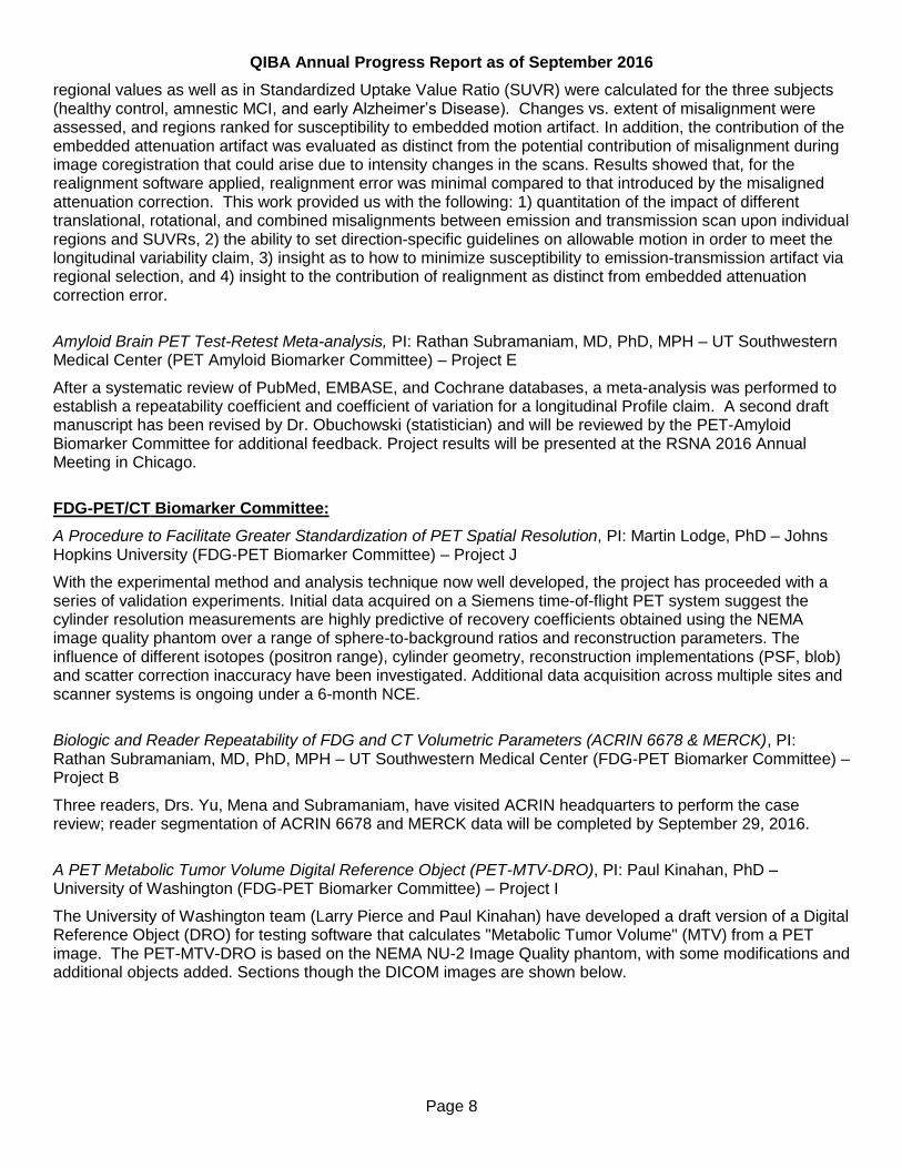
A PET Metabolic Tumor Volume Digital Reference Object (PET-MTV-DRO), PI: Paul Kinahan, PhD – University of Washington (FDG-PET Biomarker Committee) – Project I
The University of Washington team (Larry Pierce and Paul Kinahan) have developed a draft version of a Digital Reference Object (DRO) for testing software that calculates "Metabolic Tumor Volume" (MTV) from a PET image. The PET-MTV-DRO is based on the NEMA NU-2 Image Quality phantom, with some modifications and additional objects added. Sections though the DICOM images are shown below.
QIBA Annual Progress Report as of September 2016
Page 9
Two sections through the PET-MTV-DRO. All objects are three dimensional, and one object as indicated has variable SUVs.
Noise-free and noisy images have been generated, and the PET-MTV-DRO is currently being prepared for distribution within the FDG-PET/CT Biomarker Committee for testing on different software analysis packages. After testing, it will be used as a reference check for the Round-5 groundwork project "Biologic and Reader Repeatability of FDG and CT Volumetric Parameters" (PI: Rathan Subramaniam), Project B.
SPECT Biomarker Committee:
There were two (2) groundwork projects approved for funding during 3Q2016:
1. “Multi-center Phantom Study to Characterize Bias and Precision of Quantitative 123I SPECT”. The objective is to better characterize the optimum acquisition parameters and reconstruction methods for estimating the specific binding ratio (SBR) in 123I ioflupane SPECT with higher precision and reduced bias. Currently, there are insufficient data to make strong recommendations on image acquisition and reconstruction parameters in the Profile. Studies using a physical striatal phantom will launch at two sites that use SPECT/CT systems and reconstruction software from two different vendors (Siemens and GE). The overall aim is to improve differentiation between disease and non-disease groups and to improve sensitivity for assessing changes in longitudinal studies in neurodegenerative disease.
2. “I-123 DAT Scan Digital Reference Object Development”. The goal is to design and construct a prototype brain Digital Reference Object (DRO). This phantom will have properties that are appropriate for testing software used to characterize I-123 ioflupane uptake in the striatum and quantify the striatal specific bind ratio. The deliverables will help characterize design specifications for conformance with the SPECT DAT scan Profile, and to test current commercial and academic I-123 DAT scan analysis software for conformance in a consistent fashion.
US SWS Biomarker Committee:
Analysis of Sources of US SWS Measurement Inter-System Variability, PI: Mark Palmeri, MD, PhD – Duke University (SWS Ultrasound Biomarker Committee) – Projects F1, F2
Experimental data from all of the sites that participated in the Phase II phantom study are being analyzed, and preliminary statistical analyses are quantifying site and system variables. Phantom temperature dependencies have been characterized. Raw SWS data from the clinical study performed at MGH are being analyzed for viscoelastic effects.
Viscoelastic digital phantom datasets have been augmented to include more parameters and have been uploaded to the QIDW US SWS Community. Phase aberration was studied as a confounder effect and found to have a more detrimental effect on SWS measurements by increasing the bias in measured values.
Ongoing groundwork studies underway to support further refinement of the SWS Profile v2.0 are:
a. Continued studies by manufacturers of SWS ultrasound systems using the QIBA-supplied digital phantom will allow the manufacturers to further optimize their software (and hardware, if needed) to reach the performance levels specified in the claims section of the Profile. The manufacturers will then revise their data acquisition procedures as needed for inclusion in Appendix D of the Profile. Initial

QIBA Annual Progress Report as of September 2016
Page 10
Profile-based acquisition testing results will be supplied to manufacturers to help them determine the adequacy of existing acquisition hardware/software to help them with the process of system modification.
b. Self-funded SWS acquisition at the two Profile test sites will be used to assist manufacturers in making any needed system modifications as part of item (a) above. The acquisitions and protocol deviation testing will be used to further refine the Profile and to add information on effects of protocol deviations to the conformance section of the Profile. This type of information is critical to effectively use the Profile in the real world where errors are common.
c. Results from the Round-5 Project “Analysis of Sources of US SWS Measurement Inter-System Variability” headed by Mark Palmeri, coupled with new work at Mayo Clinic (Round 6 project) will be used by manufacturers to decrease measurement bias and variability by correcting for viscoelastic effects. The magnitude of viscoelastic effects will be quantified and correction of SWS values or acquisition at the same frequency will be employed and specified in the Profile.
US Volume Blood Flow Biomarker Committee:
Examination of Flow Phantom as Reference Standard for Validation of Ultrasound Volume Blood Flow Measurement, PI: Oliver Kripfgans, PhD - University of Michigan
This project will collect preliminary data using the prototype physical phantom. Initial tests have been performed at the University of Michigan campus. These data were used before the actual round robin testing to refine the prototype phantom Gammex provided in response to our specifications. A document has been created to specify information required to perform the volume flow computation.
US CEUS Biomarker Committee: Two labs (U Washington, U Texas at Dallas) are preparing the phantom set-up for the initial measurements as described in the QIBA CEUS Biomarker Committee Proposal (currently published on the QIBA Wiki).
Cross-Modality Groundwork Project:
Aggregated Measures of Agreement for QIB Validation: An Open Source Toolkit, PI: Daniel Barboriak, MD – Duke University – Project K The purpose of this project is develop open-source software to calculate aggregated measures of agreement in order to facilitate image analysis algorithm development, comparative analysis of algorithm output, and demonstrate technical compliance. This project will further develop a toolkit that will calculate the following statistics: concordance correlation coefficient, root mean square deviation, total deviation index, Bland-Altman limits of agreement, and Sigma analysis based on estimates of allowable total error. An existing open-source package, QIBA DRO Evaluation Tool (QDET), developed by Hendrik Laue in a previous groundwork project, is being used as a starting point. This package’s source code has been downloaded, and preliminary analysis of it has been conducted. The software requires some Python modules, such as WxPython and Matplotlib, that are not part of the standard Python installation. These additional modules have been obtained and installed. Python has several ways to package and release software as an executable application; these are currently being investigated to determine which to use. We have successfully continued updating the capabilities of the QDET program. The program now accepts text as input, and we have validated that the RMSD, CCC, TDI and Bland-Altman statistics obtained from text images are identical to those obtained from corresponding image data. These statistics have been verified against statistics obtained from the R software package. Scripts demonstrating how to use QDET to tune software parameters and to rank performance of competing algorithms have been completed.

QIBA Annual Progress Report as of September 2016
Page 11
OBJECTIVE 4. DEVELOP AND EMPLOY PHYSICAL AND/OR VIRTUAL (DIGITAL) REFERENCE OBJECTS NEEDED FOR ASSESSMENT OF IMAGING BIOMARKER VARIABILITY AND/OR TO DEMONSTRATE COMPLIANCE WITH QIBA PROFILES.
Selected specifics:
CT Volumetry Biomarker Committee:
Reference Image Set for Quantification Conformance of Algorithmic Lesion Characterization, PI: Ehsan Samei, PhD – Duke University (CT Volumetry Biomarker Committee) – Project C
Development of a two-part study composed of 1) a reference image dataset with synthetic and virtually inserted lesions, and 2) a collection of clinical patient cases with real and virtually inserted lesions has begun. A standard chest CT protocol has been used with the reference image dataset. Clinical cases used in the previous QIBA Group 3A challenge have been identified for use. Virtual lesion insertion techniques previously used in a pilot study have been identified for application in the current framework.
Phantoms for CT Volumetry of Hepatic Metastasis PI: Binsheng Zhao, Columbia University Co-I: Nicholas Petrick, Center for Devices and Radiological Health, FDA Co-I: Qin Li, Center for Devices and Radiological Health, FDA The purpose of this project is to evaluate the performance of low-contrast lesion volumetry in hepatic CT as a function of various imaging acquisition parameters and to determine if the current CT Volumetry Profile generalizes to soft-tissue lesions. An anthropomorphic abdominal phantom with two removable liver inserts was designed. The liver inserts, designed to correspond to arterial and portal-venous phase imaging, contain 19 synthetic lesions with varying diameters (6-40 mm), shapes, and contrasts (10-65 HU). The phantoms were scanned on two commercial CT scanners across a set of imaging protocols (4 slice thicknesses, 3 effective mAs, 2 convolution kernels, 2 pitches). All scans were analyzed using a matched-filter estimator for volume estimation and a subset of portal venous phase scans were analyzed using a semi-automatic segmentation algorithm. The results showed that lesions with lower contrast and size ≤ 10 mm were generally not measurable and were excluded for further analyses. The matched-filter volume estimation approach showed strong linearity in the measurements for lesions ≥20 mm and estimated biases were low (-3% to 3%). The repeatability of the measurements were generally small for higher exposure scanning and > 0.6 mm slice thicknesses with repeatability coefficients (RCs) ranging from about 8% to 18%. RCs increased substantially for the lowest dose, 50 mAs , and 0.6 mm slice thickness (up to 75%). A similar trend was observed for the semi-automatic segmentation algorithm but with increased bias and RC magnitudes. The results show that liver lesion volumetry is strongly dependent on lesion size, contrast, acquisition dose and their interactions. The overall performances were similar for images reconstructed with thicker slices, and clinically used pitches, kernels and doses. Conditions that yielded repeatable measurements were identified and they generally agreed with the QIBA Profile requirements, although the findings do suggest some potential refinements to these guidelines specific to soft-tissue lesions.
PDF-MRI Biomarker Committee:
The DWI-MRI Task Force has completed its literature review and the development and dissemination of a NIST-traceable DWI MRI Phantom (along with associated data analysis software) funded by a NIBIB Round-3 project. The DWI-MRI Profile is expected to be released for public comment by late-2016. Key aspects of the draft Profile have been implemented in collaborative studies with the EORTC / IMI and with a group of São Paulo neuroradiologists leading a multicenter, multivendor clinical trial of DWI in glioblastoma patients to allow initial field testing of Profile recommendations. A manuscript is being drafted on the round-robin study with the DWI phantom.
DWI-DRO Development for ADC Analysis, PI: Dariya Malyarenko, PhD – University of Michigan (PDF-MRI Biomarker Committee / DWI Task Force) – Project G Development of this DWI-DRO is completed and suitable to assess robustness of DWI analysis software packages used for quantitative ADC map generation. Modeled tissue properties span ADC = 0.1 to 3.5x10⁻³
mm2/s and SNR = 0 to 100 (of b=0 image) within a DICOM-compliant DWI-DRO at b-values=0, 500, 800, 2000

QIBA Annual Progress Report as of September 2016
Page 12
s/mm2 (see Objective 3). Design and use of the DRO to test one representative analysis software package (GE FuncTool) is represented in the figure below.
Dynamic Susceptibility Contrast MRI Phantom, PI: Ona Wu, PhD – Harvard University / Massachusetts General Hospital (PDF-MRI Biomarker Committee / DSC-MRI Task Force) – Project H Two phantom prototypes to estimate reproducibility across imaging sites have been designed. A high-level generalized cross-vendor imaging protocol usable on clinical MRIs has been developed and will be utilized to characterize the DSC phantoms and assess repeatability on multiple scanner platforms. Phantom components are being tested for stability and suitable magnetic properties such as susceptibility range and T1. Software tools are also being developed for analyzing data under a 6-month NCE.
fMRI Biomarker Committee:
Three different rounds of fMRI DROs have been developed or are under development. A Round 1 (NIBIB funded Year 2013-14) groundwork project involved generating 20 empirically-derived DROs, which were then downloaded and analyzed at 8 QIBA fMRI Biomarker Committee sites; comparing results from different sites helped develop analysis standards for Profile v1.0. A Round 2 (NIBIB funded Year 2014-15) project generated synthetic DROs to model one of three major forces of variance: head motion, neurovascular uncoupling, or variable task performance. A third (unfunded) round of DRO development is ongoing to generate higher resolution DROs that combine empirically-derived head motion, neurovascular uncoupling, and task performance variability. Examples of Round 1 and 2 DROs are already available via the QIDW, and Round-3 DROs should be available early in 2017 for use in field tests.
FDG-PET/CT Biomarker Committee:
One of the funded groundwork data collection projects funded development of a virtual DRO for PET volumetric measurements. The University of Washington team (Drs. Larry Pierce and Paul Kinahan) have developed a draft version of a DRO for testing software that calculates "Metabolic Tumor Volume" (MTV) from a PET image. The PET-MTV-DRO is based on the NEMA NU-2 Image Quality phantom, with some modifications and additional objects added. Noise-free and noisy images have been prepared, and the PET-MTV-DRO is currently being prepared for distribution within the FDG-PET/CT Biomarker Committee for testing on different software analysis packages. See Objective 3 report above for details. PET-Amyloid Biomarker Committee:
A task force of the committee is working to identify the performance requirements needed from a DRO (or set of DROs) to address an identified gap in methodology needed for image analysis workstation conformance testing. Implementing this methodology is critical to assessing reproducibility and linearity of the software analysis tools. Complicating this exercise is the practical requirement to assess specific brain regions (target

QIBA Annual Progress Report as of September 2016
Page 13
and reference background) based on mapping correlation to either anatomic imaging for the specific subject or to a reference database. The task force is also incorporating results from the 2015-2016 Round-5 project A to inform these requirements.
SPECT Biomarker Committee:
The first working specifications for a “fillable” phantom have now been developed for deployment in multi-center environments. Testing of the prototypes will begin during 4Q2016. This groundwork project is being developed to characterize the linearity of the confidence intervals surrounding precision and bias, which are suspected of increasing with disease severity (because the signal decreases).
A digital reference object (DRO) based on a high resolution human brain MRI image is being developed based on the successful versions developed by the FDG PET/CT and PET-Amyloid groups.
Plans for a solid phantom, with a traceable source of Tellurium-123 as a proxy for Iodine-123, are being considered in collaboration with subject matter experts from the National Institute of Standards and Technology (NIST).
US Volume Blood Flow Biomarker Committee:
Specifications for vessel depth and lumen diameter have been determined. A prototype physical phantom has been created by a participating phantom manufacturer. Pre-testing of this phantom has been performed at the University of Michigan campus. Results were discussed with the committee and a list of changes were established to create a phantom suitable for the proposed round-robin study involving nine research labs.
In addition, efforts are underway to create a uniform software module to process flow image data from any participating manufacturer of ultrasound scanners. Besides the original two manufacturers, three additional manufacturers have expressed interest to join this effort and are currently modifying their systems to share required data for flow estimation processing.
OBJECTIVE 5. DEVELOP PROCEDURES AND PROCESSES FOR HARDWARE AND SOFTWARE MANUFACTURERS TO DEMONSTRATE CONFORMANCE WITH QIBA PROFILES.
Selected specifics:
CT Volumetry Biomarker Committee:
The committee has prepared a checklist of actions for each “actor” to establish Profile conformance and divided into activities related to 1) patient handling activities, 2) scan acquisition and reconstruction, 3) image quality checks, and 4) segmentation and analysis. Procedures for claiming conformance have been completed and are now part of the Profile.
Conformance assessment will be conducted through a challenge to the imaging science community. Each participant is to use their segmentation algorithm to perform volume estimation on lesions with locations that are provided a priori. This will involve performing image-based segmentation on datasets generated using (1) an anthropomorphic phantom with synthetic and virtually inserted nodules, and (2) clinical images containing real lung lesions and virtually inserted lesion models.
CT Lung Density Biomarker Committee:
Committee members are now analyzing results from a second round of testing using a modified COPDGene phantom, named “QIBA-SRM.” This modified version of the COPDGene phantom contains NIST-certified foam standards that fall within the ranges expected for CT densities of lung parenchyma. The committee has developed models for harmonizing CT lung density measures across different scanner manufacturers and models. The correction model derived from the first round of scans achieved consistent performance within 2 Hounsfield Units across four vendors (GE, Siemens, Philips, and Toshiba) and is currently under review for

QIBA Annual Progress Report as of September 2016
Page 14
publication. The second round of phantom scans included a greater number of scanner models and an updated anthropomorphic phantom to test commercially available CT dose reduction using automated exposure control and iterative reconstruction at sites in Iowa and Wisconsin.
PDF-MRI Biomarker Committee:
The conformance section of the DWI Profile has been drafted according to the revised Profile template, and is under review by the DWI Task Force. Analogous to other QIBA Profiles, such as DCE-MRI v1.0, conformance elements include scanner hardware, key personnel and image analysis procedures. The DTI and DSC efforts are fairly early in their Profile development, but conformance will be addressed as claims and protocols become more firm; scope and style will be consistent with the DWI and DCE Profiles.
fMRI Biomarker Committee:
The Round 1-5 groundwork projects on reproducibility and DROs will identify reproducibility benchmarks for motor and language fMRI scanning that can be used as goals for conformance testing by manufacturers of fMRI task and analysis software. Currently there are no standards for such software, nor benchmarks for evaluating the quality of data produced by different tasks.
FDG-PET/CT Biomarker Committee:
Representatives of each of the four vendors of PET/CT scanners systematically reviewed and commented upon their current and future ability to achieve conformance with the FDG-PET/CT Profile as part of the field-testing project. Specific comments and suggestions from the vendors regarding feasibility and practicality were provided to the committee, which will discuss and incorporate appropriate changes, potentially as part of the technically confirmed document draft to be released by 2017.
PET-Amyloid Biomarker Committee:
By better understanding the effect of subject motion (both between PET and anatomic acquisition as well as across multi-timeframe PET emission acquisitions) on SUVR calculation, a deliverable from the 2015-16 Round-5 Project A will inform performance requirements for when head motion needs to be corrected and how it needs to be corrected during the reconstruction on the image acquisition devices. If no correction is performed during the reconstruction, then these same performance requirements can inform image analysis workstation vendors of the amount and type of head motion that is tolerable such that the data still conform to the Profile claim.
SPECT Biomarker Committee:
Hardware:
Groundwork projects are being constructed that will characterize the linearity of the confidence intervals surrounding precision and bias, which are suspected of increasing with disease severity, that is, increasing with decreasing signal-to-noise ratios, as the signal decreases in patients with Parkinson’s disease. This groundwork can be conducted with the fillable and solid phantoms that are being designed.
Software:
In addition to analyzing the phantom and DRO data, it is anticipated the SPECT Biomarker Committee will be able to assemble a test set from patients and matched controls with which all software vendors can assess their analytical processes. There are several potential sources of human data that might be donated. This might become yet another example of the SPECT Biomarker Committee moving rapidly based on trails blazed by other groups, as this project would be constructed to resemble the software comparison exercise pioneered by the CT Volumetry Biomarker Committee.

QIBA Annual Progress Report as of September 2016
Page 15
US SWS Biomarker Committee:
Requirements for demonstrating conformance are under discussion. It is currently standard operating procedure for manufacturers to demonstrate performance of operational modes, such as blood flow imaging, with phantoms. Therefore, it is anticipated that phantom tests of shear wave speed estimation under various operating conditions will be an acceptable test for vendor conformance.
With the completion of SWS Profile v2.0, work has begun on high-level checklists for use by those implementing the Profile. A modified version of these checklists will be used to score site conformance to the Profile by audit of the checklists used during each SWS acquisition. Completion of detailed subchecklists will be possible when manufacturers complete their specific instructions and submit them for inclusion in Appendix D.
System conformance will be monitored using standard QIBA calibrated phantoms. The Mayo clinic has agreed to be in charge of testing/calibrating the phantoms to be distributed for equipment conformance testing. There is considerable interest on the part of CIRS and other phantom producers in supplying QIBA conformance test phantoms for use by sites desiring to be QIBA SWS compliant.
The SWS committee has decided that a modified version of the test procedure used in the Phase II Phantom tests will be used for system conformance testing.
US VBF Biomarker Committee:
The procedures and results of the multisite, multisystem groundwork project will provide a good basis for future testing of manufacturers’ systems.
US CEUS Biomarker Committee: Discussions have been initiated with equipment and software suppliers in order to keep the procedures relevant and implementable. Two of the Task Force teams formed (imaging systems requirements and quantification analysis software) include experts and representatives/engineers from imaging equipment and quantification software industries.
OBJECTIVE 6. COLLECT IMAGES AND ASSOCIATED DATA FOR A QIB DATA WAREHOUSE OR OTHER PUBLIC DATA REPOSITORIES, AND PERFORM ANALYSIS ON THE DATA TO SERVE QIB COMMITTEES AND THE BROADER IMAGING COMMUNITY.
Note that, in general, the data from each of the Committee efforts and funded projects have been, or will be, uploaded to the Quantitative Imaging Data Warehouse (QIDW) identified in the Methodology for Objective 6.
Selected specifics:
CT Modality:
Reference Image Set for Quantification Conformance of Algorithmic Lesion Characterization, PI: Ehsan Samei, PhD – Duke University (CT Volumetry Biomarker Committee) – Project C
The Kyoto Kagaku Lungman Phantom with synthetic nodule image and metadata is presently hosted by the FDA, and annotations files have been prepared for upload to the QIDW. Reference image dataset and associated metadata will be uploaded to the QIDW community upon completion of the groundwork project.
MR Modality:
Quantitating Clinical fMRI Mapping of Language: Center, Spatial Extent, and Relative Strength of Active Areas, PI: James Voyvodic, PhD – Duke University (fMRI Biomarker Committee) – Projects D1, D2
This Round-5 language fMRI project will allow the upload of representative human fMRI data sets (in anonymous form) to the QIDW to support our reproducibility findings. We will include examples of 2 language tasks from different subjects, representing different quantitative levels of reproducibility metrics.

QIBA Annual Progress Report as of September 2016
Page 16
DWI-DRO Development for ADC Analyses, PI: Dariya Malyarenko, PhD –University of Michigan (PDF-MRI Biomarker Committee) – Project G
Initial compatibility of the DWI-DRO was performed by University of Michigan investigators in application of select DICOM readers and analysis packages (GE FuncTool, Philips, QIBA_Phan, Osirix, IDL, MatLab). Currently, the DWI-DRO and User Manual reside on the QIDW, though greater use and visibility may be achieved via links on the QIBA-Wiki.
Multi-site/vendor scans of QIBA DWI phantom and DWI Phantom Analysis Software (QIBA_Phan), supported in prior year Groundwork Projects, also reside on the QIBA QIDW and serve as a resources for the broader quantitative imaging community.
PET-Amyloid Biomarker Committee:
The committee is discussing ideas on how to obtain a set of well-analyzed, anonymized subject data (normal and abnormal) as well as a DRO series of 30 or more image volumes that can then be uploaded to the QIDW and made available for image analysis workstation conformance testing as per the methodology described in the Image Analysis Conformance Section of the Profile.
Ultrasound Modality:
Analysis of Sources of US SWS Measurement Inter-System Variability, PI: Mark Palmeri, MD, PhD – Duke University (SWS Ultrasound Biomarker Committee) – Projects F1, F2
Deidentified clinical images have been uploaded to the QIDW. The procedure for accomplishing the uploads is complex and work to simplify the process is underway. All imaging and point analysis data relating to SWS estimation in the upcoming conformance phantom calibrations and clinical Profile test acquisitions will be uploaded to the QIDW after appropriate deidentification and after full IRB approval.
US VBF Biomarker Committee:
The planned uniform software module to process flow image data will allow any participating manufacturer to compare the standard analysis with software developed for and used in their ultrasound systems. Hopefully, raw data will also be made available to test their analysis.
OBJECTIVE 7. PROVIDE SUPPORT FOR THE QIB COLLABORATION PROGRAM STAFF (OUTSIDE ORGANIZATION STAKEHOLDERS AND QIB COALITION MEMBERS), PROJECT MANAGEMENT, MEETINGS, TRAVEL, AND CONFERENCE CALLS. Support for all the above committee work, funded project management meetings, conference calls and travel continues to be administered and provided by the RSNA/QIBA staff, QIBA Chair / Vice-chair, and Scientific Liaisons. Various Biomarker Committees have worked to extend supported QIBA efforts beyond that available from RSNA and NIBIB by providing support for implementation of QIBA procedures in separately supported clinical trials. Substantial support for scheduling of the online meetings and one face-to-face meeting of the Ultrasound Volume Blood Flow Biomarker Committee and its Task Forces is now provided by the American Institute of Ultrasound in Medicine (AIUM).

QIBA Annual Progress Report as of September 2016
Page 17
B. COMBINED LIST OF GROUNDWORK PROJECTS APPROVED BY STEERING COMMITTEE ACROSS THE OBJECTIVES FOR ROUND-5 (2015-2016) FUNDING.
Project Number
Biomarker Cmte
Project Title Investigator
A PET Amyloid
Analyses to Support Amyloid Imaging Profile Development
Dawn Matthews, ADM Diagnostics, LLC
B FDG-PET Biologic and Reader Repeatability of FDG and CT Volumetric Parameters (ACRIN 6678 & MERCK)
Rathan Subramaniam, MD, PhD, MPH, Johns Hopkins University
C CT Volumetry
Reference Image Set for Quantification Conformance of Algorithmic Lesion Characterization
Ehsan Samei, PhD, Duke University
D1, D2 fMRI Quantitating Clinical fMRI Mapping of Language: Center, Spatial Extent, and Relative Strength of Active Areas
James Voyvodic, PhD, Duke University
Jay J. Pillai, MD, Johns Hopkins Univ
E PET Amyloid
Amyloid Brain PET Test-Retest Meta-analysis
Rathan Subramaniam, MD, PhD, MPH, Johns Hopkins University
F1, F2 SWS US Analysis of Sources of US SWS Measurement Inter-System Variability
Mark Palmeri, MD, PhD, Duke Univ
Shigao Chen, PhD, Mayo Clinic
G PDF-MRI (DWI TF)
DWI-DRO Development for ADC Analysis
Dariya Malyarenko, PhD, University of Michigan
H PDF-MRI Dynamic Susceptibility Contrast MRI Phantom
Ona Wu, PhD, Harvard/Mass General Hospital
I FDG-PET A PET Metabolic Tumor Volume Digital Reference Object (PET-MTV-DRO)
Paul Kinahan, PhD, University of Washington
J FDG-PET A Procedure to Facilitate Greater Standardization of PET Spatial Resolution
Martin Lodge, PhD, Johns Hopkins University
K Cross Modality
Aggregated Measures of Agreement for QIB Validation: An Open Source Toolkit
Daniel Barboriak, MD, Duke University
L Lung Density
Investigation of Methods of Volume Correction for Lung Density CT
Sean Fain, PhD, University of Wisconsin
C. GENERAL PROGRESS ON ACTIVITIES BEYOND THE FUNDED PROJECTS
Additional updates from the committees are as follows.
CT Volumetry Biomarker Committee
The CT Profile describing measurements of change in tumor volume for advanced disease (the “CTV” Profile) has been updated to align with the Metrology Work Group definitions, and extensive additional public comments have been incorporated. The claims have been updated to reflect the committee consensus on appropriate thresholds of performance based on the state of the current methods and technology, as demonstrated in prior groundwork projects.

QIBA Annual Progress Report as of September 2016
Page 18
A protocol for a field test of the QIBA Profile is being re-evaluated to incorporate considerations of funding and logistics. The intent is to first carry out a feasibility assessment to determine whether sites can take the QIBA Profile and execute its requirements. The goal after this will be to collect data on the precision of clinical lesion measurements so the precision can be combined with prior information on bias to provide a more complete description of measurement variability.
A CT liver phantom has been designed and fabricated. Scans have been performed on the phantom, and analysis of the resulting scan data has been carried out.
The algorithm challenge group (Task Force 3A) has prepared a manuscript for publication and secured permission to publish. A publication from the phantom data project is in the revision process after submission. The group dedicated to this project is now organizing to provide support for the upcoming “field test” of the CT volumetry biomarker profile.
The Lung Nodule Assessment in CT Screening Task Force has been working to ensure that the small nodule claims are consistent with the established claims of the advanced disease CT Volumetry Profile. Published results and unpublished data from members of the group have been used to inform development of claim details. The committee has collaborated with manufacturer representatives to obtain technical parameter guidance for individual scanner models for quantitative applications. The Small Nodule Profile was released for public comment in 3Q2016; comments are currently being addressed.
CT Lung Density Biomarker Committee
A draft Profile and claim development for a lung density protocol are in progress, based on critical evaluation of the literature. The acquisition and reconstruction specification of CT images has been completed and is being evaluated by a working group of vendor scientists who are developing compliance procedures using a modified COPDGene Phantom named “QIBA-SRM”. The image analysis section of the Profile is nearly complete.
The group has developed recommendations for pulmonary quantitative CT (qCT) protocols to be used on multiple vendor scanners using automatic exposure control (AEC) and iterative reconstruction (IR). These protocols should guide efforts to lower CT dose for ongoing and future clinical research qCT studies of the lungs focused on measures of parenchymal density. These protocols may be used in conjunction with low-dose screening for lung cancer, and have been already implemented in the ongoing COPDGene study.
A Task Force of CT vendor scientists has been formed to develop a compliance checklist and to suggest changes to the acquisition and reconstruction parameter specifications in efforts to mitigate measurement bias. The Task Force has organized a project that involves scanning the same QIBA-SRM Phantom using three radiation doses on two models of each vendor’s CT scanners. This work has led to a submitted manuscript under review that reports a method to reduce bias in CT density measurements across scanner makes and models. The CT Vendor Task Force has also completed a second round of scanning and is planning to complete data analysis and report results by November 2016.
The Lung Density Biomarker Committee has completed a meta-analysis of the CT lung density repeatability literature, thus finalizing their measurement repeatability claim for assessing emphysema progression. The meta-analysis and Round-5 (Project L) groundwork will be the basis for a submission of a manuscript for publication in the peer-reviewed literature.
PDF-MRI Biomarker Committee
The majority of PDF-MRI activities are conducted within its respective Task Forces. Task Force groups exist within the PDF-MRI Biomarker Committee for DCE-, DWI-, DTI-, and DSC-MRI. Single Task Force updates are presented to the full PDF-MRI Biomarker Committee on a rotating basis. Discussion with leaders in the

QIBA Annual Progress Report as of September 2016
Page 19
Arterial Spin Labeling (ASL) perfusion field are also ongoing to develop a plan for forming an ASL Task Force in collaboration the European Society of Radiology’s European Imaging Biomarker Alliance (EIBALL).
The DCE-MRI Task Force is presently focused on defining systematic literature review procedures, and their application for select organ/tumor sites, to support the DCE-MRI v2.0 Profile claims. Even within a given organ site, the literature often reveals a broad range of key acquisition parameters (e.g., temporal sampling rate) with incomplete description of methodology. Relatively uniform multi-site/-platform DCE-MRI methodology was achieved in the ACRIN 6701 test/retest prostate clinical trial used to field-test the DCE v1.0 Profile. Moreover, an automated software analysis package was developed and applied to data acquired on the QIBA DCE-MRI Phantom for site qualification in the ACRIN 6701 study. The analysis software and user manual have been uploaded to the QIDW, along with example data from scanners from three major MR system vendors and the associated reports produced by the software. In addition, an open-source software package to facilitate comparison of parametric images generated by different DCE-MRI analysis packages when utilizing the DRO created as part of a previously funded groundwork project is also available on the QIDW. This software is capable of importing 2D and 3D DICOM images, or binary data formats, as well as imaging formats such as TIFF and PNG. It generates difference and ratio maps (exportable as PNG), scatter diagrams and box-plots, and ANOVA statistics to more easily compare analysis packages. These resources are available on the QIBA QIDW for future clinical trials and the broader quantitative imaging community.
The DWI TF is recasting its Profile language into the revised QIBA Profile template. In 2014-15 the DWI Task Force successfully completed scans of its physical “QIBA DWI phantom”, where all datasets were analyzed using the “QIBA_Phan” software developed as a prior groundwork project and available on the QIDW. In 2016, DWI Task Force members also completed development of a DWI DRO. Analogous to the DWI Task Force, the DSC Task Force is currently developing a physical phantom, protocol, and corresponding analysis software supported under QIBA groundwork project contracts. DTI Task Force leaders have previously developed a novel isotropic plus anisotropic physical diffusion phantom. This phantom, allows ground-truth measurements of key DTI metrics: mean diffusivity (MD), fractional anisotropy (FA), radial diffusivity (RD) and axial diffusivity (AD). Task Force members will be scanning this phantom in the coming year to evaluate intra-/inter-platform variance of these metrics.
fMRI Biomarker Committee
The fMRI Biomarker Committee continues work on v1.0 of its Profile for pre-surgical mapping of eloquent brain tissue. Refinements to the clinical claims and context were made, particularly the acquisition guidelines, as well as accompanying appendices with detailed performance specifications. Likewise, members are in the process of completing Section 3, Profile Details, specific to the mapping of motor cortex. To inform conformance procedures, members are conducting groundwork studies focused on software analysis specifications.
The fMRI Bias Task Force meets bi-weekly to focus on the issue of bias in the fMRI measurand. This activity informs the Profile claims definition and guides development of methodological sequences for image analysis that best achieve the claims.
fMRI-DRO testing was completed at 8 sites, all analyzing the same bilateral hand motion and language mapping DROs but with each site employing its own standard fMRI processing and analysis workflow. The activation map results accompanied by data analysis forms describing workflow were collected from each site. For the period through September 2015, generation and testing of advanced DROs for head motion in fMRI were performed. These include DROs from various combinations of selected empirical and synthetic datasets wherein amplitude and spatial distribution of task-related fMRI signals and associated fMRI noise were controlled. By fully specifying “ground truth” in this way, subsequent post-processing and display methods can be tested for the ability to accurately recover the original signal distributions and to quantify any inaccuracies that might be present. These DROs, containing realistic task signal and noise variability (including motion, performance, and neurovascular uncoupling sources of variance), have been uploaded to the QIDW. These can be used for conformance testing and comparison of fMRI analysis and correction methods for coping with the variance of the BOLD signal in the primary motor cortex as a function of presence or absence of NVU.

QIBA Annual Progress Report as of September 2016
Page 20
Members of the fMRI Biomarker Committee contribute to the DICOM Working Group 16 fMRI task force. The proposed DICOM work item will build on recent quantitative imaging support added to the standard, with new elements created as necessary to represent fMRI acquisition, activation maps, and task paradigms. The functional requirements incorporated by WG-16 fMRI were drawn from work done in the QIBA fMRI Biomarker Committee.
FDG-PET/CT Biomarker Committee
An FDG-PET/CT Profile Field Test was performed to thoroughly examine the feasibility and practicality of the QIBA Profile in the specific context of three academic PET imaging centers using imaging equipment from three different manufacturers. The field test resulted in Profile revisions and identified impractical or ambiguous specifications, and initiated discussions regarding how to formalize this QIBA profile revision workflow procedure. A reduced list of 36 specifications was extracted from the QIBA FDG-PET Profile that can serve as a simple checklist for imaging sites to determine their QIBA conformance. This distillation from the much longer set of QIBA Profile specifications was based on feasibility and relevance to quantification. Profile specifications relevant to PET/CT devices were sent to the four manufacturers to evaluate their own system’s compatibility with the Profile.
A follow-up field test was initiated, in which the site-relevant Profile specifications were evaluated for feasibility at 11 sites (academic and non-academic). Additional incompatibilities between Profile specifications and imaging site practices were identified. From this, follow-up discussions amongst committee members were held to determine whether individual specifications should be modified to be more compatible with sites’ practices, or whether sites should be encouraged to modify their practices.
A similar field test specifically aimed at PET/CT manufacturers and software developers was initiated. Each of the four manufacturers did an extensive review of the Profile for potential for conformance. Robust and informative discussions revealed that different manufacturers sometimes used different approaches to meet the same quantitative ends. Potential modifications to the Profile are currently under discussion as the committee decides whether these alternate approaches still meet the claim requirements. This is the nearly final step towards the Technically Confirmed version of the Profile projected for release in 2017.
A QIBA/NIBIB-funded DRO extension project increased the functionality of the previous PET-CT DRO in efforts to test for (a) PET/CT display alignment, (b) SUVpeak calculation, and (c) Region of Interest (ROI) fidelity.
In related efforts, a draft manuscript titled “Summary of the QIBA Profile for FDG-PET/CT as an Imaging Biomarker Measuring Response to Cancer Therapy” is in its final editing phases prior to submission.
PET-Amyloid Biomarker Committee
The committee has made substantive progress in drafting a Profile whereby 18F-Amyloid tracers may be used in clinical trials for assessing subjects with cognitive impairment. The Profile is based on a longitudinal claim and uses the change in SUVR as the measurand. There continues to be excellent participation on the teleconferences by members of all 18F-PET radiotracer manufacturers and equipment manufacturers as well as key subject matter experts from clinical, academic, imaging core lab, medical physics and systems engineering backgrounds. The draft Profile is currently undergoing the final phase of internal committee review. The claim language has been revised based on results from the literature meta-analysis (2015-16 Round-5 project E). The Profile is slated for distribution for public review during 4Q2016.
Looking beyond the first version of the Profile release, the committee is discussing items such as Profile feasibility testing, the incorporation of PET-MR imaging, an improved DRO for image analysis conformance testing, the effect that different PET reconstruction algorithms have on the SUVR measurand, and the development of a Profile for imaging tau proteins.
QIBA Annual Progress Report as of September 2016
Page 21
SPECT Biomarker Committee
Driven by mounting evidence that quantification adds value to SPECT, and the accelerating production of commercial software packages to capture that value, a large group of physicians and scientists, from industry, academia, and governmental agencies, have now begun developing a SPECT Profile. International enthusiasm for participation has been particularly strong from Japan and several European Union states. The initial focus is on dopamine transporter brain scans to assist in the evaluation of patients with Parkinsonian symptoms. Use cases that are expected to follow include quantification of trans-arterially administered macro aggregated albumin for radiotherapy planning in patients with liver metastases, and targeted theranostics for selecting candidates for treatment with companion therapeutics in the emerging fields of antibody and small molecule drug conjugates. Four Task Forces, led by deep subject matter experts, hold regular meetings to define the first Profile and claim language. Although this is a relatively new initiative, solid progress has been made toward the completion of the first Profile (v1.0), which is expected to be released to public comment by the end of 4Q2016.
US SWS Biomarker Committee
The initial goal of the SWS US Biomarker Committee was to develop a QIBA Profile for a single biomarker: ultrasound shear wave speed (SWS) as a measure of liver stiffness which correlates with the degree of liver fibrosis/cirrhosis present. Major efforts center on completing groundwork studies and publishing results, continuing to understand and account for sources of bias in SWS estimation with ultrasound imaging systems, continuing to determine sources of variance in these estimates, minimizing those contributions, and finalizing the draft Protocol and Profile documents.
Three recent areas of groundwork efforts were: (1) validation of simulations and phantoms mimicking elastic and viscoelastic properties of liver, (2) comparison of SWS measurements in uniform liver-mimicking phantoms using ultrasound imaging systems, the established US non-imaging system, and, initially, MR elastography, and (3) determination of sources of measurement variability in shear-wave elasticity techniques. Physical and digital phantoms are part of these efforts, as well as of the conformance procedures.
Simulation data sets have been developed and posted on the QIDW for use by research groups and manufacturers. The goal is to find approaches that allow different ultrasound systems to achieve the same SWS results from data generated using appropriate simulated visco-elastic materials. Simulated data representing elastic (lossless) and viscoelastic (tissue-mimicking) media have been released for download by interested parties, and several manufacturers have begun to look at the materials to determine if it is technically and economically feasible to analyze test data using their proprietary software. A comprehensive comparison of simulation results obtained with two common commercial finite element modeling software has been performed, and the corresponding code to process the data are available on GitHub. If this plan is successful, use of digital reference objects (DROs) to analyze ways to achieve better agreement in SWS values will become a real possibility.
Data have been acquired to investigate sources of variance from comorbidity, biological variability, and measurement methods that might affect shear wave speed estimate correlation with fibrosis. Studies were based on a literature analysis of 1,548 publications from which 102 SWS papers included a study of one or more confounding factors. A further analysis of the potential for steatosis and/or inflammation to affect the correlation of SWS with liver fibrosis was performed using results obtained for 242 subjects.
A standardized plan for archiving clinical and phantom data into the QIDW is being devised and will be included as an appendix to the QIBA Profile.
A first draft of the QIBA ultrasound Profile “SWS Estimation of Liver Fibrosis” has been created. Distribution within and approval by the Biomarker Committee is pending while the Profile document is converted to the new document template provided by the Process Committee and system-dependent methods descriptions are provided by the participating manufacturers. A standardized SWS data collection report form has been developed for inclusion in the Profile appendices.

QIBA Annual Progress Report as of September 2016
Page 22
Although the initial goal of the US SWS Biomarker Committee was to develop SWS estimates specifically for diagnosis and grading of liver fibrosis, recent discussions within the QIBA Process Committee and the Steering Committee have changed the focus of QIBA Biomarker work toward provision of quantitative biomarkers with known accuracy and precision, rather than focusing on use in a specific diagnostic task. One important reason for this change is that confirmation of biomarker use for clinical diagnosis requires extensive clinical testing, such as a large multicenter study, which is beyond the current resources of QIBA. Researchers and/or interested clinical organizations could take the QIBA Profile and test it in a multicenter trial to confirm its performance for a specific clinical or research task (such as fibrosis grading). The SWS Biomarker Committee and the Profile drafting task force are now in the process of determining how the SWS Profile will be modified to reflect more general use of the biomarker while still retaining information specific to use for liver fibrosis. This may require additions to the standard template geared towards providing clinical and research use information specific for liver fibrosis, for example, tables of proposed SWS thresholds for assigninig fibrosis grades to livers based on SWS results
Additionally, collaboration between the SWS Biomarker Committee and the MR Elastography Biomarker Committee to assure consistent SWS results between the two modalities is underway. One significant step in that direction is the decision by the US SWS committee to use MRE values for SWS and loss moduli as the “gold standard” against which US SWS results will be judged. For this reason, it was decided that the phantoms used to determine US system performance will be calibrated by MRE and the MRE values will be converted to expected US SWS values for each phantom by correcting for the difference in shear wave frequency. Alternatively, the MRE tests will be conducted at a shear wave frequency close to that used by typical ultrasound systems so direct comparisons of US and MR SWS estimates is possible. All these procedures will appear as part of the acceptance and conformance process outlined in the SWS Profile. This work will be conducted while the work on technical verification of the Profile is underway.
Various Biomarker Committees
Several biomarker committees have worked to extend support for QIBA beyond that available from RSNA and NIBIB. A major contribution has been provision of support for implementation of QIBA procedures in separately supported clinical trials. Martialing the internal resources of committed companies, hospital systems, and researchers has been an integral part of the QIBA process. Substantial support for scheduling of the online meetings and one face-to-face meeting of the Ultrasound Volume Blood Flow Biomarker Committee and its Task Forces is now provided by the American Institute of Ultrasound in Medicine (AIUM). However, all this growing effort in development and validation of QIBs would not continue at anywhere near its present rate, without the core support of RSNA and NIBIB.