Embed Size (px)
Citation preview
Inhibition of Pluripotency Networks by the Rb Tumor Suppressor Restricts Reprogramming and Tumorigenesis
Michael S. Kareta1,2,3,6, Laura L. Gorges1,2, Sana Hafeez3,6, Bérénice A. Benayoun2,7, Samuele Marro3,6, Anne-Flore Zmoos1,2, Matthew J. Cecchini8, Damek Spacek1,2, Luis F.Z. Batista4,5,6, Megan O’Brien1,2, Yi-Han Ng3,6, Cheen Euong Ang3,6, Dedeepya Vaka1,2, Steven E. Artandi4,5,6, Frederick A. Dick8, Anne Brunet2,7, Julien Sage1,2,9,*, and Marius Wernig3,6,9,*
1Department of Pediatrics, Stanford University, Stanford, CA 94305, USA
2Department of Genetics, Stanford University, Stanford, CA 94305, USA
3Department of Pathology, Stanford University, Stanford, CA 94305, USA
4Department of Medicine, Stanford University, Stanford, CA 94305, USA
5Department of Biochemistry, Stanford University, Stanford, CA 94305, USA
6Stanford Institute for Stem Cell Biology and Regenerative Medicine, Stanford University, Stanford, CA 94305, USA
7Paul F. Glenn Laboratories for the Biology of Aging, Stanford University, Stanford, CA 94305, USA
8London Regional Cancer Program, Children’s Research Institute, Western University, London, ON N6A 4L6, Canada
SUMMARY
Mutations in the retinoblastoma tumor suppressor gene Rb are involved in many forms of human
cancer. In this study, we investigated the early consequences of inactivating Rb in the context of
cellular reprogramming. We found that Rb inactivation promotes the reprogramming of
© 2015 Elsevier Inc.*Correspondence: [email protected] (J.S.), [email protected] (M.W.).9Co-senior author
ACCESSION NUMBERSThe data for RNA and ChIP sequencing have been deposited in NCBI’s Gene Expression Omnibus and are accessible through GEO Series accession number GSE40594 (http://www.ncbi.nlm.nih.gov/geo/query/acc.cgi?acc=GSE40594).
SUPPLEMENTAL INFORMATIONSupplemental Information for this article includes six figures, five tables, and Supplemental Experimental Procedures and can be found with this article online at http://dx.doi.org/10.1016/j.stem.2014.10.019.
AUTHOR CONTRIBUTIONSM.S.K. was responsible for the experiment design, execution, data analysis, and manuscript preparation. L.G. and L.F.Z.B. aided in the initial reprogramming assays. S.H. and M.O. aided with the efficiency assays. B.A.B. performed the broad domain analysis. S.M. performed Southern blotting. D.S. performed histone ChIP-qPCR. Y.H.N. made the chimeric mice. C.E.A. performed high-throughput imaging for cell seeding. A.-F.Z. bred the mice. M.J.C. generated the ΔG mouse line and isolated MEFs. D.V. advised on sequence analysis. S.A. advised on the human reprogramming. F.A.D. advised on Rb domain mutagenesis and manuscript review. A.B. advised on the broad domain analysis. J.S. and M.W. were equally responsible for supervision of research, data interpretation, and manuscript preparation.
HHS Public AccessAuthor manuscriptCell Stem Cell. Author manuscript; available in PMC 2015 April 08.
Published in final edited form as:Cell Stem Cell. 2015 January 8; 16(1): 39–50. doi:10.1016/j.stem.2014.10.019.
Author M
anuscriptA
uthor Manuscript
Author M
anuscriptA
uthor Manuscript
differentiated cells to a pluripotent state. Unexpectedly, this effect is cell cycle independent, and
instead reflects direct binding of Rb to pluripotency genes, including Sox2 and Oct4, which leads
to a repressed chromatin state. More broadly, this regulation of pluripotency networks and Sox2 in
particular is critical for the initiation of tumors upon loss of Rb in mice. These studies therefore
identify Rb as a global transcriptional repressor of pluripotency networks, providing a molecular
basis for previous reports about its involvement in cell fate pliability, and implicate misregulation
of pluripotency factors such as Sox2 in tumorigenesis related to loss of Rb function.
INTRODUCTION
The retinoblastoma gene product Rb is a potent suppressor of the tumorigenic process and is
genetically or functionally inactivated in most, if not all, human cancers by direct mutation
or alterations in upstream pathway members (Burkhart and Sage, 2008). Loss of Rb function
has been directly implicated in initiation of cancers, including retinoblastoma, osteosarcoma,
and small cell lung carcinoma in patients, as well as pituitary and thyroid tumors in mice
(Chinnam and Goodrich, 2011). Rb controls multiple cellular functions, including
proliferation, survival, differentiation, metabolism, and genomic stability, but how loss of
Rb initiates cancer is still not completely understood (reviewed in Chinnam and Goodrich,
2011; Manning and Dyson, 2012; Nicolay and Dyson, 2013; Viatour and Sage, 2011).
Induced pluripotent stem cells (iPSCs) and embryonic stem cells (ESCs) share some
similarities to cancer cells, including the capacity to bypass senescence and form tumors
upon transplantation (Goding et al., 2014). Accordingly, some genes often associated with
cancer, such as Myc (Nakagawa et al., 2008; Wernig et al., 2008), p53 (Krizhanovsky and
Lowe, 2009), and telomerase (Batista et al., 2011; Park et al., 2008), have been implicated in
cellular reprogramming. Additionally, two reprogramming factors, Oct4 and Sox2, can be
oncogenic in some cellular contexts (Hochedlinger et al., 2005; Lu et al., 2010; Rudin et al.,
2012; Sarkar and Hochedlinger, 2013).
Given previous observations that Rb could be involved in cellular dedifferentiation (Calo et
al., 2010; McEvoy et al., 2011) and given the similarities between reprogramming and some
aspects of tumorigenesis, we set out to identify mechanisms by which Rb suppresses
dedifferentiation using iPSC reprogramming as a cellular system. We found that Rb
regulates the reprogramming of fibroblasts to iPSCs. Surprisingly, this phenomenon is not
due to changes in the cell cycle. Instead, Rb globally represses pluripotency networks in
somatic cells, thereby rendering cells more amenable to reprogramming, including in the
absence of exogenous Sox2. Importantly, pituitary hyperplasia driven by Rb loss in mice is
blocked by Sox2 deletion, expanding the functional interaction between Rb and Sox2 to
cancer.
RESULTS
Rb Loss Promotes Reprogramming to iPSCs
To test whether Rb regulates the reprogramming process, we infected wild-type and Rb−/−
mouse embryonic fibroblasts (MEFs) with lentiviruses harboring the traditional four
Kareta et al. Page 2
Cell Stem Cell. Author manuscript; available in PMC 2015 April 08.
Author M
anuscriptA
uthor Manuscript
Author M
anuscriptA
uthor Manuscript
reprogramming factors (4F), Oct4, Sox2, Klf4, and c-Myc. Based on the appearance of
alkaline phosphatase (AP)+ colonies, we saw more efficient reprogramming in Rb mutant
MEFs than in WT cells, suggesting that loss of Rb facilitates reprogramming (Figures 1A
and 1B). A similar increase in AP+ colonies was observed with human fibroblasts
expressing shRNAs to knock down human RB (shRB) (Figure 1A and Figure S1A available
online). Because AP is not a specific marker of pluripotency, we then used Oct4-NeoR
knockin MEFs in combination with Rb knockdown (Figure S1B) and counted neomycin-
resistant colonies. To avoid errors due to reseeding of daughter colonies, we performed a 96-
well assay and quantified the wells that contained iPSC colonies rather than the number of
colonies themselves. In this stringent assay, the reprogramming efficiency was increased in
Rb-deficient cells to a similar degree to that reported in cells with a loss of p53 (Figures 1B
and 1C) (Krizhanovsky and Lowe, 2009). Notably, Rb loss decreased the plating efficiency
of MEFs (Figure S1C), illustrating our underestimation of the efficiency of reprogramming
upon Rb loss. Reprogramming was increased by multiple hairpins against Rb (Figure 1B)
upon acute deletion of Rb in Rblox/lox conditional knockout MEFs (cKO MEFs) after
adenoviral Cre (Ad-Cre) delivery (Figure 1D) and after treatment with 4-hydroxytamoxifen
(4OHT) in Rosa26CreER cKO MEFs (Figure S1D and S1E). Conversely, overexpression of
Rb reduced the reprogramming efficiency (Figure 1E). MEFs deficient for p107 or p130,
two factors closely related to Rb, showed no changes in their reprogramming efficiency
(Figure 1F). Triple knockout MEFs for the entire Rb family (TKOs) did not reprogram to
iPSCs, presumably due to an unidentified stress response that leads to increased apoptosis
during reprogramming (Figure S1F).
To further determine the kinetics of reprogramming in the absence of Rb, we reprogrammed
Oct4-GFP knockin MEFs after Rb knockdown. Colonies re-expressing endogenous Oct4
(GFP+) appeared significantly more quickly in cells with low Rb levels (Figure 1G).
Utilizing SSEA1 expression as an early marker of reprogramming (Brambrink et al., 2008),
we observed that Rb loss increases reprogramming as early as days 4 and 6 (Figure 1H,
Figure S1G). To test the time requirement for the exogenous factors to trigger full
reprogramming, we used Oct4-GFP;M2rtTA MEFs infected with a doxycycline (Dox)-
inducible 4F lentiviral construct. After a total of 15 days and varying the length of Dox
treatment, the number of GFP+ iPSC colonies was counted. Decreased Rb levels led to a
significant reduction in the time requirement for 4F expression to achieve reprogramming
(Figure 1I). Together, these data demonstrate that Rb loss accelerates and enhances iPSC
generation from fibroblasts, indicating that Rb normally restricts this process.
Rb Loss Does Not Accelerate the Cell Cycle during Reprogramming
One explanation for the increased reprogramming observed in Rb-deficient cells is a faster
cell cycle, as in p53 mutant cells (Hanna et al., 2009). We determined the doubling time of
control and Rb knockdown MEFs in the first 10 days of the process to avoid the
confounding effects of increased proliferation due to enhanced reprogramming in Rb mutant
cells. Notably, no significant change in the growth rate of the 4F-infected MEFs was
observed upon Rb knockdown (Figure 2A), as was previously observed in Rb-deficient
MEFs (Dannenberg et al., 2000; Sage et al., 2000) (see below). Similarly, there was no
change in the S phase fraction in the shRb-infected MEFs, although, at day 6 there was a
Kareta et al. Page 3
Cell Stem Cell. Author manuscript; available in PMC 2015 April 08.
Author M
anuscriptA
uthor Manuscript
Author M
anuscriptA
uthor Manuscript
trend to a longer G2 and shorter G1 (Figures 2B and 2C, Figure S2A), possibly a
consequence of increased reprogramming because a shorter G1 correlates with the
acquisition of pluripotency (Conklin and Sage, 2009). Annexin V staining did not show a
significant difference in apoptosis (Figure 2D, Figure S2B). To quantify proliferation
specifically in reprogramming cells, we focused on SSEA1+ cells and used
Carboxyfluorescein diacetate succinimidyl ester (CFSE) pulse-chase staining to determine
the number of cell divisions after the induction of reprogramming (Koche et al., 2011).
Using an automated analysis, we confirmed that SSEA1+ cells are proliferative (Figure S2C
and S2D) and, importantly, we found no change in the number of doublings between control
and Rb knockdown populations (Figure 2E). Thus, the enhanced reprogramming efficiency
and kinetics observed in the absence of Rb cannot be simply explained by changes in cell
cycle profiles.
Rb Binds to and Represses Pluripotency Genes
Next we sought to explore how the absence of the Rb protein might render MEFs more
amenable to reprogramming. Because Rb is a transcriptional coregulator, we determined the
consequence of Rb loss on gene expression. To this end, cKO MEFs were infected with Ad-
Cre or Ad-GFP (Figure S3A and S3B) and infected by the 4F or an empty vector; 3 days of
selection after 4F infection, the RNA was sequenced. We used k-means clustering to divide
genes that change upon 4F expression, and then we further subdivided each cluster for
changes by the further loss of Rb (Figure 3A, Figures S3C and S3D, Table S1 available
online). In all these clusters, loss of Rb led to changes in the expression of cell cycle genes.
However, both positive (e.g., Mcm3, Pola1, and Cdk1) and negative (e.g., p107, Cdkn2c,
and Brca1) cell cycle regulators were upregulated, which may explain why loss of Rb
function does not promote cell division under these conditions (Figures 3A, Figure S3C and
S3D). We used gene set enrichment analysis (GSEA) to determine the consequences of Rb
deletion on MEF or iPSC gene sets. We found a significant enrichment for the iPSC-specific
gene signature in cells without Rb compared to Rb wild-type cells upon 4F expression
(Figure 3B and Table S1). Interestingly, using RT-qPCR, we observed a small but
significant increase in Sox2 and Oct4 expression in Rb-deficient MEFs even without 4F
infection (Figure 3C). As expected, the absolute expression of pluripotency genes was still
much lower (<0.5%) than in ESCs (Figure 3D). Loss of the entire Rb family did not further
increase the expression of these pluripotency genes, in contrast to cell cycle genes (Figures
1D and 3C). Slight OCT4 and SOX2 induction was also observed in human cells in response
to RB knockdown (Figure 3E). Flow cytometry of Sox2-GFP MEFs after acute knockdown
of Rb showed that the Sox2 induction observed in bulk analysis is caused by slight induction
in many cells and not strong induction in a few cells (Figure 3F). Thus, loss of Rb leads to a
global derepression of components of the pluripotency transcriptional program, leading to
only a subtle gene induction in the majority of cells.
To determine if the repressive action of Rb on pluripotency genes might be direct, we
performed Rb chromatin immunoprecipitation (ChIP) assays under the same conditions as
the RNA sequencing profiling. Interrogating a few selected genomic regions by ChIP-qPCR,
we observed that Rb binds to the promoter regions of Sox2 and Oct4 as well as classical cell
cycle genes (Figure 4A). Global analysis of Rb ChIP sequencing data showed that the
Kareta et al. Page 4
Cell Stem Cell. Author manuscript; available in PMC 2015 April 08.
Author M
anuscriptA
uthor Manuscript
Author M
anuscriptA
uthor Manuscript
majority of the genes identified as being bound by Rb are associated with the cell cycle and
most are upregulated upon loss of Rb, consistent with its established role as a transcriptional
repressor (Figure S4A and S4B). Genome-wide data also confirmed robust Rb binding at the
Sox2 and Oct4 loci (Figure 4B, Table S2). Moreover, we found Rb binding to the regulatory
regions of a number of genes involved in reprogramming and pluripotency, an observation
supported by the analysis of human Rb ChIP sequencing data sets (Chicas et al., 2010)
(Figure S4C). Remarkably, at the Sox2 locus, Rb was primarily bound to the downstream
enhancer (SRR2; Figure 4B and Figure S4D), the active enhancer in pluripotent lineages
(Tomioka et al., 2002).
We next sought to determine if binding of Rb to pluripotency genes might play a role in a
developmental context. When we examined expression data of both wild-type and Rb family
mutant mouse ESCs before and after differentiation into embryoid bodies (EBs), we found a
significant defect in the silencing of pluripotency genes (listed in Figure S4C) upon
differentiation (Figures 4C and 4D). Additionally, Rb progressively binds to both Oct4 and
Sox2 during EB differentiation, correlating with their silencing (Figure 4E). Together, these
experiments indicate that the binding of Rb to pluripotency factors, including Sox2 and
Oct4, may both contribute to their silencing in differentiated cells and restrict their re-
expression during the reprogramming process.
Because Rb typically requires a binding partner to be recruited to DNA, we sought to
investigate how Rb is localized to pluripotency genes. Previous studies have suggested that
E2F proteins bind to Sox2 and Oct4 (Julian et al., 2013; Xu et al., 2007). To explore if direct
interactions between Rb and E2Fs at these loci were involved in gene repression, we used
MEFs in which mutations at either the E2F-binding pocket (ΔG allele) or the LxCxE-
binding cleft (ΔL allele) were knocked in to the endogenous Rb locus (Figure 4F) (Isaac et
al., 2006; Talluri et al., 2010). We found a specific increase in the reprogramming efficiency
of DG mutants (Figure 4G), correlating with increased levels of Oct4 and Sox2 (Figure 4H),
similar to Rb deletion (Figure 3C). Thus, interactions through the E2F-binding pocket are
required for the silencing of pluripotency genes by Rb.
Rb Contributes to the Maintenance of the Chromatin State at Pluripotency Genes in Fibroblasts
Based on these observations and the known interactions between Rb and regulators of
chromatin structure (Longworth and Dyson, 2010), we determined whether loss of Rb
function affects the chromatin at pluripotency genes. At both Oct4 and Sox2, we observed a
significant increase in the acetylation of histone H3 (H3Ac) in the proximal promoter
regions upon loss of Rb (Figure S5A). The extent of this increase was similar to what we
observed for Rb canonical targets such as Ccna2 (Figure S5A). However, H3K4me3 was not
increased at the Oct4 or Sox2 promoter, unlike at Ccna2, which may reflect the difference
between enhanced gene activation of an already expressed E2F target upon Rb loss and the
slight increase of basal transcription that we observed for Oct4 and Sox2 (Figures 3C and
3D). Additionally, we found a significant loss of H3K27me3 at Oct4 (Figure S5A).
Using ChIP sequencing to gain a genome-wide perspective on histone changes upon Rb loss,
we detected significantly increased levels of both H3Ac and H4K4me3 and a decrease of
Kareta et al. Page 5
Cell Stem Cell. Author manuscript; available in PMC 2015 April 08.
Author M
anuscriptA
uthor Manuscript
Author M
anuscriptA
uthor Manuscript
H3K27me3 at multiple pluripotency genes, including Oct4, Nanog, and Sox2 (Figure 5A,
Figure S5B). Increased activating marks including H3Ac and H3K4me3 were highly
correlated (Figure S5C) and found predominantly at cell cycle genes and known Rb targets
such as p107 (Figure 5A, Table S3); however, increased activating marks were also
observed at both Sox2 and Nanog (Figure 5A). Notably, the region with increased H3Ac and
H3K4me3 downstream of Sox2 is not the SRR2 enhancer, but rather a region that is bound
by both p300 and CTCF and can interact with the SRR2 enhancer (Dixon et al., 2012).
GSEA indicated that an iPSC-specific gene set is significantly enriched among the genes
that gain H3Ac and H3K4me3 to a similar degree as a gene set specific for Rb loss (Figure
5B).
A recent study described the existence of broad H3K4me3 domains that mark genes
essential for cell identity and function (Benayoun et al., 2014). We observed a change in
broad domain organization upon loss of Rb (Figure 5C, Figures S5D and S5E). Strikingly,
an unbiased analysis of genes that showed increased H3K4me3 domain breadth showed that
these genes were enriched to be direct transcriptional targets of the core pluripotency
network (e.g., OCT4 and NANOG; Table S3). The same enrichment for transcriptional
targets of OCT4, SOX2, KLF4, NANOG, and TCF3 was observed in Rb-deficient cells
when we analyzed increases in H3K4me3 breadth specifically in the set of H3K4me3-
marked genes (Figure 5D). Peaks broadened by loss of Rb were domains more likely to be in
the top 5% broadest domains in mouse ESCs (Figures 5E and 5F). Thus, the absence of Rb
in MEFs generates a chromatin landscape that more closely resembles that of an ESC. Thus,
Rb loss not only leads to derepression of some core pluripotency genes, but also specifically
alters the chromatin of downstream targets for these pluripotency factors, possibly
facilitating the transcription of a global pluripotency program and its implementation.
The silencing of chromatin at key pluripotency genes by Rb is consistent with the known
interactions of Rb with key repressors (Longworth and Dyson, 2010). To interrogate the
recruitment of such factors to Oct4 and Sox2, we performed ChIP experiments in Rb wild-
type and mutant MEFs. These experiments showed a strong correlation between cofactor
recruitment and the histone mark changes observed. As mentioned above, Rb loss decreased
H3K27me3 at a region spanning the distal enhancers to the promoter of Oct4 (Figure 5A),
which correlated with a decrease of EZH2 recruitment to this region (Figure 5G). The
increased H3Ac at the proximal promoters of Oct4 and Sox2 observed in Rb mutant cells by
ChIP-qPCR (Figure S5A) corresponded to a significant decrease of HDAC recruitment to
these loci (Figures 5G and 5H). It should be noted, however, that the change in H3Ac at the
proximal promoters of Oct4 and Sox2 was not identified by ChIP-eq, presumably due to the
low levels of this mark escaping detection when analyzed on a genomic scale. Finally,
because p27 was reported to repress Sox2 (Li et al., 2012), we determined p27 localization at
the DNA in wild-type, p130−/−; p107−/−, and Rb family TKO MEFs. We found p27 binding
to the downstream enhancer of Sox2 even in the absence of p130 and p107, but not in the
TKO MEFs, indicating that the recruitment of p27 at the Sox2 locus can also be mediated by
Rb (Figure S5F).
Kareta et al. Page 6
Cell Stem Cell. Author manuscript; available in PMC 2015 April 08.
Author M
anuscriptA
uthor Manuscript
Author M
anuscriptA
uthor Manuscript
Rb Functionally Regulates Oct4 and Nanog, and Its Loss Can Functionally Replace Sox2 in Reprogramming
While the levels of derepression of pluripotency genes observed in Rb-deficient fibroblasts
are small, we wondered if they may be sufficient to bypass the need for exogenous
reprogramming factors. We infected Oct4-NeoR MEFs with three-factor combinations of the
reprogramming factors and a hairpin against Rb. With just Oct4, Klf4, and c-Myc, several
neomycin-resistant clones were readily obtained in Rb knockdown cells, but not in control
cells. Fewer clones were observed with Oct4 and Klf4 alone in the Rb knockdown than with
c-Myc or all four factors (Figure S6A and S6B). No iPSC colonies were observed when
either Klf4 or Oct4 were omitted. These RbKD OKM clones, in which the absence of Sox2
transgene was verified (Figure S6C and S6D), had a similar morphology to that of iPSCs
reprogrammed with the four factors and expressed endogenous Oct4, Sox2, Nanog, and AP
activity (Figure 6A). Additionally the Rb-deficient iPSCs reprogrammed without Sox2
formed teratomas with all three germ layers (Figure S6E) and contributed to a host embryo
forming chimeric mice (Figure S6F). Thus, acute loss of Rb can functionally replace Sox2
during iPSC reprogramming.
To determine whether Rb could control the expression of pluripotency factors beyond Sox2,
using another system, we tested if Rb loss might prime Oct4 and Nanog for activation. We
employed the CRISPR-on approach (Cheng et al., 2013) to directly target the transcriptional
activator VP64 to the Oct4 and Nanog promoters and observed a significantly enhanced
expression from these genes when Rb was absent (Figure 6B). Therefore, while Rb loss did
not compensate for exogenous Oct4 to generate fully reprogrammed iPSCs, Rb regulates the
Oct4 locus physiologically.
Sox2 Is Required for the Development of Tumors Initiated by Loss of Rb
Based on emerging evidence that Sox2 plays oncogenic roles (Lu et al., 2010; Rudin et al.,
2012), we determined if the derepression of Sox2 observed in Rb-deficient cells might be
relevant to cancer initiation. To test this idea, we crossed Rosa26CreER Rblox/lox mice to
Sox2lox/lox mice. As expected (Jacks et al., 1992), we observed a significantly enlarged
pituitary at 9 weeks after tamoxifen injection in Rosa26CreER Rblox/lox mice. Strikingly,
concomitant loss of Sox2 completely blocked this cancer phenotype (Figures 6C and 6D),
independent of gender (Figure S6G and S6H). Accordingly, loss of Sox2 completely
inhibited cell proliferation in the pituitary of Rb-mutant mice (Figures 6E and 6F, Figure
S6I). These experiments indicate that Sox2 is critical for cancer initiation upon loss of Rb in
the mouse pituitary gland.
DISCUSSION
The retinoblastoma tumor suppressor Rb is commonly inactivated in human cancer. While
Rb has been extensively studied, its mechanisms of action remain incompletely understood.
Here, using iPSC reprogramming as a cellular system, we identified a new facet of Rb tumor
suppressor function in which Rb restricts the expression of pluripotency genes and their
downstream targets, thereby preventing cellular dedifferentiation and transformation.
Kareta et al. Page 7
Cell Stem Cell. Author manuscript; available in PMC 2015 April 08.
Author M
anuscriptA
uthor Manuscript
Author M
anuscriptA
uthor Manuscript
The degree of Rb involvement in direct regulation of pluripotency genes and downstream
targets is particularly noteworthy. Although on the individual gene level, the transcriptional
changes in pluripotency networks observed after Rb inactivation are subtle, a sizeable
number of genes are affected. The genome-wide summation of these individually small
effects results in a global transcriptionally permissive state and could explain the increased
cell pliability of Rb-deficient cells that was observed before (Calo et al., 2010; McEvoy et
al., 2011). In fact, that low levels of pluripotency genes, particularly Sox2, may have strong
phenotypic effects is not surprising because iPSCs often have few genomic integrations of
Sox2 proviruses (Meissner et al., 2007; Wernig et al., 2007), and low Sox2 is in fact
favorable for high-quality iPSCs (Carey et al., 2011). Our findings also provide a molecular
basis for related cellular phenotypes directly or indirectly involving Rb and previously
assumed to be caused by changes in the cell cycle (Banito et al., 2009; Li et al., 2010; Liu et
al., 2009). Notably, the effect on reprogramming was specific for Rb and was not observed
for the closely related genes p107 and p130. Much like Rb, both of these factors restrict cell
cycle progression (Dannenberg et al., 2000; Sage et al., 2000), yet are rarely mutated in
cancer. Therefore, the unique ability of Rb among its family members to suppress
dedifferentiation may provide an explanation for its crucial role in suppressing cancer
formation while its cell cycle checkpoint role is compensated more potently by the usually
intact p107 and p130 proteins.
We found that the regulation of target genes by Rb correlates with the recruitment of histone
modifying complexes and the subsequent regulation of histone marks. While loss of Rb
resulted in increases of H3Ac and H3K4me3 at cell cycle genes, pluripotency genes,
including Oct4, Sox2, and Nanog, mostly gained H3Ac and lost H3K27me3. This effect of
Rb loss on pluripotency genes therefore more likely represents a derepression resulting in
increased basal transcription rather than full activation, which is associated with robust
promoter H3K4me3. While this derepression is general to pluripotency genes, we know that
not all genes respond to the same degree because the increase of total expression of Sox2 is
markedly higher than that of Oct4, and Rb loss could only replace Sox2 and not Oct4 to
derive iPSCs. This may be explained by additional regulatory mechanisms restricting Oct4
expression, including high levels of DNA methylation at the Oct4 locus (Mikkelsen et al.,
2008), or a lack of LIN28+ regulation of Oct4 mRNA in MEFs (Qiu et al., 2010).
Nevertheless, loss of Rb enhances the expression of both Oct4 and Nanog when forced by a
strong transcriptional activator, supporting a direct role for Rb in the regulation of these
genes. Additional evidence linking cell cycle regulators to pluripotency genes is starting to
emerge. For instance, the Cdk2 inhibitor p27 also controls the pluripotency of ESCs by
direct inhibition of Sox2 (Li et al., 2012) (see also below). This regulation is mediated by the
same enhancer that we found to be bound and regulated by Rb, suggesting possible
interactions between p27 and Rb on the chromatin. Future studies may elucidate whether
such interactions exist genome-wide or are specific for this enhancer.
While we observed changes in the chromatin landscape of MEFs after Rb loss, we expected
that perturbation of the LxCxE binding cleft would have a reprogramming phenotype similar
to loss of Rb. Mice harboring these knockin mutations have shown defects in chromatin
modifications in other settings (Bourgo et al., 2011; Talluri et al., 2010). However, our data
Kareta et al. Page 8
Cell Stem Cell. Author manuscript; available in PMC 2015 April 08.
Author M
anuscriptA
uthor Manuscript
Author M
anuscriptA
uthor Manuscript
show that interactions through this surface are not required for the silencing of Sox2 and
Oct4. This is consistent with reports that in differentiated tissues, Rb-dependent silencing is
normal in LxCxE mutant mice (Andrusiak et al., 2013; Talluri et al., 2010). Furthermore, a
dominant-negative DP1 (which blocks E2F transactivation) can rescue increased acetylation
observed upon Rb loss (Andrusiak et al., 2013). This presents two models whereby Rb might
regulate heterochromatin. First, HDACs interact with Rb in LxCxE-in-dependent ways, so
the longstanding model of direct HDAC recruitment is correct but at least partly LxCxE
independent. Alternatively, Rb repression of E2Fs allows HDAC recruitment through other
neighboring contacts. In this way, the effects on chromatin are genetically Rb dependent but
the biochemical mechanism is distinct from simply being recruited by Rb. Regardless of
which paradigm explains our data, E2F appears to be central to the mechanism of Rb
regulation of Sox2 and Oct4.
Regulation of Sox2 at its upstream enhancer by the Rb family/ E2F and at its downstream
enhancer by p21 has already been shown to direct the fate choices of neural stem cells
(Julian et al., 2013; Marqués-Torrejón et al., 2013). We observe that Rb represses Sox2 at its
downstream, pluripotency-specific enhancer as cells differentiate. Moreover, the loss of this
repression in the pituitary induces hyperplasia in a Sox2-dependent manner. It is noteworthy
that Sox2 is expressed in the retina (Li et al., 2012), in which loss of Rb is tumor initiating.
Amplification of Sox2 has also been shown to be a driver mutation in human small cell lung
cancer, a cancer initiated by loss of RB and p53 (Rudin et al., 2012). Although inactivation
of Rb, by either mutation or repression, is common to nearly all cancers, it is only initiating
in a few cell types. This difference may rely in part on the initiating cell’s ability to
reactivate Sox2 transcriptional networks. The extent by which Rb regulation of Sox2 limits
tumors in humans is still unknown, but our observations support the idea that cellular
reprogramming assays can be used as a cellular system to probe the molecular mechanisms
of cancer initiation.
EXPERIMENTAL PROCEDURES
Cell Lines
The cell lines used were as follows: Rblox/lox (cKO), Oct4-GFP+/−, Oct4-Neo+/−, B6;129S-
Sox2tm2Hoch/J (Sox2-GFP) and M2rtTA+/−;Oct4-GFP+/− (Brambrink et al., 2008; Burkhart
et al., 2010a; Meissner et al., 2007; Wernig et al., 2007). Rb1ΔL/ΔL MEFs have been reported
previously (Isaac et al., 2006). Rb1ΔG/ΔG MEFs express an R461E, K542E mutant and its
properties have previously been reported (Cecchini and Dick, 2011; Cecchini et al., 2014).
Reprogramming and Differentiation Assays
For the 96-well efficiency assays, shRNA-infected cells were selected by 3 days of growth
in puromycin, infected with the 4F, and plated at a density of 100 cells per well into 96-well
plates containing 1,000 g-irradiated feeder cells per well. Wells with AP+ cells and proper
morphology (after 15 days) were reported to remove potential contribution of the seeding of
daughter colonies. EBs were formed when we plated trypsinized ESCs in nonadherent plates
at a density of 5.5 × 10⁵ cells/ml in DMEM with 10% serum. After 3 days they were plated
Kareta et al. Page 9
Cell Stem Cell. Author manuscript; available in PMC 2015 April 08.
Author M
anuscriptA
uthor Manuscript
Author M
anuscriptA
uthor Manuscript
onto gelatin-coated tissue culture plates. After 5 days they were maintained in serum free
conditions.
CFSE Analysis
CFSE staining was performed using the CellTrace™ Violet Cell Proliferation Kit
(Invitrogen #C34557). Cells were trypsinized and washed with PBS, and then CFSE was
added to 5 µM. They were incubated in the dark at 37°C for 20 min and then the reaction
was quenched with media for 5 min. The cells were then washed, cultured, and later
analyzed by FACS.
RNA and ChIP Sequencing
ChIP was performed as previously described (Burkhart et al., 2010b). Antibodies are listed
in Table S5. Sequencing libraries were generated using the NEBNext DNA Sample Prep
Master Mix Set (New England Biolabs) and purified using Agencourt Ampure XP beads
(Beckman Coulter). Library quality was assessed on an Agilent 2100 Bioanalyzer and
sequenced to generate single-end 50 bp reads using the Illumina HiSeq platform. We
analyzed ChIP data by mapping the reads using Bowtie2 and identified peaks using MACS2
for the histone ChIP sequencing and CisGenome for the Rb ChIP. For expressional
profiling, purified RNA was processed into cDNA with the Ovation RNA-Seq System V2
(NuGEN) and sonicated using a Covaris S2 Sonicator. RNA sequencing data were analyzed
with the Tuxedo suite. H3K4me3 breadth remodeling was performed as previously
described (Benayoun et al., 2014). See Supplemental Experimental Procedures for additional
information.
Pituitary Sectioning and Staining
To isolate the pituitary, we fixed the whole head overnight in Bouin’s Fixative, removed it,
fixed it overnight in 4% paraformaldehyde, and then dehydrated it in 70% ethanol. Antigen
retrieval on the sections was performed using the Trilogy solution (Cell Marque) for 15 min
in a pressure cooker. The sections were then washed in PBS + Tween20 (PBST) and fixed
for 1 hr in PBST with 10% normal horse serum (NHS). They were incubated with Ki67
antibody (Table S5) overnight at 4°C in PBST with 5% NHS. After being washed in PBS,
they were incubated with the secondary antibody in PBS with 5% NHS. Sections were then
washed with PBS, stained with DAPI, and mounted. We identified cells by using
CellProfiler (http://www.cellprofiler.org) to count nuclei on the DAPI channel, while Ki67
was scored manually in a blinded manner.
Supplementary Material
Refer to Web version on PubMed Central for supplementary material.
ACKNOWLEDGMENTS
We would like to thank Gary Mantalas, Ben Passarelli, and Steven Quake for sequencing; Helen Blau for critical reading of the manuscript; Moritz Mall for the CRISPR vectors; and Jamie Imam for assistance with teratoma assays. This work was supported by PHS Grant #T32 CA09151, awarded by the NCI; DHHS (M.S.K.); the Spectrum Child Health Child Health Research Institute (M.S.K.); CIHR MD/PhD (M.J.C.); the Lucile Packard Foundation for Children’s Health (J.S.); and the CIRM (CIRM RB1-01385 -J.S.). F.A.D. is the Wolfe Senior
Kareta et al. Page 10
Cell Stem Cell. Author manuscript; available in PMC 2015 April 08.
Author M
anuscriptA
uthor Manuscript
Author M
anuscriptA
uthor Manuscript
Fellow in Tumor Suppressor Genes at Western University, M.W. is a New York Stem Cell Foundation-Robertson Investigator and a Tashia and John Morgridge Faculty Scholar, and J.S. is the Harriet and Mary Zelencik Scientist in Children’s Cancer and Blood Diseases.
REFERENCES
Andrusiak MG, Vandenbosch R, Dick FA, Park DS, Slack RS. LXCXE-independent chromatin remodeling by Rb/E2f mediates neuronal quiescence. Cell Cycle. 2013; 12:1416–1423. [PubMed: 23574720]
Banito A, Rashid ST, Acosta JC, Li S, Pereira CF, Geti I, Pinho S, Silva JC, Azuara V, Walsh M, et al. Senescence impairs successful reprogramming to pluripotent stem cells. Genes Dev. 2009; 23:2134–2139. [PubMed: 19696146]
Batista LF, Pech MF, Zhong FL, Nguyen HN, Xie KT, Zaug AJ, Crary SM, Choi J, Sebastiano V, Cherry A, et al. Telomere shortening and loss of self-renewal in dyskeratosis congenita induced pluripotent stem cells. Nature. 2011; 474:399–402. [PubMed: 21602826]
Benayoun BA, Pollina EA, Ucar D, Mahmoudi S, Karra K, Wong ED, Devarajan K, Daugherty AC, Kundaje AB, Mancini E, et al. H3K4me3 breadth is linked to cell identity and transcriptional consistency. Cell. 2014; 158:673–688. [PubMed: 25083876]
Bourgo RJ, Thangavel C, Ertel A, Bergseid J, McClendon AK, Wilkens L, Witkiewicz AK, Wang JY, Knudsen ES. RB restricts DNA damage-initiated tumorigenesis through an LXCXE-dependent mechanism of transcriptional control. Mol. Cell. 2011; 43:663–672. [PubMed: 21855804]
Brambrink T, Foreman R, Welstead GG, Lengner CJ, Wernig M, Suh H, Jaenisch R. Sequential expression of pluripotency markers during direct reprogramming of mouse somatic cells. Cell Stem Cell. 2008; 2:151–159. [PubMed: 18371436]
Burkhart DL, Sage J. Cellular mechanisms of tumour suppression by the retinoblastoma gene. Nat. Rev. Cancer. 2008; 8:671–682. [PubMed: 18650841]
Burkhart DL, Ngai LK, Roake CM, Viatour P, Thangavel C, Ho VM, Knudsen ES, Sage J. Regulation of RB transcription in vivo by RB family members. Mol. Cell. Biol. 2010a; 30:1729–1745. [PubMed: 20100864]
Burkhart DL, Wirt SE, Zmoos AF, Kareta MS, Sage J. Tandem E2F binding sites in the promoter of the p107 cell cycle regulator control p107 expression and its cellular functions. PLoS Genet. 2010b; 6:e1001003. [PubMed: 20585628]
Calo E, Quintero-Estades JA, Danielian PS, Nedelcu S, Berman SD, Lees JA. Rb regulates fate choice and lineage commitment in vivo. Nature. 2010; 466:1110–1114. [PubMed: 20686481]
Carey BW, Markoulaki S, Hanna JH, Faddah DA, Buganim Y, Kim J, Ganz K, Steine EJ, Cassady JP, Creyghton MP, et al. Reprogramming factor stoichiometry influences the epigenetic state and biological properties of induced pluripotent stem cells. Cell Stem Cell. 2011; 9:588–598. [PubMed: 22136932]
Cecchini MJ, Dick FA. The biochemical basis of CDK phosphorylation-independent regulation of E2F1 by the retinoblastoma protein. Biochem. J. 2011; 434:297–308. [PubMed: 21143199]
Cecchini MJ, Thwaites MJ, Talluri S, MacDonald JI, Passos DT, Chong JL, Cantalupo P, Stafford PM, Saenz-Robles MT, Francis SM, et al. A retinoblastoma allele that is mutated at its common E2F interaction site inhibits cell proliferation in gene-targeted mice. Mol. Cell Biol. 2014; 34:2029–2045. [PubMed: 24662053]
Cheng AW, Wang H, Yang H, Shi L, Katz Y, Theunissen TW, Rangarajan S, Shivalila CS, Dadon DB, Jaenisch R. Multiplexed activation of endogenous genes by CRISPR-on, an RNA-guided transcriptional activator system. Cell Res. 2013; 23:1163–1171. [PubMed: 23979020]
Chicas A, Wang X, Zhang C, McCurrach M, Zhao Z, Mert O, Dickins RA, Narita M, Zhang M, Lowe SW. Dissecting the unique role of the retinoblastoma tumor suppressor during cellular senescence. Cancer Cell. 2010; 17:376–387. [PubMed: 20385362]
Chinnam M, Goodrich DW. RB1, development, and cancer. Curr. Top. Dev. Biol. 2011; 94:129–169. [PubMed: 21295686]
Conklin JF, Sage J. Keeping an eye on retinoblastoma control of human embryonic stem cells. J. Cell. Biochem. 2009; 108:1023–1030. [PubMed: 19760644]
Kareta et al. Page 11
Cell Stem Cell. Author manuscript; available in PMC 2015 April 08.
Author M
anuscriptA
uthor Manuscript
Author M
anuscriptA
uthor Manuscript
Dannenberg JH, van Rossum A, Schuijff L, te Riele H. Ablation of the retinoblastoma gene family deregulates G(1) control causing immortalization and increased cell turnover under growth-restricting conditions. Genes Dev. 2000; 14:3051–3064. [PubMed: 11114893]
Dixon JR, Selvaraj S, Yue F, Kim A, Li Y, Shen Y, Hu M, Liu JS, Ren B. Topological domains in mammalian genomes identified by analysis of chromatin interactions. Nature. 2012; 485:376–380. [PubMed: 22495300]
Goding CR, Pei D, Lu X. Cancer: pathological nuclear reprogramming? Nat. Rev. Cancer. 2014; 14:568–573. [PubMed: 25030952]
Hanna J, Saha K, Pando B, van Zon J, Lengner CJ, Creyghton MP, van Oudenaarden A, Jaenisch R. Direct cell reprogramming is a stochastic process amenable to acceleration. Nature. 2009; 462:595–601. [PubMed: 19898493]
Hochedlinger K, Yamada Y, Beard C, Jaenisch R. Ectopic expression of Oct-4 blocks progenitor-cell differentiation and causes dysplasia in epithelial tissues. Cell. 2005; 121:465–477. [PubMed: 15882627]
Isaac CE, Francis SM, Martens AL, Julian LM, Seifried LA, Erdmann N, Binné UK, Harrington L, Sicinski P, Bérubé NG, et al. The retinoblastoma protein regulates pericentric heterochromatin. Mol. Cell. Biol. 2006; 26:3659–3671. [PubMed: 16612004]
Jacks T, Fazeli A, Schmitt EM, Bronson RT, Goodell MA, Weinberg RA. Effects of an Rb mutation in the mouse. Nature. 1992; 359:295–300. [PubMed: 1406933]
Julian LM, Vandenbosch R, Pakenham CA, Andrusiak MG, Nguyen AP, McClellan KA, Svoboda DS, Lagace DC, Park DS, Leone G, et al. Opposing regulation of Sox2 by cell-cycle effectors E2f3a and E2f3b in neural stem cells. Cell Stem Cell. 2013; 12:440–452. [PubMed: 23499385]
Koche RP, Smith ZD, Adli M, Gu H, Ku M, Gnirke A, Bernstein BE, Meissner A. Reprogramming factor expression initiates widespread targeted chromatin remodeling. Cell Stem Cell. 2011; 8:96–105. [PubMed: 21211784]
Krizhanovsky V, Lowe SW. Stem cells: The promises and perils of p53. Nature. 2009; 460:1085–1086. [PubMed: 19713919]
Li F, He Z, Shen J, Huang Q, Li W, Liu X, He Y, Wolf F, Li CY. Apoptotic caspases regulate induction of iPSCs from human fibroblasts. Cell Stem Cell. 2010; 7:508–520. [PubMed: 20887956]
Li H, Collado M, Villasante A, Matheu A, Lynch CJ, Cañamero M, Rizzoti K, Carneiro C, Martínez G, Vidal A, et al. p27(Kip1) directly represses Sox2 during embryonic stem cell differentiation. Cell Stem Cell. 2012; 11:845–852. [PubMed: 23217425]
Liu Y, Clem B, Zuba-Surma EK, El-Naggar S, Telang S, Jenson AB, Wang Y, Shao H, Ratajczak MZ, Chesney J, Dean DC. Mouse fibroblasts lacking RB1 function form spheres and undergo reprogramming to a cancer stem cell phenotype. Cell Stem Cell. 2009; 4:336–347. [PubMed: 19341623]
Longworth MS, Dyson NJ. pRb, a local chromatin organizer with global possibilities. Chromosoma. 2010; 119:1–11. [PubMed: 19714354]
Lu Y, Futtner C, Rock JR, Xu X, Whitworth W, Hogan BL, Onaitis MW. Evidence that SOX2 overexpression is oncogenic in the lung. PLoS ONE. 2010; 5:e11022. [PubMed: 20548776]
Manning AL, Dyson NJ. RB: mitotic implications of a tumour suppressor. Nat. Rev. Cancer. 2012; 12:220–226. [PubMed: 22318235]
Marqués-Torrejón MA, Porlan E, Banito A, Gómez-Ibarlucea E, Lopez-Contreras AJ, Ferná ndez-Capetillo O, Vidal A, Gil J, Torres J, Fariñas I. Cyclin-dependent kinase inhibitor p21 controls adult neural stem cell expansion by regulating Sox2 gene expression. Cell Stem Cell. 2013; 12:88–100. [PubMed: 23260487]
McEvoy J, Flores-Otero J, Zhang J, Nemeth K, Brennan R, Bradley C, Krafcik F, Rodriguez-Galindo C, Wilson M, Xiong S, et al. Coexpression of normally incompatible developmental pathways in retinoblastoma genesis. Cancer Cell. 2011; 20:260–275. [PubMed: 21840489]
Meissner A, Wernig M, Jaenisch R. Direct reprogramming of genetically unmodified fibroblasts into pluripotent stem cells. Nat. Biotechnol. 2007; 25:1177–1181. [PubMed: 17724450]
Kareta et al. Page 12
Cell Stem Cell. Author manuscript; available in PMC 2015 April 08.
Author M
anuscriptA
uthor Manuscript
Author M
anuscriptA
uthor Manuscript
Mikkelsen TS, Hanna J, Zhang X, Ku M, Wernig M, Schorderet P, Bernstein BE, Jaenisch R, Lander ES, Meissner A. Dissecting direct reprogramming through integrative genomic analysis. Nature. 2008; 454:49–55. [PubMed: 18509334]
Nakagawa M, Koyanagi M, Tanabe K, Takahashi K, Ichisaka T, Aoi T, Okita K, Mochiduki Y, Takizawa N, Yamanaka S. Generation of induced pluripotent stem cells without Myc from mouse and human fibroblasts. Nat. Biotechnol. 2008; 26:101–106. [PubMed: 18059259]
Nicolay BN, Dyson NJ. The multiple connections between pRB and cell metabolism. Curr. Opin. Cell Biol. 2013; 25:735–740. [PubMed: 23916769]
Park IH, Zhao R, West JA, Yabuuchi A, Huo H, Ince TA, Lerou PH, Lensch MW, Daley GQ. Reprogramming of human somatic cells to pluripotency with defined factors. Nature. 2008; 451:141–146. [PubMed: 18157115]
Qiu C, Ma Y, Wang J, Peng S, Huang Y. Lin28-mediated post-transcriptional regulation of Oct4 expression in human embryonic stem cells. Nucleic Acids Res. 2010; 38:1240–1248. [PubMed: 19966271]
Rudin CM, Durinck S, Stawiski EW, Poirier JT, Modrusan Z, Shames DS, Bergbower EA, Guan Y, Shin J, Guillory J, et al. Comprehensive genomic analysis identifies SOX2 as a frequently amplified gene in small-cell lung cancer. Nat. Genet. 2012; 44:1111–1116. [PubMed: 22941189]
Sage J, Mulligan GJ, Attardi LD, Miller A, Chen S, Williams B, Theodorou E, Jacks T. Targeted disruption of the three Rb-related genes leads to loss of G(1) control and immortalization. Genes Dev. 2000; 14:3037–3050. [PubMed: 11114892]
Sarkar A, Hochedlinger K. The sox family of transcription factors: versatile regulators of stem and progenitor cell fate. Cell Stem Cell. 2013; 12:15–30. [PubMed: 23290134]
Talluri S, Isaac CE, Ahmad M, Henley SA, Francis SM, Martens AL, Bremner R, Dick FA. A G1 checkpoint mediated by the retinoblastoma protein that is dispensable in terminal differentiation but essential for senescence. Mol. Cell. Biol. 2010; 30:948–960. [PubMed: 20008551]
Tomioka M, Nishimoto M, Miyagi S, Katayanagi T, Fukui N, Niwa H, Muramatsu M, Okuda A. Identification of Sox-2 regulatory region which is under the control of Oct-3/4-Sox-2 complex. Nucleic Acids Res. 2002; 30:3202–3213. [PubMed: 12136102]
Viatour P, Sage J. Newly identified aspects oftumor suppression by RB. Dis. Model. Mech. 2011; 4:581–585. [PubMed: 21878458]
Wernig M, Meissner A, Foreman R, Brambrink T, Ku M, Hochedlinger K, Bernstein BE, Jaenisch R. In vitro reprogramming of fibroblasts into a pluripotent ES-cell-like state. Nature. 2007; 448:318–324. [PubMed: 17554336]
Wernig M, Meissner A, Cassady JP, Jaenisch R. c-Myc is dispensable for direct reprogramming of mouse fibroblasts. Cell Stem Cell. 2008; 2:10–12. [PubMed: 18371415]
Xu X, Bieda M, Jin VX, Rabinovich A, Oberley MJ, Green R, Farnham PJ. A comprehensive ChIP-chip analysis of E2F1, E2F4, and E2F6 in normal and tumor cells reveals interchangeable roles of E2F family members. Genome Res. 2007; 17:1550–1561. [PubMed: 17908821]
Kareta et al. Page 13
Cell Stem Cell. Author manuscript; available in PMC 2015 April 08.
Author M
anuscriptA
uthor Manuscript
Author M
anuscriptA
uthor Manuscript
Highlights
• The Rb tumor suppressor inhibits reprogramming of fibroblasts to iPSCs
• The effect of Rb on reprogramming is independent of cell-cycle regulation
• Rb promotes assembly of repressive chromatin at pluripotency network genes
• Deletion of Sox2 prevents cancer initiation upon loss of Rb in mice
Kareta et al. Page 14
Cell Stem Cell. Author manuscript; available in PMC 2015 April 08.
Author M
anuscriptA
uthor Manuscript
Author M
anuscriptA
uthor Manuscript
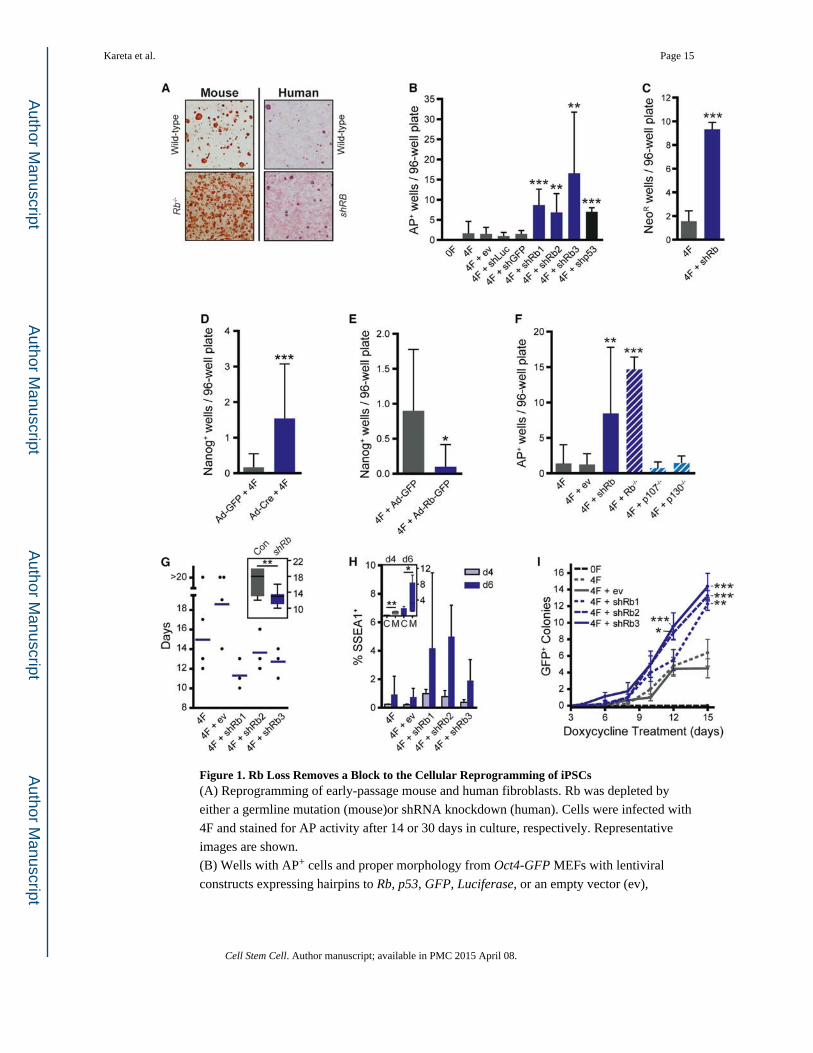
Figure 1. Rb Loss Removes a Block to the Cellular Reprogramming of iPSCs(A) Reprogramming of early-passage mouse and human fibroblasts. Rb was depleted by
either a germline mutation (mouse)or shRNA knockdown (human). Cells were infected with
4F and stained for AP activity after 14 or 30 days in culture, respectively. Representative
images are shown.
(B) Wells with AP+ cells and proper morphology from Oct4-GFP MEFs with lentiviral
constructs expressing hairpins to Rb, p53, GFP, Luciferase, or an empty vector (ev),
Kareta et al. Page 15
Cell Stem Cell. Author manuscript; available in PMC 2015 April 08.
Author M
anuscriptA
uthor Manuscript
Author M
anuscriptA
uthor Manuscript
subjected to the 4F (see Experimental Procedures). Significance was assessed by a t test to
the 4F-infected sample (n ≥ 3).
(C) Efficiency of iPSC formation was tested using Oct4-NeoR MEFs followed by treatment
with G418 on day 15 and AP staining after 5 days of selection. Significance was assessed by
a t test to the 4F sample.
(D) Efficiency of iPSC formation was tested using cKO MEFs stained for AP activity and
Nanog expression to identify iPSC colonies. Significance was assessed by a t test (n = 3).
(E) Efficiency of iPSC formation was tested with Rb overexpression by infection with Ad-
Rb-GFP or Ad-GFP. Significance was assessed by a t test (n = 2).
(F) Efficiency was tested for Rb knockdown MEFs and knockout MEFs for each of the
pocket proteins. Significance was assessed by a t test to the 4F sample (n ≥ 3).
(G) Kinetics of reprogramming was determined by infecting shRb-infected Oct4-GFP MEFs
with 4F. Plates were scored by the earliest GFP+ colony appearance up to day 20.
Significance was assessed by a t test comparing the controls against the three shRb hairpins
(n ≥ 3).
(H) Analysis of early reprogramming events by SSEA1 expression in shRb (M) or control
(C) infected MEFs 4 and 6 days after 4F (n = 3).
(I) Dox-dependence assay where shRb-infected Oct4-GFP MEFs were infected with 4F on
day 0 and treated with Dox on day 1. The cells were switched to Dox-free media at regular
intervals and GFP+ colonies were screened on day 15 (n ≥ 3).
All plots, unless noted, display the mean ± SD where *p < 0.05, **p < 0.01, and ***p <
0.001. See also Figure S1.
Kareta et al. Page 16
Cell Stem Cell. Author manuscript; available in PMC 2015 April 08.
Author M
anuscriptA
uthor Manuscript
Author M
anuscriptA
uthor Manuscript
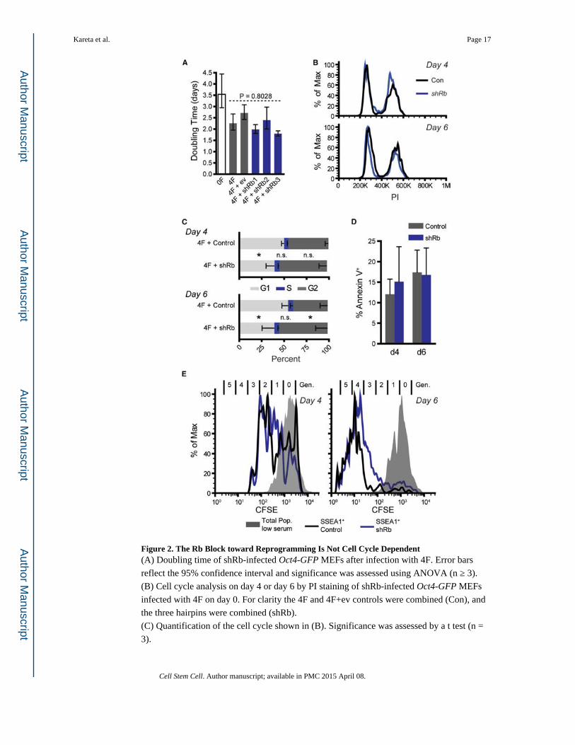
Figure 2. The Rb Block toward Reprogramming Is Not Cell Cycle Dependent(A) Doubling time of shRb-infected Oct4-GFP MEFs after infection with 4F. Error bars
reflect the 95% confidence interval and significance was assessed using ANOVA (n ≥ 3).
(B) Cell cycle analysis on day 4 or day 6 by PI staining of shRb-infected Oct4-GFP MEFs
infected with 4F on day 0. For clarity the 4F and 4F+ev controls were combined (Con), and
the three hairpins were combined (shRb).
(C) Quantification of the cell cycle shown in (B). Significance was assessed by a t test (n =
3).
Kareta et al. Page 17
Cell Stem Cell. Author manuscript; available in PMC 2015 April 08.
Author M
anuscriptA
uthor Manuscript
Author M
anuscriptA
uthor Manuscript
(D) Percent apoptosis as determined by Annexin V staining of Oct4-GFP MEFs either 4 or 6
days after 4F infection (n = 3).
(E) Number of generations (Gen) after 4F evaluated by CFSE staining of shRb-infected
MEFs. MEFs were infected with 4F on day 0 and stained with CFSE on day 1. On days 4 or
6, the cells were stained with anti-SSEA1 and analyzed by FACS. The gray curve represents
CFSE-stained MEFs grown in 0.5% serum to induce quiescence. The black and the blue
curves represent the CFSE histogram for the SSEA1+ control or shRb samples, respectively
(n = 3).
All plots display the mean ±SD unless noted where *p < 0.05. See also Figure S2.
Kareta et al. Page 18
Cell Stem Cell. Author manuscript; available in PMC 2015 April 08.
Author M
anuscriptA
uthor Manuscript
Author M
anuscriptA
uthor Manuscript
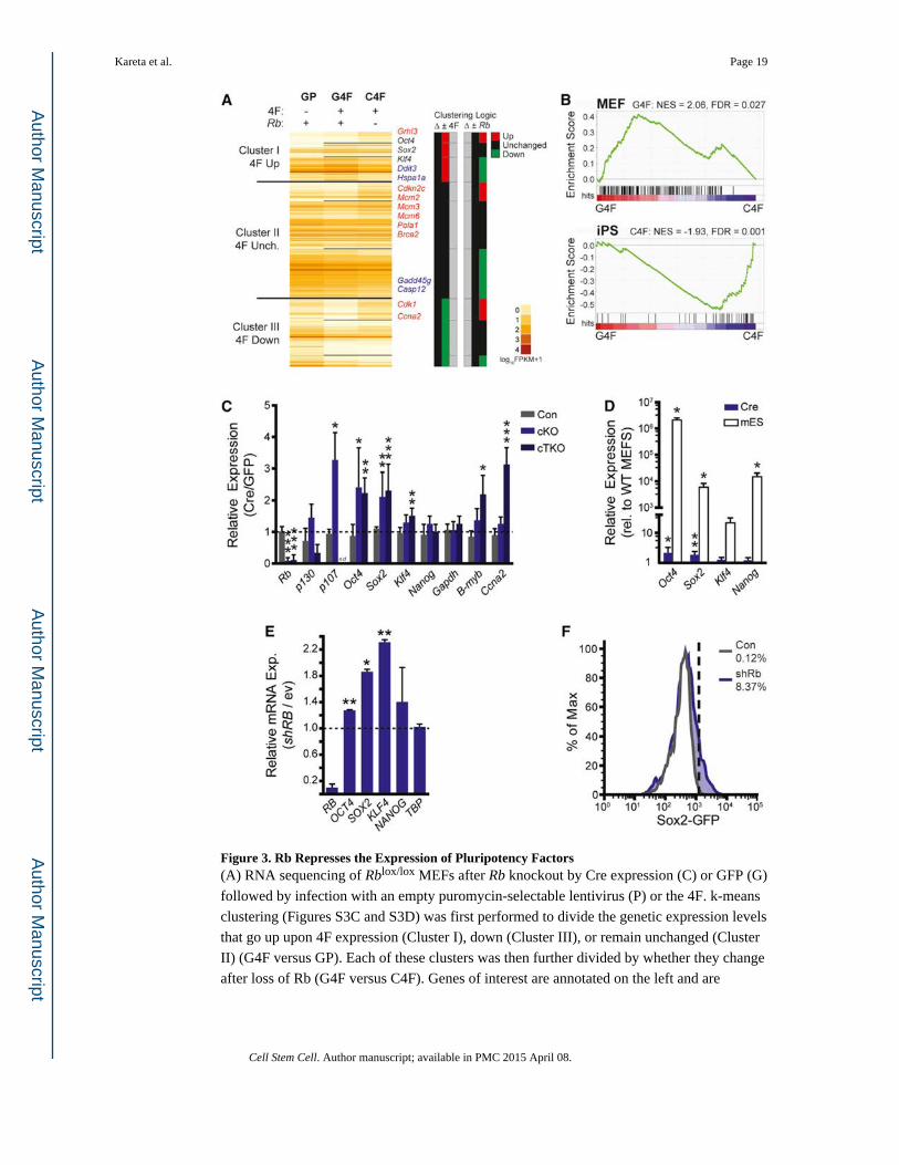
Figure 3. Rb Represses the Expression of Pluripotency Factors(A) RNA sequencing of Rblox/lox MEFs after Rb knockout by Cre expression (C) or GFP (G)
followed by infection with an empty puromycin-selectable lentivirus (P) or the 4F. k-means
clustering (Figures S3C and S3D) was first performed to divide the genetic expression levels
that go up upon 4F expression (Cluster I), down (Cluster III), or remain unchanged (Cluster
II) (G4F versus GP). Each of these clusters was then further divided by whether they change
after loss of Rb (G4F versus C4F). Genes of interest are annotated on the left and are
Kareta et al. Page 19
Cell Stem Cell. Author manuscript; available in PMC 2015 April 08.
Author M
anuscriptA
uthor Manuscript
Author M
anuscriptA
uthor Manuscript
marked red, blue, or black depending on whether their expression levels go up, down, or
stay unchanged, respectively, in the C4F set (upon loss of Rb). See also Table S1.
(B) GSEA profiles comparing the G4F and C4F set to expression profiles of matched MEF
and iPSCs (Table S1). The green graph shows the running enrichment score for the gene set
as it is calculated running down the genes from the RNA sequencing (Figure 4A) that are
ranked by their enrichment in either the G4F or C4F sets (red/ blue heatmap). Normalized
enrichment scores (NESs) and false discovery rates (FDRs) for the gene sets are reported.
(C) Gene expression in control (Con, Rb+/+; p130+/lox; p107+/+), cKO (Rblox/lox; p130+/+;
p107+/+), and cTKO (Rblox/lox; p130lox/lox; p107−/−) MEFs as assessed by RT-qPCR.
Expression is graphed as the relative expression of Ad-Cre-infected MEFs compared to that
of Ad-GFP normalized to Arppo. Statistical significance was determined by a paired t test of
the Ad-Cre and Ad-GFP ΔCt values. nd, no data.
(D) Expression measured by RT-qPCR for either Rb cKO MEFs infected with Ad-GFP or
Ad-Cre (n = 4) or mESCs (n = 3), shown relative to the Ad-GFP cells. Statistical
significance was determined by a paired t test of the Ad-Cre MEFs and an unpaired t test for
the mESCs.
(E) Expression measured by RT-qPCR is graphed as the relative expression of shRB-in-
fected human fibroblasts compared to those infected with an empty vector (ev) and
normalized to GAPDH. Statistical significance was determined by a paired t test of the
knockdown and control ΔCt values; n = 2.
(F) FACS analysis of GFP in Sox2-GFP knockin MEFs after shRb infection compared to
controls. Dashed line demarcates GFP+ gate.
All plots display the mean ± SD where *p < 0.05, **p < 0.01, and ***p < 0.001. See also
Figure S3.
Kareta et al. Page 20
Cell Stem Cell. Author manuscript; available in PMC 2015 April 08.
Author M
anuscriptA
uthor Manuscript
Author M
anuscriptA
uthor Manuscript
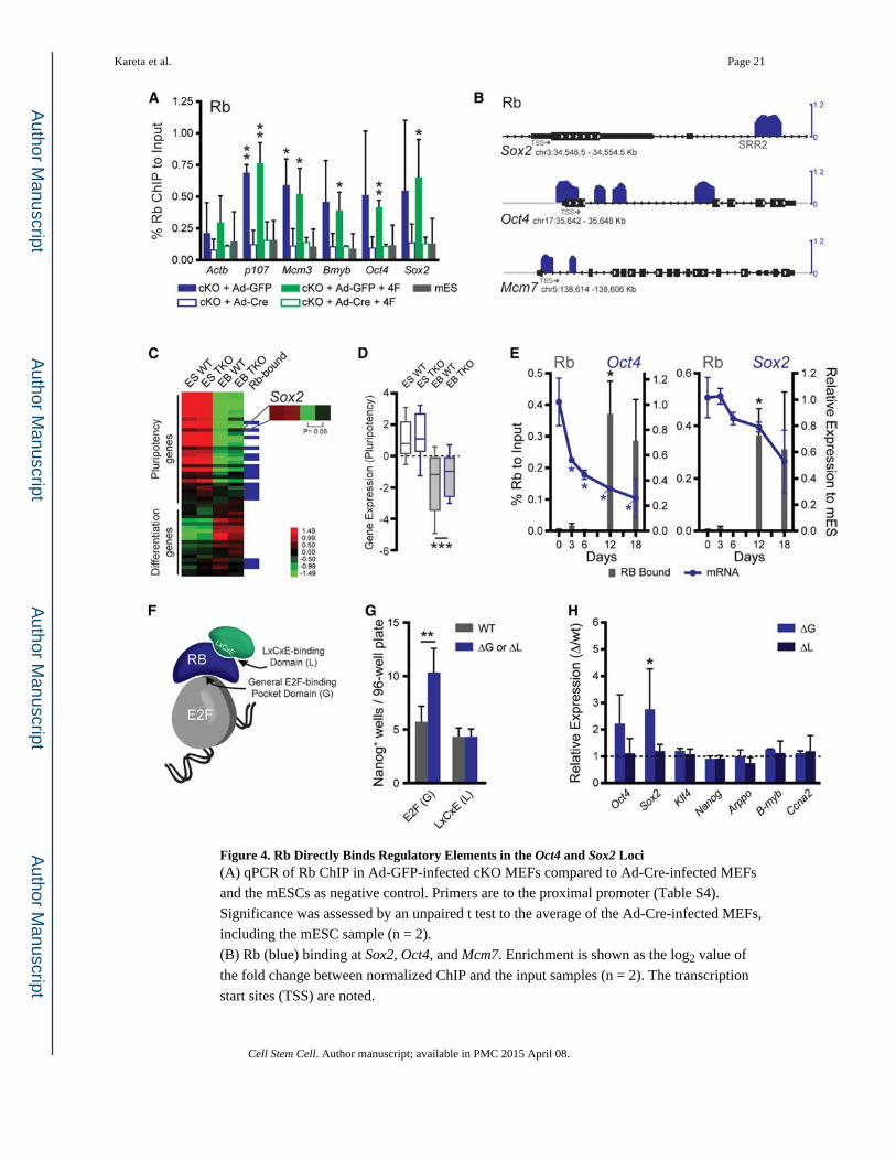
Figure 4. Rb Directly Binds Regulatory Elements in the Oct4 and Sox2 Loci(A) qPCR of Rb ChIP in Ad-GFP-infected cKO MEFs compared to Ad-Cre-infected MEFs
and the mESCs as negative control. Primers are to the proximal promoter (Table S4).
Significance was assessed by an unpaired t test to the average of the Ad-Cre-infected MEFs,
including the mESC sample (n = 2).
(B) Rb (blue) binding at Sox2, Oct4, and Mcm7. Enrichment is shown as the log2 value of
the fold change between normalized ChIP and the input samples (n = 2). The transcription
start sites (TSS) are noted.
Kareta et al. Page 21
Cell Stem Cell. Author manuscript; available in PMC 2015 April 08.
Author M
anuscriptA
uthor Manuscript
Author M
anuscriptA
uthor Manuscript
(C) Gene expression by microarray of WT and TKO mESCs after their differentiation into
embryoid bodies (n = 3). Pluripotency genes (Figure S4D) and a selection of differentiation
markers from endoderm, mesoderm, ectoderm, and trophectoderm are shown. These are also
scored for their status as direct Rb targets.
(D) The average expression of the pluripotency genes shown in (C). Plots show the mean
(horizontal line), the 25th to the 75th percentile (box), and the extent of the data (bars).
Significance was tested by a paired t test.
(E) Rb ChIP in mESCs (d0) and after their subsequent differentiation into EBs (d3, d6, d12,
and d18). Binding is shown (gray bars, left axis) as percent input normalized to a control
locus to further account for cell number. Expression from the locus (either Oct4 or Sox2) is
shown relative to the levels in the mESCs (blue line, right axis). Significance was
determined by an unpaired t test to the respective d0 values for the ChIP and qPCR sets.
(F) Schematic of Rb interaction surfaces including the general E2F binding surface (G) and
the LxCxE-binding domain (L), which interacts with other silencing factors.
(G) Reprogramming efficiency of the ΔG or ΔL mutant MEFs compared to those derived
from a wild-type littermate. Efficiency was screened by AP and Nanog staining.
Significance was determined by a paired t test.
(H) Gene expression in the ΔG and ΔL MEFs compared to wild-type MEFs as assessed by
RT-qPCR. Expression is graphed as the relative expression of the mutant to the wild-type.
Statistical significance was determined by a paired t test of the ΔCt values.
All plots, unless noted, display the mean ± SD where *p < 0.05, **p < 0.01, and ***p <
0.001. See also Figure S4.
Kareta et al. Page 22
Cell Stem Cell. Author manuscript; available in PMC 2015 April 08.
Author M
anuscriptA
uthor Manuscript
Author M
anuscriptA
uthor Manuscript
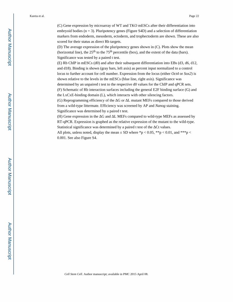
Figure 5. Rb Loss Perturbs the Chromatin State of Pluripotency Genes by Modulating Histone Modifier Recruitment(A) Histone modification profiles for H3Ac (light green), H3K4me3 (dark green), and
H3K27me3 (red) displayed as a heatmap. The bottom track indicates regions of significant
changes for each mark by their respective color.
(B) GSEA results showing enrichment of iPSC and Rb gene sets upon gain of H3
acetylation and H3K4me3. Data shown are the rank of the gene set relative to all identified
gene sets, the normalized enrichment score (NES), and the false discovery rate (FDR).
Kareta et al. Page 23
Cell Stem Cell. Author manuscript; available in PMC 2015 April 08.
Author M
anuscriptA
uthor Manuscript
Author M
anuscriptA
uthor Manuscript
(C) Examples of genes that show an increase (blue) or decrease (gray) in H3K4me3 domain
breadth upon loss of Rb. Black bars represent the extent of the domains.
(D) Significance for enrichment of transcription factor targets at genes that gained
substantial H3K4me3 breadth in MEFs upon loss of Rb against the expected genome-wide
value from 1,000 random samplings expressed as — log10 (p value) in one-sided Wilcoxon
tests.
(E) Examples of a gene in which the H3K4me3 peak breadth in wild-type MEFs (gray) is
broadened upon loss of Rb (blue), compared to mESCs (green). Horizontal black bars
represent the extent of the peak.
(F) Fold-enrichment for overlap with top 5% broadest H3K4me3 domains (“buffer
domains”) for genes that lost or gained substantial H3K4me3 breadth in MEFs upon loss of
Rb. Enrichment and significance (one-sided Wilcoxon tests) were calculated against
expected genome-wide values from 1,000 random samplings.
(G) ChIP-qPCR of HDAC and EZH2 in Ad-GFP- and Ad-Cre-infected MEFs plotted
relative to the mESCs as a negative control to account for relative primer strength. qPCR
was performed with primers to the DE, PE, proximal promoter (PP), and first exon (E1) of
Oct4. Significance was determined by a t test.
(H) ChIP-qPCR of HDAC binding shown as in Figure 5F, shown for the SRR1 and SRR2
enhancers, the PP, and the E1 of Sox2.
All plots, unless noted, display the mean ± SD where *p < 0.05 and ***p < 0.001. See also
Figure S5.
Kareta et al. Page 24
Cell Stem Cell. Author manuscript; available in PMC 2015 April 08.
Author M
anuscriptA
uthor Manuscript
Author M
anuscriptA
uthor Manuscript
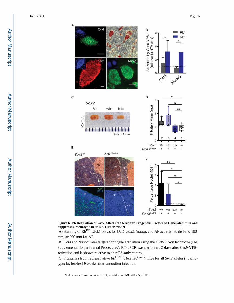
Figure 6. Rb Regulation of Sox2 Affects the Need for Exogenous Factors to Generate iPSCs and Suppresses Phenotype in an Rb Tumor Model(A) Staining of RbKD OKM iPSCs for Oct4, Sox2, Nanog, and AP activity. Scale bars, 100
mm, or 200 mm for AP.
(B) Oct4 and Nanog were targeted for gene activation using the CRISPR-on technique (see
Supplemental Experimental Procedures). RT-qPCR was performed 5 days after Cas9-VP64
activation and is shown relative to an rtTA-only control.
(C) Pituitaries from representative Rblox/lox; Rosa26CreER mice for all Sox2 alleles (+, wild-
type; lx, lox/lox) 9 weeks after tamoxifen injection.
Kareta et al. Page 25
Cell Stem Cell. Author manuscript; available in PMC 2015 April 08.
Author M
anuscriptA
uthor Manuscript
Author M
anuscriptA
uthor Manuscript
(D) Box and whisker plots of the pituitary size from mice of the genotypes in Figure 6C,
including Rblox/lox; Rosa+/+ mice as controls.
(E) Pituitary sections of Rblox/lox; Rosa26CreER mice that are either Sox2+/+ or Sox2lox/lox 9
weeks after tamoxifen injection, stained with H&E (top) or Ki67 (bottom). The Pars
Nervosa (PN), Pars intermedia (PI), Pars distalis (PD), and residual cleft (rc) are shown.
The green channel was exposed equally between the two images. Representative images are
shown.
(F) Quantification of Ki67 in Rblox/lox; Rosa26CreER pituitaries with the shown Sox2
genotypes (+, wild-type; lx, lox/lox) 9 weeks after tamoxifen injection. Percentages were
calculated from four pituitaries, except the CreER− control, which had n = 3. Dashed line
indicates the number of positive cells counted in the secondary-only control.
All plots, unless noted, display the mean ± SD where *p < 0.05, **p < 0.01, and ns = not
specified. See also Figure S6.
Kareta et al. Page 26
Cell Stem Cell. Author manuscript; available in PMC 2015 April 08.
Author M
anuscriptA
uthor Manuscript
Author M
anuscriptA
uthor Manuscript


![Guidelines for flag State inspections under the Maritime ... · 4 MEFS-Guidelines(Rev.)-[2008-09-0144-8]-En.doc/v4 Interim Maritime Labour Certificate; Declaration of Maritime Labour](https://img.pdfslide.us/doc/110x75/5f05dc8e7e708231d41514e8/guidelines-for-flag-state-inspections-under-the-maritime-4-mefs-guidelinesrev-2008-09-0144-8-endocv4.jpg)