Embed Size (px)
Citation preview

Quantification of Facial Skeletal Shape Variation in Fibroblast Growth Factor Receptor-Related Craniosynostosis Syndromes
Yann Heuzé1, Neus Martínez-Abadías1, Jennifer M. Stella1, Eric Arnaud2, Corinne Collet3, Gemma García Fructuoso4, Mariana Alamar4, Lun-Jou Lo5, Simeon A. Boyadjiev6, Federico Di Rocco2, and Joan T. Richtsmeier1,*
1Department of Anthropology, Pennsylvania State University, University Park, Pennsylvania
2Craniofacial Surgery Unit, Department of Pediatric Neurosurgery, Hôpital Necker-Enfants Malades, University Paris V, Paris, France
3Laboratoire de Biochimie et de Biologie Moléculaire, INSERM U606, Paris, France
4Servei de Neurocirurgia, Hospital Sant Joan de Deu, Barcelona, Spain
5Department of Plastic and Reconstructive Surgery, Chang Gung Memorial Hospital, Chang Gung University, Taoyuan, Taiwan
6Section of Genetics, Department of Pediatrics, University of California Davis, Sacramento, California
Abstract
Background—fibroblast growth factor receptor (FGFR) -related craniosynostosis syndromes are
caused by many different mutations within FGFR-1, 2, 3, and certain FGFR mutations are
associated with more than one clinical syndrome. These syndromes share coronal craniosynostosis
and characteristic facial skeletal features, although Apert syndrome (AS) is characterized by a
more dysmorphic facial skeleton relative to Crouzon (CS), Muenke (MS), or Pfeiffer syndromes.
Methods—Here we perform a detailed three-dimensional evaluation of facial skeletal shape in a
retrospective sample of cases clinically and/or genetically diagnosed as AS, CS, MS, and Pfeiffer
syndrome to quantify variation in facial dysmorphology, precisely identify specific facial features
pertaining to these four syndromes, and further elucidate what knowledge of the causative FGFR
mutation brings to our understanding of these syndromes.
Results—Our results confirm a strong correspondence between genotype and facial phenotype
for AS and MS with severity of facial dysmorphology diminishing from Apert FGFR2S252W to
Apert FGFR2P253R to MS. We show that AS facial shape variation is increased relative to CS,
although CS has been shown to be caused by numerous distinct mutations within FGFRs and
reduced dosage in ERF.
© 2014 Wiley Periodicals, Inc.*Correspondence to: Joan T. Richtsmeier, Department of Anthropology, The Pennsylvania State University, Carpenter Building, University Park, PA 16802., [email protected] Martínez-Abadías present address is European Molecular Biology Laboratory (EMBL) - Center for Genomic Regulation (CRG) Systems Biology Research Unit, CRG, Barcelona, Spain
HHS Public AccessAuthor manuscriptBirth Defects Res A Clin Mol Teratol. Author manuscript; available in PMC 2014 May 21.
Published in final edited form as:Birth Defects Res A Clin Mol Teratol. 2014 April ; 100(4): 250–259. doi:10.1002/bdra.23228.
Author M
anuscriptA
uthor Manuscript
Author M
anuscriptA
uthor Manuscript

Conclusion—Our quantitative analysis of facial phenotypes demonstrate subtle variation within
and among craniosynostosis syndromes that might, with further research, provide information
about the impact of the mutation on facial skeletal and nonskeletal development. We suggest that
precise studies of the phenotypic consequences of genetic mutations at many levels of analysis
should accompany next-generation genetic research and that these approaches should proceed
cooperatively.
Keywords
genotype-phenotype correspondence; midfacial retrusion; suture fusion; morphogenesis; diagnosis
Introduction
Cell–cell signaling through fibroblast growth factors and their receptors (FGF/FGFR) plays
fundamental roles in development (Montero et al., 2000; Ornitz, 2005) and has been
proposed as a key system in the evolution of the vertebrate head (Bertrand et al., 2011). The
FGF/FGFR signaling system is implicated in the control of basic processes (e.g.,
proliferation, differentiation, migration, polarity, adhesion, apoptosis) in cells destined to
become various tissues including bone and cartilage (Ornitz and Itoh, 2001; Dorey and
Amaya, 2010; Li et al., 2013). Mutations in FGFR1, 2 and 3 can lead to varied impairments
of skeletal development as demonstrated by the FGFR-related craniosynostosis syndromes
(i.e., Apert [AS], Beare-Stevenson, Crouzon [CS], Crouzon with acanthosis nigricans,
Jackson-Weiss, Muenke [MS], and Pfeiffer [PS] syndromes). These syndromes are
characterized by premature fusion of one or several cranial vault sutures associated with
skull dysmorphology and potentially presenting with malformations affecting the limbs,
upper airway, brain, spine, heart, and/ or lungs (Cohen and MacLean, 2000).
With the exception of MS, all FGFR-related craniosynostosis syndromes were originally
defined phenotypically. Consequently, diagnosis of FGFR-related craniosynostosis
syndromes is based on clinical findings (Robin et al., 1998) (e.g., craniosynostosis,
dysmorphic facial features, limbs appearance) and, when possible, confirmed by genetic
testing. This is important as genetic and phenotypic variation within and among
craniosynostosis syndromes results in the now well-known lack of a one-toone
correspondence between a given FGFR mutation and a specific skull shape. Some of the
FGFR-related craniosynostosis syndromes can be caused by many different mutations
within one or more of the FGFRs (e.g., PS and CS), while others are caused by a unique
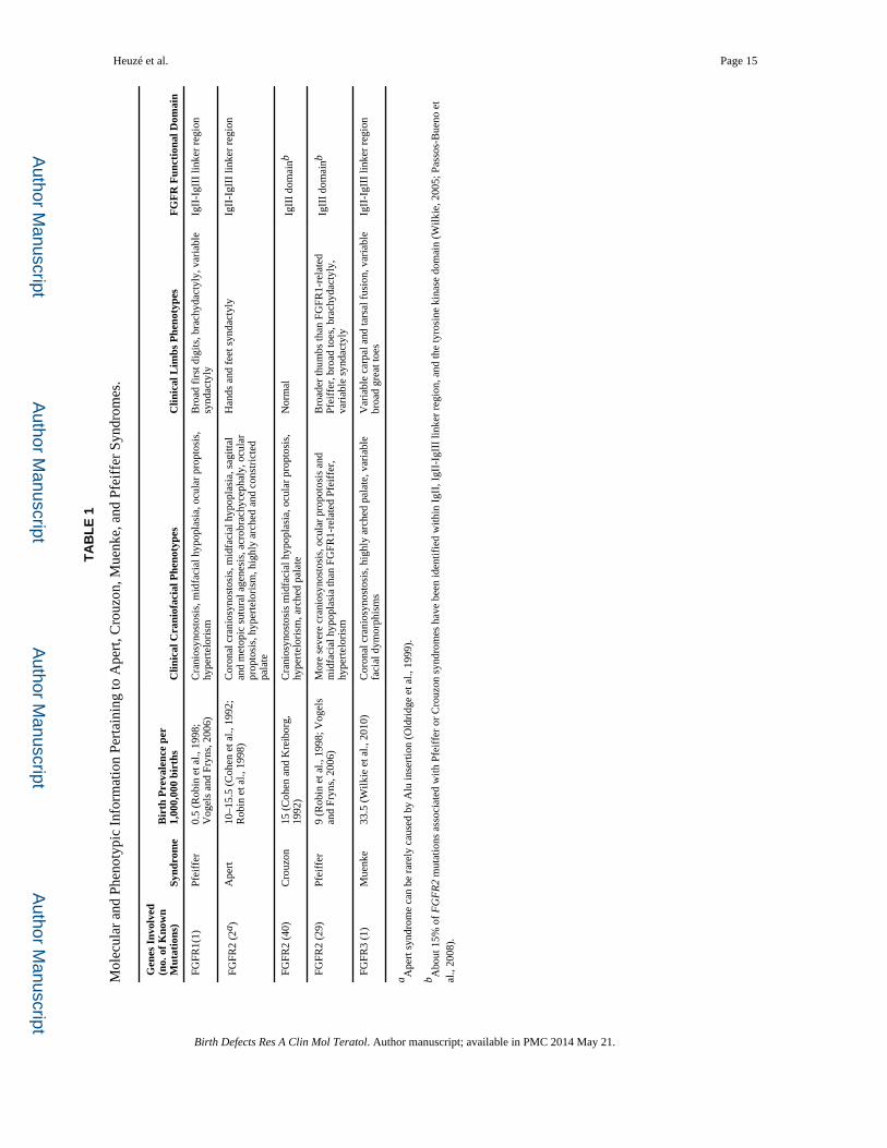
mutation (e.g., MS), or a very small set of mutations (e.g., AS) (Table 1). In addition, there
are certain mutations in FGFR2 that have been associated with more than one clinical
syndrome (e.g., CS and PS) (Passos-Bueno et al., 2008). Consequently, in many cases
clinical diagnosis is problematic.
The craniofacial phenotypes of AS, CS, MS, PS are highly variable. Craniofacial
phenotypes of AS can include varying degrees of midfacial retrusion. It is reported that AS
cases carrying the FGFR2S252W mutation have a more severe facial phenotype relative to AS
patients who carry the FGFR2P253R mutation while the FGFR2P253R group has more severe
limb anomalies (Slaney et al., 1996; Lajeunie et al., 1999; von Gernet et al., 2000).
Heuzé et al. Page 2
Birth Defects Res A Clin Mol Teratol. Author manuscript; available in PMC 2014 May 21.
Author M
anuscriptA
uthor Manuscript
Author M
anuscriptA
uthor Manuscript

Craniofacial phenotypes of CS can vary from normal, to facial skeletal dys-morphologies
without calvarial craniosynostosis, to cloverleaf skull malformation. In the majority of cases,
several cranial sutures are prematurely fused at birth, although on occasion, the phenotypic
features of CS may be absent at birth and evolve gradually during the first few years of life
(Lajeunie et al., 1999; Connolly et al., 2004; Hoefkens et al., 2004). Variation in the severity
of the craniofacial phenotype and limb anomalies of PS has led to the creation of three
clinical subtypes (Cohen, 1993). Finally, the craniofacial phenotype of MS is
characteristically variable and ranges from normal to severe (Doherty et al., 2007).
Facial phenotype is one of the key clinical findings used in differential diagnosis among the
craniosynostosis syndromes. Although AS is characterized by a more dys-morphic facial
skeleton relative to CS, MS, or PS (Cohen and MacLean, 2000), all of these syndromes
share characteristic facial skeletal features (i.e., Crouzonoid face), including midfacial
retrusion, hypertelorism, proptosis (secondary to orbital dysmorphogenesis), high-arched
palate, flattened malar region, and beaked nose (Table 1) (Robin et al., 1998; Johnson and
Wilkie, 2011). Midfacial retrusion or hypoplasia is defined as the posterior positioning
and/or vertical shortening of the infraorbital and perialar regions, or increased concavity of
the face and/or reduced nasolabial angle representing underdevelopment of the maxillary
height (decreased midface height) or depth (retrusion of the maxilla) (Allanson et al., 2009).
This definition allows for a wide range of variation and currently, midfacial retrusion
represents a catch-all diagnosis for many craniofacial conditions that can result from distinct
molecular causes and novel developmental dynamics. Midfacial retrusion is potentially the
most challenging clinical manifestation of many of the FGFR-related craniosynostosis
syndromes affecting oral health, feeding, and airway function (Cunningham et al., 2007;
Johnson and Wilkie, 2011) but is relatively uncommon in MS (Ridgway et al., 2011).
Here, we perform a detailed three-dimensional (3D) evaluation of facial skeletal shape in a
large retrospective sample (n = 43) of cases clinically and/or genetically diagnosed as AS,
CS, MS, and PS to quantify variation in severity of facial dysmorphology, and precisely
identify specific facial features pertaining to one or several of these four syndromes. First,
we investigate the genotype phenotype correspondence for the segment of our sample
(19/43) with known genetic mutations and subsequently add the remaining cases to further
elucidate what knowledge of the causative FGFR mutation brings to our understanding of
these syndromes.
Materials and Methods
SUBJECTS
We amassed pre-operative computed tomography (CT) images of children aged 0 to 23
months acquired by several medical centers in France, United States, Taiwan, and Spain
over the past 10 years. Our sample consists of images of 43 individuals genetically and/or
clinically diagnosed with AS, CS, MS, or PS, and 20 unaffected individuals (Tables 2 and
3). All collected images were anonymized and no information other than sex, age at the time
of the CT exam, and causative mutation were communicated to us. No data on the ethnicity
or geographic origin of the patients were available. The coronal suture is prematurely fused
in 38 of 43 craniosynostosis syndrome cases while three CS individuals and one AS case do
Heuzé et al. Page 3
Birth Defects Res A Clin Mol Teratol. Author manuscript; available in PMC 2014 May 21.
Author M
anuscriptA
uthor Manuscript
Author M
anuscriptA
uthor Manuscript

not present with craniosynostosis of any vault suture (Table 3). The causative FGFR1–3
mutation was identified in 19 cases. Diagnosis of the remaining cases (N = 24) is based
solely on clinical evaluation. The unaffected sample consists of images of children without
craniosynostosis who underwent CT scanning for conditions not associated with
craniosynostosis (e.g., seizures, suspected brain anomalies). Use of the CT images was
approved by the Institutional Review Boards of the Pennsylvania State University and the
participating institutions and the images were acquired in accordance with institutional
guidelines.
MORPHOMETRIC DATA AND ANALYSIS
Skulls were reconstructed from the CT images using a threshold that enabled visualization
of bone. A set of 39 anatomical landmarks was defined and located on the 3D reconstruction
of the skull of each individual and the corresponding x, y, z coordinates were recorded with
Avizo 6 (Visualization Sciences Group, SAS). In addition to the anatomical landmarks, a
total of 168 semilandmarks were defined on 11 predefined curves (92 curve semiland-
marks) and four surface patches (76 surface semiland-marks) on each skull (Supp. Fig. S1,
which is available online; for details, see Heuzé et al., 2010 and Heuzé et al., 2012).
Semilandmarks present “deficient” coordinates and were slid according to a sliding
algorithm that minimizes the bending energy (Bookstein, 1997; Gunz et al., 2005) to define
their final location on the defined curves or surfaces. Once slid, semilandmarks acquire a
geometric correspondence across individuals so that comparative analyses can be conducted.
The 3D coordinates of semilandmarks were computed using Viewbox 4 (dHAL software,
Athens, Greece).
Shape information of each individual defined on the basis of landmarks and semilandmarks
was extracted using general Procrustes analysis, a procedure that superimposes
configurations of landmarks by shifting them to a common position, rotating and scaling
them to a standard size until a best fit of corresponding landmarks is achieved (Rohlf and
Slice, 1990; Dryden and Mardia, 1998). A Procrustes average shape (PAS) for a defined
group was computed as the coordinate-wise average of the coordinates of the resulting
centered, scaled, and rotated landmarks (Mitteroecker and Gunz, 2009). Distinct Procrustes
superimpositions were used for the computation of PAS for specific skull anatomical units
(Supp. Fig. S1). The PAS triangular mesh can be obtained by warping the triangular mesh of
one case to the landmark set corresponding to the PAS. The covariance matrix of the
Procrustes shape coordinates was analyzed by principal components analysis (PCA)
(Jolliffe, 2002) to reduce the dimensionality of the dataset. PCA transforms the variables
entered into the analysis into a set of new variables, the principal components (PCs), which
are uncorrelated with each other. The first PC accounts for the maximum possible amount of
variation and each successive PC in turn accounts for the maximum possible amount of
variation that remains. Unlike discriminant analyses, PCA does not consider a priori
knowledge of internal structure of the data and just rotates the data to find new axes (i.e.,
directions in the morphospace) that explain the maximum of morphological variation in the
sample. PCA was used here to assess if the specific combination of morphological variables
that explain most variation is also able to successfully separate individuals into groups of
known membership (e.g., known syndrome, mutation). The PCs contain the loadings for the
Heuzé et al. Page 4
Birth Defects Res A Clin Mol Teratol. Author manuscript; available in PMC 2014 May 21.
Author M
anuscriptA
uthor Manuscript
Author M
anuscriptA
uthor Manuscript

linear combinations of the original variables and can be visualized as shape deformation. A
simulation of a continuous shape deformation based on the available data can be obtained by
warping the PAS triangular mesh along the negative (or positive) direction of each principal
component. To facilitate visualization of shape differences between two PAS, we present a
color map produced by comparing the corresponding surface warps. The color map has been
computed in Avizo 6 (Visualization Sciences Group, VSG) and corresponds to the vector
field computed by the difference of the vertex positions of corresponding vertices in both
PAS surface warps.
Shape differences among groups of syndromic and unaffected individuals were estimated
using Procrustes distances. The Procrustes distance is measured as the square root of the
sum of squared distances between corresponding landmarks of two shape configurations
after Procrustes superimposition (Dryden and Mardia, 1998; Mitteroecker and Gunz, 2009).
For each comparison, the Procrustes distance between the mean shapes of the two groups
considered was computed and a permutation test with 10,000 rounds was performed to test
for statistical significance.
Potential influence of age on shape was explored by computing a multivariate regression of
shape (Drake and Klingenberg, 2008) on age. The residuals of the multivariate regression
were analyzed in a new PCA where the effect of age is removed.
Results
GENOTYPE PHENOTYPE CORRESPONDENCE FOR AS AND MS
Specifics of the mutations carried by the 19 genotyped individuals in our sample are given in
Table 3 with most identified mutations associated with AS and MS. When the shape of the
whole skull is analyzed using PCA of Procrustes coordinates, the first and second PCs
together account for 66% of the total shape variation. Shape variation of the FGFR2S252W
AS, the FGFR2P253R AS, and the FGFR3P250R MS cases overlap and these groups fail to
separate on the PC1 versus PC2 plot (Supp. Fig. S2A). Procrustes distances separating the
mean skull shapes of the FGFR2S252W AS group, FGFR2P253R AS group, and FGFR3P250R
MS group are not significant (Table 4). However, when only data from the facial skeleton
are analyzed by PCA, a clear separation between individuals carrying the FGFR2S252W,
FGFR2P253R, or FGFR3P250R mutations is revealed along PC1, which accounts for 46.9%
of shape variation (Fig. 1A). The Procrustes distance separating facial shapes of the
FGFR2S252W AS group and the FGFR3P250R MS group is significant, but the Procrustes
distances between the FGFR2P253R AS group and the FGFR2S252W AS group and the
FGFR2P253R AS group and the FGFR3P250R MS group are not (Table 4). The anatomical
regions of the facial skeleton showing the most intense shape changes along PC1 are the
frontal and zygomatic bones, the alveolar process of the maxilla and the most posterior
aspect of the palate (Fig. 1B). AS cases carrying the FGFR2S252W mutation, corresponding
to the lowest scores on PC1, are characterized by increased facial width, larger orbits,
posterior positioning, and vertical shortening of the maxilla and zygomatic, reduced perialar
region, and a v-shaped palate with shorter length; a facial shape characteristic of midfacial
retrusion. AS cases carrying the FGFR2P253R mutation and MS cases are also located along
the negative end of PC1, although not at the extreme. These groups separate from the
Heuzé et al. Page 5
Birth Defects Res A Clin Mol Teratol. Author manuscript; available in PMC 2014 May 21.
Author M
anuscriptA
uthor Manuscript
Author M
anuscriptA
uthor Manuscript
FGFR2S252W group and from each other. According to this analysis, the FGFR2S252W group
is characterized by the most severe facial phenotype with intensity of facial dysmorphology
relatively diminished in the FGFR2P253R and FGFR3P250R groups. These results confirm a
strong correspondence between genotype and phenotype that enables differentiation of AS
and MS on the basis of facial skeletal shape.
The PCA computed using the residuals of the multivariate regression of shape on age
provided a very similar arrangement of the individuals indicating that the effect of age on
facial and skull shape was not the main signal of the first two PCs (Supp. Figs. S2, S3).
FACIAL PHENOTYPIC VARIATION OF PATIENTS GENETICALLY AND/OR CLINICALLY DIAGNOSED WITH AS, CS, MS, OR PS
Differential diagnosis of the remaining AS, CS, and PS cases was based solely on clinical
findings of craniosynostosis, specific dysmorphic facial features, and/or limb anomalies. CS
and PS have been reported to display relatively similar facial phenotypes but distinct limb
phenotypes. Consequently, variation of facial skeletal shape of CS and PS are expected to
overlap. Although the AS facial phenotype is reported as being more severe than that of CS
and PS, little quantitative information is available for comparative variation in facial shape
among AS, CS, MS and PS. We quantified shape variation of the 43 individuals diagnosed
genetically and/or clinically with FGFR-related craniosynostosis syndromes (Fig. 2A). The
relative position of the genotyped individuals remains similar to the previous analysis that
included only those patients with a genetic diagnosis (Fig. 1A), indicating that the addition
of the syndromic cases that are clinically diagnosed does not change the general pattern of
shape variation. Indeed, facial skeletal shape differences between the positive and negative
ends of PC1 (results not shown) are the same as those reported in Figure 1B. Clinically
diagnosed cases of CS occupy an intermediate position between AS and the unaffected
individuals, while PS cases overlap with AS, CS, MS, and the unaffected individuals. The
Procrustes distances separating the mean facial skeletal shapes of the syndromic groups are
all significant with the exception of the PS-MS and PS-CS distances (Table 4). When
considering the whole skull, the PC1 versus PC2 plot (59.2% of shape variation) of the skull
shape analysis failed to separate the different syndromes (Supp. Fig. S5A) similar to our
findings for the genotyped cases only, but the mean skull shapes of the syndromic groups
displayed significant inter-group differences with the exception of the comparison of MS
with AS and MS with PS (Table 4). No particular structure related to sex is observed in the
PC1 versus PC2 plot of facial shape or skull shape (Supp. Figs. S4A, S5A).
The effect of age on facial and skull shape was not the main signal recorded on the
corresponding PC1 versus PC2 plot as shown by the PCAs computed on the basis of the
residuals of the multivariate regression of shape on age which displayed very similar
arrangement of the individuals (Supp. Figs. S4, S5). To further define the anatomical
location of the differences in facial shape among the FGFR-related craniosynostosis
syndromes, we compared each of the syndrome specific consensus shapes (PAS) with that
of all unaffected individuals (Fig. 2B). When compared with the unaffected individuals, AS
and MS share similar dysmorphic features but those of MS are less severe and include some
degree of asymmetry due to the presence of two MS cases with right unilateral coronal
Heuzé et al. Page 6
Birth Defects Res A Clin Mol Teratol. Author manuscript; available in PMC 2014 May 21.
Author M
anuscriptA
uthor Manuscript
Author M
anuscriptA
uthor Manuscript

craniosynostosis (Table 3). The anatomical regions of AS and MS displaying the most
intense shape differences when compared with the unaffected individuals are the superior
and inferior lateral borders of the orbits formed by the frontal and the zygomatic bones
respectively, the alveolar process of the maxilla, and the most posterior aspect of the palate
formed by the palatine bone. These shape differences that characterize AS and MS are very
similar to those observed in Figure 1B and result in larger orbits, larger interorbital distance,
reduced body and alveolar process of the maxilla, posterior positioning and vertical
shortening of the infraorbital region, shorter primary palate, and shorter and superiorly
projected secondary palate. The asymmetric shape differences revealed in the CS face are
due to the CS cases with right unilateral coronal craniosynostosis (Table 3). Although the
shape differences between CS and unaffected individuals involve the superolateral border of
the orbits, the alveolar process of the maxilla, and the most posterior aspect of the palate,
there is little shape difference local to the most inferior and lateral border of the orbit formed
by the zygomatic bone. The shape of the nasal bridge is affected in CS but not in AS.
Finally, when compared with unaffected individuals, PS displays features similar to those of
CS. One key difference, however, is the reduced shape change of the alveolar process of the
maxilla and the more intense shape change of the superior and lateral border of the orbit in
PS relative to CS.
In summary, the nasal bridge and anterior-most aspect of the zygomatic are the anatomical
regions that best differentiate AS and MS faces from those of PS and CS.
Discussion
The shape of the facial skeleton is a challenging clinical symptom and critical diagnostic
feature that, along with craniosynostosis and limbs anomalies, is used to clinically diagnose
FGFR-related craniosynostosis syndromes. Although based on a relatively small sample of
genotyped patients (n = 19), our results reveal distinct facial phenotypes associated with
FGFR2S252W, FGFR2P253R, and FGFR3P250R mutations. Such robust genotype phenotype
correspondence for AS and MS does not exist when the entire skull is analyzed. Our results
show a quantitative scale of severity of facial dysmorphology and particularly midfacial
retrusion that is strongest in cases carrying the FGFR2S252W mutation, relatively decreased
in the FGFR2P253R group and further decreased in MS. The anatomical regions displaying
the most intense shape differences among AS and MS when compared with unaffected
individuals are the frontal and zygomatic contributions to the orbit, and the palate. Our
results are in agreement with previous studies of humans with AS and mouse models for AS
in which the facial skeleton, particularly the palate, was found to be the most profoundly
dysmorphic cranial feature, especially in the FGFR2S252W group (Slaney et al., 1996;
Lajeunie et al., 1999; Von Gernet et al., 2000; Martínez-Abadías et al., 2010, 2013; Wang et
al., 2010).
Three individual cases merit discussion. The CS case carrying the FGFR2F276V mutation
displayed no sign of craniosynostosis at the age of 2 months. Because of the absence of
craniosynostosis, this case plots with the unaffected cases in the skull shape analysis (Supp.
Fig. S5A), while in the facial skeletal shape analysis this individual plots with the other
syndromic cases (Figs. 1A, 2A). The facial phenotype and the absence of characteristic limb
Heuzé et al. Page 7
Birth Defects Res A Clin Mol Teratol. Author manuscript; available in PMC 2014 May 21.
Author M
anuscriptA
uthor Manuscript
Author M
anuscriptA
uthor Manuscript
anomalies prompted the diagnosis of CS for this individual, even though the FGFR2F276V
mutation has also been associated with PS (Passos-Bueno et al., 2008). These findings
prompt us to propose that the FGFR2F276V mutation functions to contribute to facial
dysmorphology, perhaps by causing premature closure of facial sutures, but does not
necessarily cause premature closure of the cranial vault sutures. The case carrying the
FGFR2A172F mutation that is specific to PS and known to be associated with severe limb
phenotypes (Wilkie et al., 2002; Cohen, 2004), has a facial phenotype similar to that of AS
(Figs. 1A, 2A). Finally, the FGFR2C342R mutation is typically characterized by severe
phenotypes (Lajeunie et al., 2006) and frequently associated with PS, although also with CS.
Our case displays bilateral craniosynostosis of the coronal and lambdoid sutures and in our
analysis is the second most severe facial phenotype after the PS case carrying the
FGFR2F276V mutation.
While genetic causes of CS include at least 47 distinct mutations mostly within FGFR2
(Passos-Bueno et al., 2008), and reduced dosage of ERF (Twigg et al., 2013), the
quantitative range of facial shape variation for our total CS sample is small relative to AS
(Fig. 2A), which is caused by only two mutations in 99% of cases. Obviously, a small
fraction of the mutations causative for CS occurs at high incidence, and only a subset of
these is most likely represented in our sample. Still, we can reasonably assume that some of
the cases clinically diagnosed with CS carry a different mutation than the genetically
diagnosed CS case included in the present study (i.e., FGFR2F276V). Finally, the relatively
small range of MS facial shape variation revealed by our analysis contrasts with current
knowledge of variation in MS with phenotypic consequences ranging from normal to severe
(Doherty et al., 2007). This most likely represents an ascertainment bias in that our sample is
limited to individuals who sought treatment at a medical facility and would not include
individuals with normal appearance or with a mild phenotype not requiring reconstructive
surgery. Consequently, our MS sample most likely represents the more severe end of the MS
phenotypic continuum.
Mutations causative for FGFR-related craniosynostosis syndromes do not always determine
clearly distinguishable craniofacial phenotypes and variation is substantial. Our analyses
show that, even if the mean shapes of the different syndromic groups are significantly
different (as measured by the inter-group Procrustes distances), the within-group variation is
large, particularly for skull shape, impeding separation between groups (as shown by the
PCAs of the skull shape). It is worth noting that our precise, quantitative description of the
similarities and differences between the 3D facial phenotypes of AS, MS, CS, and PS leads
to groupings that are similar to those obtained when groups are defined on the basis of the
affected functional domain of the specific FGFR (Table 1) (Cunningham et al., 2007).
Indeed, AS, MS, and FGFR1-related PS are caused by mutations within the IgII-IgIII linker
region, while the majority of CS and FGFR2-dependent PS are caused by mutations within
the IgIII domain. The mechanistic effect of mutations within the IgII-IgIII linker region
result in altered ligand-binding specificity and/or affinity (Anderson et al., 1998; Yu et al.,
2000; Ibrahimi et al., 2001, 2004), while mutations within the IgIII domain result in aberrant
intermolecular disulfide bonds between unpaired cysteine residues generating constitutive
activation of the receptor (Neilson and Friesel, 1996; Mangasarian et al., 1997; Robertson et
Heuzé et al. Page 8
Birth Defects Res A Clin Mol Teratol. Author manuscript; available in PMC 2014 May 21.
Author M
anuscriptA
uthor Manuscript
Author M
anuscriptA
uthor Manuscript

al., 1998). Although it is tempting to see a direct relationship between molecular data and
our data on the corresponding craniofacial phenotypes, in the present study we identified
two PS cases with facial shapes similar to AS and MS carrying FGFR2 mutations; the
FGFR2A172F mutation within the IgII domain and the FGFR2C342R mutation within the IgIII
domain, instead of the anticipated FGFR1 mutation within the IgII-IgIII linker region.
Current knowledge suggests the importance of mechanisms of interaction among genes and
modulation of gene expression by regulatory relationships in the making of clinical
phenotypes. These dynamics, which are becoming clearer for some mutations (Sharma et al.,
2013) will most likely provide the mechanistic explanation for levels of penetrance observed
in patients carrying craniosynostosis mutations.
Facial reconstructive surgery, necessary in the more severe craniosynostosis cases is aimed
at improving function and cosmetic appearance. Our analysis of facial skeletal
dysmorphology in craniosynostosis syndromes defines distinct phenotypes and patterns of
variation for diagnostic groups based on genetic and clinical information, and this should be
no surprise. What our analysis brings to the field is quantitative evidence that facial
dysmorphogenesis within these syndromes is not a generic event. Rather, each case of
craniosynostosis results from change(s) in specific (and potentially diverse) signaling and
regulatory cascades, and these changes have multiple consequences for developmental
trajectories and phenotypic variation. The overall similarity in facial morphology among
craniosynostosis syndromes (i.e., the Crouzonoid face) underscores the fact that generally
similar phenotypes can be produced by different mutations in genes within the same or
similar gene expression cascades. The differences in intensity of shared dysmorphologies
observed in facial phenotypes of AS, MS, PS, and CS (Figs. 1, 2) may, with further data,
provide key information about exactly which developmental pathways are being activated in
a specific individual. Ideally, the synthesis of quantitative phenotypes, precise knowledge of
the molecular consequences of specific mutations and their impact on developmental
dynamics will reveal the mechanisms underlying craniosynostosis phenotypes providing
valuable information to clinicians and surgeons managing these patients. The critical upshot
would be to predict precise, individual phenotypes given genetic information (Houle, 2010).
Unraveling the complex relationship between genotype and phenotype requires levels of
precision in analysis of phenotypes comparable to those used in modern genetic analysis.
Our suggestion is that equally precise phenotypic analyses should accompany next-
generation genetic research and that these approaches should proceed cooperatively, both in
the study of animal models and humans. Not only will surgical planning be improved but
new opportunities and limitations for the development of therapeutic interventions will be
revealed.
Acknowledgments
This study was funded in part by the Centers for Disease Control and Prevention, the National Science Foundation, the National Institutes of Health, a Children’s Miracle Network Endowed Chair, and the American Recovery and Reinvestment Act: K23 DE00462, R03 DE016342, R01 DE016886, M01-RR00052, R01 DE018500, 3R01 DE18500-02S1, R01 DE022988; 5R01 DD000350, BCS 0725227.
We thank all study participants and their families and the following persons who participated in the CT image collection and management: Jeffrey Marsh, St. Johns Mercy HealthCare system; Jayesh Panchal, University of
Heuzé et al. Page 9
Birth Defects Res A Clin Mol Teratol. Author manuscript; available in PMC 2014 May 21.
Author M
anuscriptA
uthor Manuscript
Author M
anuscriptA
uthor Manuscript

Oklahoma; Alex Kane, Children’s Medical Center Dallas; Benjamin Carson and Craig VanderKolk, Johns Hopkins Medical Institutions; Caroline Robson and Joan Stoler, Children’s Hospital Boston; Pedro Sanchez-Lara, Children’s Hospital of Los Angeles; James Boggan, University of California Davis.
References
Allanson JE, Cunniff C, Hoyme HE, et al. Elements of morphology: standard terminology for the head and face. Am J Med Genet A. 2009; 149A:6–28. [PubMed: 19125436]
Anderson J, Burns HD, Enriquez-Harris P, et al. Apert syndrome mutations in fibroblast growth factor receptor 2 exhibit increased affinity for FGF ligand. Hum Mol Genet. 1998; 7:1475–1483. [PubMed: 9700203]
Bertrand S, Camasses A, Somorjai I, et al. Amphioxus FGF signaling predicts the acquisition of vertebrate morphological traits. Proc Natl Acad Sci U S A. 2011; 108:9160–9165. [PubMed: 21571634]
Bookstein FL. Landmark methods for forms without landmarks: morphometrics of group differences in outline shape. Med Image Anal. 1997; 1:225–243. [PubMed: 9873908]
Cohen, MM. FGFs/FGFRs and association disorders. In: Epstein, CJ.; Erickson, R.; Wynshaw-Boris, A., editors. Inborn errors of development. Oxford: Oxford University Press; 2004. p. 380-400.
Cohen, MM.; MacLean, RE. Craniosynostosis: diagnosis, evaluation, and management. 2. Oxford: Oxford University Press; 2000.
Cohen MM Jr. Pfeiffer syndrome update, clinical subtypes, and guidelines for differential diagnosis. Am J Med Genet. 1993; 45:300–307. [PubMed: 8434615]
Cohen MM Jr, Kreiborg S. Birth prevalence studies of the Crouzon syndrome: comparison of direct and indirect methods. Clin Genet. 1992; 41:12–15. [PubMed: 1633640]
Cohen MM Jr, Kreiborg S, Lammer EJ, et al. Birth prevalence study of the Apert syndrome. Am J Med Genet. 1992; 42:655–659. [PubMed: 1303629]
Connolly JP, Gruss J, Seto ML, et al. Progressive postnatal craniosynostosis and increased intracranial pressure. Plast Reconstr Surg. 2004; 113:1313–1323. [PubMed: 15060342]
Cunningham ML, Seto ML, Ratisoontorn C, et al. Syndromic craniosynostosis: from history to hydrogen bonds. Orthod Craniofac Res. 2007; 10:67–81. [PubMed: 17552943]
Doherty ES, Lacbawan F, Hadley DW, et al. Muenke syndrome (FGFR3-related craniosynostosis): expansion of the phenotype and review of the literature. Am J Med Genet A. 2007; 143A:3204–3215. [PubMed: 18000976]
Dorey K, Amaya E. FGF signalling: diverse roles during early vertebrate embryogenesis. Development. 2010; 137:3731–3742. [PubMed: 20978071]
Drake AG, Klingenberg CP. The pace of morphological change: historical transformation of skull shape in St Bernard dogs. Proc Biol Sci. 2008; 275:71–76. [PubMed: 17956847]
Dryden, IL.; Mardia, KV. Statistical shape analysis. Chichester: Wiley; 1998.
Gunz, P.; Mitteroecker, P.; Bookstein, FL. Semilandmarks in three dimensions. In: Slice, DE., editor. Modern morphometrics in physical anthropology. New York: Kluwer Academic/Plenum Publishers; 2005. p. 73-98.
Heuzé Y, Boyadjiev SA, Marsh JLK, et al. New insights into the relationship between suture closure and craniofacial dysmorphology in sagittal nonsyndromic craniosynostosis. J Anat. 2010; 217:85–96. [PubMed: 20572900]
Heuzé Y, Martínez-Abadías N, Stella JM, et al. Unilateral and bilateral expression of a quantitative trait: asymmetry and symmetry in coronal craniosynostosis. J Exp Zoolog B Mol Dev Evol. 2012; 318:109–122.
Hoefkens MF, Vermeij-Keers C, Vaandrager JM. Crouzon syndrome: phenotypic signs and symptoms of the postnatally expressed subtype. J Craniofac Surg. 2004; 15:233–240. discussion 241–242. [PubMed: 15167238]
Houle D. Numbering the hairs on our heads: the shared challenge and promise of phenomics. Proc Natl Acad Sci U S A. 2010; 107(Suppl 1):1793–1799. [PubMed: 19858477]
Heuzé et al. Page 10
Birth Defects Res A Clin Mol Teratol. Author manuscript; available in PMC 2014 May 21.
Author M
anuscriptA
uthor Manuscript
Author M
anuscriptA
uthor Manuscript

Ibrahimi OA, Eliseenkova AV, Plotnikov AN, et al. Structural basis for fibroblast growth factor receptor 2 activation in Apert syndrome. Proc Natl Acad Sci U S A. 2001; 98:7182–7187. [PubMed: 11390973]
Ibrahimi OA, Zhang F, Eliseenkova AV, et al. Biochemical analysis of pathogenic ligand-dependent FGFR2 mutations suggests distinct pathophysiological mechanisms for craniofacial and limb abnormalities. Hum Mol Genet. 2004; 13:2313–2324. [PubMed: 15282208]
Johnson D, Wilkie AOM. Craniosynostosis. Eur J Hum Genet. 2011; 19:369–376. [PubMed: 21248745]
Jolliffe, IT. Principal component analysis. New York: Springer; 2002.
Lajeunie E, Cameron R, El Ghouzzi V, et al. Clinical variability in patients with Apert’s syndrome. J Neurosurg. 1999; 90:443–447. [PubMed: 10067911]
Lajeunie E, Heuertz S, El Ghouzzi V, et al. Mutation screening in patients with syndromic craniosynostoses indicates that a limited number of recurrent FGFR2 mutations accounts for severe forms of Pfeiffer syndrome. Eur J Hum Genet. 2006; 14:289–298. [PubMed: 16418739]
Li X, Young NM, Tropp S, et al. Quantification of shape and cell polarity reveals a novel mechanism underlying malformations resulting from related FGF mutations during facial morphogenesis. Hum Mol Genet. 2013; 22:5160–5172. [PubMed: 23906837]
Mangasarian K, Li Y, Mansukhani A, Basilico C. Mutation associated with Crouzon syndrome causes ligand-independent dimerization and activation of FGF receptor-2. J Cell Physiol. 1997; 172:117–125. [PubMed: 9207932]
Martínez-Abadías N, Holmes G, Pankratz T, et al. From shape to cells: mouse models reveal mechanisms altering palate development in Apert syndrome. Dis Model Mech. 2013; 6:768–779. [PubMed: 23519026]
Martínez-Abadías N, Percival C, Aldridge K, et al. Beyond the closed suture in apert syndrome mouse models: evidence of primary effects of FGFR2 signaling on facial shape at birth. Dev Dyn. 2010; 239:3058–3071. [PubMed: 20842696]
Mitteroecker P, Gunz P. Advances in geometric morphometrics. Evol Biol. 2009; 36:235–247.
Montero A, Okada Y, Tomita M, et al. Disruption of the fibroblast growth factor-2 gene results in decreased bone mass and bone formation. J Clin Invest. 2000; 105:1085–1093. [PubMed: 10772653]
Neilson KM, Friesel R. Ligand-independent activation of fibroblast growth factor receptors by point mutations in the extracellular, transmembrane, and kinase domains. J Biol Chem. 1996; 271:25049–25057. [PubMed: 8798788]
Oldridge M, Zackai EH, McDonald-McGinn DM, et al. De novo alu-element insertions in FGFR2 identify a distinct pathological basis for Apert syndrome. Am J Hum Genet. 1999; 64:446–461. [PubMed: 9973282]
Ornitz DM. FGF signaling in the developing endochondral skeleton. Cytokine Growth Factor Rev. 2005; 16:205–213. [PubMed: 15863035]
Ornitz DM, Itoh N. Fibroblast growth factors. Genome Biol. 2001; 2:REVIEWS3005. [PubMed: 11276432]
Passos-Bueno MR, Serti Eacute AE, Jehee FS, et al. Genetics of craniosynostosis: genes, syndromes, mutations and genotype-phenotype correlations. Front Oral Biol. 2008; 12:107–143. [PubMed: 18391498]
Ridgway EB, Wu JK, Sullivan SR, et al. Craniofacial growth in patients with FGFR3Pro250Arg mutation after fronto-orbital advancement in infancy. J Craniofac Surg. 2011; 22:455–461. [PubMed: 21403567]
Robertson SC, Meyer AN, Hart KC, et al. Activating mutations in the extracellular domain of the fibroblast growth factor receptor 2 function by disruption of the disulfide bond in the third immunoglobulin-like domain. Proc Natl Acad Sci U S A. 1998; 95:4567–4572. [PubMed: 9539778]
Robin, NH.; Falk, MJ.; Haldeman-Englert, CR. GeneReviews™ [Internet]. In: Pagon, RA.; Adam, MP.; Bird, TD.; Dolan, CR.; Fong, CT.; Stephens, K., editors. FGFR-related craniosynostosis syndromes. Seattle: University of Washington; 1998–2011. Available at: http://www.ncbi.nlm.nih.gov/books/NBK1455 [Accessed December 17, 2012]
Heuzé et al. Page 11
Birth Defects Res A Clin Mol Teratol. Author manuscript; available in PMC 2014 May 21.
Author M
anuscriptA
uthor Manuscript
Author M
anuscriptA
uthor Manuscript
Rohlf F, Slice D. Extensions of the Procrustes method for the optimal superimposition of landmarks. Syst Zool. 1990; 39:40–59.
Sharma VP, Fenwick AL, Brockop MS, et al. Mutations in TCF12, encoding a basic helix-loop-helix partner of TWIST1, are a frequent cause of coronal craniosynostosis. Nat Genet. 2013; 45:304–307. [PubMed: 23354436]
Slaney SF, Oldridge M, Hurst JA, et al. Differential effects of FGFR2 mutations on syndactyly and cleft palate in Apert syndrome. Am J Hum Genet. 1996; 58:923–932. [PubMed: 8651276]
Twigg SRF, Vorgia E, McGowan SJ, et al. Reduced dosage of ERF causes complex craniosynostosis in humans and mice and links ERK1/2 signaling to regulation of osteogenesis. Nat Genet. 2013; 45:308–313. [PubMed: 23354439]
Vogels A, Fryns J-P. Pfeiffer syndrome. Orphanet J Rare Dis. 2006; 1:19. [PubMed: 16740155]
Von Gernet S, Golla A, Ehrenfels Y, et al. Genotype-phenotype analysis in Apert syndrome suggests opposite effects of the two recurrent mutations on syndactyly and outcome of craniofacial surgery. Clin Genet. 2000; 57:137–139. [PubMed: 10735635]
Wang Y, Sun M, Uhlhorn VL, et al. Activation of p38 MAPK pathway in the skull abnormalities of Apert syndrome Fgfr2(+P253R) mice. BMC Dev Biol. 2010; 10:22. [PubMed: 20175913]
Wilkie AOM. Bad bones, absent smell, selfish testes: the pleiotropic consequences of human FGF receptor mutations. Cytokine Growth Factor Rev. 2005; 16:187–203. [PubMed: 15863034]
Wilkie AOM, Byren JC, Hurst JA, et al. Prevalence and complications of single-gene and chromosomal disorders in craniosynostosis. Pediatrics. 2010; 126:e391–e400. [PubMed: 20643727]
Wilkie AOM, Patey SJ, Kan S-H, et al. FGFs, their receptors, and human limb malformations: clinical and molecular correlations. Am J Med Genet. 2002; 112:266–278. [PubMed: 12357470]
Yu K, Herr AB, Waksman G, Ornitz DM. Loss of fibroblast growth factor receptor 2 ligand-binding specificity in Apert syndrome. Proc Natl Acad Sci U S A. 2000; 97:14536–14541. [PubMed: 11121055]
Heuzé et al. Page 12
Birth Defects Res A Clin Mol Teratol. Author manuscript; available in PMC 2014 May 21.
Author M
anuscriptA
uthor Manuscript
Author M
anuscriptA
uthor Manuscript

FIGURE 1. Genotype phenotype correspondence in FGFR-related craniosynostosis syndromes. A: Placement of the syndromic cases and unaffected individuals on PC1 and PC2 in the shape
space (principal components analysis of the Procrustes shape coordinates) when analyzing
all landmarks and semilandmarks measured on the facial skeleton. B: Shape changes
associated with PC1 when analyzing the facial skeleton. The warped facial skeleton in red
corresponds to the facial shape of the FGFR2S252W group while the warped facial skeleton
in gray corresponds to the facial shape of the unaffected individuals.
Heuzé et al. Page 13
Birth Defects Res A Clin Mol Teratol. Author manuscript; available in PMC 2014 May 21.
Author M
anuscriptA
uthor Manuscript
Author M
anuscriptA
uthor Manuscript

FIGURE 2. Facial phenotypic variation of patients genetically and/or clinically diagnosed with AS, CS,
MS, or PS. A: Placement of the syndromic cases and unaffected individuals on PC1 and
PC2 in the shape space (principal components analysis of the Procrustes shape coordinates)
when analyzing all landmarks and semilandmarks measured on the facial skeleton. Filled
circles: patients genetically diagnosed; open circles: patients clinically diagnosed. B: Lateral, anterior and inferior views of the mean shape differences between the facial
skeleton of AS, MS, PS, CS and unaffected individuals. Colors represent the magnitude of
shape differences between the consensus shape (PAS) of each syndrome (AS, MS, PS, or
CS) and the unaffected PAS computed as the difference of the vertex positions of
corresponding vertices in both triangular meshes.
Heuzé et al. Page 14
Birth Defects Res A Clin Mol Teratol. Author manuscript; available in PMC 2014 May 21.
Author M
anuscriptA
uthor Manuscript
Author M
anuscriptA
uthor Manuscript
Author M
anuscriptA
uthor Manuscript
Author M
anuscriptA
uthor Manuscript
Heuzé et al. Page 15
TA
BL
E 1
Mol
ecul
ar a
nd P
heno
typi
c In
form
atio
n Pe
rtai
ning
to A
pert
, Cro
uzon
, Mue
nke,
and
Pfe
iffe
r Sy
ndro
mes
.
Gen
es I
nvol
ved
(no.
of
Kno
wn
Mut
atio
ns)
Synd
rom
eB
irth
Pre
vale
nce
per
1,00
0,00
0 bi
rths
Clin
ical
Cra
niof
acia
l Phe
noty
pes
Clin
ical
Lim
bs P
heno
type
sF
GF
R F
unct
iona
l Dom
ain
FGFR
1(1)
Pfei
ffer
0.5
(Rob
in e
t al.,
199
8;
Vog
els
and
Fryn
s, 2
006)
Cra
nios
ynos
tosi
s, m
idfa
cial
hyp
opla
sia,
ocu
lar
prop
tosi
s,
hype
rtel
oris
mB
road
fir
st d
igits
, bra
chyd
acty
ly, v
aria
ble
synd
acty
lyIg
II-I
gIII
link
er r
egio
n
FGFR
2 (2
a )A
pert
10–1
5.5
(Coh
en e
t al.,
199
2;
Rob
in e
t al.,
199
8)C
oron
al c
rani
osyn
osto
sis,
mid
faci
al h
ypop
lasi
a, s
agitt
al
and
met
opic
sut
ural
age
nesi
s, a
crob
rach
ycep
haly
, ocu
lar
prop
tosi
s, h
yper
telo
rism
, hig
hly
arch
ed a
nd c
onst
rict
ed
pala
te
Han
ds a
nd f
eet s
ynda
ctyl
yIg
II-I
gIII
link
er r
egio
n
FGFR
2 (4
0)C
rouz
on15
(C
ohen
and
Kre
ibor
g,
1992
)C
rani
osyn
osto
sis
mid
faci
al h
ypop
lasi
a, o
cula
r pr
opto
sis,
hy
pert
elor
ism
, arc
hed
pala
teN
orm
alIg
III
dom
ainb
FGFR
2 (2
9)Pf
eiff
er9
(Rob
in e
t al.,
199
8; V
ogel
s an
d Fr
yns,
200
6)M
ore
seve
re c
rani
osyn
osto
sis,
ocu
lar
prop
otos
is a
nd
mid
faci
al h
ypop
lasi
a th
an F
GFR
1-re
late
d Pf
eiff
er,
hype
rtel
oris
m
Bro
ader
thum
bs th
an F
GFR
1-re
late
d Pf
eiff
er, b
road
toes
, bra
chyd
acty
ly,
vari
able
syn
dact
yly
IgII
I do
mai
nb
FGFR
3 (1
)M
uenk
e33
.5 (
Wilk
ie e
t al.,
201
0)C
oron
al c
rani
osyn
osto
sis,
hig
hly
arch
ed p
alat
e, v
aria
ble
faci
al d
ymor
phis
ms
Var
iabl
e ca
rpal
and
tars
al f
usio
n, v
aria
ble
broa
d gr
eat t
oes
IgII
-IgI
II li
nker
reg
ion
a Ape
rt s
yndr
ome
can
be r
arel
y ca
used
by
Alu
inse
rtio
n (O
ldri
dge
et a
l., 1
999)
.
b Abo
ut 1
5% o
f F
GF
R2
mut
atio
ns a
ssoc
iate
d w
ith P
feif
fer
or C
rouz
on s
yndr
omes
hav
e be
en id
entif
ied
with
in I
gII,
IgI
I-Ig
III
linke
r re
gion
, and
the
tyro
sine
kin
ase
dom
ain
(Wilk
ie, 2
005;
Pas
sos-
Bue
no e
t al
., 20
08).
Birth Defects Res A Clin Mol Teratol. Author manuscript; available in PMC 2014 May 21.

Author M
anuscriptA
uthor Manuscript
Author M
anuscriptA
uthor Manuscript
Heuzé et al. Page 16
TABLE 2
Distribution of the Type of Data by Medical Center.
CT Images DNA
SC Unaffected SC Unaffected
Hôpital Necker Enfants Malades, Paris, France 15 0 15 0
Chang Gung Memorial Hospital, Taiwan 9 0 0 0
Children’s Hospital Saint Louis, Washington University, MO 6 9 0 0
Johns Hopkins Hospital, Baltimore, MD 3 3 1 0
Children’s Hospital of Los Angeles, CA 3 0 1 0
Hospital Sant Joan de Deu, Barcelona, Spain 3 0 0 0
Children’s Hospital Oklahoma, Oklahoma University, OK 2 8 0 0
The Children’s Medical Center, Dayton, OH 1 0 1 0
Mercy General Medical Center, Sacramento, CA 1 0 1 0
Boston Children’s Hospital, MA 0 0 0 0
University of California Davis, Sacramento, CA 0 0 0 0
Total 43 20 19 0
CT, computed images; SC, syndromic craniosynostosis.
Birth Defects Res A Clin Mol Teratol. Author manuscript; available in PMC 2014 May 21.

Author M
anuscriptA
uthor Manuscript
Author M
anuscriptA
uthor Manuscript
Heuzé et al. Page 17
TABLE 3
Distribution of Age, Sex, Type of Craniosynostosis and Identified Mutations by Phenotype.
N (F;M)Mean Age in Months (SD) Type of Craniosynostosis (N) Genetic Data (N)
Apert 21 (11;10) 6.3 (6.6) BCS (16); BCS+LULS (2); BCS+RULS (1); RUCS (1); None (1)
FGFR2S252W (7); FGFR2P253R (3)
Crouzon 9 (3;6) 6.0 (3.3) BCS (1); BCS+BLS+SS (1); BLS (1); RUCS (2); RUCS+BLS+SS (1); None (3)
FGFR2F276V (1)
Pfeiffer 7 (5;2) 7.3 (7.9) BCS (2); BCS+BLS (1); BCS+SS (1); LUCS (2); LUCS+SS (1)
FGFR2A172F (1); FGFR2C342R (1)
Muenke 6 (3;3) 4.8 (2.6) BCS (3); BCS+LULS (1); RUCS (2) FGFR3P250R (6)
Unaffected 20 (10;10) 10.7 (6.8) None (20)
BCS, bicoronal synostosis; BLS, bilambdoid synostosis; SS, sagittal synostosis; RUCS, right unicoronal synostosis; LUCS, left unicoronal synostosis; LULS, left unilambdoid synostosis; None, no sign of craniosynostosis.
Birth Defects Res A Clin Mol Teratol. Author manuscript; available in PMC 2014 May 21.

Author M
anuscriptA
uthor Manuscript
Author M
anuscriptA
uthor Manuscript
Heuzé et al. Page 18
TABLE 4
Procrustes Distances between Groups and Corresponding p-Values after Permutation Test with 10,000
Rounds.
Faces of the genotyped cases
FGFR2P253R FGFR2S252W FGFR3P250R
FGFR2S252W 0.074 (0.1564)
FGFR3P250R 0.0915 (0.1169) 0.0866 (0.0033)
None 0.1414 (0.0004) 0.1601 (<0.0001) 0.0964 (<0.0001)
Skulls of the genotyped cases
FGFR2P253R FGFR2S252W FGFR3P250R
FGFR2S252W 0.0531 (0.407)
FGFR3P250R 0.06 (0.379) 0.0598 (0.0872)
None 0.1462 (0.0001) 0.1491 (<0.0001) 0.1225 (<0.0001)
Faces of the genotyped and clinically diagnosed cases
Apert Crouzon Muenke Pfeiffer
Crouzon 0.0908 (<0.0001)
Muenke 0.0661 (0.0104) 0.0788 (0.004)
Pfeiffer 0.0774 (0.0011) 0.0612 (0.0863) 0.0661 (0.1254)
Unaffected 0.1339 (<0.0001) 0.0795 (<0.0001) 0.0963 (<0.0001) 0.0829 (<0.0001)
Skulls of the genotyped and clinically diagnosed cases
Apert Crouzon Muenke Pfeiffer
Crouzon 0.105 (<.0001)
Muenke 0.0484 (0.0951) 0.095 (0.0001)
Pfeiffer 0.0681 (0.003) 0.0647 (0.0082) 0.0672 (0.0713)
Unaffected 0.1459 (<0.0001) 0.0888 (<0.0001) 0.1225 (<0.0001) 0.1016 (<0.0001)
Birth Defects Res A Clin Mol Teratol. Author manuscript; available in PMC 2014 May 21.