Embed Size (px)
Citation preview

CLINICAL SCIENCES
HeterotopicBone Formation:Clinical,Laboratory,and Imaging CorrelationJoseph A. Orzel and Thomas G. Rudd
DepartmentofNuclear Medicine,HarborviewMedicalCenter;andUniversityof Washington,Seattle, Washington
The clinicalfindings,laboratorydata,radiographs,andradionuclidestudiesof 50patientsreferredforevaluationof possibleheterotopicboneformation(HBF)werereviewed. HBF begins approximately 17 days following injury or neurologic insult,heralded by an acute rise in serum alkaline phosphatase (SAP), and increasedvascularityonthree-phaseradionuclideboneimaging(RNBI).RNBIsoft-tissueuptakeis evident at 24 days and radiographic calcification is visible 1 wk later. Clinical signsandsymptomsoccurrelativelylate inthe courseof disease.HBFmimicsthrombophlebitisandshouldbe consideredinall patientsreferredforvenographyifthe clinicalsituationisappropriate.SerialSAPmeasurementsandthree-phaseRNBIshouldallowearlydefinitivediagnosisinvirtuallyall cases.
J Nucl Med 26:125—132,1985
1-11eterotopicboneformation(HBF)ormyositisossificans is a disorder characterized by an initial inflammatory lesion of muscle and other soft tissues followedby heterotopic ossification (1 ,2). Although the etiologyis uncertain, it is believed to result from transformationof primitive mesenchymal derived cells present in softtissue, into bone forming cells in response to a variety ofstimuli (3). It can occur as a rare progressive congenitalform, but most commonly is seen after direct muscletrauma and as a complication of paralysis from spinalcord or brain insult (4,5). It complicates total hip arthroplasty in up to 53% ofcases (6). Mature HBF consists of cortical and trabecular architecture and marrowelements (7).
HBF causes considerable morbidity due to swelling,pain, and loss of range of motion of the affected extremity with resultant delay in rehabilitation (8). HBFmay result in ankylosis of affected joints, requiringsurgical excision of heterotopic bone to restore range ofmotion (9). The early inflammatory phase of this illnesscauses additional morbidity by clinically mimickingtumor, infection, and thrombophlebitis (JO).
Contrast arteriography shows intense hyperemia inacute (early) HBF which diminishes as maturation oc
Received July 19, 1984; revision accepted Oct. 26, 1984.
For reprintscontact:ThomasG. Rudd, MD, Dept.of Radiology,GroupHealthHospital,20116thE.,Seattle,WA 98112.
curs (11). Radionuclide bone imaging (RNBI) has beenutilized in the evaluation of HBF and three-phaseimaging has been advocated for early diagnosis and serialmonitoring of disease activity (12).
Surgical resection of heterotopic bone has been employed to free ankylosed joints and entrapped nerves,however, this approach is frequently plagued by recurrence of HBF, especially ifsurgery is not deferred untilthe metabolic activity has stabilized or decreased (9).
Medical treatment of HBF has included the use offorceful range of motion, steroids, aspiration, calcitonin,local irradiation, and oral diphosphonates (13—16).Theoral diphosphonates appear to be the most promisingtherapeutic agents and have been effective when startedearly in the course of HBF or when used prophylacticallyto prevent recurrence of surgically excised heterotopicbone (17). The mechanism of action is not certain butavailable evidence suggests they prevent calcification ofhydroxyapatite bone matrix (18).
Though RNBI has been widely utilized for evaluatingI-IBF since the original report by Suzuki (19), no protocol for optimum evaluation of HBF exists. In addition,the relationship of RNBI to other diagnostic modalitiessuch as serum alkaline phosphatase (SAP) and radiography is not well-defined. To explore these relationshipsand to optimize the effectiveness of RNBI for evaluatingI-IBF, we conducted a review of the clinical history, radionuclide imaging studies, laboratory chemistries, radiographs, and therapeutic response of patients referred
125volume26 •Number2 •February1985
by on October 26, 2017. For personal use only. jnm.snmjournals.org Downloaded from

CharacteristicN(%)Sex,
averageageMale-29.6yr30(70)Female-
32.1yr13(30)RaceWhite35
(82)Black5(12)Hispanic1
(2)NativeAmerican1(2)Asian1
(2)Nature
ofinjurySpinalcordtrauma27(63)Paraplegics17(40)closedheadinjury7(16)Periperal
trauma6(14)Gerebralvascularinsult2(5)Burn1
(2)Gause
ofinjuryMotorvehicleaccidentorfall35(81)Gun-shotwound5(12)Stroke2
(5)Gasolineburn1 (2)
to our imaging department for diagnosis and evaluationof HBF during a 2-yr interval.
METHODS
Patient materialThe medical records, pertinent laboratory chemistries,
RNBI studies, and radiographs of all patients referredto our imaging department for the initial diagnosis orevaluation of HBF during a 2-yr interval were reviewed.The average clinical follow-up from initial RNBI tocompletion of the review was 22.5 mo. The date andnature of the precipitating insult, patient populationcharacteristics, clinical course, laboratory data, andresponse to therapy were documented from the medicalrecord.
RNBI techniqueAll studies were performed with an Anger LFOV 37
tube scintillation camera and high resolution parallelhole collimator using a I5% energy window centered onthe 140 keY 99mTcphotopeak. The patient was positioned with the region of clinical suspicion under thecamera and 25 mCi of [99mTc]hydroxymethy1enedi@phosphonate were injected as a bolus into a large peripheral vein or central venous catheter. Serial 4 secfirst-pass images were acquired for 1 mm followed byimmediate blood-pool and delayed static images (3—4hr) of all areas of possible HBF.
Areas of abnormal soft-tissue diphosphonate uptakewere imaged with computer data acquisition using a 128by 128 matrix. Regions of interest were manually drawnover HBF lesions and a contralateral normal controlregion. The average number of counts per pixel in thesematched regions was calculated and the ratio of HBF tocontrol activity was determined for each HBF lesion.Computerized image acquisition was not obtained in allpatients.
Review protocolRNBI and radiographs were reviewed without
knowledge of the original interpretation or the clinicalcourse and any disagreement in interpretation was resolved by consensus of the authors. The RNBI studies,including first-pass, blood-pool, and delayed static images were evaluated for hyperemia and abnormal softtissue uptake, subjectively comparing symptomatic andcontralateral normal regions. No attempt was made tograde the abnormal findings but qualitative comparisonswere made on serial studies. RNBI was correlated withthe onset of clinical signs and symptoms of disease,laboratory chemistries (SAP and serum calcium), andradiographic evidence of HBF. The effect of oral diphosphonate therapy was evaluated by clinical response,effect on RNBI, and radiographic evidence ofcalcification.
TABLEIPatientcharacteristicsandPrecipitatingInjury
in43 Casesof HBF
RESULTS
Patient populationOf 50 patients referred for evaluation, 43 were proven
to have HBF. As shown in Table 1, HBF affected a widevariety of races and occurred over a wide age range( I6-86 yr). The typical patient was a 29-yr-oldwhitemale paraplegic, injured in a fall or high speed motorvehicle accident. This series contained no elective hiparthroplasty patients due to the acute trauma and rehabilitation orientation of our institution. The largenumber of spinal cord injury patients in our series isconsistent with the known incidence of HBF in thispopulation (20).
Clinical findingsThe relative frequency of the clinical signs and
symptoms of HBF is shown in Table 2. The typical patient had multiple findings and pain was common whensensation was intact. Symptomatic disease occurred laterthan the earliest biochemical evidence of HBF diseaseactivity. The inflammatory nature and resultant extremity swelling ofearly HBF frequently resulted in anincorrect initial clinical diagnosis of thrombophiebitis,cellulitis, or osteomyelitis. In 23 of43 patients with HBF,
126 Orzel and Rudd TheJournalof NuclearMedicine
by on October 26, 2017. For personal use only. jnm.snmjournals.org Downloaded from

Sign/symptomPatients(43)
N(%)Stiffness/loss
ROM21(49)Swelling/erythema/warmth16(37)Pain15(35)Other
(fever,abnormallab)9 (21)
RegionLesions(81)
N(%)PatIents(43)
N(%)N,(%)N,(%)Hip45(55.5)33(76.7)Shoulder11(13.6)8(18.6)Proximal
thigh10(12.3)10(23.3)Elbow10(12.3)8(18.6)Distal
thigh/knee5(6.2)3(7.0)Leg1(1.2)1(2.3)
TABLE2SignsandSymptomsof AcuteHBF
TABLE3Distribution of 81 HBFLesions in 43 Patients
the original clinical diagnosis was deep venous thrombosis, resulting in a request for RNV as the initial diagnostic study.
RNBI investigationof HBFEighty-nine studies were performed in 50 patients.
HBF was confirmed in 43 of these 50 patients and correctly excluded in the remaining seven. Although allpatients with HBF had abnormal RNBI, in 15 of 43patients the first-pass and blood-pool portions of thethree-phase study were much more impressive than softtissue uptake. In three of these 43 patients only thefirst-pass or blood-pool images were sufficiently abnormal to suspect HBF (Fig. 1). Soft-tissue uptake, diagnostic of HBF, was confirmed in these three patientswithin 1 wk by repeat RNBI. Failure to perform firstpass and blood-pool images would have resulted in three
to 15 false-negative studies and a fall in sensitivity tobetween 65% and 93%. Two studies were performedwhen the SAP was within normal limits, though rising,and these studies were clearly abnormal.
Eighty-one HBF lesions were detected in the 43 patients with HBF. This represents 1.9 lesions per patientwith a distribution and relative frequency as illustratedin Table 3. The hip region was the most common locationof HBF. No lesions were detected above the level ofspinal cord injury, on the contralateral normal side inhemiparesis, or remote from the site of direct soft-tissuetrauma. This distribution is well described in the literature (20).
Eleven patients had radiographs at sufficiently frequent intervals to determine the temporal relationship
FIGURE1Initial three-phase RNBI (a—c)16days following acute cord injurywith paraplegiademonstrateshyperemia but minimal soft-tissueuptake (arrow). Repeat study (dolayed static image only) 1 wk latershows diagnostic soft-tissueuptake(d)
b
dC
127volume26 •Number2 •February1985
by on October 26, 2017. For personal use only. jnm.snmjournals.org Downloaded from

SERUM ALK 4, CALCIUM, AND IMAGING CORRELATION IN HBF
Positive RNBI studies
3-phase+j ;4. Staticonly
:,Serumcalcium ‘I
PeakAlk4 359 U/L(3.5 x normal)
Upper limit Alk 4@lower limitfor Ca4@
4Injury
0 1 2 3 4 5 6 7 8 9 10 11 12 13141516 171819 2021 222324
Timeinweekspostinjury
FIGURE2SAPandcalciumbehaviorInacuteHBFandrelationshipto RNBIandradiographicstudies
between abnormal RNBI and radiographic soft tissuecalcification. Radiographic documentation followedRNBI by an average of 15 days (range 6—21days). Nolesion seen radiographically was missed by RNBI andthe true extent of HBF was consistently underestimatedradiographically in the acute stage.
Six patients were followed serially with an average offour RNBI studies (including computer acquired images) at intervals of 5 to 6 mo. HBF/normal bone activity ratios were followed serially as suggested by Tanaka Ct al. (21 ). Maturation of heterotopic bone andresponse to oral diphosphonate therapy resulted in decreasing ratios which generally correlated with subjectiveimage interpretation of disease activity.
Serum alkaline phosphataseSufficient data were available in 35 of 43 patients with
HBF to comment on the SAP in acute HBF and itstemporal relationship to clinical signs and symptoms andresults of RNBI and radiography. SAP levels (353exams) are summarized in Fig. 2.
SAP was abnormal in all patients, rising briskly anaverage of 7 wk before signs and symptoms led to clinicalsuspicion of HBF. The average peak SAP was 355 U/i
(3.5 times normal) with a range of 1.3 to 13.4 timesnormal.In twocasestheSAPwasrisingbutstillwithinthe normal range when RNBI documented early HBFand in fiveof43 patients the SAP had returned to normalwhen the diphosphonate study showed persistently increased HBF metabolic activity. Peak values of SAP didnot correlate well with the number or extent of HBFlesions; however, a persistently high value generally indicated extensive and active disease.
Serum calciumIn 24 of 43 patients with HBF 149 paired determi
nations of total serum calcium and albumin were available. Ionized calcium was estimated from total calciumand albumin values (22). Serum calcium was transientlydepressed an average of 0.7 mg/dl below the lower limitof normal (range 0.2 to 2.2 mg/dl) in 23 of 24 patients.This occurred an average of 5.3 days followinginjury andpersisted an average of 9.9 days. Serum calcium returnedto normal before any rise in SAP. Tetany, electrocardiographic changes, or other complications of hypocalcemia, were not sought or documented. Serum calciumwas normal in all seven patients without HBF.
TheJournalof NuclearMedicine128 OrzelandRudd
by on October 26, 2017. For personal use only. jnm.snmjournals.org Downloaded from

FIGURE3RNV (a)demonstratesfocal narrowing(arrow)at site of HBF
a showninradiograph(b).Retrogradefemoralcontrastvenogram (c) documents extrInsic venous compression
thrombosis. All 28 of these patients had antecedent orsubsequent proof of active HBF and 12 of these 28 patients (42.9%) had abnormal RNV suggesting deep yenous disease. On review, it was apparent that seven of the12 abnormal studies were probably due to extrinsiccompression by active HBF, though this was confirmed
,1
,‘@@
.@
OTHER RADIONUCLIDE STUDIES
RadionuclidevenographyOf the 50 patients referred for evaluation of possible
HBF 28 (56%) had radionuclidevenography(RNV) onone or more occasions for suspicion of deep venous
I
129volume 26 •Number 2 •February 1985
by on October 26, 2017. For personal use only. jnm.snmjournals.org Downloaded from
ba
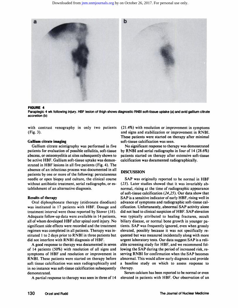
FIGURE4Paraplegic 4 wk following injury. HBFlesion of thigh ShOWSdiagnostic RNBIsoft-tissue uptake (a)and avid gallium citrateaccretion(b)
with contrast venography in only two patients(Fig. 3).
Gallium citrate imagingGallium citrate scintigraphy was performed in five
patients for evaluation of possible cellulitis, soft-tissueabscess, or osteomyelitis at sites subsequently shown tobe active HBF. Gallium soft-tissue uptake was demonstrated in HBF lesions in all five patients (Fig. 4). Theabsence of an infectious process was documented in allpatients by one or more of the following: percutaneousneedle or open biopsy and culture, the clinical coursewithout antibiotic treatment, serial radiographs, or establishment of an alternative diagnosis.
Results of therapyOral diphosphonate therapy (etidronate disodium)
was instituted in 17 patients with HBF. Dosage andtreatment interval were those reported by Stover (16).Adequate follow-up data were available in 14 patients,all ofwhom developed HBF after spinal cord injury. Nosignificant side effects were recorded and the treatmentregimen was completed in all patients. Therapy was instituted I to 2 days prior to RNBI in three patients butdid not interfere with RNBI diagnosis of HBF.
A good responseto therapy was documented in sevenof 14 patients (50%) with resolution of all signs andsymptoms of HBF and resolution or improvement inRNBI. These patients were started on therapy beforesoft tissue calcification was seen radiographically andin no instance was soft-tissue calcification subsequentlydemonstrated.
A partial response to therapy was seen in three of 14
(21 .4%) with resolution or improvement in symptomsand signs and stabilization or improvement in RNBI.These patients were started on therapy after minimalsoft-tissue calcification was seen.
No significant response to therapy was demonstratedby RNBI and serial radiographs in four of 14 (28.6%)patients started on therapy after extensive soft-tissuecalcification was documented radiographically.
DISCUSSION
SAP was originally reported to be normal in HBF(23). Later studies showed that it was invariably abnormal, rising at the time of radiographic appearanceof soft-tissue calcification (24,25). Our data show thatSAP is a sensitive indicator ofearly HBF, rising well inadvance of symptoms and radiographic soft-tissue calcification. Unfortunately, abnormal SAP activity alonedid not lead to clinical suspicion of HBF. SAP elevationwas typically attributed to healing fractures, occultbiliary disease, or normal bone growth in younger patients. SAP was frequently ignored, even when grosslyelevated, possibly because it was not specifically requested but was measured incidentally along with moreurgent laboratory tests. Our data suggest SAP is a reliable screening study for HBF, and we recommend following the SAP during the period of increased risk, reserving RNBI for confirmation when the SAP becomesabnormal. This would allow early diagnosis and providea baseline study on which to assess response totherapy.
Serum calcium has been reported to be normal or evenelevated in patients with HBF. Our observation of an
TheJournalof NuclearMedicine130 OrzelandRudd
by on October 26, 2017. For personal use only. jnm.snmjournals.org Downloaded from

4 IS
b
dC
FIGURE5Quadraplegic 5 wk following injury.RNVperformedwith[9@Tc]diphosphonate(a,b)demonstratesmild irregularityand narrowingof femoralvein(arrows).Immediateblood-pool(C)anddelayedstatic images(d)are dlagnosticof HBF,accountingfor yenous abnormality
efficacy of this combined study has not beenestablished.
The clinical presentation of acute HBF frequentlysuggested a variety of infectious processes resulting inrequests for gallium citrate imaging. While gallium localization in HBF has not been described, it is not surprising that gallium accretion occurs in the inflammatorylesion of early HBF and in the bone and marrow dcments of mature HBF.
Diphosphonates are potent in vivo and in vitro inhibitors ofcalcification and our data, though from a smallseries, are in good agreement with the literature on theclinical effectiveness of these agents in the medicaltreatment of HBF (17). If treatment is to be effective itshould ideally be instituted before there is radiographicevidence of heterotopic ossification.
HBF is a relatively common disease in the setting ofneurologic insult, soft-tissue trauma, and joint arthroplasty. On the basis of this review, we can make certainsuggestions to optimize the diagnosis and managementof HBF.
1. Three-phase RNBI should be done in all patientssuspected of HBF, even if an abnormal or normal butrising SAP is the only evidence of disease activity.
2. The first-pass study should examine the area of
clinical suspicion and immediate blood-pool imagesshould examine this and all other areas of possible disease distribution.
acute depression in serum calcium has not been previously reported. Although the average depression inserum calcium observed in acute HBF was not great, thisfinding was so consistent that we speculate it may be theearliest biochemical indicator of HBF and might playa causative role in the pathogenesis of HBF. Depressionof total and ionized calcium of a similar magnitude wasdemonstrated by Clowes and Simcone in surgical andtraumatized patients (26), suggesting the alternativeexplanation that hypocalcemia may be a nonspecificresult of trauma, unrelated to the development of HBF.A prospectivestudy of ionizedcalcium in patientsat riskfor HBF is needed to resolve this issue.
Our previous demonstration that extrinsic venouscompression from HBF can affect the RNV (27),suggests caution should be used in interpreting RNV inpatients at risk far both HBF and thrombophlebitis.Abnormal RNV might require contrast venography fordefinitive diagr@psisof thrombosis; conversely, a normalRNV should immediately suggest the possibilityof HBF.One practical approach in such patients would be to inject a bone imaging agent through the dorsal veins of thefeet to obtain an RNV (Fig. 5). This can be followed byimmediate blood-pool and delayed static images of allpossible areas of I-IBF. If RNV abnormalities cannot beattributed to extrinsic compression by HBF, the diagnosisof venous thrombosis is more secure. This approachsacrifices the first-pass information and the diagnostic
131volume26 •Number2 •February1985
a è.
I
by on October 26, 2017. For personal use only. jnm.snmjournals.org Downloaded from

3. Delayed static images of all areas susceptible toHBF are indicated. Ifstatic images are negative, in thepresence of hyperemia, repeat RNBI should be performed in I wk.
4. Radiographs may be obtained to demonstrate cvidence of soft-tissue calcification but should not be reliedon for early diagnosis.
5. If oral diphosphonate therapy is to be used, itshould be started at the first symptom of disease or theearliest observed rise in SAP, pending imagingconfirmation.
6. HBF should be considered in the differential of allpatients referred for venography for suspected thrombophlebitis if the clinical setting is appropriate.
Finally, prospective studies are needed to define therelationship of serum calcium changes to the onset ofI-IBF and to evaluate the clinical utility ofserial RNBIstudies (quantitative and qualitative) in monitoringdisease activity.
REFERENCES
1. Hardy AG, DicksonJW: Pathological ossification intraumatic paraplegia. J Bone Joint Surg 45B:76-87,1963
2. SilverJR:Heterotopicossification:A clinicalstudyofits possible relationship to trauma. Paraplegia 7:220—230, 1969
3. Buring K: On the origin ofcells in heterotopic bone formation. C/in Orthop 110:293-302, 1975
4. Smith R: Myositis ossificansprogressiva:A review ofcurrent problems.Semin Arthritis Rheum 4:369—381,I975
5. Rossier AB, Bussat PH, Infante F, Ct al: Current factson para-osteo-arthropathy(POA). Paraplegia 2:36-78,I973
6. Nollen AJG, SlooffTJJH: Para-articular ossificationsafter total hip replacement. Ada Orthop Scand 44:230—241,1973
7. Miller LF, O'Neill C: Myositis ossificans in paraplegics.J Bone JointSurg 31-A:283-294,I949
8. BerrolS: Heterotopicossificationin casesof head injury.WestJMed 140:605-606,1984
9. Stover SL, Niemann KMW, Miller JM: Disodiumetidronate in the prevention of postoperative recurrence ofheterotopicossificationin spinalcord injury patients.JBoneJointSurg 58-A:683-688,1976
10. GoldmanAB:Myositisossificanscircumscripta:A benign lesion with a malignant differential diagnosis. AmJRoentgenol 126:32-40, 1976
I 1. Yaghmai I: Myositis ossificans: Diagnostic value of arteriography. Am J Roentgenol 128:81 1-816, 1977
I2. FreedJH, DreisbachiN, Hahn H, et al: The useof thethreephasebonescanintheearlydiagnosisofheterotopicossification (HO) and in the evaluation of Didroneltherapy. Proceedings ofthe Seventh Annual ScientificMeeting of the American Spinal Injury Association,March 26-30, 1981, New Orleans, pp 63-64
I3. MolloyJC, McGuirk RA:Treatment of traumatic myositis ossificans circumscripta: Use of aspiration andsteroids.JTrauma 16:851—857,1976
14. NaftchiNE, ViauAT, SellGH, Ctal:Spinalcordinjury:Effect of thyrocalcitonin on periarticular bone formationin three subjects. Arch Phys Med Rehabil 60:280—283,1979
15. Coventry MB, Scanlon PW: The use of irradiation todiscourageectopicbone. J BoneJoint Surg 63A:201-208,1981
16. Stover SL, Hahn HR. Miller JM: Disodium etidronatein the prevention of heterotopic ossification followingspinal cord injury. Paraplegia 14:146—156, 1976
I7. Finerman GAM, Stover SL: Heterotopic ossificationfollowing hip replacement or spinal cord injury. Twoclinical studies with EHDP. Metab Bone Dis Rel Res4,5:337—342, 1981
I8. Plasmans CMT, KuypersW, Slooff TJJH: The effectof ethane-1-hydroxy-1,1-diphosphonicacid (EHDP) onmatrix induced ectopic bone formation. C/in Orthop132:233—243, 1978
19. Suzuki Y, Hisada K,Takeda M: Demonstrationof myositisossificansby 99mTcpyrophosphatebonescanning.Radiology111:663-664,1974
20. StoverSL, Hataway Ci, Zeiger HE: Heterotopicossification in spinalcord-injuredpatients. Arch Phys MedRehabil56:199-204,1975
21 . Tanaka 1, Rossier AB, Hussey RW, et al: Quantitativeassessmentof para-osteo-arthropathyand its maturationon serial radionuclide bone images. Radiology 123:217—221,1977
22. Aurbach GD, Marx Si, Speigel AM: Parathyroid hormone, calcitonin, and the calciferols. In Textbook ofEndocrinology, Williams RH, ed. Philadelphia, W. B.Saunders, 1981,pp 924-925
23. Venier LH, Ditunno iF, Jr: Heterotopic ossification inthe paraplegic patient. Arch Phys Med Rehabil 52:475—479,1971
24. Nicholas ii: Ectopic bone formation in patients withspinalcord injury.ArchPhys MedRehabil 54:354-359,I973
25. Furman R, Nicholas ii, Jivoff L: Elevation of the serumalkaline phosphatase coincident with ectopic-bone formation in paraplegic patients. J Bone Joint Surg 52A:1131—1137,1970
26. Clowes GHA Jr, Simeone FA: Acute hypocalcenia insurgical patients.AnnSurg 146:530—541,1957
27. Orzel JA, Rudd TG, Nelp WB: Heterotopicbone formation (myositis ossificans) and lower extremity swellingmimickingdeep venous thrombosis. J Nucl Med 25:1105—1107,1984
TheJournalof NuclearMedicine132 OrzelandRudd
by on October 26, 2017. For personal use only. jnm.snmjournals.org Downloaded from

1985;26:125-132.J Nucl Med. Joseph A. Orzel and Thomas G. Rudd Heterotopic Bone Formation: Clinical, Laboratory, and Imaging Correlation
http://jnm.snmjournals.org/content/26/2/125This article and updated information are available at:
http://jnm.snmjournals.org/site/subscriptions/online.xhtml
Information about subscriptions to JNM can be found at:
http://jnm.snmjournals.org/site/misc/permission.xhtmlInformation about reproducing figures, tables, or other portions of this article can be found online at:
(Print ISSN: 0161-5505, Online ISSN: 2159-662X)1850 Samuel Morse Drive, Reston, VA 20190.SNMMI | Society of Nuclear Medicine and Molecular Imaging
is published monthly.The Journal of Nuclear Medicine
© Copyright 1985 SNMMI; all rights reserved.
by on October 26, 2017. For personal use only. jnm.snmjournals.org Downloaded from