Embed Size (px)
Citation preview
INSECTS OF MICRON SIA
Heteroptera: Enicocepha idae1
By ROBERT L. USINGERUNIVERSITY OF CALIFORNIA, BERKELEY
and PEDRO WYGODZINSKYINSTITUTO MIGUEL LILLO, TUCUM.AN, ARGENTINA
INTRODUCTION
Enicocephalidae, or gnat bugs, are perhaps the most uni ue of all Heteroptera. The wings are not heteropterous at all but are tota ly membranous.The head is long and usually strongly bilobed with compound yes on the frontlobe and ocelli on the hind lobe.
Enicocephalids were formerly grouped with reduviids [R uter, 1910, ActaSoc. Sci. Fenn. 37 (3) : 1-171] but are now recognized as dis inct from Reduvioidea on the bases of different venation, eggs without ca s, distinct malegenitalia, and other characters. The true position of Enico ephalidae in thephylogenetic system of Hemiptera is not known.
The biology of Enicocephalidae has been summarized in wo monographs(Jeannel, 1941, Soc. Ent. France, Ann. 110:273-368; Usi ger, 1945, Ent.Soc. Am., Ann. 38: 321-342) and further details are given b Carayon [1950,Mus. Nat. Rist. Natur. Paris, Bull. II, 22 (6) : 739-745; 1951, Soc. Ent.France, Bull. 1951: 39-41]. The essential points are as foUos: Nymphs andadults occur under stones, beneath bark, and in rotten logs, nd in humus inthe soil. They are predaceous and apparently polyphagous. T eir most strikinghabit is their swarming, which takes place usually in the aft moon and commonly during or after rains. The swarms are exactly like th se of chironomidmidges and not unlike those of may flies and some other in ects. Both sexesswarm.
Apterous Enicocephalidae were first recorded by Ender ein (1904, Zoo1.Anzeiger 27: 783-788) for a curious genus found on Crozet I land by the German South Polar Expedition. Recently, other apterous f rms have been
1 This represents, in small part, Results of Professor T. Esaki's Micrones an Expeditions (19361940), No. 102.
Insects of Microne~Vol.7, 0.5,1960
DISTRIBUTION
Enicocephalids are rare in collections and ar generally overlooked, even,in such regions as the Hawaiian Islands and Eu ope, where the insect faumh~~:is well known. Therefore, generalizations base on the not very numerousi;;tspecimens of two genera and four species now kn wn from Micronesia are not.?!justifiable. All that can be said is that Oncyloco is is a tropicopolitan genu$~with its greatest development in Africa, the Orie t, and Australasia and witifa few species in Central artd South America. One species is known from Fiif(and several have been described from New Guine , the Philippines, and neigh",boring islands. N esenicocephalus is more restric ed, with one species inHawaiian Islands, one in the Philippine Islands, and an undescribed spe·,before us from New Guinea. Present knowledge ndicates that within Mi.nesia each Oncylocotis is a one-island endemic. esenicocephalus dybasi :likewise prove to be a one-island endemic if, as e suspect, the single fe',from Palau proves to represent a distinct species.
described by Woodward [1956, Roy. Soc. New ealand, Trans. 84 (2) : 391430] in New Zealand. Also, a dimorphic genus, Alienates, was described byBarber (1953, Am. Mus. Nov. 1614: 1-4) from imini Island.
Two types of wing polymorphism are seen in the Micronesian collections:One, a simple type of brachyptery (Oncylocoti ); the other, an interestingcase of sexual dimorphism (Nesenicocephalus) . n the latter case, the malesare fully macropterous, whereas the females hav no wings. Since the thoraxis normally developed, it is possible that the wi s are shed. (We have seensuch shedding of wings in a South American e icocephalid, upon which weshall report in detail in another publication.)
The illustrations for this paper were drawn y Wygodzinsky from specimens in liquid, as the hairs are apt to hide the tr e outlines of the body partswhen dry. Furthermore, in these generally soft- died insects, dry specimenshave a tendency to shrink. Coloring, howeve , should be examined anddescribed in dry specimens.
The United States Office of Naval Researc , the Pacific Science Board(National Research Council), the National Scie ce Foundation, and BerniceP. Bishop Museum have made this survey and p blication of the results possible. Field research was aided by a contract tween the Office of NavalResearch, Department of the Navy, and the Na ional Academy of Sciences,NR 160-175.
The following symbols indicate the instituti ns in which specimens arestored: US (United States National Museum), ISHOP (Bishop Museum),KU (Kyushu University), and CM (Chicago Na ral History Museum).
The formula 1 + 1, 3 + 3, et cetera, used in keys and descriptions refersto spines or bristles bilaterally arranged, 1, 3,et etera, on each sige.
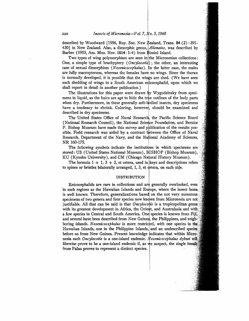
220
Body elongate. Head bilohed. Rostrum and antennae four-segmented. Eyes and ocellidistinct in Micronesian species. Pronotum trilobed in Micronesian species, and ventrallywith fore coxal cavities open behind. Prosternum without stridulatory sulcus. Front wingsentirely membranous with apical discal cell closed and basal discal present (Oncylocotis)or absent (Nesenicocephalus). Scent gland orifice lacking on metapleura, a single orificepresent at middle of third visible abdominal tergite. Front legs more or less incrassate, thetibiae slightly to strongly widened apically with a small projection on inner apex bearingspecialized spine-like setae; similar spine-like setae also on undersurface of tarsus subapically. Front tarsi one-segmented with two claws in Micronesian species; middle andhind tarsi two-segmented with two claws.
Usinger, Wygodzinsky, Ferris-Heteroptera
Distribution of Micronesian Enicocephalidae:1"
.; MICRONESIAN ISLANDGROUPS
Caroline
.:!!.
'"'£
a <'" "'C .:: " "'" :s 0.
.~~ '" 0- '0 ". '"~ ~ r:en -;; .. .. 0 "Po< >< () f-< Po< i:4- - - :- -
1. Nesenicocephalus dybasi* ? X2. Oncylocotis swezeyi X3. O. gracilis* X4. O. esakii* X5. O. capitonis* X
• Described as new.
SYSTEMATICS
FAMILY ENICOCEPHALIDAE
221
..~
,,: ~
.:~
. ~
.,1.'\"
2.
3.
KEY TO MICRONESIAN GENERA AND SPECIES OF ENICOCEPHALIDAE
Wings reduced or a,bsent 2Wings complete h 3Wings reduced to short but distinct pads j body more than 4 mm Oncylocotis sp.Wings deciduous or absent j body about 1 mm hNesenicocephaluaBasal discal cell lacking; apical discal cell truncate distally, veins with one row
of setae only (fig. 1, d) ; projection of fore tibia with three straight and onec6nspicuously curved spines, 1+1 similar, though shorter, spines on tarsusbefore apex (fig. 1, g); small, about 1 mm Nesenicocephalus dybasi
Basal discal cell present; apical diocal cell pointed distally, veins with a doublerow of setae (fig. 2, a, c) ; projection of fore tibia with numerous straight spinesonly, similar spines on fore tarsus ventrally (fig. 2, e) ; larger, 4.5-8 mm.(Oncylocotis) .h h h..hhh hh : 4
4. Fourth antennal segment distinctly longer than third (fig. 2, a). Small, 4.5 mm.Legs brown with knees ochraceous. Genital capsule of male subsemicircular(fig. 2, g, i) h h h Oncylocotia swezeyi
".i
Insects of Micronesia-Vol. 7, No.5, 1960
Genus Nesenicocephalus Usinger
N esenicocephalus Usinger, 1939, Hawaiian Ent. Soc., Proc. 10 (2) : 268-270.Very small, dark-colored species with relatively slender legs and with apical discal
cell closed and basal discal cell absent. Males fully winged. Females with wings absentor deciduous. Venation of hind wings complete.
A tendency toward reduction of fore wing is shown not only by the lack of basal .,discal cell, but by the absence of free area between anal vein and wing border on posterior'two-thirds of wing border. .
Type species: Nesenicocephalus hawaiienS"is Usinger.
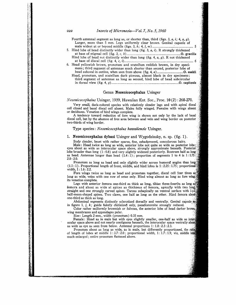
1. Nesenicocephalus dybasi Usinger and Wygodzinsky, n. sp. (fig. 1).Body slender, beset with rather sparse, fine, subadpressed, concolorous hairs. -tMale; Head twice as long as wide, anterior lobe not quite so wide as posterior lobe;:¥
eyes about as wide as interocular space above, strongly approximate beneath. Posterior:/"lobe broader than long (1 : 0.8) and very slightly widened posteriorly. Rostrum half as long~\':'i)as head. Antennae longer than head (1.4: I); proportion of segments 1 to 4 is 1: 1.75.;.~?2.0: 2.0. .:;',:
Pronotum as long as head and only slightly wider across humera1 angles than Ion·(1.1 ; 1). Proportional length of front, middle, and hind lobes is 1: 2.25 : 1.75; proportiwidth, 1 : 1.6: 2.2.
Fore wings twice as long as head and pronotum together, discal cell four timeslong as wide, veins with one row of setae only. Hind wing almost as long as fore w'its venation complete.
Legs with anterior femora one-third as thick as long, tibiae three-fourths as long'femora and about as wide at apices as thickness of femora, apically with two Istraight and one strongly curved spines. Tarsus subapically on ventral surface with 1half-moon-shaped spines. Two claws, one half as long as the other. Hind femora aone-third as thick as long. .
Abdominal segments distinctly sclerotized dorsally and ventrally. Genital capsule.:in figure I, j, k; guide faintly chitinized only, pseudosternite strongly reduced. ,"
Color rather uniformly brownish or fulvous, the anterior lobe of head darker brawing membranes and appendages paler.
Size: Length 2 mm., width (pronotum) 0.35 mm. .Female: Head as in male but with eyes slightly smaller, one-half as wide as in .:'
ocular space above and not nearly contiguous beneath, the interocular space ventrally a .as wide as eye as seen from below. Antennal proportions 1 : 1.8: 2.3 : 2.3.
Pronotum about as long as wide, as in male, but differently proportioned, theof length of lobes at middle 1: 3.7: 2.0; proportional width, 1: 1.7: 1.9, viz, middle tmuch enlarged; entire pronotum flattened above.
Fourth antennal segment as long as, or shorter than, third (figs. 3, a, i; 4, a, g).Larger, more than 5 mm. Legs uniformly clear brown. Genital capsule ofmale widest at or heyond middle (figs. 3, h; 4, j, m} : 5
S. Hind lobe of head distinctly wider than long (fig. 3, a, i). R strongly thickenedat base of stigmal cell (fig. 3, C, k} O. gracilis
Hind lobe of head not distinctly wider than long (fig. 4, a, g). R not thickenedat base of discal cell (fig. 4, C, i} 6
6. Head yellowish brown, pronotum and scutellum reddish brown, in dry specimens; third segment of antennae much shorter than second, posterior lobe ofhead suboval in outline, when seen from above (fig. 4, a} O. esakU
Head, pronotum, and scutellum dark piceous, almost black in dry specimens;third segment of antennae as long as second, hind lobe of head subcircularin dorsal view (fig. 4, g} O. capitonis '
222
,"}
,Ii
":,("is
223
~o
Usinger, W ygodzinsky, Ferris-Heteroptera
FIGURE l.-Nesenicocephalus dybasi. a-k, male: a, head, pron(ltum, and scutellum,dorsal view; b, head, lateral view; c, head, seen from below; d, fore wing; e, hind wing;j, fore leg, outlines; g, apex of tibia and tarsus of fore leg; h, hind leg; i, bristles ofabdominal tergites; j, genital region, seen from above; k, genital capsule, seen frombehind. I-p, female: I, head and thorax, seen from above; 111, head, lateral view; n, foreleg; 0, hind leg; p, genital region, ventral view.
\:~;,
Insects of Micronesiev-Vol. 7, No. 5;1960224
Genus Oneyloeotis Stal
Oncylocotis Stal, 1855, Ofv. K. Vet.-Akad., Forh. 12: 44.Dicephalus Kirby, 1891, Linn. Soc. London, Jour. 24: 117.Sphigmocephalus Enderlein, 1904, Zoo1. Anzeiger 27: 785.Didymocephalus Jeannel, 1941, Soc. Ent. France, Ann. 110: 335.
Robust species with pubescence of erect hairs. Middle lobe of pronotum with deemedian longitudinal sulcus terminating before posterior constriction in short, transverdepression. Lateral discs of middle lobe each with distinct, tripartite depression. Frwings' fully developed or more or less brachypterous, but with basal discal cell presenand apical discal cell closed. . .t,
ifType species: Oncylocotis nasutus Stal. \~
Usinger's'interpretation (1945, Ent. Soc. Am., Ann. 38: 321-342) of this!:;':large and important genus is different from that of Jeannel (1941) and has not~~
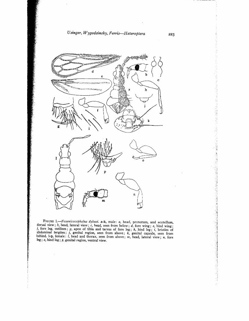
Meso- and metanota completely exposed. Wings lacking (apparently shed). Scutellumabout one-third as long as pronotum, the sides concave and apex broad. Metanotum aboutas long as scutellum, with depressed area at middle and raised areas on either side converging posteriorly.
Legs incrassate, fore femora half as thick as long, tibiae one-third as wide at apices }as long. Hind femora also incrassate, half as thick as long.
Abdomen membranous. Center of last stemite slightly salient, its margin somewhat "~I
more chitinized. ,;"Color much as in male but with last two antennal segments white, and saclike "',
abdomen pale-colored. :::.Size as in male.
i\Holotype, male (US 64520), Ponape, north slope, Mt. Kupwuriso, 300- ,I,
450 m., Mar. 11, 1948, beating vegetation, Dybas. Allotype, female, Mt. Temwetemwensekir, 150-450 m., Mar. 23, 1948, Dybas. Paratypes (CM, BISHOP), same data as for holotype but Mar. 8, 1948; female, Nanipil, Net District, Feb. 27, 1948, Dybas.
DISTRIBUTION: Caroline Is. (Ponape).This species is named for ,H. S. Dybas, the assiduous collector who is per
sonally responsible for gathering nearly all of the enicocephalids known fromMicronesia. It is the smallest thus far described in the genus. N. philippinensisis much larger and darker with pale wing bases and reddish costal marginsand has erect white hairs on head and pronotum. N. hawaiiensis is slightlylarger and is also darker with white hairs; it differs further in the smaller eyes,longer second antennal segment, longer hind lobe of pronotum, and shorter, .broader discal cell; it is only 2.5 times as long as wide.
A single wingless female is also at hand from Garakayo (Ngergoi), Palau,collected August 7, 1945, under bark, by Dybas. This specimen is similar toN. dybasi in most respects but has rather long white pubescence. It is notincluded in the paratypes. Also, a last instar nymph is before us from the eastcoast of Peleliu Island, Palau, collected January 26, 1948, by Dybas.
225Usinger, Wygodzinsky, Ferris-Heteroptera
i: been followed by Villiers in several papers of recent date. The type of Oncylo~;cotis has been studied and falls well within the range of variation seen in~';'species from diverse parts of the world. Even if this interpretation is rejected,i Jeannel's name cannot be used because Didymocephalus Jeannel (1941) and{Sphigmocephalus Enderlein (1904) are isogenotypic, each having as its type;'Henicocephalus basalis Westw90d (= Henicocephalus curculio Karsch) ..,
.,;,
I'.j
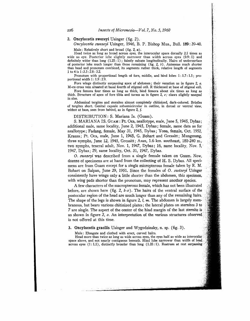
FIGURE 2.-0ncylocotis swezeyi, a-i, male: G, whole insect, s~en from above, bristlesof wings not shown; b, head, lateral view; c, setae of vein of fore wing; d, fore leg; e,apex of tibia and tarsus of fore leg; j, hind leg; g, abdomen, ventral view, membranousareas stippled; h, bristles of abdominal tergite; i, genital region, seen from below; j,genital capsule, seen from behind. k-o, female: k, head, lateral view; I,. fore leg; m, hindleg; n, abdomen (dorsal surface left side, ventral right side) ; 0, center of posterior borderof last sternite.
Insects of Micronesiar-Vol. 7, No.5, 1960
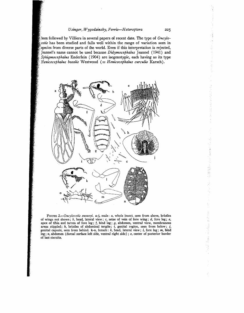
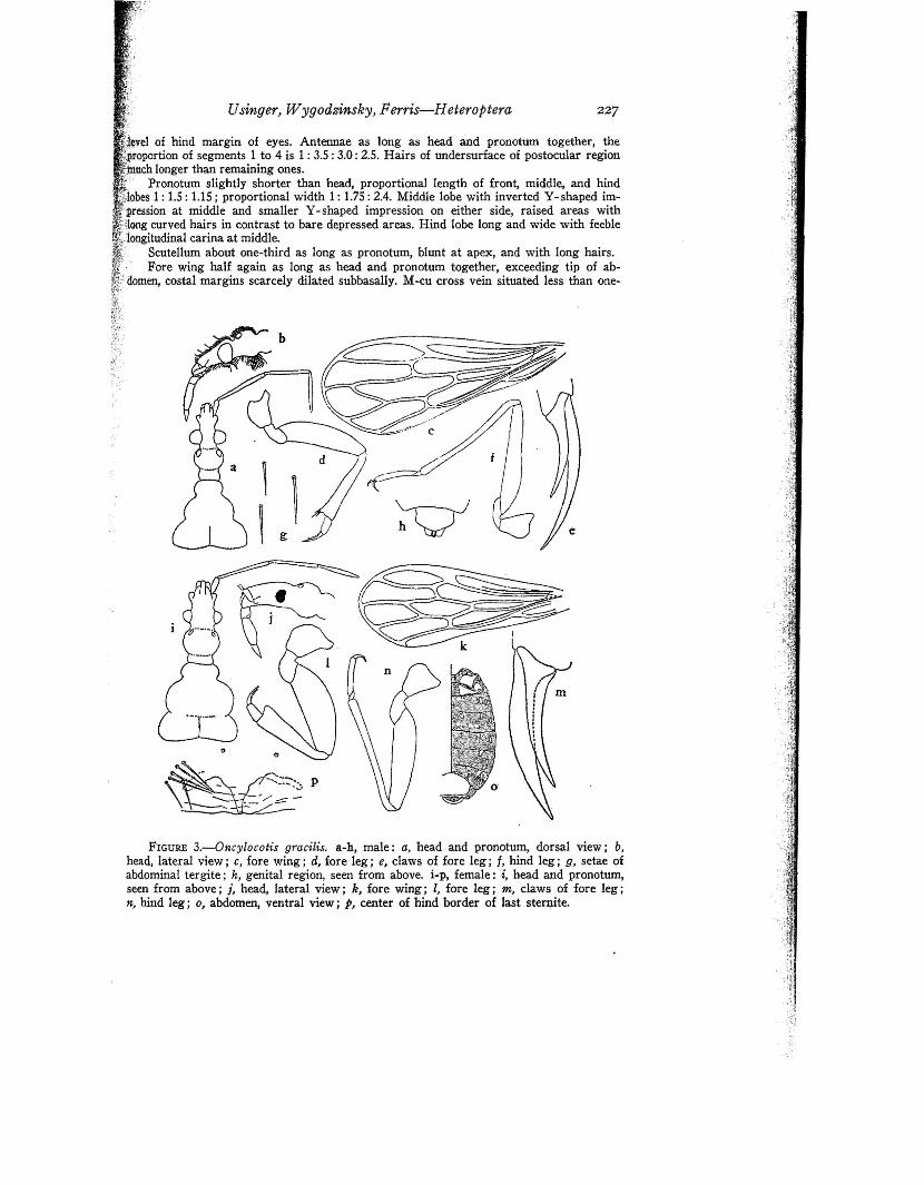
3. Oncylocotis gracilis Usinger and Wygodzinsky, n. sp. (fig. 3).Male: Elongate and clothed with erect, curved hairs. :,,-~
Head more than twice as long as wide across eyes, the eyes half as wide as interoculal::::;~.
space above, and not n~a~ly contiguous beneath. Hind lobe narrower than width of h~r!i!across eyes (1: 1.1), distinctly broader than long (1.35: 1). Rostrum at rest surpassIng'"
_~,:-,~,:",:;;",;,;,;';";';' -··ii··i-i-."i·-iii··iiii·-·-i-iiii·ii.i'••i-iii·-.-.'•• m ~. _ .~. •• ~_ - ......~-. - _.. --- -_ .• ' "'-••_.
.. - ..,,-.-
DISTRIBUTION; S. Mariana Is. (Guam).S. MARIANA IS. GUAM : Pt. Oca, neallotype, male, June 5,1945, Dybas;
additional male, same locality, June 2, 1945, Dybas; female, same data as forneallotype j Fadang, female, May 31, 1945, Dybas; Yona, female, Oct. 1952,Krauss; Pt. Oca, male, June 1, 1945, G. Bohart and Gressitt; Mungmung,three nymphs, June 12, 1945, Gressitt; Asan, 1.6 km. southeast, 180-240 m.,two nymphs, teneral adult, Nov. 1, 1947, Dybas; 16, same locality, Nov. 5,'1947, Dybas; 29, same locality, Oct. 31, 1947, Dybas.
O. swesryi was described from a single female taken on Guam. Now,dozens of specimens are at hand from the collecting of H. S. Dybas: All specimens are from Guam except for a single micropterous female taken by R. M.Bohart on Saipan, June 29, 1951. Since the females of O. swBzeyi Usingerconsistently have wings only a little shorter than the abdomen, this specimen,with wing pads shorter than the pronotum, may represent another species.
A few characters of the macropterous female, which has not been illustratedbefore, are shown here (fig. 2, k-o). The hairs of the ventral surface of thepostocular region of the head are much longer than any of the remaining hairs..,The shape of the legs is shown in figure 2, t m. The abdomen is largely mem- .branous, but bears various chitinized plates; the lateral plates on sternites 3 to ',:;7 are single. The aspect of the center of the hind margin of the last sternite is ;.!}as shown in figure 2, o. An interpretation of the various structures observed.:{"is not offered at this time. .';
2. Oncylocotis swezeyi Usinger (fig. 2).Oncylocotis swezeyi Usinger, 1946, B. P. Bishop Mus., Bull. 189: 39-40.Male: Relatively short and broad (fig. Z, a).Head twice as long as broad across eyes, the interocular space dorsally 2.5 times as
wide as eye. Posterior lobe slightly narrower than width across eyes (0.9: 1) anddefinitely wider than long (1.25: 1) ; faintly sulcate longitudinally. Hairs of undersurfaceof posterior lobe much longer than those remaining (fig. 2, b). Antennae much shorterthan head and pronotum combined, its segments rather, thick, relative length of segments1 to 4 is 1 :3.3: 2.8: 3.2.
Pronotum with proportional length of fore, middle, and hind lobes 1: 1.7: 1.5 i proportional width 1 : 1.9: 2.9.
Fore wings distinctly surpassing apex of abdomen i their venation as in figure 2, a.M-cu cross vein situated at basal fourth of stigmal cell. R thickened at base of stigmal cell.
Fore femora four times as long as thick, hind femora about six times as long asthick. Structure of apex of fore tibia and tarsus as in figure 2, e; claws slightly unequalin size.
Abdominal tergites and sternites almost completely chitinized, dark-colored. Bristlesof tergites short. Genital capsule subsemicircular in outline, in dorsal or ventral view,widest at base, seen from behind, as in figure 2, j.
226
1;·
227Usinger, W ygodzinsky, Ferris-Heteroptera
FIGURE 3.-0ncylocotis gracilis. a-h, male: a, head and pronotum, dorsal view; b,head, lateral view; c, fore wing; d, fore leg; e, claws of fore leg; f, hind leg; g, setae ofabdominal tergite; h, genital region, seen from above. i-PI female: i, head and pronotum,seen from above; j, head, lateral view; k, fore wing; 1, fore leg; m, claws of fore leg;n, hind leg; 0, abdomen, ventral view; p, center of hind border of last sternite.
,vel of hind margin of eyes. Antennae as long as head and pronotum together, theportion of segments 1 to 4 is 1 : 3.5 : 3.0: 2.5. Hairs of undersurface of postocular regionch longer than remaining ones.
Pronotum slightly shorter than head, proportional length of front, middle, and hind'bes 1 : 1.5 : 1.15; proportional width 1 : 1.75 : 2.4. Middle lobe with inverted Y- shaped im
ssion at middle and smaller Y- shaped impression on ~ither side, raised areas with'long curved hairs in contrast to bare depressed areas. Hind lobe long and wide with feebleiongitudinal carina at middle.. Scutellum about one-third as long as pronotum, blunt at apex, and with long hairs.
Fore wing half again as long as head and pronotum together, exceeding tip of abdomen, costal margins scarcely dilated subbasally. M-cu cross vein situated less than one-
Insects of Micronesiar-Vol. 7, No.5, 1960
fourth from base of stigmal cell, much proximad of level of inner posterior angle of apicaldiscal cell. R thickened at base of stigmal cell. Veins with two rows of curved hairs.
Legs only moderately incrassate. Fore femora slightly curved, somewhat more thanfour times as long as thick. Fore tibia slightly shorter than femur (0.85: 1). Claws of forelegs conspicuously unequal in size. Hind femora 6 times as long as wide.
Abdominal stemites and tergites completely chitinized, pigmentation not very intense.Genital segment subtrapezoidal when seen from above or below, widest slightly beyondmiddle seen from behind as in O. swezeyi.
Color brown; the head, except anteriorly, pronotumat middle, and fore wings, exceptat base, darker brown, the rest of body and appendages light brown; legs uniformlycolored.
Size: Length 5.2-5.3 mm., width of pronotum 1.1 mm., width across fore wings 1.5 rom.
Holotype, male (KU), Kusaie, Lelo, Dec. 24, 1937, Esaki. Paratype, male(US), Kusaie, Mt. Tafeayat, 300-360 m., Aug. 20, 1946, Townes.
DISTRIBUTION: Caroline Is. (Kusaie).Two additional specimens are at hand (Kusaie, Mt. Matante, 380 m., south
slope, Mar. 4, 1953, Clarke; Kusaie, Mt. Tafeayat, 518 m., Feb. 9, 1953,Clarke). They differ from the above in pronotal proportions (broader middlelobe). They are females, but not included as paratypes. The head is shown infigure 3, i, j; the eyes are only about one-third of the interocular space above,and the hairs of the ventral surface of the posterior lobe are much longer thanthe remaining ones. The femora of fore legs are more strongly swollen thanthose of the males, their length three times their maximum width; the clawsare only slightly unequal in size. The hind legs are shown in figure 3, n.The abdomen is largely membranous, having dorsal chitinized plates as inO. swezeyi, but sternites 3 to 7 each have 2 + 2 small sclerotized plate~ laterally. Structure of center of hind border of last sternite as in figure 3, p.
This species differs from O. swezeyi in its larger size (intermediatetween O. swezeyi and O. esakii), the paler color, the shorter fourth antenna!segment, and the characters given in the key.
4. Oncylocotis esakii Usinger and Wygodzinsky, n. sp. (fig. 4, a-f).Male: Elongate, the surface clothed with erect hairs with curved tips.Head distinctly shining (dry specimens), slightly more than twice as long as wide-;:,
across eyes (l: 0.45). Eyes relatively small, less than half as wide as interocular space,above (1: 2.3), widely separated beneath, far removed from level of dorsal and ventraC;;surface of head in lateral view. Hind lobe narrower than width across eyes (1: 1.3), about::'as long as broad, suboval in shape in dorsal view. Antennae nearly as long as head an4:':pronotum together. Proportion of segments 1 to 4 is 1: 3.0-3.3: 2.1-2.2: 1.9-2.0. Rostrum at",~
rest reaching to level of hind margin of eyes. Hairs of undersurface of postocular portioq",,;',':of head much longer than other hairs.
Pronotum shorter than head (0.85: 1), wider than long (1.1: 1), the middle lobe,;~I,
forming an inverted Y-shaped impression on either side,· the depressions bare in contrastp,/'to pubescent raised areas. Hind lobe relatively long and broad, with feeble longitudinal':;::carina at middle. Posterior margin shallowly emarginate. Proportional length of for~ ..~'middle, and hind lobes is 1 :2.0: 2.0; proportional width, 1:1.65 : 3.2. c'.>',
Scutellum one-third as long as pronotum, raised at middle and broad at apex, w',-, ",exceptionally long curved hairs apically.
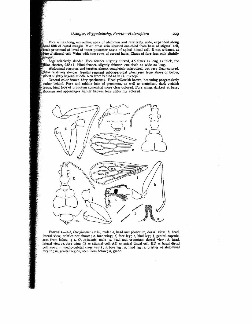
FIGURE 4.-a-£, Oncylocotis esakii, male: a, head and pronotum, dorsal view; b, head,lateral view, bristles not shown i C, fore wing j d, fore leg; e, hind leg; f, genital capsule,seen from below. g-n, O. capitonis, male: 0, head and pronotum, dorsal view j h, head,lateral view; i, fore wing (8 = stigmal cell, AD =apical discal cell, BD =basal discalcell, m-cu = medio-cubital cross vein) j j, fore leg; k, hind leg; I, bristles of abdominaltergite; m, genital region, seen from below; K, guide.
229
//
Usinger} Wygodzinsky} Ferris-Heteroptera
Fore wings long, exceeding apex of abdomen and relatively wide, expanded alongal fifth of CQstal margin. M-cu cross vein situated one-third from base of stigmal cell,
;qiuch proximad of level of inner posterior angle of apical di'scal cell. R not widened atc" , e of stigmal cell. Veins with two rows of curved hairs. Claws of fore legs only slightly·,@equal.',1 Legs relatively slender. Fore femora slightly curved, 4.5 times as long as thick, the-- ,', iae shorter, 0.85 : 1. Hind femora slightly thinner, one-sixth as wide as long. '
Abdominal sternites and tergites almost completely sclerotized, but very Clear-cQlored.• ' etae relatively slender. Genital segment subtrapezoidal when seen from above or below,;,~dest slightly beyond middle seen from behind as in O. S'lI'euyi.Y: General color brown (dry specimens). Head yellowish brown, becoming progressively';,.darker behind. Fore and middle lobe of pronotum, as well as scutell1,U1l, dark reddish':brown, hind lobe of pronotum somewhat more clear-colored. Fore wings darkest at base;
·;:ibdomen and appendages lighter brown, legs uniformly colored.
, .,i'
~;, .',.I" ~
I
iI
ii
!! '
Insects of Micronesiar-Vol. 7, No.5, 1960
Holotype, male (KU), Ponape, Nipot, July 20, 1939, Esaki. Paratypes,two males, same data as for holotype; four males (CM, BISHOP), Ponape,summit, Mt. Kupwuriso, 600 m., Mar. 10, 1948, beating vegetation, Dybas.
DISTRIBUTION: Caroline Is. (Ponape).This species is dedicated to Professor Teiso Esaki, early student of Enico
cephalidae and pioneer collector in Micronesia, whose recent death deprivedthe world of one of its leading entomologists.
5. Oncylocotis capitonis Usinger and Wygodzinsky2, n. sp. (fig. 4, g-n).Surface of head dull (dry specimens) ; its hind lobe subcircular in outline when seen
from above, more strongly elevated in lateral aspect.Proportion of antennal segments 1 'to 4 is 1: 3.5 : 3.5 : 3.0, viz, third segment as long
as second (considerably shorter than second in O. esakii).M-cu cross vein situated nearer middle of stigmal cell, almost at level of inner
posterior angle of apical discal cell. R narrow at base of stigmal cell, as in O. esakii (fig.4, i).
Color (dry specimens) : Head, pronotum, and scutellum dark piceous, almost black;anteocular portion orange brown.
Fore femur thicker, less than 4 times as wide as long.
Holotype, male (US 64632), Ponape, Mt. Beirut (Pairot), about 600 m.,Mar. 13, 1948, beating vegetation, Dybas. Paratype, Ponape, Mt. Nahnalaud,about 600 m., Mar. 18, 1948, Dybas.
DISTRIBUTION: Caroline Is. (Ponape).This species agrees in most characters with O. esakii, but differs in some
characters which seem to fall outside the usual range of variability in thisgroup, as described above.
• Capito, -on;s, one having a luge bead.