Embed Size (px)
Citation preview
Biochemical Systematics and Ecology 26 (1998) 495—509
Heterogeneity of SSU and LSU rDNA sequencesof Alexandrium species
David Walsh!, Rosalind A. Reeves", David J. Saul!,Russell D. Gray#, Lincoln MacKenzie$, Patricia R. Bergquist%,*,
Peter L. Bergquist!,"
!Centre for Gene Technology, School of Biological Sciences, University of Auckland, New Zealand"School of Biological Sciences, Macquarie University, Sydney, New South Wales 2109, Australia
#Department of Psychology, University of Auckland, New Zealand$Cawthron Institute, Nelson, New Zealand
%Centre for Marine Science, School of Biological Sciences, University of Auckland, P. Bag 92019, Auckland,New Zealand
Received 1 May 1997; accepted 24 May 1997
Abstract
With the growth of aquaculture, toxic phytoplankton have become a significant problem. Atpresent, the identification of specific algal species within blooms requires considerable expertise,and the methods are both costly and time consuming. It is clear that alternative diagnostic testsmust be developed that allow rapid and simple assessments to be made on site. We report onthe identification of unique sequences within the SSU and LSU rDNA genes of Alexandriumspecies that provide data for the design of species- and genus-specific molecular probes. Inaddition we examine some aspects of the phylogeny of Alexandrium isolates from New Zealandwaters. ( 1998 Elsevier Science Ltd. All rights reserved.
Keywords: Alexandrium; Toxic phytoplankton; Algal bloom; Molecular probe; Phylogeny
1. Introduction
Problems caused by toxic phytoplankton blooms have become significant with thegrowing emphasis on aquaculture world-wide. The New Zealand shellfish aquacul-ture industry was required to cease harvesting for an extended period in 1993 in many
*Corresponding author. Tel.: 0064 09 373 7599 X77; fax: 0064 093 737 417
0305-1978/98/$19.00 ( 1998 Elsevier Science Ltd. All rights reserved.PII: S0305-1978(98)00006-4
areas because of an algal bloom around the coast which included several toxic species.In many locations, most of the species known to produce troublesome toxins arepresent in small numbers over much of the year, and high levels of toxins producedduring a bloom may be retained selectively and differentially in bivalve tissue afteralgal numbers decline. The identification and discrimination of toxic and non-toxicalgal species relies largely on visual observations by light microscopy and scanningelectron microscopy. It is clear that identification could be made more rapid andpositive if genetic differences between families, genera and species could be exploitedby constructing molecular probes for use in rapid molecular diagnostic methods(reviewed by Anderson, 1995). We have used as a model a member of the Dino-phyceae, Alexandrium, which is the causal agent of Paralytic Shellfish Poisoning(PSP). We have initiated a search for suitable discriminatory sequences for probeconstruction and have reported previously on the sequences of the rRNA intergenicregions of the same Alexandrium isolates used in this study (Spalter et al., 1997).Adachi et al. (1996) have also reported on the diversity of sequences in the intergenicregions of a large sample of Alexandrium tamarense/A. catenella/A. fundyense and A.affine isolates. There is general agreement between the two research groups withrespect to the molecular phylogeny of Alexandrium sp. although there is only oneisolate in common that was sequenced in the two reports. As part of our search forsuitable diagnostic sequences, the current paper describes molecular diversity in thegenes coding for the large and small sub-unit ribosomal RNAs between the Alexan-drium species and isolates.
Woese and his colleagues (reviewed in Woese, 1987) have pioneered the use ofribosomal RNAs in identifying organisms and inferring their natural relationships.Ribosomal RNA sequence analysis is the method of choice for elucidatingphylogenetic relationships between micro-organisms because of its high informationcontent and differing degrees of sequence conservation, which allows distant as well asclose systematic relationships to be distinguished. We have employed a combinationof PCR-amplified SSU (18S) and LSU (28S) rRNA genes with cycle sequencing toprovide a simple and reliable method for obtaining data for the design of molecularprobes and for the inference of the organisms’ phylogeny.
Several groups have reported on variations in SSU rRNA gene sequences thatdistinguish groups and strains of Alexandrium species (Destombe et al., 1992; Scholinand Anderson, 1993, 1994). Destombe et al. (1992) showed that two Alexandriumspecies differentiated on the basis of minor morphological characteristics had diver-gent SSU rRNA sequences. Scholin and Anderson (1994) demonstrated that therewere clear differences in SSU rRNA genes to distinguish otherwise apparently closelyrelated Alexandrium species or populations as the result of sequencing studies andRFLP analysis. Their results allowed the division of the A. tamarense/A. catenella/A. fundyense complex into species clusters defined by geographic origins rather thanmorphotype designations, and in addition, revealed the presence of an apparentlynon-functional pseudogene in North American isolates that was useful for trackingthe spread of populations regionally and globally.
Amann et al. (1991) have reported on the use of LSU sequences to devise discrimi-natory probes to distinguish three closely related strains of the symbiotic bacterium
496 D. Walsh et al./Biochemical Systematics and Ecology 26 (1998) 495—509

Holospora and although sequence analysis of LSU rRNA is less well developed thanSSU, it is an obvious target for fine discrimination between closely related organisms.Lenaers et al. (1991) have inferred a molecular phylogeny of dinoflagellates usingsequence data for the divergent domains D1 and D8 of LSU rRNA and Scholin et al.(1994) have found LSU sequences to be valuable taxonomic and biogeographicalmarkers for population resolution. Recently, Scholin and Anderson (1996) havereported that RFLP analysis of PCR-amplified LSU rDNAs are useful geneticmarkers for distinguishing between isolates of Alexandrium in the A. tamarense/A. catenella/A. fundyense complex at less cost and effort than full DNA sequenceanalysis. Since no sequence information was available for Alexandrium ostenfeldii(a causative agent of PSP), or for any other Alexandrium species from the NewZealand region, we have accumulated partial LSU and complete SSU sequences fora number of isolates from different areas as well as appropriate non-New Zealandstrains to act as controls. We have also sequenced ribosomal RNA genes from severalspecies that could be confused with Alexandrium.
2. Materials and methods
The species and isolates of Alexandrium sequenced in this work have been describedpreviously (Spalter et al., 1997). Cultures of Fragilidinium sublobosum and Cachinonahallii were provided from the Lincoln MacKenzie collection at Cawthron Institute,Nelson, New Zealand.
2.1. Algal cultures and DNA preparation
Isolates of Alexandrium were maintained and genomic DNA extracted as describedin Tyrrell et al. (1996).
2.2. Polymerase chain reaction (PCR) amplification
Approximately 10 ng of genomic DNA was added to a PCR mixture containing0.25 mM of each dNTP, 3.0 mM MgCl2, 50 mM KCl, 15 pmoles of primers P1 andP2 (see results), 1.25 units of Taq DNA polymerase (AmpliTaqTM Cetus Perkin Elmer,Melbourne, Australia) in a reaction volume of 50 lL. The reaction conditions were:[94°C/1 min]]1, [94°C/1 min., 55°C/1 min, 72°C/2 min]]35, [72°C/2 min]]1.
2.3. Cloning
Two cloning strategies were used: PCR products were either inserted directly intoSmaI-cut M13mpl0, or initially inserted into pCRII (Invitrogen Corp, San Diego, CA,USA) and then transferred to M13mpl8 and M13mpl9. Extraction and preparation ofsingle-stranded template DNA for sequencing was performed as described in Tyrrellet al. (1996).
D. Walsh et al./Biochemical Systematics and Ecology 26 (1998) 495—509 497
2.4. Sequence analysis
Sequencing reactions were carried out using an ABI Catalyst robotic workstationand an ABI 373A automated DNA sequencer using dye-primer chemistry and theuniversal M13 primer. Both strands of at least two different recombinants weresequenced to check for PCR errors generated in the amplification procedure. Tem-plate preparation has been described previously (Saul et al., 1993: Tyrrell et al., 1996).Sequences were edited and manipulated using the GCG package (Devereux et al.,1984). Multiple sequence alignment was performed using PILEUP (Feng andDoolittle, 1987) using a low gap penalty and then manually adjusted to providealignments based on rRNA secondary structure.
2.5. Development of a colorimetric analysis for Alexandrium minutum
Four reverse complement primers were synthesised (see Fig. 1). One pair wascapable of binding to all dinoflagellates and the other was specific for Alexandrium.One member of each pair of primers was labelled at its 5@-end with biotin and theother member of the pair was labelled with fluorescein. The anti-fluorescein-coupledhorseradish peroxidase system in the presence of diammonium 2,2@-azino-di-[3-ethylbenzthiazoline sulfonate] (ABTS) was employed to produce a positive greencolour reaction. Biotinylated primer in TBS buffer plus 0.1% bovine serum albumen(total volume, 250 ll) was added to each well of a streptavidin-coated microtiter plate(NUNC, Naperville, IL, USA) and incubated at room temperature for 30 min. Thewells were washed five times with TBS buffer containing 0.1% Tween 20. 500 ng offluorescein-labelled primer was added to diluted anti-fluorescein horseradish per-oxidase and incubated at 37°C for 30 min with shaking. The nucleic acid template (in5]SSC) was added and the mixture apportioned into the wells of the microtiter plate.The wells were then washed three times with 5 x SSC and ABTS added and incubationcontinued for 30 min at 37°C. A positive reaction gave a green colour which was readat 405 nm on a microtitre plate reader.
2.6. Phylogenetic analysis
After alignment, the sequences were analysed using the program paup* 4.0d59.Cachonina hallii and Fragilidinium subglobosum were used as the outgroups for theSSU data and the LSU data. In morphological terms, Cachonina hallii and Fragilidin-ium subglobosum are good outgroups in that they are clearly discriminated fromAlexandrium although in size and superficial appearance in natural water samplesthey are very similar. A common problem in phylogenetic analyses is that theoutgroup taxa can be too genetically remote from the ingroup. If character statechanges on this basal branch are high enough phylogenetic signal is lost, and therooting of the tree becomes effectively random. To check whether C. hallii andF. subglobosum were appropriate outgroups pairwise transition:transversion ratioswere calculated using Paup*. As there are only two possible transitional changes andfour possible transversion, at saturation the transition:transversion ratio would be 0.5
498 D. Walsh et al./Biochemical Systematics and Ecology 26 (1998) 495—509
(assuming equal base composition; Rodrigo, et al., 1994). In the parsimony analysisall the characters were equally weighted. Alignment gaps were treated as missingcharacters. The random trees option (100 000 random trees) was used to test for thepresence of significant phylogenetic signal by examining the skewness (g
1) of the
distribution of random tree lengths (Hillis, 1991). Significance values for the (g1)
statistic were taken from Hillis and Huelsenbeck (1992). The branch-and-boundoption of PAUP* was used to find the most parsimonious tree(s). Bootstrap analysis(Felsenstein, 1985) was performed to evaluate the degree of support for the group-ings found in the most parsimonious tree(s). 2000 bootstrap replicates were used,each with a branch-and-bound search. The Kishino—Hasegawa test for significantdifferences in tree typology was used to evaluate likely alternatives to the mostparsimonious trees.
3. Results
3.1. Sequencing of genes for SSU ribosomal RNAs
The complete sequences of SSU rDNA has been completed on both strands fromfor the following isolates of micro-algae:
A. minutum (Anakoha Bay isolate); A. minutum (Croisilles Harbour isolate); A.margalefi (Bream Bay isolate); A. ostenfeldii (Kaitaia isolate); A. ostenfeldii (Timaruisolate); A. ostenfeldii (Wellington isolate), as described previously (Spalter et al., 1997).In addition, the complete sequences of the SSU rRNA genes were determined forF. subglobosum, C. halli and A. tamarense (Ipswich, MA, USA) The sequence of theA. tamarense gene was determined because of a number of uncertainty codes withinthe database submission. At the time of the research, the only available sequence hadbeen determined by the reverse transcriptase method which results in many unresol-ved bases. All sequences presented in this paper were been confirmed on both strands.The data are not repeated here but can be obtained from GenBank under accessionnumbers—SSU: A. ostenfeldii, U27500; A. minutum, U27499; A. margalefi, U27498;F. subglobosum, AF033869; C. hallii, AF033865. LSU: A. ostenfeldii, AF033533;A. minutum, AF033532; A. margalefi, AF033531; F. subglobosum, AF033868; C. hallii,AF033867.
Sequence alignment indicates that geographically separated isolates of the samespecies have very similar SSU sequences but that there are distinct differences betweenindividual species. An example is shown in Fig. 1 of an alignment of the sequencesbetween positions 481 and 560, including other information on microalgae depositedin the database. The region used for the construction of a fluorescein-labelled specifichybridisation probe is indicated (see later).
3.2. Partial sequencing of LSU ribosomal RNA genes
Scholin et al. (1994) showed that the sequences of the D1 and D2 hypervariabledomains of the LSU rRNA gene were a valuable taxonomic and biogeographical
D. Walsh et al./Biochemical Systematics and Ecology 26 (1998) 495—509 499
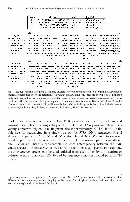
Fig. 1. Sequence lineup of regions of suitable diversity for probe construction to discriminate Alexandriumspecies. Primers used in for the detection of Alexandrium SSU gene sequences are shown 5@ to 3@ in the toptable and reversed at the positions to which they bind on the target sequences. Coordinates refer to theposition in the Alexandrium SSU gene sequence. A. minutum (A)"Anakoha Bay isolate, (C)"CroisillesHarbour isolate; A. ostenfeldii (T)"Timaru isolate, (W)"Wellington isolate, K"Kaitaia isolate;A. margalefi is Bream Bay isolate; A. tamarense is Ipswich, MA, USA isolate.
marker for Alexandrium species. The PCR primers described by Scholin andco-workers amplify as a single fragment the D1 and D2 regions and their inter-vening conserved region. The fragment size (approximately 670 bp) is of a suit-able size for sequencing in a single run on the 373A DNA sequencer. Fig. 2shows an alignment of the D1 and D2 regions for all New Zealand Alexandriumisolates plus a North American isolate of A. tamarense plus Fragilidiniumand Cachonina. There is considerable sequence heterogeneity between the indi-vidual species of Alexandrium as well as with the other algal species. For example,the Alexandrium species can be distinguished from each other by an insertion ordeletion event at positions 462-486 and by sequence variation around position 710(Fig. 2)
&&&&&&&&&&&&&&&&&&&&&&&&&&&&&&&&&&&&&&&&&&&&&"Fig. 2. Alignment of the partial DNA sequences of LSU rRNA genes from selected micro-algae. Thedifferences between the sequences are highlighted by reverse font. Single letter abbreviations for individualisolates are explained in the legend for Fig. 1.
500 D. Walsh et al./Biochemical Systematics and Ecology 26 (1998) 495—509

D. Walsh et al./Biochemical Systematics and Ecology 26 (1998) 495—509 501

3.3. Development of a diagnostic test
Trials were undertaken to determine whether it was feasible to detect the presenceof specific Alexandrium species using a colorimetric approach. For the method, fourprimers were designed: one pair was capable of binding all dinoflagellates (based onthe data available in the GenBank database at the time) and the other pair was specificfor Alexandrium sp. (Fig. 1).
The assay strategy adopted is shown in Fig. 3. In summary, the procedure relies onthe ability of an immobilised oligonucleotide to capture DNA or RNA with the LSUsequence. Once captured, this DNA/RNA hybrid can bind a second oligonucleotidewhich is fluorescein-labelled. Although fluorescein can be detected by excitation withUV, the signal is well below the level acceptable for sensitive detection. Consequently,an additional step was employed whereby an anti-fluorescein antibody-peroxidase(POD) conjugate was added. With an appropriate substrate, a colour reaction can beobtained. To test the sensitivity of this method, single-stranded DNA was made from
Fig. 3. The assay System used for the detection of specific SSU sequences.
502 D. Walsh et al./Biochemical Systematics and Ecology 26 (1998) 495—509

M13 recombinants containing rDNAs of several of the Alexandrium species. Theresults showed that Alexandrium could be differentiated from other dinoflagellates butas much as 100 ng of target DNA was required before a significant signal could bedetected. Without amplification, this signal would equate to roughly 108 cells. Clearly,a 106-fold amplification is needed for detection of suitable numbers of cells if the genesare used as the targets. The use of probes complementary to the cellular rRNA wouldincrease the sensitivity by a factor of 103—104, which may still be too low fora predictive field test of use to the industry. However, as reviewed in the discussion, therRNA gene concentration for this number of cells would be well within the rangeachieved by the Qb system that we propose to use in the future.
3.4. Phylogenetic analysis
As there was only extremely minor variation between the different geographicisolates of the New Zealand samples of A. ostenfeldii and A. minutum these sequenceswere combined into majority rule consensus sequences. For the SSU data the pairwisetransition : transversion ratios between C. hallii and F. subglobosum and the other taxaranged in 1.68—3.43, and for the LSU data they ranged in 0.86—1.26, indicating that inboth cases the comparisons were not saturated. C. hallii and F. subglobosum are thusappropriate outgroups for both data sets.
The SSU sequences contained 1462 constant sites, 237 variable sites that wereparsimony-uninformative, and 122 parsimony-informative sites. Parsimony analysisfound two shortest trees (Fig. 4) of length 445, with a consistency index (CI) of 0.88,and a retention index (RI) of 0.66. The random tree length distribution was signifi-cantly skewed, with a g
1index of !1.57. The two shortest trees differed in the
placement of A. minutum, A. ostenfeldii and A. margalefi In one of the two mostparsimonious trees these taxa form a monophyletic group that is a sister taxa to theother two Alexandrium taxa. In the second most parsimonious tree, A. minutum,A. ostenfeldii and A. margalefi are paraphyletic to the A. fundyensii/A. tamarense clade.There was strong bootstrap support for the monophyly of the Alexandrium taxa andthe A. fundyensii/A. tamarense clade.
The LSU sequences contained 340 constant sites, 221 variable sites that wereparsimony uninformative, and 192 parsimony informative sites. Parsimony analysisfound one shortest tree (Fig. 5) of length 709, with a CI 0.77, and an RI of 0.70. Therandom tree length distribution was significantly skewed, with a g
1index of !0.67.
Interestingly the A. minutum sample from New Zealand and the previously publishedsequence (Zardoya et al., 1995) do not form a monoplyletic group in the mostparsimonious LSU tree. The Zardoya sample of A. minutum is most closely related toA. lusitanicum (100% bootstrap support). Constraining the A. minutum samples to bemonophyletic results in a tree that is 17 steps longer than the shortest tree. Thistopology is also significantly worse (Kishino—Hasegawa test, s.d."6.37, t"2.67,p(0.05). The topology of the shortest LSU tree was perfectly congruent with oneof the SSU trees (Fig. 4a) for the taxa they have in common. Although the monophylyof the Alexandrium taxa received only moderate bootstrap support, the groupingof the Alexandrium taxa into two major clades received strong bootstrap support.
D. Walsh et al./Biochemical Systematics and Ecology 26 (1998) 495—509 503

Fig. 4. The two most parsimonious trees for the SSU data (tree length"445, CI"0.88, RI"0.66). Thepercentage of times a branch occurred in the 2000 bootstrap replications is shown above that branch.Genera: A"Alexandrium; Ca"Cachonina; F"Fragilidinium.
Constraining the Alexandrium species to the best possible topology consistent withthe SSU tree that does not split Alexandrium in this way (Fig. 4b) results in two treesthat are 14 steps longer than the shortest LSU tree. This topology is significantlylonger (Kishino—Hasegawa test, s.d."6.61 and 6.91, t"2.12 and 2.02, p(0.05). Asthe tree reported by Spalter et al. (1997) based on ITS sequences also separatedAlexandrium into two groups this inference seems soundly based. The incongruenttopology found in one of the SSU trees (Fig. 4b) is likely to be due to the lack ofresolution possible with this data set.
4. Discussion
The SSU sequence data gave a reasonable indication of the phylogenetic relation-ships of the Alexandrium species studied and provided us with target sequences to testmolecular probing strategies. However, the close similarity between the sequenceswould cause concern if SSU rRNA was used as a target for a diagnostic detectionstrategy. Although, species-specific differences can be found within the SSU genes,
504 D. Walsh et al./Biochemical Systematics and Ecology 26 (1998) 495—509

Fig. 5. The most parsimonious tree for the LSU data (tree length"709, CI"0.77, RI"0.60). Thepercentage of times a branch occurred in the 2000 bootstrap replications is shown above that branch.A"Alexandrium; Ca"Cachonina; F"Fragilidinium. A. minutumZ refers to the isolated described byZardoya et al. (1995).
there is a significant chance that other Alexandrium species exist which would sharethe target sequences. As a consequence of this consideration, we shifted our attentionto the LSU rRNA gene. The D1 and D2 regions of the LSU are some of themost rapidly evolving regions of the eukaryotic rRNA genes. Accelerated rates ofnucleotide substitutions within these domains have allowed the identification ofspecies-specific and in some cases, strain-specific, sequences for the identificationof taxonomic and biogeographical questions regarding Alexandrium (Lanaers et al.,1991). LSU partial sequence analysis allowed Scholin et al. (1994) to divide theAlexandrium tamarense/A. fundyense/A. catenella complex into a number of geo-graphical types based on a phylogenetic analysis. This group could be distinguishedfrom A. minutum, A. affine and A. andersoni on the basis of sequence variations (largelysmall deletions) and RFLP analyses. They were able to conclude that the A. tamarensecomplex is composed of a number of genetically distinct strains whose morphologicalfeatures do not adequately describe their relationships. We used the same primers forthe D1 and D2 regions as reported by Scholin et al. (1994) and showed that first, thesamples of A. ostenfeldii collected from several locations in New Zealand did not show
D. Walsh et al./Biochemical Systematics and Ecology 26 (1998) 495—509 505
geographical variation, and secondly, that there were several insertions/deletions thatallowed the differentiation of A. ostenfeldii from all other recorded Alexandriumspecies as well as providing targets for specific probes. The LSU rDNA sequencingconfirmed our previous results on the intergenic spacer region (Spalter et al., 1997)and SSU sequencing results reported in this paper. However, the substantial LSUsequence differences found between morphologically similar strains should allow lesspossibility of ambiguity and more specificity with probe sequences than the smallerdifferences seen in the SSU sequences.
Although in places the bootstrap support is not high, the ITS-(Spalter et al., 1997),SSU- and LSU-generated trees largely agree. In addition, the trees are generally inaccord with those derived from conventional taxonomic characters, although someinteresting relationships are indicated. The clustering of A. lusitanicum, A minutumand A. andersonii agrees with existing taxonomic groupings; distinctions betweenthese species are based on only slight morphological differences. The grouping ofA. ostenfeldii with A. minutum is curious as they are very different in terms of size andmorphology and probably the most easily distinguished of all the Alexandrium spp.The association of A. margalefi with this group is also interesting as it has a sufficientlydistinctive morphology that it was until relatively recently assigned to a differentgenus (Goniodoma).
A. tamarense, A. fundyense, A. excavatum and A. catenella again form a naturalgroup in terms of their gross morphological similarity. Balech (1995) considers thatA. tamarense and A. excavatum are synonymous though he adds, ‘‘2everything is notresolved and some questions still persist.’’ Morphologically, A. fundyense andA. catenella are more closely related to each other than to A. tamarense and A.excavatum since they both lack a ventral pore on the apical plate (a small butseemingly good, conservative taxanomic character). A. catenella is mainly distin-guished by its propensity to form long chains. A. affine also forms long chains andquite closely closely resembles A. catenella but also has some subtle but distinctivefeatures. A. affine’s looser association with the tamarense/fundyense/excavatum/catenellagroup is reasonable.
Some of the rRNA genes from the strains we have sequenced have been examinedby others for phylogenetic puroposes. Lenaers et al. (1991) have used reverse tran-scriptase sequencing of the D1 and D8 domains of the LSU rRNA in constructinga molecular phylogeny of the Pyrrophyta. Their results were on a broader scale thanthose of Scholin et al. (1994) but they demonstrate a clear separation of two Alexan-drium species, A. tamarense and A. catenella . The A. tamarense used was the sameisolate sequenced both by Scholin et al. (1994) and ourselves but it is not clear whetherthe A. catenella strain is, in fact, A. fundyense BGt1, the sequence of which wasreported by Scholin et al. (1994). We suspect this interpretation to be correct fromcomparison of the two phylogenetic trees. However, this result has no influence onany comparative analysis of our data.
Other workers have also investigated the possibility of using sequence-specificprobes for discriminating between toxic and non-toxic microalgal species and isolates.The implicit goal is to provide a rapid field-based identification system as an alterna-tive to the current time-consuming laboratory-based identification techniques that
506 D. Walsh et al./Biochemical Systematics and Ecology 26 (1998) 495—509
commonly employ scanning electron microscopy (SEM). Miller and Scholin (1996)have reported the use of specific fluorescent LSU-specific probes for the identificationof Pseudo-nitzschia by whole cell hybridisation and on methods for field identificationusing immobilised probes in a sandwich hybridisation assay (Scholin et al., 1996).Probes for Pseudo-nitzschia LSU discriminated amongst cultured isolates of a numberof species with relative ease in a whole-cell hybridisation assay using ethanol-preserved material. A probe to a conserved chloroplast sequence showed hybridisa-tion with all isolates and demonstrated that the oligonucleotide had access to theinternal chloroplast DNA sequences. The whole cell hybridisation procedure has clearbenefits in terms of time and cost-effectiveness in comparison to SEM of cleanedPseudo-nitzschia frustules.
The colorimetric test using the fluorescein-labelled primer and the conjugatedanti-fluorescein-horse-radish peroxidase was able to differentiate between thedifferent Alexandrium species. However, it was insufficiently sensitive to be usefulas a field indicator of harmful algal blooms (‘Red Tides’, Anderson, 1994) forthe cultivated shellfish industry. We ascertained that there was binding of thecapture probe onto the streptavidin-coated microtitre tray wells and we testedthe effects of changing the concentrations of primers, target, and reactants, as wellas changing the number and extent of the washes, but we were unable to increasethe sensitivity of the assay. We decided not to pursue this form of assay because ofthe high backgrounds encountered and the poor reproducibility of results, whichmade it unsuited to a field assay situation. Our assay for Alexandrium described inthe Results section was performed with DNA copies of the SSU rRNA gene.From these results, we suggest that even the 103—104-fold amplification expectedusing ribosomal RNA as target would not have offered the necessary sensitivityto allow detection of Alexandrium strains. The sensitivity could be increased onlyat the expense of a substantial increase in complexity of the assay in inexperi-enced hands by the introduction of, for example, a PCR amplification step. Theadvent of new techniques provided the opportunity of using the sequence data inthe development of an assay which provided sensitivity by amplification of thesuccessfully hybridised probe rather than the target sequence. As a result, we haveturned our attention to the use of isothermal systems coupled with an increasedsensitivity of detection achieved by, for example, amplification of the successfullyhybridised probe sequences (Tyagi et al., 1996) and their specific visual identificationusing Molecular Beacons (Tyagi and Kramer, 1996). Our results with this techniqueusing the discriminatory LSU sequences described here will be reported in a sub-sequent paper.
Acknowledgements
This work was supported by grants from the Foundation for Research, Science andTechnology, Wellington, New Zealand, and the University of Auckland ResearchGrants Fund. We would also like to thank David Swofford for kindly providing usaccess to a pre-release version of PAUP* 4.
D. Walsh et al./Biochemical Systematics and Ecology 26 (1998) 495—509 507

References
Adachi, M., Sako, Y., Ishida, Y., 1996. Analysis of Alexandrium (Dinophyceae) species using sequences of the5.8S ribosomal DNA and internal transcribed spacer regions. J. Phycol. 32, 424—432.
Amann, R., Springer, N., Ludwig, W., Gortz, H.-D., Schleifer, K.-H., 1991. Identification in situ andphylogeny of uncultured bacterial endosymbionts. Nature 351, 161—164.
Anderson, D. M., 1994. Red Tides. Scientific American. August, pp. 52—58.Anderson, D. M., 1995. Identification of harmful algal species using molecular probes: an
emerging perspective. In: Lassus, P., Erard, E., Gentien, P., Marcaillou, C. (Eds.), HarmfulMarine Algal Blooms, Technique et Documentation-Lavoisier, Intercept Ltd. Andover, England,pp. 3—13.
Balech, E., 1995. The genus Alexandrium Halim (Dinoflagellata). Sherkin Island Marine Sation, SherkinIsland, Cc Cork Ireland, 151pp.
Destombe, C., Cembella, A. D., Murphy, C. A., Ragan, M. A., 1992. Nucleotide sequence of the 18Sribosomal RNA genes from the marine dinoflagellate Alexandrium tamarense (Gonyaulacales, Dino-phyta). Phycologia 31, 121—124.
Devereux, J., Haeberli, P., Smithies, O., 1984. A comprehensive set of sequence analysis programs for theVAX. Nucl. Acids Res. 12, 387—395.
Felsenstein, J., 1985. Confidence limits on phylogenies, an approach using the bootstrap. Evolution 39,783—791.
Feng, D. F., Doolittle, R. F., 1987. Progressive sequence alignment as a prerequisite to correct phylogenetictrees. J. Mol. Evol. 25, 351—360.
Hillis, D. M., 1991. Discrimination between phylogenetic signal and random noise in DNA sequences. InMiyamoto, M. M., Cracraft, J. (Eds.). Phylogenetic analysis of DNA sequences. Oxford University Press,New York, pp. 278—294.
Hillis, D. M., Huelsenbeck, J. P., 1992. Signal, noise, and reliability in molecular phylogenetic analysis.J. Hered. 83, 189—195.
Lenaers, G., Scholin, C., Bhaud, Y., Saint-Hilaire, D., Herzog, M., 1991. A molecular phylogeny ofdinoflagellate protists (Pyrrhophyta) inferred from the sequence of 24S rRNA divergent domains D1 andD8. J. Mol. Evol. 32, 53—63.
Miller, P. E., Scholin, C. A., 1996. Identification of cultured Pseudo-nitzschia (Bacillariophyceae) usingspecies-specific LSU rRNA-targeted flourescent probes. J. Phycol. 32, 646—655.
Rodrigo, A. G., Bergquist, P. R., Bergquist, P. L., 1994. Inadequate support for an evolutionary linkbetween the Metazoa and the Fungi. Systematic Biology, 43, 578—584.
Saul, D. J., Rodrigo, A. G., Reeves, R. A., Williams, L. C., Borges, K. M., Morgan, H. W., Bergquist, P. L.,1993. Phylogeny of twenty ¹hermus isolates constructed from 16S rRNA gene sequence data. Int. J. Syst.Bacteriol. 43, 754—760.
Scholin, C. A., Anderson, D. M., 1993. Population analysis of toxic and nontoxic Alexandrium species usingribosomal RNA signature sequences. In: Smayda, T. J., Shimizu, Y. (Eds.), Toxic Phytoplankton Bloomsin the Sea, Elsevier, Amsterdam, pp. 95—102.
Scholin, C. A, Anderson, D. M., 1994. Identification of group- and strain-specific genetic markers forglobally-distributed Alexandrium (Dinophyceae) I. RFLP analysis of SSU rRNA genes. J. Phycol. 30,744—754.
Scholin, C. A., Anderson, D. M., 1996. LSU rDNA-based RFLP assays for discriminating species andstrains of Alexandrium (Dinophyceae). J. Phycol. 32, 1022—1035.
Scholin, C. A., Herzog, M., Sogin, M., Anderson, D.M., 1994. Identification of group- and strain-specificgenetic markers for globally-distributed Alexandrium (Dinophyceae) II. Sequence analysis of a fragmentof the LSU rRNA gene. J. Phycol. 30, 999—1011.
Scholin, C. A., Marin, R. III, Miller, P. E., 1996. DNA probe-based assays for rapid detection of toxic algalspecies in environmental samples. J. Phycol. 32, Suppl., p. 43.
Spalter R. A., Walsh, D., Saul, D. J., Reeves, R. A., Gray, R. D., Bergquist, P. L., MacKenzie, L., Bergquist,P. R., 1997. Sequence heterogeneity of the ribosomal RNA intergenic region for the detection ofAlexandrium species. Biochem. System. Ecol., 25, 231—239.
508 D. Walsh et al./Biochemical Systematics and Ecology 26 (1998) 495—509

Tyagi, S., Landegren, U., Tazi, M., Lizardi, P. M., Kramer, F. R., 1996. Extremely sensitive background-freegene detection using binary probes and Qb replicase. Proc. Nat. Acad. Sci. U. S. A. 93, 5395—5400.
Tyagi, S., Kramer, F. R., 1996. Molecular beacons: probes that fluoresce upon hybridization. NatureBiotechnol. 14, 303—308.
Tyrrell, J. V., Bergquist, P. R., Gray, R., MacKenzie, L., Bergquist, P. L. 1996. Phylogeny of theRaphidophytes Heterosigma akashiwo and Chatonella antiqua using ‘V4’ domain SSU sequences. Bio-chem. Syst. Ecol. 24, 221—235, 1996.
Woese, C. R., 1987. Bacterial evolution. Microbiol. Rev. 51, 221—271.Zardoya, R. Costas, E., Lopezrodas, V., Garridoptertierra, A., Bautista, J. M., 1995. Revised dinoflagellate
phylogeny inferred from molecular analysis of large-subunit ribosomal RNA gene sequences. J. Mol.Evol. 41, 637—645.
D. Walsh et al./Biochemical Systematics and Ecology 26 (1998) 495—509 509


![Saizen [somatropin (rDNA origin) for injection] … · Saizen® [somatropin (rDNA origin) for injection] cool.click](https://img.pdfslide.us/doc/110x75/5b8977fc7f8b9abe1e8db089/saizen-somatropin-rdna-origin-for-injection-saizen-somatropin-rdna-origin.jpg)