Embed Size (px)
Citation preview
© 2
017
Nat
ure
Am
eric
a, In
c., p
art
of
Sp
rin
ger
Nat
ure
. All
rig
hts
res
erve
d.
Nature GeNetics ADVANCE ONLINE PUBLICATION �
l e t t e r s
Neuroblastoma is a tumor of the peripheral sympathetic nervous system�, derived from multipotent neural crest cells (NCCs). To define core regulatory circuitries (CRCs) controlling the gene expression program of neuroblastoma, we established and analyzed the neuroblastoma super-enhancer landscape. We discovered three types of identity in neuroblastoma cell lines: a sympathetic noradrenergic identity, defined by a CRC module including the PHOX2B, HAND2 and GATA3 transcription factors (TFs); an NCC-like identity, driven by a CRC module containing AP-� TFs; and a mixed type, further deconvoluted at the single-cell level. Treatment of the mixed type with chemotherapeutic agents resulted in enrichment of NCC-like cells. The noradrenergic module was validated by ChIP-seq. Functional studies demonstrated dependency of neuroblastoma with noradrenergic identity on PHOX2B, evocative of lineage addiction. Most neuroblastoma primary tumors express TFs from the noradrenergic and NCC-like modules. Our data demonstrate a previously unknown aspect of tumor heterogeneity relevant for neuroblastoma treatment strategies.
Nearly one in six people who die of a cancer in childhood have a neuroblastoma, a tumor of the peripheral sympathetic nervous sys-tem1. Several genes, including MYCN2, ALK3–6 and TERT7,8, have
been shown to act as major drivers of neuroblastoma oncogenesis. In this work, we have determined the transcriptional CRCs9 that govern the gene expression program of neuroblastoma. CRCs, which can be defined by super-enhancer mapping of H3K27 acetylation (H3K27ac) and further sequence motif analysis, provide integrative information about cell identity9,10.
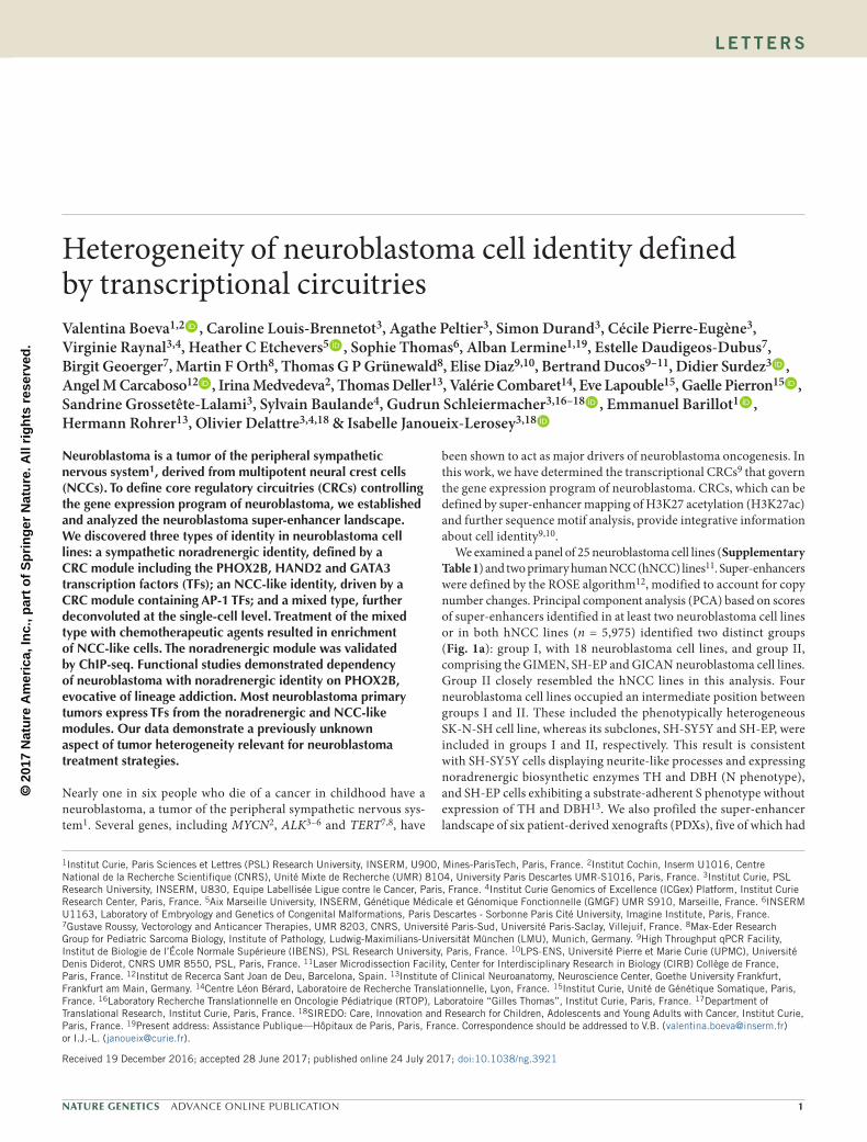
We examined a panel of 25 neuroblastoma cell lines (Supplementary Table 1) and two primary human NCC (hNCC) lines11. Super-enhancers were defined by the ROSE algorithm12, modified to account for copy number changes. Principal component analysis (PCA) based on scores of super-enhancers identified in at least two neuroblastoma cell lines or in both hNCC lines (n = 5,975) identified two distinct groups (Fig. 1a): group I, with 18 neuroblastoma cell lines, and group II, comprising the GIMEN, SH-EP and GICAN neuroblastoma cell lines. Group II closely resembled the hNCC lines in this analysis. Four neuroblastoma cell lines occupied an intermediate position between groups I and II. These included the phenotypically heterogeneous SK-N-SH cell line, whereas its subclones, SH-SY5Y and SH-EP, were included in groups I and II, respectively. This result is consistent with SH-SY5Y cells displaying neurite-like processes and expressing noradrenergic biosynthetic enzymes TH and DBH (N phenotype), and SH-EP cells exhibiting a substrate-adherent S phenotype without expression of TH and DBH13. We also profiled the super-enhancer landscape of six patient-derived xenografts (PDXs), five of which had
Heterogeneity of neuroblastoma cell identity defined by transcriptional circuitriesValentina Boeva1,2 , Caroline Louis-Brennetot3, Agathe Peltier3, Simon Durand3, Cécile Pierre-Eugène3, Virginie Raynal3,4, Heather C Etchevers5 , Sophie Thomas6, Alban Lermine1,19, Estelle Daudigeos-Dubus7, Birgit Geoerger7, Martin F Orth8, Thomas G P Grünewald8, Elise Diaz9,10, Bertrand Ducos9–11, Didier Surdez3 , Angel M Carcaboso12 , Irina Medvedeva2, Thomas Deller13, Valérie Combaret14, Eve Lapouble15, Gaelle Pierron15 , Sandrine Grossetête-Lalami3, Sylvain Baulande4, Gudrun Schleiermacher3,16–18 , Emmanuel Barillot1 , Hermann Rohrer13, Olivier Delattre3,4,18 & Isabelle Janoueix-Lerosey3,18
1Institut Curie, Paris Sciences et Lettres (PSL) Research University, INSERM, U900, Mines-ParisTech, Paris, France. 2Institut Cochin, Inserm U1016, Centre National de la Recherche Scientifique (CNRS), Unité Mixte de Recherche (UMR) 8104, University Paris Descartes UMR-S1016, Paris, France. 3Institut Curie, PSL Research University, INSERM, U830, Equipe Labellisée Ligue contre le Cancer, Paris, France. 4Institut Curie Genomics of Excellence (ICGex) Platform, Institut Curie Research Center, Paris, France. 5Aix Marseille University, INSERM, Génétique Médicale et Génomique Fonctionnelle (GMGF) UMR S910, Marseille, France. 6INSERM U1163, Laboratory of Embryology and Genetics of Congenital Malformations, Paris Descartes - Sorbonne Paris Cité University, Imagine Institute, Paris, France. 7Gustave Roussy, Vectorology and Anticancer Therapies, UMR 8203, CNRS, Université Paris-Sud, Université Paris-Saclay, Villejuif, France. 8Max-Eder Research Group for Pediatric Sarcoma Biology, Institute of Pathology, Ludwig-Maximilians-Universität München (LMU), Munich, Germany. 9High Throughput qPCR Facility, Institut de Biologie de l’École Normale Supérieure (IBENS), PSL Research University, Paris, France. 10LPS-ENS, Université Pierre et Marie Curie (UPMC), Université Denis Diderot, CNRS UMR 8550, PSL, Paris, France. 11Laser Microdissection Facility, Center for Interdisciplinary Research in Biology (CIRB) Collège de France, Paris, France. 12Institut de Recerca Sant Joan de Deu, Barcelona, Spain. 13Institute of Clinical Neuroanatomy, Neuroscience Center, Goethe University Frankfurt, Frankfurt am Main, Germany. 14Centre Léon Bérard, Laboratoire de Recherche Translationnelle, Lyon, France. 15Institut Curie, Unité de Génétique Somatique, Paris, France. 16Laboratory Recherche Translationnelle en Oncologie Pédiatrique (RTOP), Laboratoire “Gilles Thomas”, Institut Curie, Paris, France. 17Department of Translational Research, Institut Curie, Paris, France. 18SIREDO: Care, Innovation and Research for Children, Adolescents and Young Adults with Cancer, Institut Curie, Paris, France. 19Present address: Assistance Publique—Hôpitaux de Paris, Paris, France. Correspondence should be addressed to V.B. ([email protected]) or I.J.-L. ([email protected]).
Received 19 December 2016; accepted 28 June 2017; published online 24 July 2017; doi:10.1038/ng.3921

© 2
017
Nat
ure
Am
eric
a, In
c., p
art
of
Sp
rin
ger
Nat
ure
. All
rig
hts
res
erve
d.
2 ADVANCE ONLINE PUBLICATION Nature GeNetics
l e t t e r s
MYCN amplification (Supplementary Table 2). All PDXs clustered with group I when included in the PCA (Fig. 1a).
We then sorted super-enhancers according to median H3K27ac signal for each group (Fig. 1b,c and Supplementary Table 3). In group I, the strongest super-enhancers comprise a set of TF loci including HAND2, PHOX2A, PHOX2B, GATA2, GATA3 and the ALK oncogene locus (Fig. 1b,d,e and Supplementary Figs. 1 and 2). These findings are consistent with previous super-enhancer data on a few neuroblastoma cell lines14,15. PHOX2B, HAND2 and GATA3 participate in a complex TF network controlling normal sympathetic neuron specification and differentiation16,17. Recurrent super-enhancers in these TFs therefore appear to be a hallmark of sympathetic cell identity. Most super-enhancers of group II overlapped with super-enhancers of hNCC lines (Fig. 1c), consistent with the results of the PCA analysis.
Our analysis found MYCN super-enhancers for 10 out of 18 cell lines in group I, with or without MYCN amplification, and for three
PDXs (Supplementary Fig. 3). No groups linked to the MYCN or ALK status were identified in the PCA. Furthermore, supervised analysis of super-enhancer scores did not indicate super-enhancers associ-ated with MYCN amplification or ALK mutations (Supplementary Tables 4 and 5). Cell lines with a PHOX2B mutation were observed in group I (SH-SY5Y), group II (SH-EP) and in the intermediate group (SK-N-SH).
To detect driver TFs for groups I and II, we used i-cisTarget to find DNA sequence motifs enriched in the super-enhancers with the high-est score. For group I, this analysis identified a TAATYYAATTA bind-ing motif common to several homeobox proteins, including PHOX2B and PHOX2A (Supplementary Fig. 4). During sympathetic nerv-ous system development, PHOX2B regulates PHOX2A expression18. Both TFs are highly expressed in most neuroblastoma cell lines and primary tumors (Supplementary Fig. 5). PHOX2B was undetect-able at the transcript and protein levels in group II and hNCC lines
CLB-CAR
CLB-GA
CLB-MA
SK-N-FI
hNCC rep. 1
hNCC rep. 2
ALK SE
ALK
fa
SK−N−AS
GIMEN
SJNB12
SH−EPhNCCrep1
SK−N−SH
CHP−212
GICANhNCCrep2
MYC-amp ALKwt
ALKmutALKwt
hNCCMNA ALKmutMNA ALKwt
b
ALK
c
MYCN-amp
[0–80]CLB-CAR
CLB-GA
CLB-MA
SK-N-FI
hNCC rep. 1
hNCC rep. 2
PHOX2B SE
PHOX2B
SH−SY5Y
−1
−0.8
−0.6
−0.4
−0.2
0
0.2
0.4
0.6
0.8
1PHOX2B
GATA
3H
AN
D2
HA
ND
1K
LF7
ISL1
PH
OX
2AN
R3C
1B
HLH
E41
MA
FF
GLI
S3
IRF
1IR
F2
IRF
3F
LI1
ME
F2D
PR
RX
1R
UN
X1
RU
NX
2TB
X18
FOSL1
FOSL2
PHOX2BGATA3
HAND2HAND1
KLF7ISL1
PHOX2ANR3C1BHLHE41
MAFFGLIS3
IRF1IRF2
IRF3FLI1
MEF2DPRRX1
RUNX1RUNX2
TBX18FOSL1
FOSL2
d e
g
h
Module 2
Module 1
–0.06
–0.04
–0.02
0.00
0.02
PC
2
Group I
Group II
Cell linePDX
40,000
80,000
120,000
160,000
Med
ian
norm
aliz
ed H
3K27
ac s
core HAND2
TCF4
GATA2PHOX2BGATA3
PHOX2A
KLF7TBX2
CIC, ERF NFIB
INSM2HAND1 MYT1L
ZNF423HIF1A
Group I
–0.024
(0–80)
–0.020 –0.016 –0.012 0 20 40 60 80 100 0 20 40 60 80 100
120,000
160,000
200,000
240,000
PC1 SE rank SE rank
No overlap with hNCC SEs
Overlap with hNCC SEs
Med
ian
norm
aliz
ed H
3K27
ac s
core
AFF3
HAND2
BCL6MYC
TBX2KLF6 CIC, ERF
GLIS3 ZBTB38FOSL2
HIF1ABNC2MYBL1
HMGA2
JUN
Group II
IGR
–NB
8IG
R–N
B83
5H
SJD
–NB
011
MA
P-G
R-A
99-N
B-1
MA
P-G
R-B
25-N
B-1
MA
P-I
C-A
23-N
B-1
SJN
B6
SJN
B8
CLB
–CA
RC
LB–P
EIM
R32
LAN
–1N
206
SK
–N–B
E(2
)–C
SK
–N–D
ZT
R14
CLB
–BE
R–L
udC
LB–M
AN
B–6
9C
LB–G
AS
JNB
1S
K–N
–FI
SH
–SY
5YN
B–E
Bc1
CH
P–2
12S
K–N
–AS
SK
–N–S
HS
JNB
12G
IME
NS
H–E
PG
ICA
NhN
CC
rep
1hN
CC
rep
2
TCF7L2SMAD3BACH1STAT3STAT1MEF2AJUNMTF1IRF3IRF1GLIS3NR3C1MAFFIRF2PRRX1BHLHE41TBX18RUNX2MEF2DRUNX1FLI1FOSL2FOSL1FOXP1ETV6KLF12ZNF217RXRANFIBPHOX2ATBX2ISL1HAND1PHOX2BGATA3HAND2KLF7
Present in predicted CRCsAbsent in predicted CRCsi
VinculinPHOX2B
GIC
AN
NB
69C
HP
-212
SK
-N-S
HC
LB-B
er-L
udC
LB-G
AC
LB-M
AC
LB-P
EG
IME
NS
H-E
PS
H-S
Y5Y
SJN
B1
SJN
B6
SJN
B12
SK
-N-A
SS
K-N
-BE
(2)-
CS
K-N
-FI
0 4.001.00
ModuleModule 1Module 2
9 10 36 1 7 37 4 8 18 22 29 46 42 5 32 28 33 38 35 2 14 3 6 11 15 40 24 21 34 39 26 41 25 45 12 23 16 20 19 13 17 44 30 27 31 43Cell ID
PHOX2BGATA3HAND2HAND1KLF7ISL1PHOX2ANR3C1GLIS3IRF1IRF2PRRX1RUNX1RUNX2TBX18FOSL1FOSL2
Module
Figure 1 Super-enhancer landscape identifies various CRCs and identities in neuroblastoma cell lines. (a) PCA based on neuroblastoma and hNCC super-enhancer (SE) log scores. ALKwt, wild-type ALK; ALKmut, mutant ALK; MNA, MYCN amplification; MYC-amp, MYC amplification. (b,c) Ranked plot for the 100 SEs with the highest median H3K27ac scores in groups I (b) and II (c). Genes encoding TFs are indicated with arrows. (d,e) Tracks for ChIP-seq profiles for H3K27ac binding at ALK and PHOX2B SEs, respectively. (f) Immunoblot analysis of PHOX2B, with vinculin as a loading control, in a panel of neuroblastoma cell lines. SK-N-SH cells were from batch 1 (batches are described in Online Methods). (g) TF genes predicted to participate in a CRC with PHOX2B (top) or with a FOS or JUN family member (bottom) in neuroblastoma cell lines. TFs whose binding motifs are enriched in SEs of group I and II are shown in bold. (h) Pearson correlation matrix, based on RNA-seq data in neuroblastoma cell lines and PDXs (n = 31), for the expression values of 22 TF genes identified in CRCs of cell lines. (i) Single-cell analysis showing heterogeneity of cell identity in the SK-N-AS cell line. Expression was evaluated by RT-qPCR, and data were normalized to the SK-N-AS cell population overall.
© 2
017
Nat
ure
Am
eric
a, In
c., p
art
of
Sp
rin
ger
Nat
ure
. All
rig
hts
res
erve
d.
Nature GeNetics ADVANCE ONLINE PUBLICATION 3
l e t t e r s
but was expressed in all other neuroblastoma cell lines (Fig. 1f and Supplementary Fig. 6). There was a corresponding lack of super-enhancer at PHOX2B and no TH and DBH transcription in GIMEN, SH-EP or GICAN cells (Supplementary Fig. 7). i-cisTarget analysis showed enrichment in AP-1 motif in group II and the hNCC lines (Supplementary Figs. 8 and 9). AP-1 is a heterodimer composed of FOS and JUN family members, both of which are expressed in imma-ture hNCCs11. These results suggest that PHOX2B participates in the activity of neuroblastoma group I super-enhancers while the AP-1 TF influences the super-enhancer landscape of group II.
A CRC calling algorithm9,19 identified PHOX2B as specific to group I, and FOSL1, FOSL2 and JUN specific to group II (Supplementary Fig. 10), consistent with our i-cisTarget results. We therefore searched for TFs predicted to be in a CRC either with PHOX2B or a FOS or JUN family member (Fig. 1g). Cell lines showing an intermediate position in the PCA had a CRC that included several TFs of sets asso-ciated with PHOX2B and FOS or JUN. Most TFs of the latter set are expressed in NCCs and/or mesenchymal neural crest derivatives. The CRCs of the six PDXs were highly similar to those of the group I cell lines (Fig. 1g). Because super-enhancer strength correlated linearly
with gene expression (Supplementary Fig. 11), we used the latter to further define fully connected TF modules. This analysis identified two main TF modules distinguishing groups I and II (Fig. 1h): mod-ule 1, which included PHOX2B, GATA3 and HAND2, and module 2, which included FOSL1 and FOSL2. These modules were anticorre-lated at the gene expression level. Immunoblot analysis confirmed the coexpression of several TFs in group I or group II (Supplementary Fig. 12). Furthermore, single-cell analysis showed that the SK-N-AS and SK-N-SH cell lines were heterogeneous and comprise cells expressing TFs of module 1 or module 2 within the same population (Fig. 1i, Supplementary Fig. 13 and Supplementary Table 6).
Taken together, these data demonstrate a novel type of heterogene-ity in neuroblastoma cell lines and suggest that individual cells assume either (i) a sympathetic noradrenergic identity, characterized by a CRC module including PHOX2B, HAND2 and GATA3 and expression of the enzymes TH and/or DBH, or (ii) an NCC-like identity, character-ized by expression of a distinct module including FOS and JUN family members but lacking PHOX2B and noradrenergic marker expression. Both types of identity are observed in several heterogeneous cell lines. All cell lines with MYCN amplification except one (CHP-212) had a
−1
−0.8
−0.6
−0.4
−0.2
0
0.2
0.4
0.6
0.8
1
PH
OX
2BG
ATA
3H
AN
D2
HA
ND
1K
LF7
ISL1
PH
OX
2AN
R3C
1B
HLH
E41
MA
FF
GLI
S3
IRF
1IR
F2
IRF
3F
LI1
ME
F2D
PR
RX
1R
UN
X1
RU
NX
2TB
X18
FO
SL1
FO
SL2
PHOX2BGATA3
HAND2HAND1
KLF7ISL1
PHOX2ANR3C1BHLHE41
MAFFGLIS3
IRF1IRF2
IRF3FLI1MEF2D
PRRX1RUNX1
RUNX2TBX18
FOSL1FOSL2
ba
c
Module 1
Module 2
d
MNA tumorsNon-MNA tumors
5 6 7 8
1.0
1.5
2.0
2.5
3.0
Average expression (log FPKM)of module 1
Ave
rage
exp
ress
ion
(log
FP
KM
)of
mod
ule
2
0 2 4 6
−2
−1
0
1
2
Module 1 log FC
Mod
ule
2 lo
g F
C
2 4 6 8
2.0
2.5
3.0
3.5
4.0
4.5
Average expression (log FPKM)of module 1
Ave
rage
exp
ress
ion
(log
FP
KM
)of
mod
ule
2
hNCCrep 1hNCC
rep 2
SH-SY5Y
SH-EP
SK-N-SH
SK-N-SHCtrl 2
SK-N-SHCtrl 1
SK-N-SHcisplatin
SK-N-SHdoxorubicin
Diagnosis to relapsechange of expression
In houseGSE65303
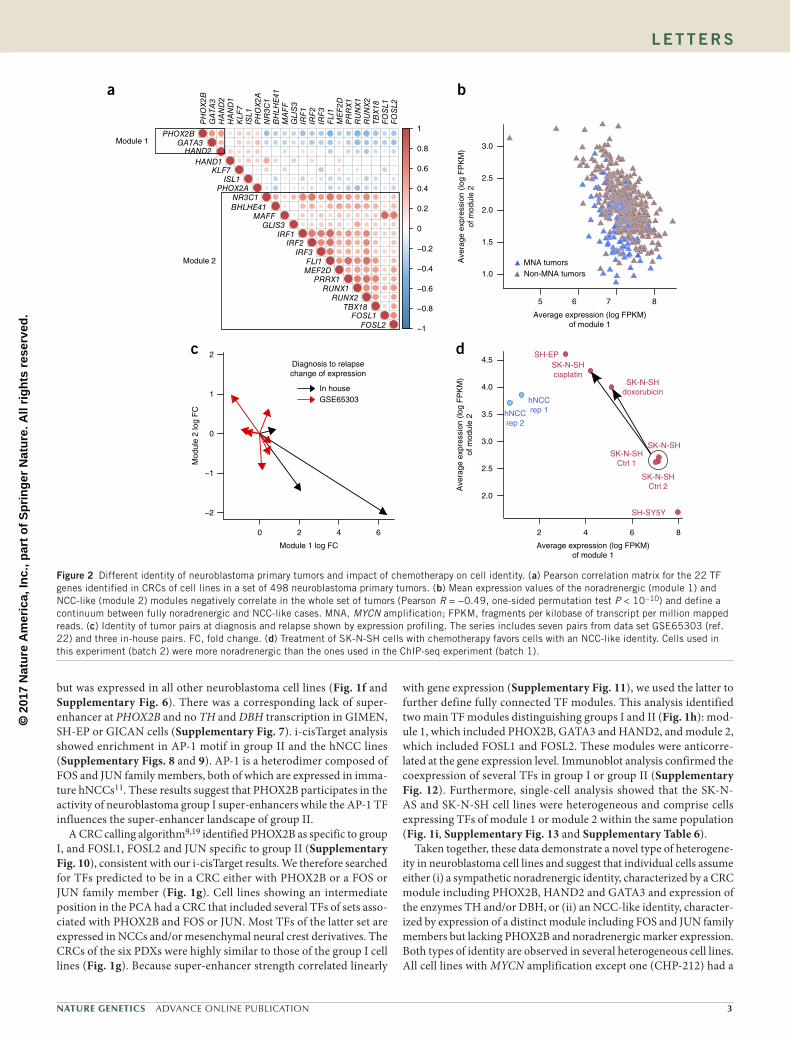
Figure 2 Different identity of neuroblastoma primary tumors and impact of chemotherapy on cell identity. (a) Pearson correlation matrix for the 22 TF genes identified in CRCs of cell lines in a set of 498 neuroblastoma primary tumors. (b) Mean expression values of the noradrenergic (module 1) and NCC-like (module 2) modules negatively correlate in the whole set of tumors (Pearson R = −0.49, one-sided permutation test P < 10−10) and define a continuum between fully noradrenergic and NCC-like cases. MNA, MYCN amplification; FPKM, fragments per kilobase of transcript per million mapped reads. (c) Identity of tumor pairs at diagnosis and relapse shown by expression profiling. The series includes seven pairs from data set GSE65303 (ref. 22) and three in-house pairs. FC, fold change. (d) Treatment of SK-N-SH cells with chemotherapy favors cells with an NCC-like identity. Cells used in this experiment (batch 2) were more noradrenergic than the ones used in the ChIP-seq experiment (batch 1).
© 2
017
Nat
ure
Am
eric
a, In
c., p
art
of
Sp
rin
ger
Nat
ure
. All
rig
hts
res
erve
d.
� ADVANCE ONLINE PUBLICATION Nature GeNetics
l e t t e r s
noradrenergic identity, whereas all three identities were represented in cell lines without MYCN amplification (Fig. 1g).
To explore whether the different identity classes seen in neurob-lastoma cell lines are also characteristic of neuroblastoma primary tumors, we studied expression data from a large set of primary tumors20 (n = 498; GEO GSE49711). Correlations between expres-sion of the TFs of each module identified in cell line CRCs were calculated for these tumors. We observed strong positive correla-tions between PHOX2B, HAND2 and GATA3 expression levels in module 1. We also detected positive intercorrelations in expression of the NCC-like module TF genes (Fig. 2a). Our analysis further indicated that expression of PHOX2B, HAND2 and GATA3 is anti-correlated with that of the NCC-like module TF genes. These results therefore confirm our observations in cell lines and further define a PHOX2B–HAND2–GATA3 noradrenergic CRC module in pri-mary neuroblastomas. Next, we used the average expression of the two modules to explore primary tumor identity. All but two tumors showed high expression of the noradrenergic module (Fig. 2b).
A continuum was observed from low to high values of the NCC-like module, suggesting heterogeneity of cell identity in primary tumors. The remaining two cases with low noradrenergic and high NCC-like module expression may correspond to rare cases with full NCC-like identity, as described for group II cell lines. Similarly to the cell lines (Supplementary Fig. 14), lower expression of the NCC-like module was observed in the majority of MYCN-amplified tumors (P = 1.01 × 10⁻¹⁰, two-sided Wilcoxon signed-rank test). A role for MYCN in downregulation of genes from this module is consistent with its promotion of peripheral neuron differentiation from multipotent avian NCC21.
Next, expression of the NCC-like and noradrenergic modules was evaluated in ten diagnosis–relapse sample pairs22. Different patterns were observed between the two disease stages (Fig. 2c). To address a possible link between heterogeneity of cell identity and treatment response, we investigated the effect of chemotherapy on the NCC-like SH-EP and noradrenergic SH-SY5Y cell lines. SH-EP cells were more resistant to the three agents used (Supplementary Fig. 15).
d
b
a
c
0
20
40
Per
cent
age
of s
uper
-enh
ance
rs
60
80
100
HAND2–PHOX2B–GATA3
HAND2–PHOX2B
HAND2–GATA3
PHOX2B–GATA3
HAND2
PHOX2B
GATA3
None
Binding by
PHOX2B ALK
e
PHOX2BHAND2GATA3
H3K27ac
GATA3
PHOX2B
HAND2
SE HAND2
SE PHOX2B
SE GATA3
HAND2
PHOX2B
GATA3
f
H3K27ac
GATA3
PHOX2B
HAND2
−1,000 −500 0 500 1,000
0.0
0.2
0.4
0.6
0.8
1.0
Distance from PHOX2B peak maximum (bp)
Ave
rage
of n
orm
aliz
ed C
hIP
−se
q de
nsity
(0–180)
(0–60)
(0–80)
(0–100)
(0–180)
(0–60)
(0–80)
(0–100)
0
1
2
Bits
1 2 3 4 5 6 7 8 9 10 11 12 13 14 15 16 17 18 1 2 3 4 5 6 7 8 9 100
1
2
Bits
0
1
2
Bits
1 2 3 4 5 6 7 8 9 10 11 12
PHOX2B HAND2 GATA3
Top100
Top100−200
Top200−300
Top300−400
Top400−500
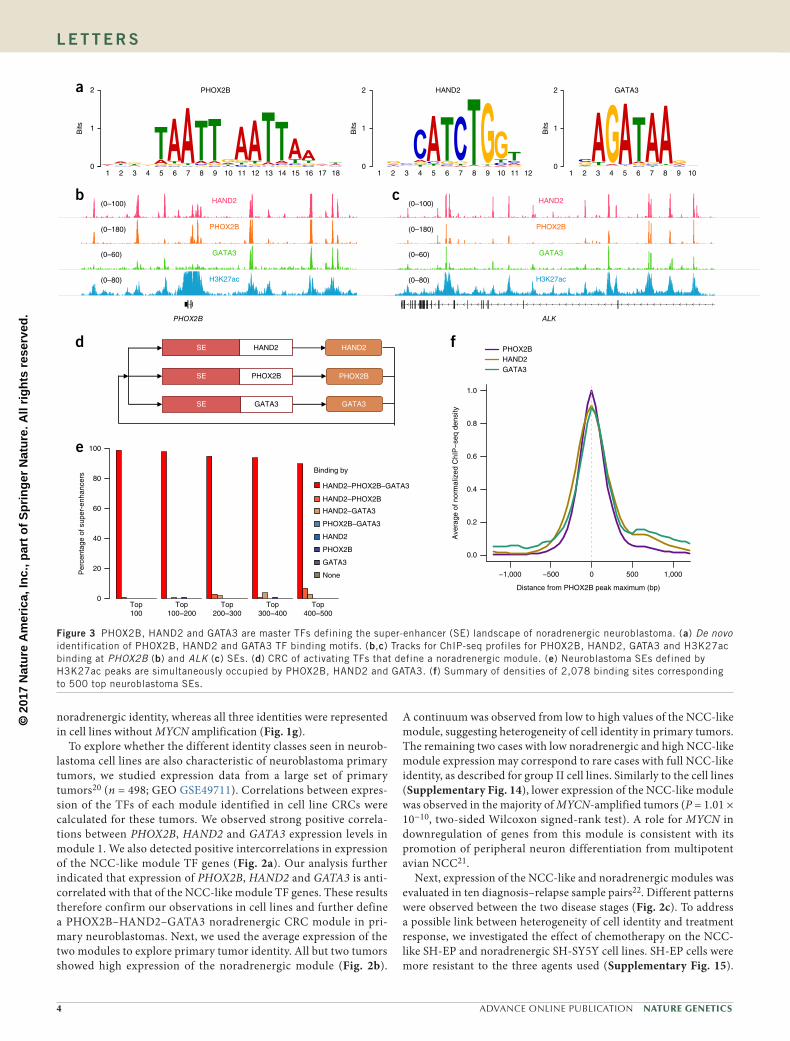
Figure 3 PHOX2B, HAND2 and GATA3 are master TFs defining the super-enhancer (SE) landscape of noradrenergic neuroblastoma. (a) De novo identification of PHOX2B, HAND2 and GATA3 TF binding motifs. (b,c) Tracks for ChIP-seq profiles for PHOX2B, HAND2, GATA3 and H3K27ac binding at PHOX2B (b) and ALK (c) SEs. (d) CRC of activating TFs that define a noradrenergic module. (e) Neuroblastoma SEs defined by H3K27ac peaks are simultaneously occupied by PHOX2B, HAND2 and GATA3. (f) Summary of densities of 2,078 binding sites corresponding to 500 top neuroblastoma SEs.
© 2
017
Nat
ure
Am
eric
a, In
c., p
art
of
Sp
rin
ger
Nat
ure
. All
rig
hts
res
erve
d.
Nature GeNetics ADVANCE ONLINE PUBLICATION �
l e t t e r s
Treatment of the parental SK-N-SH cell line with doxorubicin or cis-platin resulted in the decreased or increased expression of modules 1 and 2, respectively (Fig. 2d). Enrichment of cells with an NCC-like identity thus correlates with better drug resistance. However, we cannot exclude the possibility that treatment may also induce transdifferentiation from a noradrenergic to an NCC-like identity. The observation that tumors at relapse are not systematically enriched in NCC-like cells supports the concept of plasticity in the reversion of cell identity. This may rely on a switch from adrenergic to NCC-like identity under chemotherapy, and from NCC-like to noradrenergic after treatment. Together, these data underline the importance of tar-geting both types of cells during treatment.
Strong correlations among PHOX2B, HAND2 and GATA3 expres-sion were observed in cell lines and tumors. PHOX2B directly binds
HAND2 protein23, and PHOX2B, HAND2 and GATA3 cross-regulate during sympathetic nervous system development16. We therefore performed ChIP-seq analysis for these TFs in the CLB-GA neurob-lastoma cell line and identified binding motifs for PHOX2B, HAND2 and GATA3 (Fig. 3a). Binding regions for all three TFs corresponded to the H3K27ac peaks in the PHOX2B, GATA3, HAND2 and ALK super-enhancers and also in the MYCN super-enhancer (Fig. 3b,c and Supplementary Fig. 16). These results therefore confirm the biologi-cal existence of the noradrenergic module, showing that PHOX2B, HAND2 and GATA3 are super-enhancer regulated and bind to one another’s super-enhancers (Fig. 3d). We next investigated the occu-pancy by these TFs of 4,336 super-enhancer regions predicted in at least two neuroblastoma cell lines. We ranked super-enhancer regions according to average super-enhancer score and evaluated intersection with TF binding sites. More than 90% of the strong and recurrent neu-roblastoma super-enhancers were co-occupied by PHOX2B, HAND2 and GATA3 (Fig. 3e). Additionally, positional binding analysis showed that HAND2, PHOX2B and GATA3 bind the same ~400-bp- long regions within active regulatory regions (Fig. 3f). Together, our results demonstrate that PHOX2B, HAND2 and GATA3 are master TFs, defining the super-enhancer landscape of neuroblastoma cell lines with noradrenergic identity.
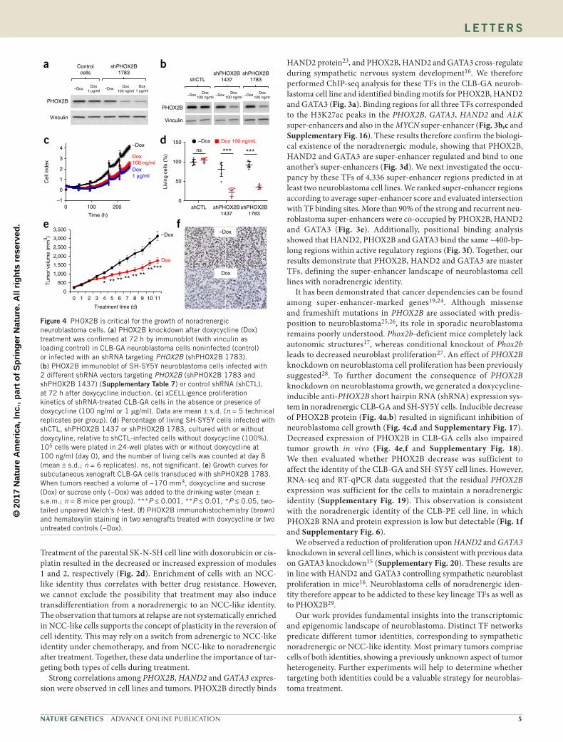
It has been demonstrated that cancer dependencies can be found among super-enhancer-marked genes19,24. Although missense and frameshift mutations in PHOX2B are associated with predis-position to neuroblastoma25,26, its role in sporadic neuroblastoma remains poorly understood. Phox2b-deficient mice completely lack autonomic structures17, whereas conditional knockout of Phox2b leads to decreased neuroblast proliferation27. An effect of PHOX2B knockdown on neuroblastoma cell proliferation has been previously suggested28. To further document the consequence of PHOX2B knockdown on neuroblastoma growth, we generated a doxycycline-inducible anti-PHOX2B short hairpin RNA (shRNA) expression sys-tem in noradrenergic CLB-GA and SH-SY5Y cells. Inducible decrease of PHOX2B protein (Fig. 4a,b) resulted in significant inhibition of neuroblastoma cell growth (Fig. 4c,d and Supplementary Fig. 17). Decreased expression of PHOX2B in CLB-GA cells also impaired tumor growth in vivo (Fig. 4e,f and Supplementary Fig. 18). We then evaluated whether PHOX2B decrease was sufficient to affect the identity of the CLB-GA and SH-SY5Y cell lines. However, RNA-seq and RT-qPCR data suggested that the residual PHOX2B expression was sufficient for the cells to maintain a noradrenergic identity (Supplementary Fig. 19). This observation is consistent with the noradrenergic identity of the CLB-PE cell line, in which PHOX2B RNA and protein expression is low but detectable (Fig. 1f and Supplementary Fig. 6).
We observed a reduction of proliferation upon HAND2 and GATA3 knockdown in several cell lines, which is consistent with previous data on GATA3 knockdown15 (Supplementary Fig. 20). These results are in line with HAND2 and GATA3 controlling sympathetic neuroblast proliferation in mice16. Neuroblastoma cells of noradrenergic iden-tity therefore appear to be addicted to these key lineage TFs as well as to PHOX2B29.
Our work provides fundamental insights into the transcriptomic and epigenomic landscape of neuroblastoma. Distinct TF networks predicate different tumor identities, corresponding to sympathetic noradrenergic or NCC-like identity. Most primary tumors comprise cells of both identities, showing a previously unknown aspect of tumor heterogeneity. Further experiments will help to determine whether targeting both identities could be a valuable strategy for neuroblas-toma treatment.
0
500
1,000
1,500
2,000
2,500
3,000
3,500
0 1 2 3 4 5 6 7 8 9 10 11
Tum
or v
olum
e (m
m3 )
Treatment time (d)
* ** ** ** ** *******
a
e
Dox1 µg/ml
Dox100 ng/ml
Dox1 µg/ml–Dox –Dox
Controlcells
shPHOX2B1783
PHOX2B
Vinculin
–1
0
1
2
3
4
0 100 200
Time (h)
–Dox
Dox100 ng/mlDox1 µg/ml
–Dox
Dox
Dox
–Dox
c d
f
Livi
ng c
ells
(%
)
shCTL shPHOX2B1437
shPHOX2B1783
0
50
100
150 –Dox Dox 100 ng/mL
*** ***ns
Cel
l ind
ex
bshCTL
PHOX2B
Vinculin
Dox100 ng/ml
–DoxDox
100 ng/ml–Dox
Dox100 ng/ml
–Dox
shPHOX2B1783
shPHOX2B1437
Figure 4 PHOX2B is critical for the growth of noradrenergic neuroblastoma cells. (a) PHOX2B knockdown after doxycycline (Dox) treatment was confirmed at 72 h by immunoblot (with vinculin as loading control) in CLB-GA neuroblastoma cells noninfected (control) or infected with an shRNA targeting PHOX2B (shPHOX2B 1783). (b) PHOX2B immunoblot of SH-SY5Y neuroblastoma cells infected with 2 different shRNA vectors targeting PHOX2B (shPHOX2B 1783 and shPHOX2B 1437) (supplementary table 7) or control shRNA (shCTL), at 72 h after doxycycline induction. (c) xCELLigence proliferation kinetics of shRNA-treated CLB-GA cells in the absence or presence of doxycycline (100 ng/ml or 1 µg/ml). Data are mean ± s.d. (n = 5 technical replicates per group). (d) Percentage of living SH-SY5Y cells infected with shCTL, shPHOX2B 1437 or shPHOX2B 1783, cultured with or without doxycyline, relative to shCTL-infected cells without doxycycline (100%). 105 cells were plated in 24-well plates with or without doxycycline at 100 ng/ml (day 0), and the number of living cells was counted at day 8 (mean ± s.d.; n = 6 replicates). ns, not significant. (e) Growth curves for subcutaneous xenograft CLB-GA cells transduced with shPHOX2B 1783. When tumors reached a volume of ~170 mm3, doxycycline and sucrose (Dox) or sucrose only (−Dox) was added to the drinking water (mean ± s.e.m.; n = 8 mice per group). ***P ≤ 0.001, **P ≤ 0.01, *P ≤ 0.05, two-tailed unpaired Welch’s t-test. (f) PHOX2B immunohistochemistry (brown) and hematoxylin staining in two xenografts treated with doxycycline or two untreated controls (−Dox).
© 2
017
Nat
ure
Am
eric
a, In
c., p
art
of
Sp
rin
ger
Nat
ure
. All
rig
hts
res
erve
d.
� ADVANCE ONLINE PUBLICATION Nature GeNetics
l e t t e r s
MeTHODsMethods, including statements of data availability and any associated accession codes and references, are available in the online version of the paper.
Note: Any Supplementary Information and Source Data files are available in the online version of the paper.
ACknOwLEDGMEnTSWe are grateful to M. Ponzoni (IRCCS Istituto Giannina Gaslini), M. Schwab (German Cancer Research Center), J. Couturier (Institut Curie) and R. Versteeg (University of Amsterdam) for providing neuroblastoma cell lines. We thank F. Tirode and C. Kamoun for help with RNA-seq analysis and alignment of NGS data, respectively; O. Blanchard for help in cell culture experiments; and M. Caly for PHOX2B immunohistochemistry. We are grateful to the animal facilities team, the Experimental Pathology Department and the Plateforme Génomique of Institut Curie. We thank N. Clément, T. Adam-de-Beaumais and B. Mallon for their help in the identification of neuroblastoma diagnosis–relapse pairs and V. Bernard for pairs validation. We thank V. Saint-André for scientific discussion, J. Maliash-Planchon and the Unité de Génétique Somatique for preparation of patient samples. We thank D. Figarella-Branger (BB-033-00097, CRB AP-HM, CRB TBM, AC-2013-1786), M. Clapisson (CRB Centre Léon Bérard, AC-2008-101), O. Minckes, C. Blanc-Fournier and N. Rousseau (CHU, Tumorothèque de Caen Basse Normandie) for providing tumor patient samples. This work was supported by grants from Institut Curie, INSERM, the Ligue Nationale contre le Cancer (Equipe Labellisée), the Société Française de Lutte contre les Cancers et les Leucémies de l’Enfant et l’Adolescent, the Institut National du Cancer (PRT-K14-061 and PHRC IC 2007-2009) and by the following associations: Association Hubert Gouin “Enfance and Cancer,” Les Bagouz à Manon, les amis de Claire, Courir pour Mathieu, Dans les pas du Géant and Olivier Chape. The MAPPYACTS protocol is supported by the Institut National du Cancer (PHRC-K14-175), the Fondation ARC (MAPY201501241), the Société Française de Lutte contre les Cancers et les Leucémies de l’Enfant et l’Adolescent (Fondation Enfants et Santé), the Fondation AREMIG and the Association Thibault BRIET. High-throughput sequencing was performed by the ICGex NGS platform of the Institut Curie, supported by the grants ANR-10-EQPX-03 (Equipex) and ANR-10-INBS-09-08 (France Génomique Consortium) from the Agence Nationale de la Recherche (Investissements d’Avenir program); by the Canceropole Ile-de-France; and by the SiRIC-Curie program -SiRIC Grant INCa-DGOS- 4654. Biomark analysis was done using the High Throughput qPCR-HD-Genomic Paris Centre platform supported by grants from Région Ile-de-France (21016711). G.S. is supported by the Annenberg Foundation (11-385). V.B. is supported by the ATIP-Avenir Program, the ARC Foundation (ARC-RAC16002KSA-R15093KS), Worldwide Cancer Research (WCR16-1294 R16100KK) and the “Who Am I?” Laboratory of Excellence ANR-11-LABX-0071, funded by the French Government through its Investissement d′Avenir program, operated by the French National Research Agency (ANR) (ANR-11-IDEX-0005-02). H.R. is supported by the Mayent-Rothschild program from Institut Curie and the Wilhelm-Sander-Stiftung. The laboratory of T.G.P.G. is supported by LMU Munich’s Institutional Strategy LMUexcellent within the framework of the German Excellence Initiative, the Mehr LEBEN für krebskranke Kinder—Bettina-Bräu-Stiftung, the Walter Schulz Foundation, the Wilhelm-Sander-Stiftung (2016.167.1) and the German Cancer Aid (DKH-111886 and DKH-70112257).
AUTHOR COnTRIBUTIOnSV.B. and I.J.-L. conceived the study, analyzed the data and wrote the manuscript. V.B. coordinated bioinformatics analysis and I.J.-L. coordinated the whole study. C.L.-B. performed in vitro experiments and ChIP experiments and participated in the study design. A.P. generated and analyzed the doxycycline-inducible anti-PHOX2B shRNA cell lines. S.D. performed the single-cell analysis and study of chemotherapeutic agents. C.P.-E. performed the in vivo experiments and contributed in vitro experiments. V.R. performed all sequencing experiments. H.C.E. and S.T. provided hNCC cell lines and V.C. provided neuroblastoma cell lines. A.L. performed alignment of RNA-seq and ChIP-seq data. E.D.-D., B.G., D.S. and A.M.C. provided neuroblastoma PDXs. I.M. performed the reproducibility analysis. E.D. and B.D. generated the Biomark data. M.F.O. and T.G.P.G. generated lentiviral particles and provided help with lentiviral infections. S.B. coordinated and supervised sequencing experiments. G.S. participated in the study design and provided the in-house pairs of diagnosis–relapse samples with the help of E.L., G.P.
and B.G. S.G.-L. participated in RNA-seq analysis. E.B. provided computational infrastructure and data storage. H.R. and T.D. provided expertise in sympathetic nervous development and TFs. I.J.-L. and O.D. provided laboratory infrastructure. I.J.-L., V.B. and O.D. provided financial support. All authors read and approved the final manuscript.
COMPETInG FInAnCIAL InTERESTSThe authors declare no competing financial interests.
Reprints and permissions information is available online at http://www.nature.com/reprints/index.html. Publisher’s note: Springer Nature remains neutral with regard to jurisdictional claims in published maps and institutional affiliations.
1. Matthay, K.K. et al. Neuroblastoma. Nat. Rev. Dis. Primers 2, 16078 (2016).2. Brodeur, G.M., Seeger, R.C., Schwab, M., Varmus, H.E. & Bishop, J.M. Amplification
of N-myc in untreated human neuroblastomas correlates with advanced disease stage. Science 224, 1121–1124 (1984).
3. Mossé, Y.P. et al. Identification of ALK as a major familial neuroblastoma predisposition gene. Nature 455, 930–935 (2008).
4. Janoueix-Lerosey, I. et al. Somatic and germline activating mutations of the ALK kinase receptor in neuroblastoma. Nature 455, 967–970 (2008).
5. George, R.E. et al. Activating mutations in ALK provide a therapeutic target in neuroblastoma. Nature 455, 975–978 (2008).
6. Chen, Y. et al. Oncogenic mutations of ALK kinase in neuroblastoma. Nature 455, 971–974 (2008).
7. Peifer, M. et al. Telomerase activation by genomic rearrangements in high-risk neuroblastoma. Nature 526, 700–704 (2015).
8. Valentijn, L.J. et al. TERT rearrangements are frequent in neuroblastoma and identify aggressive tumors. Nat. Genet. 47, 1411–1414 (2015).
9. Saint-André, V. et al. Models of human core transcriptional regulatory circuitries. Genome Res. 26, 385–396 (2016).
10. Hnisz, D. et al. Super-enhancers in the control of cell identity and disease. Cell 155, 934–947 (2013).
11. Thomas, S. et al. Human neural crest cells display molecular and phenotypic hallmarks of stem cells. Hum. Mol. Genet. 17, 3411–3425 (2008).
12. Whyte, W.A. et al. Master transcription factors and Mediator establish super-enhancers at key cell identity genes. Cell 153, 307–319 (2013).
13. Ross, R.A., Spengler, B.A. & Biedler, J.L. Coordinate morphological and biochemical interconversion of human neuroblastoma cells. J. Natl. Cancer Inst. 71, 741–747 (1983).
14. Chipumuro, E. et al. CDK7 inhibition suppresses super-enhancer-linked oncogenic transcription in MYCN-driven cancer. Cell 159, 1126–1139 (2014).
15. Oldridge, D.A. et al. Genetic predisposition to neuroblastoma mediated by a LMO1 super-enhancer polymorphism. Nature 528, 418–421 (2015).
16. Rohrer, H. Transcriptional control of differentiation and neurogenesis in autonomic ganglia. Eur. J. Neurosci. 34, 1563–1573 (2011).
17. Pattyn, A., Morin, X., Cremer, H., Goridis, C. & Brunet, J.F. The homeobox gene Phox2b is essential for the development of autonomic neural crest derivatives. Nature 399, 366–370 (1999).
18. Flora, A. et al. SP proteins and PHOX2B regulate the expression of the human PHOX2a gene. J. Neurosci. 21, 7037–7045 (2001).
19. Lin, C.Y. et al. Active medulloblastoma enhancers reveal subgroup-specific cellular origins. Nature 530, 57–62 (2016).
20. Zhang, W. et al. Comparison of RNA-seq and microarray-based models for clinical endpoint prediction. Genome Biol. 16, 133 (2015).
21. Wakamatsu, Y., Watanabe, Y., Nakamura, H. & Kondoh, H. Regulation of the neural crest cell fate by N-MYC: promotion of ventral migration and neuronal differentiation. Development 124, 1953–1962 (1997).
22. Schramm, A. et al. Mutational dynamics between primary and relapse neuroblastomas. Nat. Genet. 47, 872–877 (2015).
23. Reiff, T. et al. Neuroblastoma Phox2b variants stimulate proliferation and dedifferentiation of immature sympathetic neurons. J. Neurosci. 30, 905–915 (2010).
24. Chapuy, B. et al. Discovery and characterization of super-enhancer-associated dependencies in diffuse large B cell lymphoma. Cancer Cell 24, 777–790 (2013).
25. Trochet, D. et al. Germline mutations of the paired-like homeobox 2B (PHOX2B) gene in neuroblastoma. Am. J. Hum. Genet. 74, 761–764 (2004).
26. Mosse, Y.P. et al. Germline PHOX2B mutation in hereditary neuroblastoma. Am. J. Hum. Genet. 75, 727–730 (2004).
27. Coppola, E., d’Autréaux, F., Rijli, F.M. & Brunet, J.-F. Ongoing roles of Phox2 homeodomain transcription factors during neuronal differentiation. Development 137, 4211–4220 (2010).
28. Ke, X.-X. et al. Phox2B correlates with MYCN and is a prognostic marker for neuroblastoma development. Oncol. Lett. 9, 2507–2514 (2015).
29. Garraway, L.A. & Sellers, W.R. Lineage dependency and lineage-survival oncogenes in human cancer. Nat. Rev. Cancer 6, 593–602 (2006).

© 2
017
Nat
ure
Am
eric
a, In
c., p
art
of
Sp
rin
ger
Nat
ure
. All
rig
hts
res
erve
d.
Nature GeNeticsdoi:10.1038/ng.3921
ONLINe MeTHODsNeuroblastoma and hNCC cell lines. Neuroblastoma cell lines used in this study have been previously described30. CHP-212, IMR-32, SH-SY5Y, SK-N-AS, SK-N-BE(2)C, SK-N-DZ, SK-NF-I and SK-N-SH were obtained from the American Type Culture Collection (ATCC). CLB cell lines were derived by V. Combaret. The SH-EP and LAN-1 cell lines were kindly provided by M. Schwab and J. Couturier. Lines GIMEN, N206, SJNB1, SJNB6, SJNB8, SJNB12 and TR-14 were obtained from R. Versteeg, and line GICAN was a kind gift from M. Ponzoni. The NB69 and NB-EBc1 cell lines were obtained from the European Collection of Authenticated Cell Cultures and from the Children’s Oncology Group, respectively. One batch of SK-N-SH cells (batch 1) was used for the ChIP-seq and single-cell analysis. A second batch (batch 2) was used for the evaluation of the chemotherapeutic agents. Batch 2 was enriched in adrenergic cells. Cell line authentication was performed by comparison of the genomic copy number profile calculated from the input ChIP-seq data obtained using Control-FREEC31 with SNP array profile and STR profiling for ATCC cell lines. Cells were checked routinely by PCR for the absence of mycoplasma. Neuroblastoma cell lines were cultured at 37 °C with 5% CO2 in a humidified atmosphere in RPMI (GE Healthcare) for CLB cell lines, SH-EP, GICAN and NB69, in IMDM (Gibco) for NB-EBc1 (according to the provided conditions), or in DMEM (GE Healthcare) for other cell lines, with 10%, 15% or 20% FCS (Eurobio) and 100 µg/ml penicillin–streptomycin (Gibco). Primary hNCC lines were grown as described32 under bioethical approval PFS14-011 from the French Biomedical Agency for the use of human embryonic material. Briefly, cells were grown in Glutamax DMEM:F12 (Gibco) supplemented with 12% FCS (Eurobio), 100 µg/ml penicillin–streptomycin, 10 mM HEPES, 100 ng/ml hydrocortisone, 10 µg/ml transferrin, 400 pg/ml 3,3,5-thio-iodo-thyronine, 10 pg/ml glucagon, 100 pg/ml epidermal growth factor, 1 ng/ml insulin and 200 pg/ml fibroblast growth factor 2 (all products supplied by Sigma-Aldrich except EGF and FGF2, which were from Gibco).
PDX models. Neuroblastoma PDXs were obtained from stage L2 (MAP-IC-A23-NB-1), stage 3 (IGR-NB8) or stage 4 (IGR-N835, MAP-GR-A99-NB-1, MAP-GR-B25-NB-1 and HSJD-NB-011)33. None were related to the cell lines used. All PDXs but MAP-IC-A23-NB-1 had MYCN amplification. PDXs IGR-NB8, IGR-N835 (refs. 34,35) were obtained using female Swiss nude mice of 6–8 weeks at engraftment, whereas female NSG mice were used for MAP-GR-A99-NB-1 and MAP-GR-B25 PDXs. These PDX models are developed and maintained within the project Development of Pediatric PDX Models, approved by the experimental ethic committee 26 (CEEA26—Gustave Roussy) under the number 2015032614359689v7. The MAP-IC-A23-NB-1 (IC-pPDX-17) and HSJD-NB-011 models were obtained using female SCID mice of 10–11 weeks or female Swiss nude mice of 3–6 weeks at engraftment. Animal studies at Institut de Recerca Sant Joan de Déu were approved by the local animal care and use committee (Comité Etico de Éxperimentación Animal at Universitat de Barcelona, protocol 135/11). All experiments were performed in accordance with European legislation. MAP-IC-A23-NB-1, MAP-GR-A99-NB-1 and MAP-GR-B25-NB-1 PDXs were obtained through the MAPPYACTS protocol (ClinicalTrials.gov identifier NCT02613962).
Patient samples. Three diagnosis–relapse pairs of tumors (Pair1/2/3-Diagnosis and Relapse; all stage 4; Supplementary Table 2) were studied in this work. The relapse samples were obtained through the MAPPYACTS protocol. The MAP-GR-B25-NB-1 PDX was derived from the relapse of pair 1. Analysis of biological material from patients, including study of expression profiles of neuroblastoma samples, was approved by the Institut Curie’s Institutional Review Board. This study was authorized by the decision of the ethics com-mittees Comité de Protection des Personnes Sud-Est IV, references L07–95 and L12–171; Comité de Protection des Personnes Ile de France 1, reference 0811728; and Comité de Protection des Personnes Ile de France 3, reference 3272. Written informed consent was obtained from parents or guardians according to national law.
ChIP-seq and analysis. H3K27ac, PHOX2B, HAND2 and GATA3 chromatin immunoprecipitation (ChIP) was performed using the iDeal ChIP-seq kit for histones or iDeal ChIP-seq kit for transcription factors (Diagenode) using the following antibodies: ab4729 (rabbit polyclonal, Abcam) for H3K27ac,
sc-376997X (mouse monoclonal), sc-9409 and sc-22206X (goat polyclonal) from Santa Cruz Biotechnology for PHOX2B, HAND2 and GATA3, respec-tively. Ten million cells were cross-linked with 1% formaldehyde for 10 min followed by quenching with 125 mM glycine (final concentration) for 5 min at room temperature. Chromatin was isolated by the addition of lysis buffer, and lysates were sonicated to obtain sheared chromatin to an average length of ~300 bp. ChIP was performed with chromatin of 1 million cells for H3K27ac and 3.75 million cells for TFs. The equivalent of 1% of chromatin used for TFs was kept to quantify input and reverse cross-linked 4 h at 65 °C with proteinase K. ChIP was performed overnight at 4 °C on a rotating wheel with 1 µg of antibody for H3K27ac, 2 µg for HAND2 and 5 µg for PHOX2B and GATA3. Protein A–coated magnetic beads were precleared with antibodies for 3 h at 4 °C only for TFs. After ChIP, chromatin was eluted 30 min at room temperature and reverse cross-linked 4 h at 65 °C with proteinase K. DNA was precipitated and purified with magnetic beads with the Ipure kit (Diagenode). Before sequencing, ChIP efficiency was validated by qPCR for each antibody on specific genomic regions using powerSYBR Green Master Mix (Applied Biosystems) and compared for each primer pair to the input DNA. Primers are listed in Supplementary Table 7.
For PDX samples, frozen tumors were reduced to powder with a pestle and then resuspended in PBS. Cross-linking of chromatin was performed by adding 1% formaldehyde for 8 min with agitation on a rotating wheel. Lysis of cells, fragmentation of chromatin and ChIP were performed as described above for cell lines using the iDeal ChIP-seq kit for histones.
Illumina sequencing libraries were prepared from ChIP and input DNA using the TruSeq ChIP library preparation kit according to the manufacturer’s protocol. Briefly, DNA was subjected to consecutive steps of end repair, dA tailing and ligation to TruSeq indexed Illumina adapters. Size selection was performed only for the H3K27ac ChIP (100–600 bp). After a final amplifica-tion step of 14 cycles, the resulting DNA libraries were quantified by qPCR (KAPA Library Quantification Kit) and sequenced on the Illumina HiSeq2500 instrument (rapid run mode; single reads, 100 nt).
ChIP-seq analysis. ChIP-seq reads were mapped to the human reference genome hg19/GRCh37 using Bowtie2 v2.1.0 (ref. 36). Reads of low mapping quality (Q < 20) were discarded; duplicate reads were kept in order to detect signal in genomic amplification regions. Enriched regions (peaks) were called using HMCan v1.30 (ref. 37) with the following parameters: minimum frag-ment length, 100 bp; median fragment length, 250 bp; maximum fragment length, 400 bp; small bin length, 50 bp; large bin length, 25 kb; P value thresh-old, 0.05; merging distance, 200 bp; number of iterations, 20; final threshold, 0.1; removing duplicates, ‘false’. Regions from the hg19 ENCODE blacklist38 were excluded from the analysis. HMCan output included ChIP density pro-files corrected for the GC content and copy number bias (*.wig) and narrow and large enrichment regions further called peaks (*.bed). Density profiles were then normalized between samples with a script included in the LILY package (see below). The script rescales profiles on the basis of the median density value in the 5,000 highest peaks of each experiment, discounting the first 100 peaks, as they may correspond to amplification regions. Peaks with low signal (i.e., low HMCan score values) were also discarded on the basis of correlation values between peak length and peak signal (LILY package).
The Control-FREEC31 algorithm was applied to input samples (default parameters; input, *.bam files) to obtain copy number profiles of each cell line. These profiles matched known copy number profiles for these neurob-lastoma cell lines.
To call enhancers and super-enhancers, we used LILY, a modified version of ROSE12,39. First, large H3K27ac peaks were stitched together, using a default dis-tance of 12.5 kb, and promoter regions (±2.5 kb from the transcription start site) were excluded. Then each region received a super-enhancer score correspond-ing to the sum of normalized H3K27ac density values (already corrected for copy number and GC content bias by HMCan)37. The regions were sorted according to super-enhancer score. The threshold of the score distinguishing typical enhancers from super-enhancers was determined by ROSE. For 25 neuroblastoma cell lines, the average number of super-enhancers identified per cell line was 1,252 (s.d. 385). The highest number of super-enhancers was detected in GIMEN and SH-EP cells (1,901 and 1,819 regions, respectively).
ChIP-seq experiments for H3K27ac were performed once for every sample except for the CLB-GA cell line, for which the experiment was

© 2
017
Nat
ure
Am
eric
a, In
c., p
art
of
Sp
rin
ger
Nat
ure
. All
rig
hts
res
erve
d.
Nature GeNetics doi:10.1038/ng.3921
performed in two biological replicates. We used these replicate samples to document the reproducibility of super-enhancer calling and super-enhancer score calculation (Supplementary Fig. 21). Among the top 500 super-enhancers in replicate 1, 93% were annotated as active super-enhancers in replicate 2.
To generate a list of neuroblastoma super-enhancers, we superimposed the super-enhancer regions predicted in the 25 cell lines and excluded regions shorter than 12 kb. To avoid stitching of several neighboring super-enhancer regions into one, we separated long regions with several subpeaks into subregions, using as a threshold one-half of the median number of super-enhancers. Overall, 4,336 regions with overlapping super-enhancers detected in more than one sample were annotated as putative neuroblas-toma super-enhancers (Supplementary Table 3). Super-enhancers were assigned to the RefSeq genes (RefSeq release 78, hg19) using information about locations of topologically associating domains (TADs) in eight human cell lines40. Among all genes located in the same TAD as a super-enhancer—and therefore possibly regulated by a super-enhancer—we selected those with the highest correlation between gene expression and super-enhancer score in the full set of samples analyzed in this study (threshold = 0.361, corresponding to an adjusted P value (‘FDR’) of 0.05, Supplementary Fig. 11). Of note, each gene can have several super-enhancer regions, and each super-enhancer can be assigned to a number of genes (Supplementary Table 3). In total, neuroblastoma super-enhancers were assigned to 4,791 genes. Similarly, we detected and assigned to genes 1,639 super-enhancers active in both hNCC samples.
For further analysis, we kept only super-enhancer regions active in at least two neuroblastoma cell lines or hNCC samples (5,975 regions). This was done to remove cell-line-specific events and false positive predictions of super-enhancer regions.
PCA for 33 samples (25 neuroblastoma cell lines, 6 neuroblastoma PDXs and 2 hNCC lines) was performed on log2 values of super-enhancer scores of 5,975 super-enhancers. Supplementary Table 3 shows contributions of the super-enhancer regions to the first two principal components. Analysis of samples in the first principal components suggested their separation into group I (CLB-GA, CLB-MA, CLB-CAR, CLB-BER-Lud, CLB-PE, NB69, NB-EBc1, SJNB1, SJNB6, SJNB8, IMR-32, LAN-1, N206, SK-N-BE(2)C, SK-N-DZ, SK-N-FI, TR14, SH-SY5Y), group II (GICAN, SH-EP, GIMEN) and the intermediate group (SK-N-SH, SK-N-AS, SJNB12, CHP-212). Supplementary Table 3 includes information about fold changes and P values for the two-sided Wilcoxon test for differential analysis of super-enhancer scores between groups I and II.
To detect known TF binding motifs enriched in neuroblastoma super-enhancers (cell lines of groups I and II) and super-enhancers of hNCCs, we applied the i-cisTarget41 method to the list of 2,227, 1,850 and 1,640 valley regions in H3K27ac peaks overlapping the 100 top super-enhancers of groups I and II and hNCCs, respectively.
CRCs in the neuroblastoma cell lines, PDX samples and hNCC lines were detected by COLTRON19 on the basis of super-enhancers with the following properties: (i) super-enhancer score correlated with gene expres-sion in our set of 31 neuroblastoma samples, and (ii) super-enhancer region was detected in more than 2 cell lines in our study. We then parsed the files with ranked cliques to see whether a given TF was predicted to be involved in a CRC of a given sample. We kept TFs present in over 50% of cell lines from group I (n = 18) or group II (n = 3). This resulted in 69 TFs. From the COLTRON predictions, we excluded 17 TFs that were not asso-ciated with a super-enhancer in our analysis (Supplementary Fig. 10). As motif enrichment analysis discovered a significant enrichment in home-obox and AP-1 motifs of neuroblastoma super-enhancers (Supplementary Figs. 4 and 8), among these 52 TFs, we selected those that were predicted by COLTRON to occur in the same CRC as the homeobox TF PHOX2B or AP-1 TFs (JUN, JUNB, FOSL1 or FOSL2) in more than 50% of cell lines of group I or II. This resulted in 37 TFs (Fig. 1g). Clustering of the 37 genes (hclust, McQuitty method) on the basis of the correlation of their expression defined two modules (module 1, n = 7, includes PHOX2B; module 2, n = 15, includes FOSL1 and FOSL2) (Supplementary Fig. 22).
Motif discovery in ChIP-seq peaks of GATA3, HAND2 and PHOX2B was performed using the Position Analysis tool of the RSAT package42 (oligonu-cleotide sizes: HAND2, 5 nt; GATA3, 5 nt; PHOX2B, 8 nt).
To calculate average ChIP-seq density profiles around the PHOX2B peak maximum positions, we first extracted all 2,400-bp regions centered on PHOX2B ChIP-seq binding sites and kept those that overlapped peaks of all three TFs. We obtained 14,693 such regions throughout the human genome for the CLB-GA cell line. 2,078 of them were located within the 500 strongest neuroblastoma super-enhancers. ChIP-seq density for each TF for each region was rescaled to have a maximum value of 1 corresponding to the peak maximum. We then plotted the average rescaled density for the 2,078 regions.
RNA-sequencing and transcriptome read alignment, immunoblotting and single-cell gene expression analysis. Details of these experiments are pro-vided in the Supplementary Note.
Treatment of cell lines with chemotherapy. SH-EP and SH-SY5Y cell lines were plated in 96-well plates 2 d before the addition of cisplatin, etoposide or doxoru-bicin. Seeding densities for each cell lines were optimized to reach 80% conflu-ence in the untreated cells. Cells were treated with chemotherapeutic agents for 48 h. Cell viability was then measured using the in vitro Toxicology Assay Kit, Resazurin-based, following the manufacturer’s instructions (Sigma-Aldrich).
SK-N-SH cells were plated in 6-well plates and then treated with cisplatin (7.5 µM) or doxorubicin (100 nM) for 7 d. Medium and drugs were changed every 2 d. RNAs were extracted using NucleoSpin RNA kit (Macherey-Nagel).
Doxycycline-inducible shRNA systems. PHOX2B-specific short hairpin RNAs sh1783 and sh1437 (Supplementary Table 7) were selected from Sigma Mission shRNA library and cloned into the pLKO-Tet-On all-in-one system43 (Addgene). Lentiviral particles were produced in HEK293T cells, and CLB-GA cells were infected as previously described4. SH-SY5Y cells were incubated with viral particles for 48 h without Polybrene. Selection with puromycin (Invitrogen) at 400 ng/ml or 1 µg/ml, respectively, was performed 24 h after infection and maintained during all culture experiments, for CLB-GA and SH-SY5Y cells, respectively. PHOX2B knockdown efficacy was assessed by immunoblot 72 h after the addition of doxycycline (100 ng/ml or 1 µg/ml). For colony formation assays, 6 × 10⁴ transduced cells were plated at day 0 in 6-well dishes and stained with crystal violet at day 11 (ref. 44).
Proliferation assays. Cells were counted in real time with an xCELLigence instrument (ACEA Biosciences) monitoring impedance across gold microelec-trodes. 104 infected CLB-GA or SH-SY5Y cells were seeded per well of a 96-well plate in 200 µl medium containing doxycycline at 100 ng/ml or 1 µg/ml (quintu-plicates per group) or no doxycycline. Medium was refreshed after 48 h. For cell counting, 2 × 10⁴ infected CLB-GA or 105 infected SH-SY5Y cells were plated in 24-well plates in the presence or absence of doxycycline 100 ng/ml or 1 µg/ml. The numbers of living cells were counted at days 4, 7, 10 and 14 (triplicates per group) for CLB-GA and at day 8 (n = 5 or 6 technical replicates) for SH-SY5Y cells using a Vi-cell XR Cell Viability Analyzer (Beckman Coulter).
Xenotransplantation experiments and mice. 10 × 106 CLB-GA cells trans-duced with shRNA against PHOX2B (sh1783) were injected subcutaneously in the flanks of 6-week-old NSG mice (Charles River Laboratories) in an equal mix of PBS and Matrigel (BD Biosciences). When tumors reached a volume of around 170 mm3, mice were randomly assigned to the control (5% sucrose in drinking water) or the treatment (doxycycline (2 mg/l) and 5% sucrose in drinking water) groups. Tumor growth was monitored with a caliper every day. Mice were killed when tumors reached a volume of around 3,000 mm3 calculated as V = a/2 × b × ((a + b)/2), with a being the largest diameter and b the smallest. Experiments were conducted in accordance with the recommendations of the European Community (86/609/EEC), the French Competent Authority, and UKCCCR (guidelines for the welfare and use of ani-mals in cancer research). Approval for this study was received from Ministère de l’Education Nationale, de l′Enseignement Supérieur et de la Recherche (authorization number 5524-20 160531 1607151 v5).
siRNA and growth assays. HAND2 and GATA3 knockdown was performed with 20 nM siRNA (Hs_HAND2_3 #SI00131915, Hs_HAND2_6 #SI03046736, Hs_GATA3_7 #SI04202681 and Hs_GATA3_8 #SI04212446; control siRNA #1027281; Qiagen) using RNAimax transfection reagent (Thermo Fisher

© 2
017
Nat
ure
Am
eric
a, In
c., p
art
of
Sp
rin
ger
Nat
ure
. All
rig
hts
res
erve
d.
Nature GeNeticsdoi:10.1038/ng.3921
Scientific). The number of living cells was counted using a Vi-cell XR Cell Viability Analyzer (Beckman Coulter) (n = 5 or 6 technical replicates).
PHOX2B immunohistochemistry. Details of these experiments are provided in the Supplementary Note.
Statistical analysis. To calculate P values for Pearson correlation (null hypoth-esis, no Pearson correlation), we implemented a one-sided permutation test. The number of permutations was 104 when calculating P values for correlation between super-enhancer score and gene expression (Supplementary Fig. 11) and 106 in the test for correlation between gene expression of the noradren-ergic and NCC-like modules in the set of tumors (Fig. 2). This test does not need the data to follow the normal distribution and does not require equal variation between the groups that are statistically compared. A Life Sciences Reproducibility Summary for this paper is available.
Code availability. Code for the pipeline for super-enhancer detection from cancer ChIP-seq data is available at http://boevalab.com/LILY/.
Data availability. Raw data for cell line ChIP-seq and RNA-seq and processed data for the cell lines, tumors and PDXs are available in Gene Expression Omnibus (GEO) under accession number GSE90683. Raw data for PDX ChIP-seq and RNA-seq well as RNA-seq data for patient samples are avail-able through the European Genome-Phenome Archive (EGA) under accession code EGAS00001002505.
31. Boeva, V. et al. Control-free calling of copy number alterations in deep- sequencing data using GC-content normalization. Bioinformatics 27, 268–269 (2011).
32. Etchevers, H. Primary culture of chick, mouse or human neural crest cells. Nat. Protoc. 6, 1568–1577 (2011).
33. Monterrubio, C. et al. Targeted drug distribution in tumor extracellular fluid of GD2-expressing neuroblastoma patient-derived xenografts using SN-38-loaded nanoparticles conjugated to the monoclonal antibody 3F8. J. Control. Release 255, 108–119 (2017).
34. Vassal, G. et al. Therapeutic activity of CPT-11, a DNA-topoisomerase I inhibitor, against peripheral primitive neuroectodermal tumour and neuroblastoma xenografts. Br. J. Cancer 74, 537–545 (1996).
35. Bettan-Renaud, L., Bayle, C., Teyssier, J.R. & Benard, J. Stability of phenotypic and genotypic traits during the establishment of a human neuroblastoma cell line, IGR-N-835. Int. J. Cancer 44, 460–466 (1989).
36. Langmead, B. & Salzberg, S.L. Fast gapped-read alignment with Bowtie 2. Nat. Methods 9, 357–359 (2012).
37. Ashoor, H. et al. HMCan: a method for detecting chromatin modifications in cancer samples using ChIP–seq data. Bioinformatics 29, 2979–2986 (2013).
38. ENCODE Project Consortium. An integrated encyclopedia of DNA elements in the human genome. Nature 489, 57–74 (2012).
39. Lovén, J. et al. Selective inhibition of tumor oncogenes by disruption of super-enhancers. Cell 153, 320–334 (2013).
40. Rao, S.S.P. et al. A 3D map of the human genome at kilobase resolution reveals principles of chromatin looping. Cell 159, 1665–1680 (2014).
41. Herrmann, C., Van de Sande, B., Potier, D. & Aerts, S. i-cisTarget: an integrative genomics method for the prediction of regulatory features and cis-regulatory modules. Nucleic Acids Res. 40, e114 (2012).
42. Medina-Rivera, A. et al. RSAT 2015: Regulatory Sequence Analysis Tools. Nucleic Acids Res. 43 (W1), W50–W46 (2015).
43. Wiederschain, D. et al. Single-vector inducible lentiviral RNAi system for oncology target validation. Cell Cycle 8, 498–504 (2009).
44. Franken, N.A.P., Rodermond, H.M., Stap, J., Haveman, J. & van Bree, C. Clonogenic assay of cells in vitro. Nat. Protoc. 1, 2315–2319 (2006).
30. Schleiermacher, G. et al. Combined 24-color karyotyping and comparative genomic hybridization analysis indicates predominant rearrangements of early replicating chromosome regions in neuroblastoma. Cancer Genet. Cytogenet. 141, 32–42 (2003).