Embed Size (px)
Citation preview
Hétérogénéïté Génétique des Cancer du sein
Pr. X Pivot CHRU Besançon
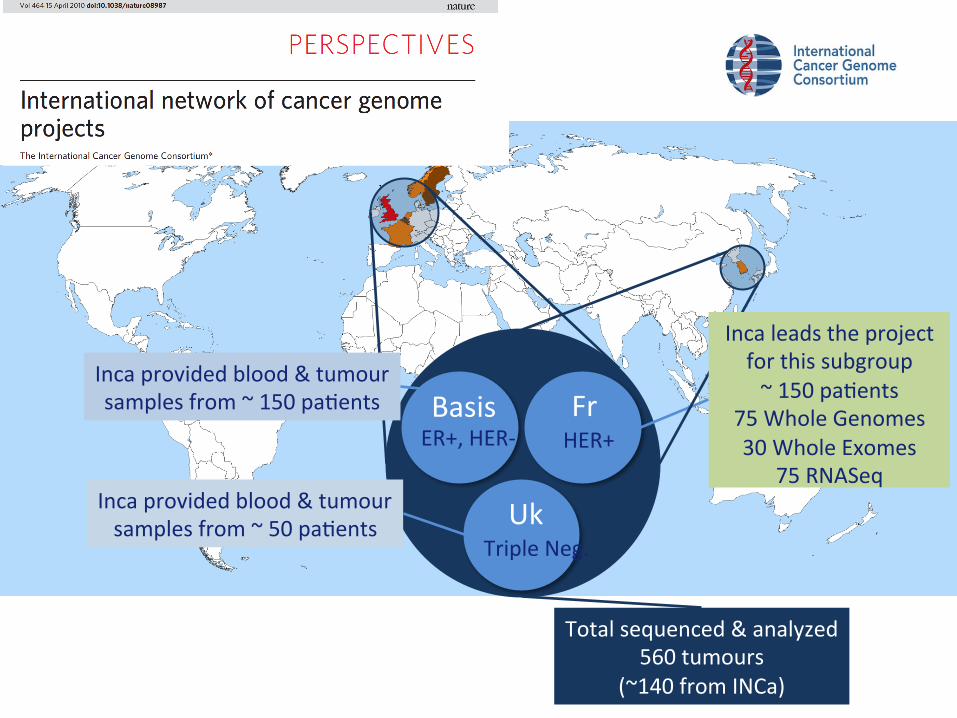
Basis ER+, HER-‐
Fr HER+
Uk Triple Neg.
Inca provided blood & tumour samples from ~ 150 paGents
Inca provided blood & tumour samples from ~ 50 paGents
Total sequenced & analyzed 560 tumours
(~140 from INCa)
Inca leads the project for this subgroup ~ 150 paGents
75 Whole Genomes 30 Whole Exomes
75 RNASeq
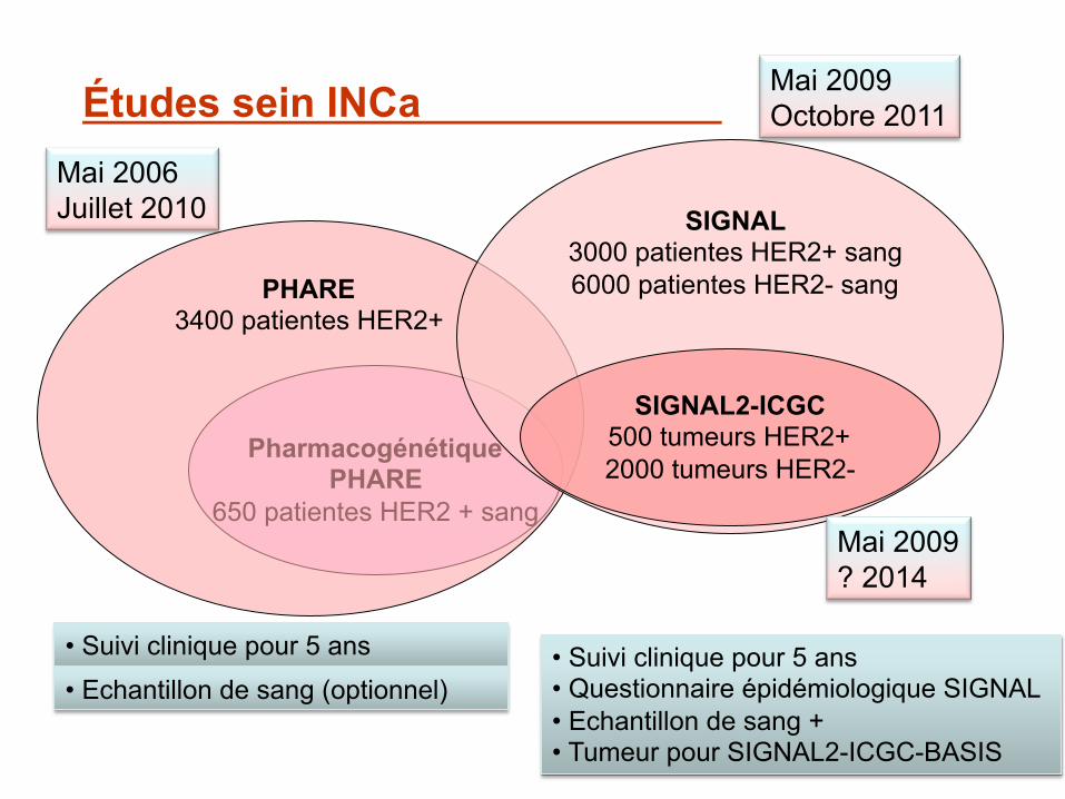
Pharmacogénétique PHARE
650 patientes HER2 + sang
Études sein INCa
PHARE 3400 patientes HER2+
SIGNAL 3000 patientes HER2+ sang 6000 patientes HER2- sang
SIGNAL2-ICGC 500 tumeurs HER2+ 2000 tumeurs HER2-
• Suivi clinique pour 5 ans
Mai 2006 Juillet 2010
Mai 2009 Octobre 2011
Mai 2009 ? 2014
• Echantillon de sang (optionnel) • Suivi clinique pour 5 ans • Questionnaire épidémiologique SIGNAL • Echantillon de sang + • Tumeur pour SIGNAL2-ICGC-BASIS
0 0 M O N T H 2 0 1 6 | V O L 0 0 0 | N A T U R E | 1
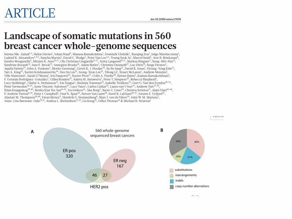
ARTICLEdoi:10.1038/nature17676
Landscape of somatic mutations in 560 breast cancer whole-genome sequencesSerena Nik-Zainal1,2, Helen Davies1, Johan Staaf3, Manasa Ramakrishna1, Dominik Glodzik1, Xueqing Zou1, Inigo Martincorena1, Ludmil B. Alexandrov1,4,5, Sancha Martin1, David C. Wedge1, Peter Van Loo1,6, Young Seok Ju1, Marcel Smid7, Arie B. Brinkman8, Sandro Morganella9, Miriam R. Aure10,11, Ole Christian Lingjærde11,12, Anita Langerød10,11, Markus Ringnér3, Sung-Min Ahn13, Sandrine Boyault14, Jane E. Brock15, Annegien Broeks16, Adam Butler1, Christine Desmedt17, Luc Dirix18, Serge Dronov1, Aquila Fatima19, John A. Foekens7, Moritz Gerstung1, Gerrit K. J. Hooijer20, Se Jin Jang21, David R. Jones1, Hyung-Yong Kim22, Tari A. King23, Savitri Krishnamurthy24, Hee Jin Lee21, Jeong-Yeon Lee25, Yilong Li1, Stuart McLaren1, Andrew Menzies1, Ville Mustonen1, Sarah O’Meara1, Iris Pauporté26, Xavier Pivot27, Colin A. Purdie28, Keiran Raine1, Kamna Ramakrishnan1, F. Germán Rodríguez-González7, Gilles Romieu29, Anieta M. Sieuwerts7, Peter T. Simpson30, Rebecca Shepherd1, Lucy Stebbings1, Olafur A. Stefansson31, Jon Teague1, Stefania Tommasi32, Isabelle Treilleux33, Gert G. Van den Eynden18,34, Peter Vermeulen18,34, Anne Vincent-Salomon35, Lucy Yates1, Carlos Caldas36, Laura van’t Veer16, Andrew Tutt37,38, Stian Knappskog39,40, Benita Kiat Tee Tan41,42, Jos Jonkers16, Åke Borg3, Naoto T. Ueno24, Christos Sotiriou17, Alain Viari43,44, P. Andrew Futreal1,45, Peter J. Campbell1, Paul N. Span46, Steven Van Laere18, Sunil R. Lakhani30,47, Jorunn E. Eyfjord31, Alastair M. Thompson28,48, Ewan Birney9, Hendrik G. Stunnenberg8, Marc J. van de Vijver20, John W. M. Martens7, Anne-Lise Børresen-Dale10,11, Andrea L. Richardson15,19, Gu Kong22, Gilles Thomas44 & Michael R. Stratton1
The mutational theory of cancer proposes that changes in DNA sequence, termed ‘driver’ mutations, confer proliferative advan-tage on a cell, leading to outgrowth of a neoplastic clone1. Some driver mutations are inherited in the germline, but most arise in
somatic cells during the lifetime of the cancer patient, together with many ‘passenger’ mutations not implicated in cancer development1. Multiple mutational processes, including endogenous and exoge-nous mutagen exposures, aberrant DNA editing, replication errors
1
We analysed whole-genome sequences of 560 breast cancers to advance understanding of the driver mutations conferring clonal advantage and the mutational processes generating somatic mutations. We found that 93 protein-coding cancer genes carried probable driver mutations. Some non-coding regions exhibited high mutation frequencies, but most have distinctive structural features probably causing elevated mutation rates and do not contain driver mutations. Mutational signature analysis was extended to genome rearrangements and revealed twelve base substitution and six rearrangement signatures. Three rearrangement signatures, characterized by tandem duplications or deletions, appear associated with defective homologous-recombination-based DNA repair: one with deficient BRCA1 function, another with deficient BRCA1 or BRCA2 function, the cause of the third is unknown. This analysis of all classes of somatic mutation across exons, introns and intergenic regions highlights the repertoire of cancer genes and mutational processes operating, and progresses towards a comprehensive account of the somatic genetic basis of breast cancer.
1Wellcome Trust Sanger Institute, Hinxton, Cambridge CB10 1SA, UK. 2East Anglian Medical Genetics Service, Cambridge University Hospitals NHS Foundation Trust, Cambridge CB2 9NB, UK. 3Division of Oncology and Pathology, Department of Clinical Sciences Lund, Lund University, Lund SE-223 81, Sweden. 4Theoretical Biology and Biophysics (T-6), Los Alamos National Laboratory, Los Alamos, NM 87545, New Mexico, USA. 5Center for Nonlinear Studies, Los Alamos National Laboratory, Los Alamos, New Mexico 87545, USA. 6Department of Human Genetics, University of Leuven, B-3000 Leuven, Belgium. 7Department of Medical Oncology, Erasmus MC Cancer Institute and Cancer Genomics Netherlands, Erasmus University Medical Center, Rotterdam 3015CN, The Netherlands. 8Radboud University, Department of Molecular Biology, Faculties of Science and Medicine, 6525GA Nijmegen, The Netherlands. 9European Molecular Biology Laboratory, European Bioinformatics Institute, Wellcome Trust Genome Campus, Hinxton, Cambridge CB10 1SD, UK. 10Department of Cancer Genetics, Institute for Cancer Research, Oslo University Hospital, The Norwegian Radium Hospital, Oslo 0310, Norway. 11K. G. Jebsen Centre for Breast Cancer Research, Institute for Clinical Medicine, University of Oslo, Oslo 0310, Norway. 12Department of Computer Science, University of Oslo, Oslo, Norway. 13Gachon Institute of Genome Medicine and Science, Gachon University Gil Medical Center, Incheon, South Korea. 14Translational Research Lab, Centre Léon Bérard, 28, rue Laënnec, 69373 Lyon Cedex 08, France. 15Department of Pathology, Brigham and Women’s Hospital, Boston, Massachusetts 02115, USA. 16The Netherlands Cancer Institute, 1066 CX Amsterdam, The Netherlands. 17Breast Cancer Translational Research Laboratory, Université Libre de Bruxelles, Institut Jules Bordet, Bd de Waterloo 121, B-1000 Brussels, Belgium. 18Translational Cancer Research Unit, Center for Oncological Research, Faculty of Medicine and Health Sciences, University of Antwerp, Antwerp, Belgium. 19Dana-Farber Cancer Institute, Boston, Massachusetts 02215, USA. 20Department of Pathology, Academic Medical Center, Meibergdreef 9, 1105 AZ Amsterdam, The Netherlands. 21Department of Pathology, Asan Medical Center, College of Medicine, Ulsan University, Ulsan, South Korea. 22Department of Pathology, College of Medicine, Hanyang University, Seoul 133-791, South Korea. 23Memorial Sloan Kettering Cancer Center, 1275 York Avenue, New York, New York 10065, USA. 24Morgan Welch Inflammatory Breast Cancer Research Program and Clinic, The University of Texas MD Anderson Cancer Center, 1515 Holcombe Boulevard., Houston, Texas 77030, USA. 25Institute for Bioengineering and Biopharmaceutical Research (IBBR), Hanyang University, Seoul, South Korea. 26Institut National du Cancer, Research Division, Clinical Research Department, 52 avenue Morizet, 92513 Boulogne-Billancourt, France. 27University Hospital of Minjoz, INSERM UMR 1098, Bd Fleming, Besançon 25000, France. 28Pathology Department, Ninewells Hospital and Medical School, Dundee DD1 9SY, UK. 29Oncologie Sénologie, ICM Institut Régional du Cancer, Montpellier, France. 30The University of Queensland, UQ Centre for Clinical Research and School of Medicine, Brisbane, Queensland 4059, Australia. 31Cancer Research Laboratory, Faculty of Medicine, University of Iceland, 101 Reykjavik, Iceland. 32IRCCS Istituto Tumori “Giovanni Paolo II”, Bari, Italy. 33Department of Pathology, Centre Léon Bérard, 28 rue Laënnec, 69373 Lyon Cédex 08, France. 34Department of Pathology, GZA Hospitals Sint-Augustinus, Antwerp, Belgium. 35Institut Curie, Department of Pathology and INSERM U934, 26 rue d’Ulm, 75248 Paris Cedex 05, France. 36Cancer Research UK Cambridge Institute, University of Cambridge, Li Ka Shing Centre, Robinson Way, Cambridge CB2 0RE, UK. 37Breast Cancer Now Toby Robin’s Research Unit, King’s College London, London SE1 9RT, UK. 38Breast Cancer Now Toby Robin’s Research Centre, Institute of Cancer Research, London SW3 6JB, UK. 39Department of Clinical Science, University of Bergen, 5020 Bergen, Norway. 40Department of Oncology, Haukeland University Hospital, 5021 Bergen, Norway. 41National Cancer Centre Singapore, 11 Hospital Drive, 169610, Singapore. 42Singapore General Hospital, Outram Road, 169608, Singapore. 43Equipe Erable, INRIA Grenoble-Rhône-Alpes, 655, Avenue de l’Europe, 38330 Montbonnot-Saint Martin, France. 44Synergie Lyon Cancer, Centre Léon Bérard, 28 rue Laënnec, Lyon Cedex 08, France. 45Department of Genomic Medicine, UT MD Anderson Cancer Center, Houston, Texas 77230, USA. 46Department of Radiation Oncology, Department of Laboratory Medicine, Radboud University Medical Center, Nijmegen 6525GA, The Netherlands. 47Pathology Queensland, The Royal Brisbane and Women’s Hospital, Brisbane, Queensland 4029, Australia. 48Department of Surgical Oncology, University of Texas MD Anderson Cancer Center, 1400 Pressler Street, Houston, Texas 77030, USA.
2
ARTICLE RESEARCH
A B
substitutions
indels
rearrangements
copy number aberrations
36%34%
10% 21%
0.0e+00 5.0e+07 1.0e+08 1.5e+08 2.0e+08 2.5e+08
050
0 Chr1
0.0e+00 5.0e+07 1.0e+08 1.5e+08 2.0e+08 2.5e+08
050
0
0.0e+00 5.0e+07 1.0e+08 1.5e+08 2.0e+08 2.5e+08
050
0 Chr2
0.0e+00 5.0e+07 1.0e+08 1.5e+08 2.0e+08 2.5e+08
050
0
0.0e+00 5.0e+07 1.0e+08 1.5e+08 2.0e+08
050
0 Chr3
0.0e+00 5.0e+07 1.0e+08 1.5e+08 2.0e+08
050
0
0.0e+00 5.0e+07 1.0e+08 1.5e+08
050
0 Chr4
0.0e+00 5.0e+07 1.0e+08 1.5e+08
050
0
0.0e+00 5.0e+07 1.0e+08 1.5e+08
050
0 Chr5
0.0e+00 5.0e+07 1.0e+08 1.5e+08
050
0
0.0e+00 5.0e+07 1.0e+08 1.5e+08
050
0 Chr6
0.0e+00 5.0e+07 1.0e+08 1.5e+08
050
0
0.0e+00 5.0e+07 1.0e+08 1.5e+08
050
0 Chr7
0.0e+00 5.0e+07 1.0e+08 1.5e+08
050
0
0.0e+00 5.0e+07 1.0e+08 1.5e+08
050
0 Chr8
0.0e+00 5.0e+07 1.0e+08 1.5e+08
050
0
0.0e+00 2.0e+07 4.0e+07 6.0e+07 8.0e+07 1.0e+08 1.2e+08 1.4e+08
050
0 Chr9
0.0e+00 2.0e+07 4.0e+07 6.0e+07 8.0e+07 1.0e+08 1.2e+08 1.4e+08
050
0
0.0e+00 2.0e+07 4.0e+07 6.0e+07 8.0e+07 1.0e+08 1.2e+08 1.4e+08
050
0 Chr10
0.0e+00 2.0e+07 4.0e+07 6.0e+07 8.0e+07 1.0e+08 1.2e+08 1.4e+08
050
0
0.0e+00 2.0e+07 4.0e+07 6.0e+07 8.0e+07 1.0e+08 1.2e+08
050
0 Chr11
0.0e+00 2.0e+07 4.0e+07 6.0e+07 8.0e+07 1.0e+08 1.2e+08
050
0
0.0e+00 2.0e+07 4.0e+07 6.0e+07 8.0e+07 1.0e+08 1.2e+08
050
0 Chr12
0.0e+00 2.0e+07 4.0e+07 6.0e+07 8.0e+07 1.0e+08 1.2e+08
050
0
0e+00 2e+07 4e+07 6e+07 8e+07 1e+08
050
0 Chr13
0e+00 2e+07 4e+07 6e+07 8e+07 1e+08
050
0
0e+00 2e+07 4e+07 6e+07 8e+07 1e+08
050
0 Chr14
0e+00 2e+07 4e+07 6e+07 8e+07 1e+08
050
0
0e+00 2e+07 4e+07 6e+07 8e+07 1e+08
050
0 Chr15
0e+00 2e+07 4e+07 6e+07 8e+07 1e+08
050
0
0e+00 2e+07 4e+07 6e+07 8e+07
050
0 Chr16
0e+00 2e+07 4e+07 6e+07 8e+07
050
0
0e+00 2e+07 4e+07 6e+07 8e+07
050
0 Chr17
0e+00 2e+07 4e+07 6e+07 8e+07
050
0
0e+00 2e+07 4e+07 6e+07
050
0 Chr18
0e+00 2e+07 4e+07 6e+07
050
0
0e+00 1e+07 2e+07 3e+07 4e+07 5e+07 6e+07
050
0 Chr19
0e+00 1e+07 2e+07 3e+07 4e+07 5e+07 6e+07
050
0
0e+00 1e+07 2e+07 3e+07 4e+07 5e+07 6e+07
050
0 Chr20
0e+00 1e+07 2e+07 3e+07 4e+07 5e+07 6e+07
050
0
0e+00 1e+07 2e+07 3e+07 4e+07
050
0 Chr21
0e+00 1e+07 2e+07 3e+07 4e+07
050
0
0e+00 1e+07 2e+07 3e+07 4e+07 5e+07
050
0 Chr22
0e+00 1e+07 2e+07 3e+07 4e+07 5e+07
050
0
0.0e+00 5.0e+07 1.0e+08 1.5e+08
050
0 Chr23
0.0e+00 5.0e+07 1.0e+08 1.5e+08
050
0
C
ER pos320
ER neg167
27 46
HER2 pos
560 whole-genome sequenced breast cancers
Extended Data Figure 1 | Landscape of driver mutations. a, Summary of subtypes of cohort of 560!breast cancers. b, Driver mutations by mutation type. c, Distribution of rearrangements throughout the genome. Black
line represents background rearrangement density (calculation based on rearrangement breakpoints in intergenic regions only). Red lines represent frequency of rearrangement within breast cancer genes.
© 2016 Macmillan Publishers Limited. All rights reserved
0 0 M O N T H 2 0 1 6 | V O L 0 0 0 | N A T U R E | 1
ARTICLEdoi:10.1038/nature17676
Landscape of somatic mutations in 560 breast cancer whole-genome sequencesSerena Nik-Zainal1,2, Helen Davies1, Johan Staaf3, Manasa Ramakrishna1, Dominik Glodzik1, Xueqing Zou1, Inigo Martincorena1, Ludmil B. Alexandrov1,4,5, Sancha Martin1, David C. Wedge1, Peter Van Loo1,6, Young Seok Ju1, Marcel Smid7, Arie B. Brinkman8, Sandro Morganella9, Miriam R. Aure10,11, Ole Christian Lingjærde11,12, Anita Langerød10,11, Markus Ringnér3, Sung-Min Ahn13, Sandrine Boyault14, Jane E. Brock15, Annegien Broeks16, Adam Butler1, Christine Desmedt17, Luc Dirix18, Serge Dronov1, Aquila Fatima19, John A. Foekens7, Moritz Gerstung1, Gerrit K. J. Hooijer20, Se Jin Jang21, David R. Jones1, Hyung-Yong Kim22, Tari A. King23, Savitri Krishnamurthy24, Hee Jin Lee21, Jeong-Yeon Lee25, Yilong Li1, Stuart McLaren1, Andrew Menzies1, Ville Mustonen1, Sarah O’Meara1, Iris Pauporté26, Xavier Pivot27, Colin A. Purdie28, Keiran Raine1, Kamna Ramakrishnan1, F. Germán Rodríguez-González7, Gilles Romieu29, Anieta M. Sieuwerts7, Peter T. Simpson30, Rebecca Shepherd1, Lucy Stebbings1, Olafur A. Stefansson31, Jon Teague1, Stefania Tommasi32, Isabelle Treilleux33, Gert G. Van den Eynden18,34, Peter Vermeulen18,34, Anne Vincent-Salomon35, Lucy Yates1, Carlos Caldas36, Laura van’t Veer16, Andrew Tutt37,38, Stian Knappskog39,40, Benita Kiat Tee Tan41,42, Jos Jonkers16, Åke Borg3, Naoto T. Ueno24, Christos Sotiriou17, Alain Viari43,44, P. Andrew Futreal1,45, Peter J. Campbell1, Paul N. Span46, Steven Van Laere18, Sunil R. Lakhani30,47, Jorunn E. Eyfjord31, Alastair M. Thompson28,48, Ewan Birney9, Hendrik G. Stunnenberg8, Marc J. van de Vijver20, John W. M. Martens7, Anne-Lise Børresen-Dale10,11, Andrea L. Richardson15,19, Gu Kong22, Gilles Thomas44 & Michael R. Stratton1
The mutational theory of cancer proposes that changes in DNA sequence, termed ‘driver’ mutations, confer proliferative advan-tage on a cell, leading to outgrowth of a neoplastic clone1. Some driver mutations are inherited in the germline, but most arise in
somatic cells during the lifetime of the cancer patient, together with many ‘passenger’ mutations not implicated in cancer development1. Multiple mutational processes, including endogenous and exoge-nous mutagen exposures, aberrant DNA editing, replication errors
1
We analysed whole-genome sequences of 560 breast cancers to advance understanding of the driver mutations conferring clonal advantage and the mutational processes generating somatic mutations. We found that 93 protein-coding cancer genes carried probable driver mutations. Some non-coding regions exhibited high mutation frequencies, but most have distinctive structural features probably causing elevated mutation rates and do not contain driver mutations. Mutational signature analysis was extended to genome rearrangements and revealed twelve base substitution and six rearrangement signatures. Three rearrangement signatures, characterized by tandem duplications or deletions, appear associated with defective homologous-recombination-based DNA repair: one with deficient BRCA1 function, another with deficient BRCA1 or BRCA2 function, the cause of the third is unknown. This analysis of all classes of somatic mutation across exons, introns and intergenic regions highlights the repertoire of cancer genes and mutational processes operating, and progresses towards a comprehensive account of the somatic genetic basis of breast cancer.
1Wellcome Trust Sanger Institute, Hinxton, Cambridge CB10 1SA, UK. 2East Anglian Medical Genetics Service, Cambridge University Hospitals NHS Foundation Trust, Cambridge CB2 9NB, UK. 3Division of Oncology and Pathology, Department of Clinical Sciences Lund, Lund University, Lund SE-223 81, Sweden. 4Theoretical Biology and Biophysics (T-6), Los Alamos National Laboratory, Los Alamos, NM 87545, New Mexico, USA. 5Center for Nonlinear Studies, Los Alamos National Laboratory, Los Alamos, New Mexico 87545, USA. 6Department of Human Genetics, University of Leuven, B-3000 Leuven, Belgium. 7Department of Medical Oncology, Erasmus MC Cancer Institute and Cancer Genomics Netherlands, Erasmus University Medical Center, Rotterdam 3015CN, The Netherlands. 8Radboud University, Department of Molecular Biology, Faculties of Science and Medicine, 6525GA Nijmegen, The Netherlands. 9European Molecular Biology Laboratory, European Bioinformatics Institute, Wellcome Trust Genome Campus, Hinxton, Cambridge CB10 1SD, UK. 10Department of Cancer Genetics, Institute for Cancer Research, Oslo University Hospital, The Norwegian Radium Hospital, Oslo 0310, Norway. 11K. G. Jebsen Centre for Breast Cancer Research, Institute for Clinical Medicine, University of Oslo, Oslo 0310, Norway. 12Department of Computer Science, University of Oslo, Oslo, Norway. 13Gachon Institute of Genome Medicine and Science, Gachon University Gil Medical Center, Incheon, South Korea. 14Translational Research Lab, Centre Léon Bérard, 28, rue Laënnec, 69373 Lyon Cedex 08, France. 15Department of Pathology, Brigham and Women’s Hospital, Boston, Massachusetts 02115, USA. 16The Netherlands Cancer Institute, 1066 CX Amsterdam, The Netherlands. 17Breast Cancer Translational Research Laboratory, Université Libre de Bruxelles, Institut Jules Bordet, Bd de Waterloo 121, B-1000 Brussels, Belgium. 18Translational Cancer Research Unit, Center for Oncological Research, Faculty of Medicine and Health Sciences, University of Antwerp, Antwerp, Belgium. 19Dana-Farber Cancer Institute, Boston, Massachusetts 02215, USA. 20Department of Pathology, Academic Medical Center, Meibergdreef 9, 1105 AZ Amsterdam, The Netherlands. 21Department of Pathology, Asan Medical Center, College of Medicine, Ulsan University, Ulsan, South Korea. 22Department of Pathology, College of Medicine, Hanyang University, Seoul 133-791, South Korea. 23Memorial Sloan Kettering Cancer Center, 1275 York Avenue, New York, New York 10065, USA. 24Morgan Welch Inflammatory Breast Cancer Research Program and Clinic, The University of Texas MD Anderson Cancer Center, 1515 Holcombe Boulevard., Houston, Texas 77030, USA. 25Institute for Bioengineering and Biopharmaceutical Research (IBBR), Hanyang University, Seoul, South Korea. 26Institut National du Cancer, Research Division, Clinical Research Department, 52 avenue Morizet, 92513 Boulogne-Billancourt, France. 27University Hospital of Minjoz, INSERM UMR 1098, Bd Fleming, Besançon 25000, France. 28Pathology Department, Ninewells Hospital and Medical School, Dundee DD1 9SY, UK. 29Oncologie Sénologie, ICM Institut Régional du Cancer, Montpellier, France. 30The University of Queensland, UQ Centre for Clinical Research and School of Medicine, Brisbane, Queensland 4059, Australia. 31Cancer Research Laboratory, Faculty of Medicine, University of Iceland, 101 Reykjavik, Iceland. 32IRCCS Istituto Tumori “Giovanni Paolo II”, Bari, Italy. 33Department of Pathology, Centre Léon Bérard, 28 rue Laënnec, 69373 Lyon Cédex 08, France. 34Department of Pathology, GZA Hospitals Sint-Augustinus, Antwerp, Belgium. 35Institut Curie, Department of Pathology and INSERM U934, 26 rue d’Ulm, 75248 Paris Cedex 05, France. 36Cancer Research UK Cambridge Institute, University of Cambridge, Li Ka Shing Centre, Robinson Way, Cambridge CB2 0RE, UK. 37Breast Cancer Now Toby Robin’s Research Unit, King’s College London, London SE1 9RT, UK. 38Breast Cancer Now Toby Robin’s Research Centre, Institute of Cancer Research, London SW3 6JB, UK. 39Department of Clinical Science, University of Bergen, 5020 Bergen, Norway. 40Department of Oncology, Haukeland University Hospital, 5021 Bergen, Norway. 41National Cancer Centre Singapore, 11 Hospital Drive, 169610, Singapore. 42Singapore General Hospital, Outram Road, 169608, Singapore. 43Equipe Erable, INRIA Grenoble-Rhône-Alpes, 655, Avenue de l’Europe, 38330 Montbonnot-Saint Martin, France. 44Synergie Lyon Cancer, Centre Léon Bérard, 28 rue Laënnec, Lyon Cedex 08, France. 45Department of Genomic Medicine, UT MD Anderson Cancer Center, Houston, Texas 77230, USA. 46Department of Radiation Oncology, Department of Laboratory Medicine, Radboud University Medical Center, Nijmegen 6525GA, The Netherlands. 47Pathology Queensland, The Royal Brisbane and Women’s Hospital, Brisbane, Queensland 4029, Australia. 48Department of Surgical Oncology, University of Texas MD Anderson Cancer Center, 1400 Pressler Street, Houston, Texas 77030, USA.
2
2 | N A T U R E | V O L 0 0 0 | 0 0 M O N T H 2 0 1 6
ARTICLERESEARCH
and defective DNA maintenance, are responsible for generating these mutations1–3.
Over the past five decades, several waves of technology have advanced the characterization of mutations in cancer genomes. Karyotype analysis revealed rearranged chromosomes and copy number alterations. Subsequently, loss of heterozygosity analysis, hybridization of cancer-derived DNA to microarrays and other approaches provided higher resolution insights into copy number changes4–8. Recently, DNA sequencing has enabled systematic characterization of the full reper-toire of mutation types including base substitutions, small insertions/deletions, rearrangements and copy number changes9–13, yielding substantial insights into the mutated cancer genes and mutational processes operative in human cancer.
As for many cancer classes, most currently available breast cancer genome sequences target protein-coding exons8,11–15. Consequently, there has been limited consideration of mutations in untranslated, intronic and intergenic regions, leaving central questions pertaining to the molecular pathogenesis of the disease unresolved. First, the role of activating driver rearrangements16–18 forming chimaeric (fusion) genes/proteins or relocating genes adjacent to new regulatory regions as observed in haematological and other malignancies19. Second, the role of driver substitutions and indels in non-coding regions of the genome20,21. Common inherited variants conferring susceptibility to human disease are generally in non-coding regulatory regions and the possibility that similar mechanisms operate somatically in cancer was highlighted by the discovery of somatic driver substitutions in the TERT gene promoter22,23. Third, which mutational processes generate the somatic mutations found in breast cancer2,24. Addressing this question has been constrained because exome sequences do not inform on genome rearrangements and capture relatively few base substitu-tion mutations, thus limiting statistical power to extract the mutational signatures imprinted on the genome by these processes24,25.
Here we analyse whole-genome sequences of 560!cases in order to address these and other questions and to pave the way to a compre-hensive understanding of the origins and consequences of somatic mutations in breast cancer.
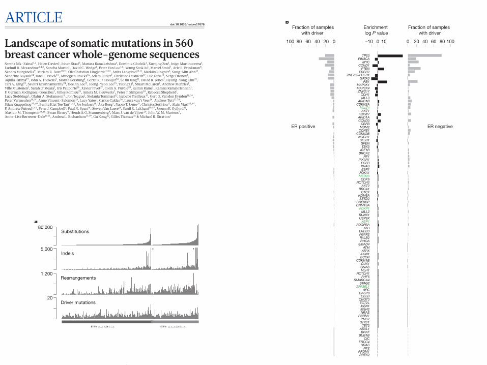
Cancer genes and driver mutationsThe whole genomes of 560 breast cancers and non-neoplastic tissue from each individual (556 female and 4 male) were sequenced (Supplementary Fig. 1, Supplementary Table 1). We detected 3,479,652!somatic base substitutions, 371,993!small indels and 77,695!rearrangements, with substantial variation in the number of each between individual samples (Fig. 1a, Supplementary Table 3). Transcriptome sequence, microRNA expression, array-based copy num-ber and DNA methylation data were obtained from subsets of cases.
To identify new cancer genes, we combined somatic substitutions and indels in protein-coding exons with data from other series12–15,26, constituting a total of 1,332!breast cancers, and searched for mutation clustering in each gene beyond that expected by chance. Five cancer genes were found for which evidence was previously absent or equivocal (MED23, FOXP1, MLLT4, XBP1, ZFP36L1), or for which the muta-tions indicate the gene acts in breast cancer in a recessive rather than in a dominant fashion, as previously reported in other cancer types (see Supplementary Methods section 7.4 for detailed descriptions). From published reports on all cancer types (http://cancer.sanger.ac.uk/census),
Enrichment log P value
0 10–10020406080100
Fraction of samples with driver
ER positive ER negative
5,000
80,000
1,200
20
Substitutions
Indels
Rearrangements
Driver mutations
Fraction of samples with driver
0 20 40 60 80
TP53PIK3CA
MYCCCND1PTEN
ERBB2ZNF703/FGFR1
GATA3RB1
MAP3K1MAP2K4ZNF217CDH1MLL3
ARID1BCDKN2AMLLT4AKT1
FBXW7ARID1ACCND3CBFBMDM2CCNE1
CDKN2BNCOR1SF3B1SPENTBX3IGF1RBRCA2
NF1PIK3R1EGFRKRASESR1
FOXA1MED23CDK6
NOTCH2AKT2
BRCA1CTCF
KDM6ASETD2
CREBBPDNMT3AFOXP1MLL2
RUNX1USP9XXBP1
PDGFRAATR
ERBB3FGFR2PALB2RHOA
SMAD4ATMATRXAXIN1BCOR
CDKN1BCUX1GNASMLH1
NOTCH1PHF6
SMARCA4STAG2
ZFP36L1APC
CASP8CBLB
CNOT3ECT2LMEN1MSH2NRAS
PBRM1PMS2STK11TET2
ASXL1BRAF
BUB1BCIC
ERCC4HRASNF2
PRDM1PREX2
ER positive ER negative
* *
b
a
100
Figure 1 | Cohort and catalogue of somatic mutations in 560!breast cancers. a, Catalogue of base substitutions, insertions/deletions, rearrangements and driver mutations in 560!breast cancers (sorted by total substitution burden). Indel axis limited to 5,000(*). b, Complete list of curated driver genes sorted by frequency (descending). Fraction of ER-positive (left, total 366) and ER-negative (right, total 194) samples carrying a mutation in the relevant driver gene presented in grey. log10 P value of enrichment of each driver gene towards the ER-positive or ER-negative cohort is provided in black. Highlighted in green are genes for which there is new or further evidence supporting these as novel breast cancer genes.
© 2016 Macmillan Publishers Limited. All rights reserved
2 | N A T U R E | V O L 0 0 0 | 0 0 M O N T H 2 0 1 6
ARTICLERESEARCH
and defective DNA maintenance, are responsible for generating these mutations1–3.
Over the past five decades, several waves of technology have advanced the characterization of mutations in cancer genomes. Karyotype analysis revealed rearranged chromosomes and copy number alterations. Subsequently, loss of heterozygosity analysis, hybridization of cancer-derived DNA to microarrays and other approaches provided higher resolution insights into copy number changes4–8. Recently, DNA sequencing has enabled systematic characterization of the full reper-toire of mutation types including base substitutions, small insertions/deletions, rearrangements and copy number changes9–13, yielding substantial insights into the mutated cancer genes and mutational processes operative in human cancer.
As for many cancer classes, most currently available breast cancer genome sequences target protein-coding exons8,11–15. Consequently, there has been limited consideration of mutations in untranslated, intronic and intergenic regions, leaving central questions pertaining to the molecular pathogenesis of the disease unresolved. First, the role of activating driver rearrangements16–18 forming chimaeric (fusion) genes/proteins or relocating genes adjacent to new regulatory regions as observed in haematological and other malignancies19. Second, the role of driver substitutions and indels in non-coding regions of the genome20,21. Common inherited variants conferring susceptibility to human disease are generally in non-coding regulatory regions and the possibility that similar mechanisms operate somatically in cancer was highlighted by the discovery of somatic driver substitutions in the TERT gene promoter22,23. Third, which mutational processes generate the somatic mutations found in breast cancer2,24. Addressing this question has been constrained because exome sequences do not inform on genome rearrangements and capture relatively few base substitu-tion mutations, thus limiting statistical power to extract the mutational signatures imprinted on the genome by these processes24,25.
Here we analyse whole-genome sequences of 560!cases in order to address these and other questions and to pave the way to a compre-hensive understanding of the origins and consequences of somatic mutations in breast cancer.
Cancer genes and driver mutationsThe whole genomes of 560 breast cancers and non-neoplastic tissue from each individual (556 female and 4 male) were sequenced (Supplementary Fig. 1, Supplementary Table 1). We detected 3,479,652!somatic base substitutions, 371,993!small indels and 77,695!rearrangements, with substantial variation in the number of each between individual samples (Fig. 1a, Supplementary Table 3). Transcriptome sequence, microRNA expression, array-based copy num-ber and DNA methylation data were obtained from subsets of cases.
To identify new cancer genes, we combined somatic substitutions and indels in protein-coding exons with data from other series12–15,26, constituting a total of 1,332!breast cancers, and searched for mutation clustering in each gene beyond that expected by chance. Five cancer genes were found for which evidence was previously absent or equivocal (MED23, FOXP1, MLLT4, XBP1, ZFP36L1), or for which the muta-tions indicate the gene acts in breast cancer in a recessive rather than in a dominant fashion, as previously reported in other cancer types (see Supplementary Methods section 7.4 for detailed descriptions). From published reports on all cancer types (http://cancer.sanger.ac.uk/census),
Enrichment log P value
0 10–10020406080100
Fraction of samples with driver
ER positive ER negative
5,000
80,000
1,200
20
Substitutions
Indels
Rearrangements
Driver mutations
Fraction of samples with driver
0 20 40 60 80
TP53PIK3CA
MYCCCND1PTEN
ERBB2ZNF703/FGFR1
GATA3RB1
MAP3K1MAP2K4ZNF217CDH1MLL3
ARID1BCDKN2AMLLT4AKT1
FBXW7ARID1ACCND3CBFBMDM2CCNE1
CDKN2BNCOR1SF3B1SPENTBX3IGF1RBRCA2
NF1PIK3R1EGFRKRASESR1
FOXA1MED23CDK6
NOTCH2AKT2
BRCA1CTCF
KDM6ASETD2
CREBBPDNMT3AFOXP1MLL2
RUNX1USP9XXBP1
PDGFRAATR
ERBB3FGFR2PALB2RHOA
SMAD4ATMATRXAXIN1BCOR
CDKN1BCUX1GNASMLH1
NOTCH1PHF6
SMARCA4STAG2
ZFP36L1APC
CASP8CBLB
CNOT3ECT2LMEN1MSH2NRAS
PBRM1PMS2STK11TET2
ASXL1BRAF
BUB1BCIC
ERCC4HRASNF2
PRDM1PREX2
ER positive ER negative
* *
b
a
100
Figure 1 | Cohort and catalogue of somatic mutations in 560!breast cancers. a, Catalogue of base substitutions, insertions/deletions, rearrangements and driver mutations in 560!breast cancers (sorted by total substitution burden). Indel axis limited to 5,000(*). b, Complete list of curated driver genes sorted by frequency (descending). Fraction of ER-positive (left, total 366) and ER-negative (right, total 194) samples carrying a mutation in the relevant driver gene presented in grey. log10 P value of enrichment of each driver gene towards the ER-positive or ER-negative cohort is provided in black. Highlighted in green are genes for which there is new or further evidence supporting these as novel breast cancer genes.
© 2016 Macmillan Publishers Limited. All rights reserved
0 0 M O N T H 2 0 1 6 | V O L 0 0 0 | N A T U R E | 1
ARTICLEdoi:10.1038/nature17676
Landscape of somatic mutations in 560 breast cancer whole-genome sequencesSerena Nik-Zainal1,2, Helen Davies1, Johan Staaf3, Manasa Ramakrishna1, Dominik Glodzik1, Xueqing Zou1, Inigo Martincorena1, Ludmil B. Alexandrov1,4,5, Sancha Martin1, David C. Wedge1, Peter Van Loo1,6, Young Seok Ju1, Marcel Smid7, Arie B. Brinkman8, Sandro Morganella9, Miriam R. Aure10,11, Ole Christian Lingjærde11,12, Anita Langerød10,11, Markus Ringnér3, Sung-Min Ahn13, Sandrine Boyault14, Jane E. Brock15, Annegien Broeks16, Adam Butler1, Christine Desmedt17, Luc Dirix18, Serge Dronov1, Aquila Fatima19, John A. Foekens7, Moritz Gerstung1, Gerrit K. J. Hooijer20, Se Jin Jang21, David R. Jones1, Hyung-Yong Kim22, Tari A. King23, Savitri Krishnamurthy24, Hee Jin Lee21, Jeong-Yeon Lee25, Yilong Li1, Stuart McLaren1, Andrew Menzies1, Ville Mustonen1, Sarah O’Meara1, Iris Pauporté26, Xavier Pivot27, Colin A. Purdie28, Keiran Raine1, Kamna Ramakrishnan1, F. Germán Rodríguez-González7, Gilles Romieu29, Anieta M. Sieuwerts7, Peter T. Simpson30, Rebecca Shepherd1, Lucy Stebbings1, Olafur A. Stefansson31, Jon Teague1, Stefania Tommasi32, Isabelle Treilleux33, Gert G. Van den Eynden18,34, Peter Vermeulen18,34, Anne Vincent-Salomon35, Lucy Yates1, Carlos Caldas36, Laura van’t Veer16, Andrew Tutt37,38, Stian Knappskog39,40, Benita Kiat Tee Tan41,42, Jos Jonkers16, Åke Borg3, Naoto T. Ueno24, Christos Sotiriou17, Alain Viari43,44, P. Andrew Futreal1,45, Peter J. Campbell1, Paul N. Span46, Steven Van Laere18, Sunil R. Lakhani30,47, Jorunn E. Eyfjord31, Alastair M. Thompson28,48, Ewan Birney9, Hendrik G. Stunnenberg8, Marc J. van de Vijver20, John W. M. Martens7, Anne-Lise Børresen-Dale10,11, Andrea L. Richardson15,19, Gu Kong22, Gilles Thomas44 & Michael R. Stratton1
The mutational theory of cancer proposes that changes in DNA sequence, termed ‘driver’ mutations, confer proliferative advan-tage on a cell, leading to outgrowth of a neoplastic clone1. Some driver mutations are inherited in the germline, but most arise in
somatic cells during the lifetime of the cancer patient, together with many ‘passenger’ mutations not implicated in cancer development1. Multiple mutational processes, including endogenous and exoge-nous mutagen exposures, aberrant DNA editing, replication errors
1
We analysed whole-genome sequences of 560 breast cancers to advance understanding of the driver mutations conferring clonal advantage and the mutational processes generating somatic mutations. We found that 93 protein-coding cancer genes carried probable driver mutations. Some non-coding regions exhibited high mutation frequencies, but most have distinctive structural features probably causing elevated mutation rates and do not contain driver mutations. Mutational signature analysis was extended to genome rearrangements and revealed twelve base substitution and six rearrangement signatures. Three rearrangement signatures, characterized by tandem duplications or deletions, appear associated with defective homologous-recombination-based DNA repair: one with deficient BRCA1 function, another with deficient BRCA1 or BRCA2 function, the cause of the third is unknown. This analysis of all classes of somatic mutation across exons, introns and intergenic regions highlights the repertoire of cancer genes and mutational processes operating, and progresses towards a comprehensive account of the somatic genetic basis of breast cancer.
1Wellcome Trust Sanger Institute, Hinxton, Cambridge CB10 1SA, UK. 2East Anglian Medical Genetics Service, Cambridge University Hospitals NHS Foundation Trust, Cambridge CB2 9NB, UK. 3Division of Oncology and Pathology, Department of Clinical Sciences Lund, Lund University, Lund SE-223 81, Sweden. 4Theoretical Biology and Biophysics (T-6), Los Alamos National Laboratory, Los Alamos, NM 87545, New Mexico, USA. 5Center for Nonlinear Studies, Los Alamos National Laboratory, Los Alamos, New Mexico 87545, USA. 6Department of Human Genetics, University of Leuven, B-3000 Leuven, Belgium. 7Department of Medical Oncology, Erasmus MC Cancer Institute and Cancer Genomics Netherlands, Erasmus University Medical Center, Rotterdam 3015CN, The Netherlands. 8Radboud University, Department of Molecular Biology, Faculties of Science and Medicine, 6525GA Nijmegen, The Netherlands. 9European Molecular Biology Laboratory, European Bioinformatics Institute, Wellcome Trust Genome Campus, Hinxton, Cambridge CB10 1SD, UK. 10Department of Cancer Genetics, Institute for Cancer Research, Oslo University Hospital, The Norwegian Radium Hospital, Oslo 0310, Norway. 11K. G. Jebsen Centre for Breast Cancer Research, Institute for Clinical Medicine, University of Oslo, Oslo 0310, Norway. 12Department of Computer Science, University of Oslo, Oslo, Norway. 13Gachon Institute of Genome Medicine and Science, Gachon University Gil Medical Center, Incheon, South Korea. 14Translational Research Lab, Centre Léon Bérard, 28, rue Laënnec, 69373 Lyon Cedex 08, France. 15Department of Pathology, Brigham and Women’s Hospital, Boston, Massachusetts 02115, USA. 16The Netherlands Cancer Institute, 1066 CX Amsterdam, The Netherlands. 17Breast Cancer Translational Research Laboratory, Université Libre de Bruxelles, Institut Jules Bordet, Bd de Waterloo 121, B-1000 Brussels, Belgium. 18Translational Cancer Research Unit, Center for Oncological Research, Faculty of Medicine and Health Sciences, University of Antwerp, Antwerp, Belgium. 19Dana-Farber Cancer Institute, Boston, Massachusetts 02215, USA. 20Department of Pathology, Academic Medical Center, Meibergdreef 9, 1105 AZ Amsterdam, The Netherlands. 21Department of Pathology, Asan Medical Center, College of Medicine, Ulsan University, Ulsan, South Korea. 22Department of Pathology, College of Medicine, Hanyang University, Seoul 133-791, South Korea. 23Memorial Sloan Kettering Cancer Center, 1275 York Avenue, New York, New York 10065, USA. 24Morgan Welch Inflammatory Breast Cancer Research Program and Clinic, The University of Texas MD Anderson Cancer Center, 1515 Holcombe Boulevard., Houston, Texas 77030, USA. 25Institute for Bioengineering and Biopharmaceutical Research (IBBR), Hanyang University, Seoul, South Korea. 26Institut National du Cancer, Research Division, Clinical Research Department, 52 avenue Morizet, 92513 Boulogne-Billancourt, France. 27University Hospital of Minjoz, INSERM UMR 1098, Bd Fleming, Besançon 25000, France. 28Pathology Department, Ninewells Hospital and Medical School, Dundee DD1 9SY, UK. 29Oncologie Sénologie, ICM Institut Régional du Cancer, Montpellier, France. 30The University of Queensland, UQ Centre for Clinical Research and School of Medicine, Brisbane, Queensland 4059, Australia. 31Cancer Research Laboratory, Faculty of Medicine, University of Iceland, 101 Reykjavik, Iceland. 32IRCCS Istituto Tumori “Giovanni Paolo II”, Bari, Italy. 33Department of Pathology, Centre Léon Bérard, 28 rue Laënnec, 69373 Lyon Cédex 08, France. 34Department of Pathology, GZA Hospitals Sint-Augustinus, Antwerp, Belgium. 35Institut Curie, Department of Pathology and INSERM U934, 26 rue d’Ulm, 75248 Paris Cedex 05, France. 36Cancer Research UK Cambridge Institute, University of Cambridge, Li Ka Shing Centre, Robinson Way, Cambridge CB2 0RE, UK. 37Breast Cancer Now Toby Robin’s Research Unit, King’s College London, London SE1 9RT, UK. 38Breast Cancer Now Toby Robin’s Research Centre, Institute of Cancer Research, London SW3 6JB, UK. 39Department of Clinical Science, University of Bergen, 5020 Bergen, Norway. 40Department of Oncology, Haukeland University Hospital, 5021 Bergen, Norway. 41National Cancer Centre Singapore, 11 Hospital Drive, 169610, Singapore. 42Singapore General Hospital, Outram Road, 169608, Singapore. 43Equipe Erable, INRIA Grenoble-Rhône-Alpes, 655, Avenue de l’Europe, 38330 Montbonnot-Saint Martin, France. 44Synergie Lyon Cancer, Centre Léon Bérard, 28 rue Laënnec, Lyon Cedex 08, France. 45Department of Genomic Medicine, UT MD Anderson Cancer Center, Houston, Texas 77230, USA. 46Department of Radiation Oncology, Department of Laboratory Medicine, Radboud University Medical Center, Nijmegen 6525GA, The Netherlands. 47Pathology Queensland, The Royal Brisbane and Women’s Hospital, Brisbane, Queensland 4029, Australia. 48Department of Surgical Oncology, University of Texas MD Anderson Cancer Center, 1400 Pressler Street, Houston, Texas 77030, USA.
2
2 | N A T U R E | V O L 0 0 0 | 0 0 M O N T H 2 0 1 6
ARTICLERESEARCH
and defective DNA maintenance, are responsible for generating these mutations1–3.
Over the past five decades, several waves of technology have advanced the characterization of mutations in cancer genomes. Karyotype analysis revealed rearranged chromosomes and copy number alterations. Subsequently, loss of heterozygosity analysis, hybridization of cancer-derived DNA to microarrays and other approaches provided higher resolution insights into copy number changes4–8. Recently, DNA sequencing has enabled systematic characterization of the full reper-toire of mutation types including base substitutions, small insertions/deletions, rearrangements and copy number changes9–13, yielding substantial insights into the mutated cancer genes and mutational processes operative in human cancer.
As for many cancer classes, most currently available breast cancer genome sequences target protein-coding exons8,11–15. Consequently, there has been limited consideration of mutations in untranslated, intronic and intergenic regions, leaving central questions pertaining to the molecular pathogenesis of the disease unresolved. First, the role of activating driver rearrangements16–18 forming chimaeric (fusion) genes/proteins or relocating genes adjacent to new regulatory regions as observed in haematological and other malignancies19. Second, the role of driver substitutions and indels in non-coding regions of the genome20,21. Common inherited variants conferring susceptibility to human disease are generally in non-coding regulatory regions and the possibility that similar mechanisms operate somatically in cancer was highlighted by the discovery of somatic driver substitutions in the TERT gene promoter22,23. Third, which mutational processes generate the somatic mutations found in breast cancer2,24. Addressing this question has been constrained because exome sequences do not inform on genome rearrangements and capture relatively few base substitu-tion mutations, thus limiting statistical power to extract the mutational signatures imprinted on the genome by these processes24,25.
Here we analyse whole-genome sequences of 560!cases in order to address these and other questions and to pave the way to a compre-hensive understanding of the origins and consequences of somatic mutations in breast cancer.
Cancer genes and driver mutationsThe whole genomes of 560 breast cancers and non-neoplastic tissue from each individual (556 female and 4 male) were sequenced (Supplementary Fig. 1, Supplementary Table 1). We detected 3,479,652!somatic base substitutions, 371,993!small indels and 77,695!rearrangements, with substantial variation in the number of each between individual samples (Fig. 1a, Supplementary Table 3). Transcriptome sequence, microRNA expression, array-based copy num-ber and DNA methylation data were obtained from subsets of cases.
To identify new cancer genes, we combined somatic substitutions and indels in protein-coding exons with data from other series12–15,26, constituting a total of 1,332!breast cancers, and searched for mutation clustering in each gene beyond that expected by chance. Five cancer genes were found for which evidence was previously absent or equivocal (MED23, FOXP1, MLLT4, XBP1, ZFP36L1), or for which the muta-tions indicate the gene acts in breast cancer in a recessive rather than in a dominant fashion, as previously reported in other cancer types (see Supplementary Methods section 7.4 for detailed descriptions). From published reports on all cancer types (http://cancer.sanger.ac.uk/census),
Enrichment log P value
0 10–10020406080100
Fraction of samples with driver
ER positive ER negative
5,000
80,000
1,200
20
Substitutions
Indels
Rearrangements
Driver mutations
Fraction of samples with driver
0 20 40 60 80
TP53PIK3CA
MYCCCND1PTEN
ERBB2ZNF703/FGFR1
GATA3RB1
MAP3K1MAP2K4ZNF217CDH1MLL3
ARID1BCDKN2AMLLT4AKT1
FBXW7ARID1ACCND3CBFBMDM2CCNE1
CDKN2BNCOR1SF3B1SPENTBX3IGF1RBRCA2
NF1PIK3R1EGFRKRASESR1
FOXA1MED23CDK6
NOTCH2AKT2
BRCA1CTCF
KDM6ASETD2
CREBBPDNMT3AFOXP1MLL2
RUNX1USP9XXBP1
PDGFRAATR
ERBB3FGFR2PALB2RHOA
SMAD4ATMATRXAXIN1BCOR
CDKN1BCUX1GNASMLH1
NOTCH1PHF6
SMARCA4STAG2
ZFP36L1APC
CASP8CBLB
CNOT3ECT2LMEN1MSH2NRAS
PBRM1PMS2STK11TET2
ASXL1BRAF
BUB1BCIC
ERCC4HRASNF2
PRDM1PREX2
ER positive ER negative
* *
b
a
100
Figure 1 | Cohort and catalogue of somatic mutations in 560!breast cancers. a, Catalogue of base substitutions, insertions/deletions, rearrangements and driver mutations in 560!breast cancers (sorted by total substitution burden). Indel axis limited to 5,000(*). b, Complete list of curated driver genes sorted by frequency (descending). Fraction of ER-positive (left, total 366) and ER-negative (right, total 194) samples carrying a mutation in the relevant driver gene presented in grey. log10 P value of enrichment of each driver gene towards the ER-positive or ER-negative cohort is provided in black. Highlighted in green are genes for which there is new or further evidence supporting these as novel breast cancer genes.
© 2016 Macmillan Publishers Limited. All rights reserved
ARTICLE RESEARCH
A
0
5
10
15
20
25
30
chromosome
!log
10(pval)
1 2 3 4 5 6 7 8 9 10 11 12 13 15 17 19 22 Y14 16 18 20 X
SF3B1
PIK3CA
PIK3CAintron GPR126
GATA3
GATA3
promoter PLEKHS1
AKT1
TP53
TP53 XBP1intergenicintergenic
B
lncRNA MALAT1
65,266,000 65,270,000 65,274,000chr11 coordinate (Mbp)
! !!! !! ! !!!!! ! ! !
! !! !! !! ! ! ! !! ! ! ! ! ! ! ! !!! !!!! ! ! ! !!!!
indel
sub
indel
sub
lncRNA NEAT1
!! ! ! ! !! ! ! !!! !
! !! ! ! ! !!!!!!!!! ! ! !! ! !!!!! ! ! !! !! !! ! !
65,190,000 65,200,000 65,210,000chr11 coordinate (Mbp)
genome viewgengengengengegengenenomeomeomeomeomomeom iviviiivieweewewweww
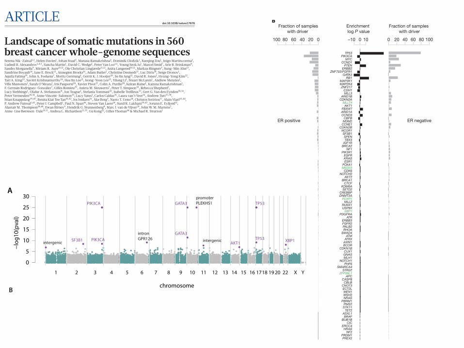
Extended Data Figure 3 | Recurrent non-coding events in breast cancers. a, Manhattan plot demonstrating sites with most significant P values as identified by binning analysis. Purple highlighted sites were also detected by the method seeking recurrence when partitioned by genomic features. b, Locus at chr11 65 Mb, which was identified by independent analyses as being more mutated than expected by chance.
Bottom, a rearrangement hotspot analysis identified this region as a tandem duplication hotspot, with nested tandem duplications noted at this site. Partitioning the genome into different regulatory elements, an analysis of substitutions and indels identified lncRNAs MALAT1 and NEAT1 (topmost panels) with significant P values.
© 2016 Macmillan Publishers Limited. All rights reserved
0 0 M O N T H 2 0 1 6 | V O L 0 0 0 | N A T U R E | 1
ARTICLEdoi:10.1038/nature17676
Landscape of somatic mutations in 560 breast cancer whole-genome sequencesSerena Nik-Zainal1,2, Helen Davies1, Johan Staaf3, Manasa Ramakrishna1, Dominik Glodzik1, Xueqing Zou1, Inigo Martincorena1, Ludmil B. Alexandrov1,4,5, Sancha Martin1, David C. Wedge1, Peter Van Loo1,6, Young Seok Ju1, Marcel Smid7, Arie B. Brinkman8, Sandro Morganella9, Miriam R. Aure10,11, Ole Christian Lingjærde11,12, Anita Langerød10,11, Markus Ringnér3, Sung-Min Ahn13, Sandrine Boyault14, Jane E. Brock15, Annegien Broeks16, Adam Butler1, Christine Desmedt17, Luc Dirix18, Serge Dronov1, Aquila Fatima19, John A. Foekens7, Moritz Gerstung1, Gerrit K. J. Hooijer20, Se Jin Jang21, David R. Jones1, Hyung-Yong Kim22, Tari A. King23, Savitri Krishnamurthy24, Hee Jin Lee21, Jeong-Yeon Lee25, Yilong Li1, Stuart McLaren1, Andrew Menzies1, Ville Mustonen1, Sarah O’Meara1, Iris Pauporté26, Xavier Pivot27, Colin A. Purdie28, Keiran Raine1, Kamna Ramakrishnan1, F. Germán Rodríguez-González7, Gilles Romieu29, Anieta M. Sieuwerts7, Peter T. Simpson30, Rebecca Shepherd1, Lucy Stebbings1, Olafur A. Stefansson31, Jon Teague1, Stefania Tommasi32, Isabelle Treilleux33, Gert G. Van den Eynden18,34, Peter Vermeulen18,34, Anne Vincent-Salomon35, Lucy Yates1, Carlos Caldas36, Laura van’t Veer16, Andrew Tutt37,38, Stian Knappskog39,40, Benita Kiat Tee Tan41,42, Jos Jonkers16, Åke Borg3, Naoto T. Ueno24, Christos Sotiriou17, Alain Viari43,44, P. Andrew Futreal1,45, Peter J. Campbell1, Paul N. Span46, Steven Van Laere18, Sunil R. Lakhani30,47, Jorunn E. Eyfjord31, Alastair M. Thompson28,48, Ewan Birney9, Hendrik G. Stunnenberg8, Marc J. van de Vijver20, John W. M. Martens7, Anne-Lise Børresen-Dale10,11, Andrea L. Richardson15,19, Gu Kong22, Gilles Thomas44 & Michael R. Stratton1
The mutational theory of cancer proposes that changes in DNA sequence, termed ‘driver’ mutations, confer proliferative advan-tage on a cell, leading to outgrowth of a neoplastic clone1. Some driver mutations are inherited in the germline, but most arise in
somatic cells during the lifetime of the cancer patient, together with many ‘passenger’ mutations not implicated in cancer development1. Multiple mutational processes, including endogenous and exoge-nous mutagen exposures, aberrant DNA editing, replication errors
1
We analysed whole-genome sequences of 560 breast cancers to advance understanding of the driver mutations conferring clonal advantage and the mutational processes generating somatic mutations. We found that 93 protein-coding cancer genes carried probable driver mutations. Some non-coding regions exhibited high mutation frequencies, but most have distinctive structural features probably causing elevated mutation rates and do not contain driver mutations. Mutational signature analysis was extended to genome rearrangements and revealed twelve base substitution and six rearrangement signatures. Three rearrangement signatures, characterized by tandem duplications or deletions, appear associated with defective homologous-recombination-based DNA repair: one with deficient BRCA1 function, another with deficient BRCA1 or BRCA2 function, the cause of the third is unknown. This analysis of all classes of somatic mutation across exons, introns and intergenic regions highlights the repertoire of cancer genes and mutational processes operating, and progresses towards a comprehensive account of the somatic genetic basis of breast cancer.
1Wellcome Trust Sanger Institute, Hinxton, Cambridge CB10 1SA, UK. 2East Anglian Medical Genetics Service, Cambridge University Hospitals NHS Foundation Trust, Cambridge CB2 9NB, UK. 3Division of Oncology and Pathology, Department of Clinical Sciences Lund, Lund University, Lund SE-223 81, Sweden. 4Theoretical Biology and Biophysics (T-6), Los Alamos National Laboratory, Los Alamos, NM 87545, New Mexico, USA. 5Center for Nonlinear Studies, Los Alamos National Laboratory, Los Alamos, New Mexico 87545, USA. 6Department of Human Genetics, University of Leuven, B-3000 Leuven, Belgium. 7Department of Medical Oncology, Erasmus MC Cancer Institute and Cancer Genomics Netherlands, Erasmus University Medical Center, Rotterdam 3015CN, The Netherlands. 8Radboud University, Department of Molecular Biology, Faculties of Science and Medicine, 6525GA Nijmegen, The Netherlands. 9European Molecular Biology Laboratory, European Bioinformatics Institute, Wellcome Trust Genome Campus, Hinxton, Cambridge CB10 1SD, UK. 10Department of Cancer Genetics, Institute for Cancer Research, Oslo University Hospital, The Norwegian Radium Hospital, Oslo 0310, Norway. 11K. G. Jebsen Centre for Breast Cancer Research, Institute for Clinical Medicine, University of Oslo, Oslo 0310, Norway. 12Department of Computer Science, University of Oslo, Oslo, Norway. 13Gachon Institute of Genome Medicine and Science, Gachon University Gil Medical Center, Incheon, South Korea. 14Translational Research Lab, Centre Léon Bérard, 28, rue Laënnec, 69373 Lyon Cedex 08, France. 15Department of Pathology, Brigham and Women’s Hospital, Boston, Massachusetts 02115, USA. 16The Netherlands Cancer Institute, 1066 CX Amsterdam, The Netherlands. 17Breast Cancer Translational Research Laboratory, Université Libre de Bruxelles, Institut Jules Bordet, Bd de Waterloo 121, B-1000 Brussels, Belgium. 18Translational Cancer Research Unit, Center for Oncological Research, Faculty of Medicine and Health Sciences, University of Antwerp, Antwerp, Belgium. 19Dana-Farber Cancer Institute, Boston, Massachusetts 02215, USA. 20Department of Pathology, Academic Medical Center, Meibergdreef 9, 1105 AZ Amsterdam, The Netherlands. 21Department of Pathology, Asan Medical Center, College of Medicine, Ulsan University, Ulsan, South Korea. 22Department of Pathology, College of Medicine, Hanyang University, Seoul 133-791, South Korea. 23Memorial Sloan Kettering Cancer Center, 1275 York Avenue, New York, New York 10065, USA. 24Morgan Welch Inflammatory Breast Cancer Research Program and Clinic, The University of Texas MD Anderson Cancer Center, 1515 Holcombe Boulevard., Houston, Texas 77030, USA. 25Institute for Bioengineering and Biopharmaceutical Research (IBBR), Hanyang University, Seoul, South Korea. 26Institut National du Cancer, Research Division, Clinical Research Department, 52 avenue Morizet, 92513 Boulogne-Billancourt, France. 27University Hospital of Minjoz, INSERM UMR 1098, Bd Fleming, Besançon 25000, France. 28Pathology Department, Ninewells Hospital and Medical School, Dundee DD1 9SY, UK. 29Oncologie Sénologie, ICM Institut Régional du Cancer, Montpellier, France. 30The University of Queensland, UQ Centre for Clinical Research and School of Medicine, Brisbane, Queensland 4059, Australia. 31Cancer Research Laboratory, Faculty of Medicine, University of Iceland, 101 Reykjavik, Iceland. 32IRCCS Istituto Tumori “Giovanni Paolo II”, Bari, Italy. 33Department of Pathology, Centre Léon Bérard, 28 rue Laënnec, 69373 Lyon Cédex 08, France. 34Department of Pathology, GZA Hospitals Sint-Augustinus, Antwerp, Belgium. 35Institut Curie, Department of Pathology and INSERM U934, 26 rue d’Ulm, 75248 Paris Cedex 05, France. 36Cancer Research UK Cambridge Institute, University of Cambridge, Li Ka Shing Centre, Robinson Way, Cambridge CB2 0RE, UK. 37Breast Cancer Now Toby Robin’s Research Unit, King’s College London, London SE1 9RT, UK. 38Breast Cancer Now Toby Robin’s Research Centre, Institute of Cancer Research, London SW3 6JB, UK. 39Department of Clinical Science, University of Bergen, 5020 Bergen, Norway. 40Department of Oncology, Haukeland University Hospital, 5021 Bergen, Norway. 41National Cancer Centre Singapore, 11 Hospital Drive, 169610, Singapore. 42Singapore General Hospital, Outram Road, 169608, Singapore. 43Equipe Erable, INRIA Grenoble-Rhône-Alpes, 655, Avenue de l’Europe, 38330 Montbonnot-Saint Martin, France. 44Synergie Lyon Cancer, Centre Léon Bérard, 28 rue Laënnec, Lyon Cedex 08, France. 45Department of Genomic Medicine, UT MD Anderson Cancer Center, Houston, Texas 77230, USA. 46Department of Radiation Oncology, Department of Laboratory Medicine, Radboud University Medical Center, Nijmegen 6525GA, The Netherlands. 47Pathology Queensland, The Royal Brisbane and Women’s Hospital, Brisbane, Queensland 4029, Australia. 48Department of Surgical Oncology, University of Texas MD Anderson Cancer Center, 1400 Pressler Street, Houston, Texas 77030, USA.
2
4 | N A T U R E | V O L 0 0 0 | 0 0 M O N T H 2 0 1 6
ARTICLERESEARCH
(CCCCAGATGGTGGG)), shifting it away from the consensus36. The association with particular mutational signatures suggests that these may also be in a region of hypermutability rather than drivers.
The WDR74 promoter showed base substitutions and indels (q value 4.6 ! 10"3) forming a cluster of overlapping mutations20 (Fig. 2a). Coding sequence driver mutations in WDR74 have not been reported. No differences were observed in WDR74 transcript levels between cancers with WDR74 promoter mutations compared to those without. Nevertheless, the pattern of this non-coding mutation cluster, with overlapping and different mutation types, is more compatible with the possibility of the mutations being drivers.
Two long non-coding RNAs, MALAT1 (q value 8.7 ! 10"11, as previ-ously reported12) and NEAT1 (q value 2.1 ! 10"2) were enriched with mutations. Transcript levels were not significantly different between mutated and non-mutated samples. Whether these mutations are driv-ers or result from local hypermutability is unclear.
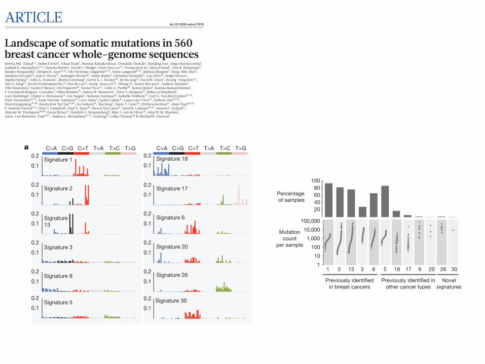
Mutational signaturesMutational processes generating somatic mutations imprint particu-lar patterns of mutations on cancer genomes, termed signatures2,24,37. Applying a mathematical approach25 to extract mutational signa-tures previously revealed five base-substitution signatures in breast cancer: signatures 1, 2, 3, 8 and 13 (refs 2, 24). Using this method for the 560!cases revealed twelve!signatures, including those previously observed and a further seven, of which five have formerly been detected in other cancer types (signatures 5, 6, 17, 18 and 20) and two are new (signatures 26 and 30) (Fig. 3a, b, 4a, Supplementary Table 21a–c, Supplementary Methods section 15). Two indel signatures were also found2,24.
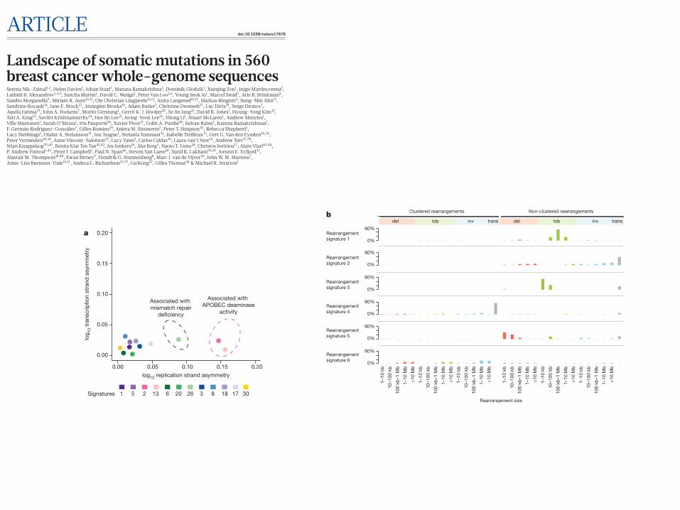
Signatures of rearrangement mutational processes have not previ-ously been formally investigated. To enable this we adopted a rear-rangement classification incorporating 32!subclasses. In many cancer genomes, large numbers of rearrangements are regionally clustered, for example in zones of gene amplification. Therefore, we first classified rearrangements into those inside and outside clusters, further subclassi-fied them into deletions, inversions and tandem duplications, and then according to the size of the rearranged segment. The final category in both groups was interchromosomal translocations.
Application of the mathematical framework used for base substitu-tion signatures2,24,25 extracted six rearrangement signatures (Fig. 4b, Supplementary Table 21). Unsupervised hierarchical clustering on the basis of the proportion of rearrangements attributed to each signature in each breast cancer yielded seven major subgroups exhibiting distinct associations with other genomic, histological or gene expression fea-tures (Fig. 5, Extended Data Figs 4–6).
Rearrangement signature 1 (9% of all rearrangements) and rear-rangement signature 3 (18% rearrangements) were characterized predominantly by tandem duplications (Fig. 4b). Tandem duplica-tions associated with rearrangement signature 1 were mostly >100 kb (Fig. 4b), and those with rearrangement signature 3 were!<10 kb (Fig. 4b, Extended Data Fig. 7). More than 95% of rearrangement signature 3 tandem duplications were concentrated in 15% of cancers (cluster D, Fig. 5), many with several hundred rearrangements of this type. Almost all cancers (91%) with BRCA1 mutations or promoter hypermethylation were in this group, which was enriched for basal-like, triple negative cancers and copy number classification of a high homologous recombination deficiency (HRD) index38–40. Thus, inac-tivation of BRCA1, but not BRCA2, may be responsible for the rear-rangement signature 3 small tandem duplication mutator phenotype.
More than 35% of rearrangement signature 1 tandem duplications were found in just 8.5% of the breast cancers and some cases had hundreds of these (cluster F, Fig. 5). The cause of this large tandem duplication mutator phenotype (Fig. 4b) is unknown. Cancers exhib-iting it are frequently TP53-mutated, relatively late diagnosis, triple- negative breast cancers, showing enrichment for base substitution signature 3 and a high HRD index (Fig. 5), but do not have BRCA1/2 mutations or BRCA1 promoter hypermethylation.
Rearrangement signature 1 and 3 tandem duplications (Extended Data Fig. 7) were generally evenly distributed over the genome. However,
a
b
0.1
0.2
0.1
0.2
0.1
0.2
0.1
0.2
Signature 18
Signature 6
Signature 20
Signature 26
0.1
0.2 Signature 17
0.1
0.2
0.1
0.2
0.1
0.2
0.1
0.2
0.1
0.2 Signature13
Signature 2
Signature 3
Signature 8
Signature 1
0.1
0.2 Signature 30
C>A C>G C>T T>A T>C T>G C>A C>G C>T T>A T>C T>G
0.1
0.2 Signature 5
0%
100%0
100,000
1 5 2 13 6 20 26 3 8 18 17Signatures 30
1
10
100
1,000
10,000
100,000
1 2 13 3 8 5 18 17 6 20 26 30
Previously identi!edin breast cancers
Previously identi!ed inother cancer types
Novel signatures
Mutationcount
per sample
Percentage of samples
100
20 40 60 80
c
Figure 3 | Extraction and contributions of base substitution signatures in 560!breast cancers. a, Twelve mutation signatures extracted using non-negative matrix factorization. Each signature is ordered by mutation class (C>A/G>T, C>G/G>C, C>T/G>A, T>A/A>T, T>C/A>G, T>G/A >C), taking immediate flanking sequence into account. For each class, mutations are ordered by 5# base (A, C, G, T) first before 3# base (A, C, G, T). b, The spectrum of base substitution signatures within 560!breast cancers. Mutation signatures are ordered (and coloured) according to broad biological groups: signatures 1 and 5 are correlated with age of diagnosis; signatures 2 and 13 are putatively APOBEC-related; signatures 6, 20 and 26 are associated with mismatch-repair deficiency; signatures 3 and 8 are associated with homologous-recombination deficiency; signatures 18, 17 and 30 have unknown aetiology. For ease of reading, this arrangement is adopted for the rest of the manuscript. Samples are ordered according to hierarchical clustering performed on mutation signatures. Top, absolute numbers of mutations of each signature in each sample. Bottom, proportion of each signature in each sample. c, Distribution of mutation counts for each signature in relevant breast cancer samples. Percentage of samples carrying each signature provided above each signature.
© 2016 Macmillan Publishers Limited. All rights reserved
4 | N A T U R E | V O L 0 0 0 | 0 0 M O N T H 2 0 1 6
ARTICLERESEARCH
(CCCCAGATGGTGGG)), shifting it away from the consensus36. The association with particular mutational signatures suggests that these may also be in a region of hypermutability rather than drivers.
The WDR74 promoter showed base substitutions and indels (q value 4.6 ! 10"3) forming a cluster of overlapping mutations20 (Fig. 2a). Coding sequence driver mutations in WDR74 have not been reported. No differences were observed in WDR74 transcript levels between cancers with WDR74 promoter mutations compared to those without. Nevertheless, the pattern of this non-coding mutation cluster, with overlapping and different mutation types, is more compatible with the possibility of the mutations being drivers.
Two long non-coding RNAs, MALAT1 (q value 8.7 ! 10"11, as previ-ously reported12) and NEAT1 (q value 2.1 ! 10"2) were enriched with mutations. Transcript levels were not significantly different between mutated and non-mutated samples. Whether these mutations are driv-ers or result from local hypermutability is unclear.
Mutational signaturesMutational processes generating somatic mutations imprint particu-lar patterns of mutations on cancer genomes, termed signatures2,24,37. Applying a mathematical approach25 to extract mutational signa-tures previously revealed five base-substitution signatures in breast cancer: signatures 1, 2, 3, 8 and 13 (refs 2, 24). Using this method for the 560!cases revealed twelve!signatures, including those previously observed and a further seven, of which five have formerly been detected in other cancer types (signatures 5, 6, 17, 18 and 20) and two are new (signatures 26 and 30) (Fig. 3a, b, 4a, Supplementary Table 21a–c, Supplementary Methods section 15). Two indel signatures were also found2,24.
Signatures of rearrangement mutational processes have not previ-ously been formally investigated. To enable this we adopted a rear-rangement classification incorporating 32!subclasses. In many cancer genomes, large numbers of rearrangements are regionally clustered, for example in zones of gene amplification. Therefore, we first classified rearrangements into those inside and outside clusters, further subclassi-fied them into deletions, inversions and tandem duplications, and then according to the size of the rearranged segment. The final category in both groups was interchromosomal translocations.
Application of the mathematical framework used for base substitu-tion signatures2,24,25 extracted six rearrangement signatures (Fig. 4b, Supplementary Table 21). Unsupervised hierarchical clustering on the basis of the proportion of rearrangements attributed to each signature in each breast cancer yielded seven major subgroups exhibiting distinct associations with other genomic, histological or gene expression fea-tures (Fig. 5, Extended Data Figs 4–6).
Rearrangement signature 1 (9% of all rearrangements) and rear-rangement signature 3 (18% rearrangements) were characterized predominantly by tandem duplications (Fig. 4b). Tandem duplica-tions associated with rearrangement signature 1 were mostly >100 kb (Fig. 4b), and those with rearrangement signature 3 were!<10 kb (Fig. 4b, Extended Data Fig. 7). More than 95% of rearrangement signature 3 tandem duplications were concentrated in 15% of cancers (cluster D, Fig. 5), many with several hundred rearrangements of this type. Almost all cancers (91%) with BRCA1 mutations or promoter hypermethylation were in this group, which was enriched for basal-like, triple negative cancers and copy number classification of a high homologous recombination deficiency (HRD) index38–40. Thus, inac-tivation of BRCA1, but not BRCA2, may be responsible for the rear-rangement signature 3 small tandem duplication mutator phenotype.
More than 35% of rearrangement signature 1 tandem duplications were found in just 8.5% of the breast cancers and some cases had hundreds of these (cluster F, Fig. 5). The cause of this large tandem duplication mutator phenotype (Fig. 4b) is unknown. Cancers exhib-iting it are frequently TP53-mutated, relatively late diagnosis, triple- negative breast cancers, showing enrichment for base substitution signature 3 and a high HRD index (Fig. 5), but do not have BRCA1/2 mutations or BRCA1 promoter hypermethylation.
Rearrangement signature 1 and 3 tandem duplications (Extended Data Fig. 7) were generally evenly distributed over the genome. However,
a
b
0.1
0.2
0.1
0.2
0.1
0.2
0.1
0.2
Signature 18
Signature 6
Signature 20
Signature 26
0.1
0.2 Signature 17
0.1
0.2
0.1
0.2
0.1
0.2
0.1
0.2
0.1
0.2 Signature13
Signature 2
Signature 3
Signature 8
Signature 1
0.1
0.2 Signature 30
C>A C>G C>T T>A T>C T>G C>A C>G C>T T>A T>C T>G
0.1
0.2 Signature 5
0%
100%0
100,000
1 5 2 13 6 20 26 3 8 18 17Signatures 30
1
10
100
1,000
10,000
100,000
1 2 13 3 8 5 18 17 6 20 26 30
Previously identi!edin breast cancers
Previously identi!ed inother cancer types
Novel signatures
Mutationcount
per sample
Percentage of samples
100
20 40 60 80
c
Figure 3 | Extraction and contributions of base substitution signatures in 560!breast cancers. a, Twelve mutation signatures extracted using non-negative matrix factorization. Each signature is ordered by mutation class (C>A/G>T, C>G/G>C, C>T/G>A, T>A/A>T, T>C/A>G, T>G/A >C), taking immediate flanking sequence into account. For each class, mutations are ordered by 5# base (A, C, G, T) first before 3# base (A, C, G, T). b, The spectrum of base substitution signatures within 560!breast cancers. Mutation signatures are ordered (and coloured) according to broad biological groups: signatures 1 and 5 are correlated with age of diagnosis; signatures 2 and 13 are putatively APOBEC-related; signatures 6, 20 and 26 are associated with mismatch-repair deficiency; signatures 3 and 8 are associated with homologous-recombination deficiency; signatures 18, 17 and 30 have unknown aetiology. For ease of reading, this arrangement is adopted for the rest of the manuscript. Samples are ordered according to hierarchical clustering performed on mutation signatures. Top, absolute numbers of mutations of each signature in each sample. Bottom, proportion of each signature in each sample. c, Distribution of mutation counts for each signature in relevant breast cancer samples. Percentage of samples carrying each signature provided above each signature.
© 2016 Macmillan Publishers Limited. All rights reserved
0 0 M O N T H 2 0 1 6 | V O L 0 0 0 | N A T U R E | 1
ARTICLEdoi:10.1038/nature17676
Landscape of somatic mutations in 560 breast cancer whole-genome sequencesSerena Nik-Zainal1,2, Helen Davies1, Johan Staaf3, Manasa Ramakrishna1, Dominik Glodzik1, Xueqing Zou1, Inigo Martincorena1, Ludmil B. Alexandrov1,4,5, Sancha Martin1, David C. Wedge1, Peter Van Loo1,6, Young Seok Ju1, Marcel Smid7, Arie B. Brinkman8, Sandro Morganella9, Miriam R. Aure10,11, Ole Christian Lingjærde11,12, Anita Langerød10,11, Markus Ringnér3, Sung-Min Ahn13, Sandrine Boyault14, Jane E. Brock15, Annegien Broeks16, Adam Butler1, Christine Desmedt17, Luc Dirix18, Serge Dronov1, Aquila Fatima19, John A. Foekens7, Moritz Gerstung1, Gerrit K. J. Hooijer20, Se Jin Jang21, David R. Jones1, Hyung-Yong Kim22, Tari A. King23, Savitri Krishnamurthy24, Hee Jin Lee21, Jeong-Yeon Lee25, Yilong Li1, Stuart McLaren1, Andrew Menzies1, Ville Mustonen1, Sarah O’Meara1, Iris Pauporté26, Xavier Pivot27, Colin A. Purdie28, Keiran Raine1, Kamna Ramakrishnan1, F. Germán Rodríguez-González7, Gilles Romieu29, Anieta M. Sieuwerts7, Peter T. Simpson30, Rebecca Shepherd1, Lucy Stebbings1, Olafur A. Stefansson31, Jon Teague1, Stefania Tommasi32, Isabelle Treilleux33, Gert G. Van den Eynden18,34, Peter Vermeulen18,34, Anne Vincent-Salomon35, Lucy Yates1, Carlos Caldas36, Laura van’t Veer16, Andrew Tutt37,38, Stian Knappskog39,40, Benita Kiat Tee Tan41,42, Jos Jonkers16, Åke Borg3, Naoto T. Ueno24, Christos Sotiriou17, Alain Viari43,44, P. Andrew Futreal1,45, Peter J. Campbell1, Paul N. Span46, Steven Van Laere18, Sunil R. Lakhani30,47, Jorunn E. Eyfjord31, Alastair M. Thompson28,48, Ewan Birney9, Hendrik G. Stunnenberg8, Marc J. van de Vijver20, John W. M. Martens7, Anne-Lise Børresen-Dale10,11, Andrea L. Richardson15,19, Gu Kong22, Gilles Thomas44 & Michael R. Stratton1
The mutational theory of cancer proposes that changes in DNA sequence, termed ‘driver’ mutations, confer proliferative advan-tage on a cell, leading to outgrowth of a neoplastic clone1. Some driver mutations are inherited in the germline, but most arise in
somatic cells during the lifetime of the cancer patient, together with many ‘passenger’ mutations not implicated in cancer development1. Multiple mutational processes, including endogenous and exoge-nous mutagen exposures, aberrant DNA editing, replication errors
1
We analysed whole-genome sequences of 560 breast cancers to advance understanding of the driver mutations conferring clonal advantage and the mutational processes generating somatic mutations. We found that 93 protein-coding cancer genes carried probable driver mutations. Some non-coding regions exhibited high mutation frequencies, but most have distinctive structural features probably causing elevated mutation rates and do not contain driver mutations. Mutational signature analysis was extended to genome rearrangements and revealed twelve base substitution and six rearrangement signatures. Three rearrangement signatures, characterized by tandem duplications or deletions, appear associated with defective homologous-recombination-based DNA repair: one with deficient BRCA1 function, another with deficient BRCA1 or BRCA2 function, the cause of the third is unknown. This analysis of all classes of somatic mutation across exons, introns and intergenic regions highlights the repertoire of cancer genes and mutational processes operating, and progresses towards a comprehensive account of the somatic genetic basis of breast cancer.
1Wellcome Trust Sanger Institute, Hinxton, Cambridge CB10 1SA, UK. 2East Anglian Medical Genetics Service, Cambridge University Hospitals NHS Foundation Trust, Cambridge CB2 9NB, UK. 3Division of Oncology and Pathology, Department of Clinical Sciences Lund, Lund University, Lund SE-223 81, Sweden. 4Theoretical Biology and Biophysics (T-6), Los Alamos National Laboratory, Los Alamos, NM 87545, New Mexico, USA. 5Center for Nonlinear Studies, Los Alamos National Laboratory, Los Alamos, New Mexico 87545, USA. 6Department of Human Genetics, University of Leuven, B-3000 Leuven, Belgium. 7Department of Medical Oncology, Erasmus MC Cancer Institute and Cancer Genomics Netherlands, Erasmus University Medical Center, Rotterdam 3015CN, The Netherlands. 8Radboud University, Department of Molecular Biology, Faculties of Science and Medicine, 6525GA Nijmegen, The Netherlands. 9European Molecular Biology Laboratory, European Bioinformatics Institute, Wellcome Trust Genome Campus, Hinxton, Cambridge CB10 1SD, UK. 10Department of Cancer Genetics, Institute for Cancer Research, Oslo University Hospital, The Norwegian Radium Hospital, Oslo 0310, Norway. 11K. G. Jebsen Centre for Breast Cancer Research, Institute for Clinical Medicine, University of Oslo, Oslo 0310, Norway. 12Department of Computer Science, University of Oslo, Oslo, Norway. 13Gachon Institute of Genome Medicine and Science, Gachon University Gil Medical Center, Incheon, South Korea. 14Translational Research Lab, Centre Léon Bérard, 28, rue Laënnec, 69373 Lyon Cedex 08, France. 15Department of Pathology, Brigham and Women’s Hospital, Boston, Massachusetts 02115, USA. 16The Netherlands Cancer Institute, 1066 CX Amsterdam, The Netherlands. 17Breast Cancer Translational Research Laboratory, Université Libre de Bruxelles, Institut Jules Bordet, Bd de Waterloo 121, B-1000 Brussels, Belgium. 18Translational Cancer Research Unit, Center for Oncological Research, Faculty of Medicine and Health Sciences, University of Antwerp, Antwerp, Belgium. 19Dana-Farber Cancer Institute, Boston, Massachusetts 02215, USA. 20Department of Pathology, Academic Medical Center, Meibergdreef 9, 1105 AZ Amsterdam, The Netherlands. 21Department of Pathology, Asan Medical Center, College of Medicine, Ulsan University, Ulsan, South Korea. 22Department of Pathology, College of Medicine, Hanyang University, Seoul 133-791, South Korea. 23Memorial Sloan Kettering Cancer Center, 1275 York Avenue, New York, New York 10065, USA. 24Morgan Welch Inflammatory Breast Cancer Research Program and Clinic, The University of Texas MD Anderson Cancer Center, 1515 Holcombe Boulevard., Houston, Texas 77030, USA. 25Institute for Bioengineering and Biopharmaceutical Research (IBBR), Hanyang University, Seoul, South Korea. 26Institut National du Cancer, Research Division, Clinical Research Department, 52 avenue Morizet, 92513 Boulogne-Billancourt, France. 27University Hospital of Minjoz, INSERM UMR 1098, Bd Fleming, Besançon 25000, France. 28Pathology Department, Ninewells Hospital and Medical School, Dundee DD1 9SY, UK. 29Oncologie Sénologie, ICM Institut Régional du Cancer, Montpellier, France. 30The University of Queensland, UQ Centre for Clinical Research and School of Medicine, Brisbane, Queensland 4059, Australia. 31Cancer Research Laboratory, Faculty of Medicine, University of Iceland, 101 Reykjavik, Iceland. 32IRCCS Istituto Tumori “Giovanni Paolo II”, Bari, Italy. 33Department of Pathology, Centre Léon Bérard, 28 rue Laënnec, 69373 Lyon Cédex 08, France. 34Department of Pathology, GZA Hospitals Sint-Augustinus, Antwerp, Belgium. 35Institut Curie, Department of Pathology and INSERM U934, 26 rue d’Ulm, 75248 Paris Cedex 05, France. 36Cancer Research UK Cambridge Institute, University of Cambridge, Li Ka Shing Centre, Robinson Way, Cambridge CB2 0RE, UK. 37Breast Cancer Now Toby Robin’s Research Unit, King’s College London, London SE1 9RT, UK. 38Breast Cancer Now Toby Robin’s Research Centre, Institute of Cancer Research, London SW3 6JB, UK. 39Department of Clinical Science, University of Bergen, 5020 Bergen, Norway. 40Department of Oncology, Haukeland University Hospital, 5021 Bergen, Norway. 41National Cancer Centre Singapore, 11 Hospital Drive, 169610, Singapore. 42Singapore General Hospital, Outram Road, 169608, Singapore. 43Equipe Erable, INRIA Grenoble-Rhône-Alpes, 655, Avenue de l’Europe, 38330 Montbonnot-Saint Martin, France. 44Synergie Lyon Cancer, Centre Léon Bérard, 28 rue Laënnec, Lyon Cedex 08, France. 45Department of Genomic Medicine, UT MD Anderson Cancer Center, Houston, Texas 77230, USA. 46Department of Radiation Oncology, Department of Laboratory Medicine, Radboud University Medical Center, Nijmegen 6525GA, The Netherlands. 47Pathology Queensland, The Royal Brisbane and Women’s Hospital, Brisbane, Queensland 4029, Australia. 48Department of Surgical Oncology, University of Texas MD Anderson Cancer Center, 1400 Pressler Street, Houston, Texas 77030, USA.
2
0 0 M O N T H 2 0 1 6 | V O L 0 0 0 | N A T U R E | 5
ARTICLE RESEARCH
there were nine locations at which recurrence of tandem duplications was found across the breast cancers and which often showed multiple, nested tandem duplications in individual cases (Extended Data Fig. 8). These may be mutational hotspots specific for these tandem duplication mutational processes, although we cannot exclude the possibility that they represent driver events.
Rearrangement signature 5 (accounting for 14% rearrangements) was characterized by deletions <100!kb. It was strongly associated with the presence of BRCA1 mutations or promoter hypermethyla-tion (cluster D, Fig. 5), BRCA2 mutations (cluster G, Fig. 5) and with rearrangement signature 1 large tandem duplications (cluster F, Fig. 5).
Rearrangement signature 2 (accounting for 22% rearrangements) was characterized by non-clustered deletions (>100!kb), inversions and interchromosomal translocations, was present in most cancers but was particularly enriched in oestrogen receptor (ER)-positive cancers with quiet copy number profiles (cluster E, GISTIC (genomic identifi-cation of significant targets in cancer) cluster 3; Fig. 5). Rearrangement signature 4 (accounting for 18% of rearrangements) was characterized by clustered interchromosomal translocations, whereas rearrangement signature 6 (19% of rearrangements) had clustered inversions and deletions (clusters A, B, C; Fig. 5).
Short segments (1–5 bp) of overlapping microhomology character-istic of alternative methods of end-joining repair were found at most rearrangements2,14. Rearrangement signatures 2, 4 and 6 were charac-terized by a peak at 1 bp of microhomology, whereas rearrangement signatures 1, 3 and 5, associated with homologous recombination DNA repair deficiency, exhibited a peak at 2 bp (Extended Data Fig. 9). Thus, different end-joining mechanisms may operate with different rearrangement processes. A proportion of breast cancers showed rear-rangement signature 5 deletions with longer (>10 bp) microhomologies involving sequences from short-interspersed nuclear elements, most commonly AluS (63%) and AluY (15%) family repeats (Extended Data Fig. 9). Long segments (more than 10 bp) of non-templated sequence were particularly enriched amongst clustered rearrangements.
Localized hypermutation: kataegisFocal base-substitution hypermutation, termed kataegis, is generally characterized by substitutions with characteristic features of signatures 2 and 13 (refs 2, 24). Kataegis was observed in 49% breast cancers, with 4% exhibiting 10 or more foci (Supplementary Table 21c). Kataegis colo-calizes with clustered rearrangements characteristic of rearrangement
signatures 4 and 6 (Fig. 4b). Cancers with tandem duplications or deletions of rearrangement signatures 1, 3 and 5 did not usually demonstrate kataegis. However, there must be additional determinants of kataegis as only 2% of rearrangements are associated with it. A rare (14 out of 1,557 foci, 0.9%) alternative form of kataegis, colocalizing with rearrange-ments but with a base-substitution pattern characterized by T>G and T>C mutations, predominantly at NTT and NTA sequences (where N can be any base A, T, C or G), was also observed (Extended Data Fig. 10). This pattern of base substitutions most closely matches signature 9 (Extended Data Fig. 10; http://cancer.sanger.ac.uk/cosmic/signatures), previously observed in B lymphocyte neoplasms and attributed to polymerase eta activity41.
Mutational signatures exhibit distinct DNA replication strand biasesThe distributions of mutations attributable to each of the 20!muta-tional signatures (12!base substitution, 2 indel and 6 rearrangement) were explored42 with respect to DNA replication strand. We found an asymmetric distribution of mutations between leading and lagging replication strands for many, but not all signatures42 (Fig. 4a). Notably, signatures 2 and 13, owing to APOBEC deamination, showed marked lagging-strand replication bias (Fig. 4a) suggesting that lagging-strand replication provides single-stranded DNA for APOBEC deamination. Of the three signatures associated with mismatch-repair deficiency (signatures 6, 20 and 26), only signature 26 exhibited replicative-strand bias, highlighting how different signatures arising from defects of the same pathway can exhibit distinct relationships with replication.
Mutational signatures associated with BRCA1 and BRCA2 mutationsOf the 560!breast cancers, 90 had germline (60) or somatic (14) inac-tivating mutations in BRCA1 (35) or BRCA2 (39) or showed methyla-tion of the BRCA1 promoter (16). Loss of the wild-type chromosome 17 or 13 was observed in 80 out of 90 cases. The latter exhibited many base substitution mutations of signature 3, accompanied by deletions of >3 bp with microhomology at rearrangement break-points, and signature 8 together with CC>AA double nucleotide substitutions. Cases in which the wild-type chromosome 17 or 13 was retained did not show these signatures. Thus signature 3 and, to a lesser extent, signature 8 are associated with absence of BRCA1 and BRCA2 functions.
Figure 4 | Additional characteristics of base substitution signatures and novel rearrangement signatures in 560!breast cancers. a, Contrasting transcriptional strand asymmetry and replication strand asymmetry between twelve base substitution signatures. b, Six rearrangement
signatures extracted using non-negative matrix factorization. Probability of rearrangement element on y axis. Rearrangement size on x axis. del,!deletion; tds,!tandem duplication; inv,!inversion; trans,!translocation.
a
1 5 2 13 6 20 26 3 8 18 17Signatures 30
b
0.20log10 replication strand asymmetry
log 10
tran
scrip
tion
stra
nd a
sym
met
ry
Associated with APOBEC deaminase
activity
Associated with mismatch repair
de!ciency
0.10 0.150.050.00
0.20
0.10
0.15
0.05
0.00
0%
60%
0%
60%
0%
60%
0%
60%
0%
60%
0%
60%
del inv transtds del inv transtds
Clustered rearrangements Non-clustered rearrangements
Rearrangementsignature 1
Rearrangementsignature 2
Rearrangementsignature 3
Rearrangementsignature 4
Rearrangementsignature 5
Rearrangementsignature 6
1–10
kb
10–1
00 k
b
100
kb–1
Mb
1–10
Mb
>10
Mb
1–10
kb
10–1
00 k
b
100
kb–1
Mb
1–10
Mb
>10
Mb
1–10
kb
10–1
00 k
b
100
kb–1
Mb
1–10
Mb
>10
Mb
1–10
kb
10–1
00 k
b
100
kb–1
Mb
1–10
Mb
>10
Mb
1–10
kb
10–1
00 k
b
100
kb–1
Mb
1–10
Mb
>10
Mb
1–10
kb
10–1
00 k
b
100
kb–1
Mb
1–10
Mb
>10
Mb
Rearrangement size
© 2016 Macmillan Publishers Limited. All rights reserved
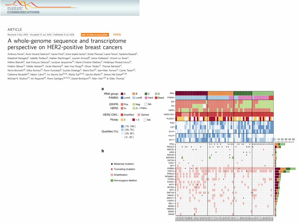
ARTICLEReceived 3 Dec 2015 | Accepted 12 Jun 2016 | Published 13 Jul 2016
A whole-genome sequence and transcriptomeperspective on HER2-positive breast cancersAnthony Ferrari1, Anne Vincent-Salomon2, Xavier Pivot3, Anne-Sophie Sertier1, Emilie Thomas1, Laurie Tonon1, Sandrine Boyault4,
Eskeatnaf Mulugeta5, Isabelle Treilleux6, Gaetan MacGrogan7, Laurent Arnould8, Janice Kielbassa1, Vincent Le Texier1,
Helene Blanche9, Jean-Francois Deleuze9, Jocelyne Jacquemier10, Marie-Christine Mathieu11, Frederique Penault-Llorca12,
Frederic Bibeau13, Odette Mariani14, Cecile Mannina15, Jean-Yves Pierga16, Olivier Tredan17, Thomas Bachelot17,
Herve Bonnefoi18, Gilles Romieu19, Pierre Fumoleau8, Suzette Delaloge11, Maria Rios20, Jean-Marc Ferrero21, Carole Tarpin22,
Catherine Bouteille23, Fabien Calvo24, Ivo Glynne Gut25,26, Marta Gut25,26, Sancha Martin27, Serena Nik-Zainal27,28,
Michael R. Stratton27, Iris Pauporte29, Pierre Saintigny30,31,32, Daniel Birnbaum33, Alain Viari1,34 & Gilles Thomas1
HER2-positive breast cancer has long proven to be a clinically distinct class of breast cancers for
which several targeted therapies are now available. However, resistance to the treatment asso-
ciated with specific gene expressions or mutations has been observed, revealing the underlying
diversity of these cancers. Therefore, understanding the full extent of the HER2-positive disease
heterogeneity still remains challenging. Here we carry out an in-depth genomic characterization of
64 HER2-positive breast tumour genomes that exhibit four subgroups, based on the expression
data, with distinctive genomic features in terms of somatic mutations, copy-number changes or
structural variations. The results suggest that, despite being clinically defined by a specific gene
amplification, HER2-positive tumours melt into the whole luminal–basal breast cancer spectrum
rather than standing apart. The results also lead to a refined ERBB2 amplicon of 106 kb and show
that several cases of amplifications are compatible with a breakage–fusion–bridge mechanism.
DOI: 10.1038/ncomms12222 OPEN
1 Synergie Lyon Cancer, Plateforme de bioinformatique ‘Gilles Thomas’ Centre Leon Berard, 28 rue Laennec, 69008 Lyon, France. 2 Institut Curie, PSL Research University, Departement dePathologie, INSERM U934, 26 rue d’Ulm, 75248 Paris, France. 3Centre Hospitalier Universitaire de Minjoz, UMR INSERM 1098, Boulevard A. Fleming, Besancon 25000, France.4 Plateforme de genomique des cancers, Centre Leon Berard, 28 rue Laennec, 69008 Lyon, France. 5 Institut Curie, UMR 3215 CNRS, Genetique et biologie du developpement, Epigenese etdeveloppement des mammiferes, U934 Inserm, 26 rue d’Ulm, 75248 Paris, France. 6Centre Leon Berard, Departement de Pathologie, 28 rue Laennec, 69008 Lyon, France. 7Departementde Biopathologie, Unite Inserm U916, Institut Bergonie, 229 cours de l’Argonne, 33076 Bordeaux, France. 8Centre Georges-Francois Leclerc et CRB Ferdinand Cabanne, 1 rue du ProfesseurMarion, Inserm U866-UBFC, 21000 Dijon, France. 9Centre d’Etude du Polymorphisme Humain (CEPH), Fondation Jean Dausset, 27 rue Juliette Dodu, 75010 Paris, France. 10 Institut Paoli-Calmettes, Departement de Pathologie, 232 Boulevard de Sainte-Marguerite, 13009 Marseille, France. 11 Institut Gustave Roussy, Comite de Pathologie Mammaire, 114 rue Edouard Vaillant,94805 Villejuif, France. 12Centre Jean Perrin, Departement de Biopathologie, EA 4677 ERTICa, Universite d’Auvergne, 58 rue Montalembert, 63000 Clermont-Ferrand, France. 13 InstitutRegional du Cancer de Montpellier (ICM), Departement de Pathologie, 208 Avenue des Apothicaires, 34298 Montpellier, France. 14 Institut Curie, PSL Research University, Service dePathologie, Centre de Ressources Biologiques, BRIF BB-0033-00048, 26 rue d’Ulm, 75248 Paris, France. 15Departement de Pathologie, Institut Bergonie, 229 cours de l’Argonne, CS 61283,33076 Bordeaux, France. 16 Institut Curie, PSL Research University, Departement d’Oncologie Medicale, Universite Paris Descartes, 26 rue d’Ulm, 75248 Paris, France. 17Centre Leon Berard,Departement de Cancerologie Medicale, 28 rue Laennec, 69008 Lyon, France. 18Department of Medical Oncology, Institut Bergonie Unicancer, University of Bordeaux, INSERM U916,CIC1401, 229 cours de l’Argonne, CS 61283, 33076 Bordeaux, France. 19 Institut Regional du Cancer de Montpellier (ICM), Oncologie Senologie, 208 Avenue des Apothicaires, 34298Montpellier, France. 20Centre Alexis Vautrin, Departement d’Oncologie Medicale, 6 Avenue de Bourgogne, 54511 Vandoeuvre Les Nancy, France. 21 Centre Antoine Lacassagne,Departement d’Oncologie Medicale, 33 Avenue de Valombrose, 06189 Nice, France. 22 Institut Paoli-Calmettes, Departement d’Oncologie Medicale, 232 Boulevard de Sainte-Marguerite,13009 Marseille, France. 23Clinique Mutualiste de Bellevue, Chirurgie Gynecologique et Mammaire, 3 rue le Verrier, 42100 Saint-Etienne, France. 24 Institut Gustave Roussy, Cancer CoreEurope, 39 rue Camille Desmoulins, Villejuif 94805, France. 25CNAG-CRG, Centre for Genomic Regulation (CRG), C/Baldiri Reixac 4, 08028 Barcelona, Spain. 26Universitat Pompeu Fabra,Placa de la Merce, 10, 08002 Barcelona, Spain. 27Wellcome Trust Sanger Institute, Hinxton, Cambridge CB10 1SA, UK. 28 East Anglian Medical Genetics Service, Cambridge UniversityHospitals NHS Foundation Trust, Cambridge CB2 9NB, UK. 29 Institut National du Cancer, Departement de Recherche Clinique, 52 Avenue Morizet, 92513 Boulogne-Billancourt, France.30 INSERM U1052-CNRS 5286, Cancer Research Center of Lyon, F-69008 Lyon, France. 31Universite de Lyon, F-69622 Lyon, France. 32Centre Leon Berard, 28 rue Laennec, 69008 Lyon,France. 33Departement d’Oncologie Moleculaire, Institut Paoli-Calmettes, Centre de Recherche en Cancerologie de Marseille, INSERM, CNRS, Aix-Marseille Universite, 232 boulevard deSainte-Marguerite, 13009 Marseille, France. 34 Equipe Erable, INRIA Grenoble-Rhone-Alpes, 655 Avenue de l’Europe, 38330 Montbonnot-Saint Martin, France. Correspondence andrequests for materials should be addressed to A.F. (email: [email protected]) or to A.V. (email: [email protected]).
NATURE COMMUNICATIONS | 7:12222 |DOI: 10.1038/ncomms12222 | www.nature.com/naturecommunications 1
hotspot mutations (Supplementary Data 2). MAP2K4 displayedHD in three ER! cases and GATA3 displayed three frameshifts.Six ERBB2 missense mutations were observed in four tumoursincluding two in the kinase domain31. To assess if modificationsof potential cis-regulatory regions could also contribute todifferences between the four expression groups, we studiedsomatic SNVs in these regions by looking at a combination ofhistone marks enriched in regulatory region and FANTOM5enhancer and promoter annotation32 (Methods). An under-representation of SNVs located in H3K4me1 and H3K27acenhancers was observed in ER! tumours (P" 8# 10$ 3, Mann–Whitney U-test) and in group A (P" 8# 10$ 3, Kruskal–Wallistest; P" 6# 10$ 4, Mann–Whitney U-test, A versus BCD)(Supplementary Fig. 2d,e).
Somatic CNVs were derived from WGS for the 64 sequencedtumours (Methods). CNV frequency profiles (SupplementaryFig. 4) were in good agreement with published profiles33,34 ofHER2! tumours including the ERBB2 amplicon as well asrecurrent CNVs found in BCs. Some CNVs were group specific:gains of 2p and 2q chromosomal arms were more frequent ingroup D (P" 1# 10$ 3 and 3# 10$ 3, respectively, Fisher’s exacttest), while loss of 11q was more frequent (P" 5# 10$ 3, Fisher’sexact test, FDR corrected) and loss of 14q was less frequent(P" 1# 10$ 2, Fisher’s exact test, FDR corrected) in group A.The median fraction of the genome altered (FGA) was 59%, anumber somewhat higher than previously reported by usingarray-CGH33 (34%). FGA was lower in group A than in the three
other groups (P" 2# 10$ 2, Kruskal–Wallis test; P" 2# 10$ 3,Mann–Whitney U-test, A versus BCD) (Supplementary Fig. 2f).
As already reported33, other regions from 17q were found to beamplified including the 17q21.32-17q21.33 region that harboursSPOP (amplified in 14% of the tumours) and KAT7 (20%) genesas well as the 17q23.1-17q24.3 region that harbours RPS6KB1(25%), PPM1D (27%), BCAS3 (20%) and DDX5 (16%) genes.These two regions appeared more frequently amplified in groupA (Supplementary Fig. 5). High-level gene amplifications wereobserved on chromosomes 8, 11 and 20 in more than 10% of thepatients and on chromosomes 1, 6 and 12 in 5–10% of thepatients (Supplementary Fig. 6). These regions included severalknown or putative oncogenes (Supplementary Data 3): 8p11.23(ZNF703 (17%), WHSC1L1 (11%), FGFR1 (11%) and PPAPDC1B(9%); 8q23.1 (RSPO2 (16%) and EIF3E (14%)); 8q24.11 (RAD21(16%)); 8q24.21 (MYC (19%)); 11q13.3 (CCND1 (22%)); 20q13.2(ZNF217 (11%)). Amplification of PPM1D and CCND1 was morefrequent in group A than in the other groups (P" 2# 10$ 3 and4# 10$ 3, respectively; Fisher’s exact test, FDR corrected).
A total of 133 firestorms35 (Methods) were detected in 27different chromosomal arms in 58 patients (90%). Althoughfirestorms were observed at least once in all chromosomal arms,they were more frequent in 17q (n" 41 tumours); 8q (n" 18)and 11q (n" 10).
A total of 24,203 somatic structural variations (SVs)were detected (Methods), with a median of 327 per sample(range 132–952) in agreement with the reported values36. The
SV17
mLum
pLum
LST
FGA
PLOIDY
HER2.CNV
HER2
PR
ER
PAM50
RNA
PI0
01P
I046
PI0
49P
I053
PI0
59P
I073
PI0
78P
I085
PI0
88P
I089
PI0
99P
I017
PI0
20P
I038
PI0
61P
I074
PI0
84P
I093
PI0
36P
I042
PI0
05P
I003
PI0
09P
I014
PI0
15P
I019
PI0
23P
I027
PI0
44P
I048
PI0
51P
I055
PI0
62P
I079
PI0
68P
I013
PI0
52P
I069
PI0
45P
I012
PI0
11P
I071
PI0
06P
I098
PI0
02P
I024
PI0
56P
I064
PI0
96P
I097
PI0
91P
I035
PI0
63P
I076
PI0
10P
I041
PI0
57P
I066
PI0
82P
I090
PI0
94P
I022
PI0
34P
I092
GNASMDM2
HMGA2MDM4MCL1
FGFR1ZNF217RAD21
ZNF703MYC
BCAS3KAT7
CCND1TOP2A
RPS6KB1PPM1DCDK12ERBB2
NF1CDH1
GATA3MAP2K4
KMT2DJAK2
KMT2CPIK3CA
TP53
0 10 20 30 40 50
Missense mutation
Truncating mutation
Amplification
Homozygous deletion
[ 0 , 25 ]
[ 25, 50 ]
[ 50, 75 ][ 75, 100 ]
Quartiles (%):
A
a
b
B C DRNA group:PAM50: LumA LumB Her2 Basal
Pos
Amplified
NANegER/PR:3+ 2+ / FISH+HER2:
> 22
GainedHER2 CNV.:
Ploidy: NA
Figure 1 | Summary of biological and genomic features of the 64 sequenced HER2! tumours. (a) (From top to bottom) RNA expression groups; PAM50subtypes; ER, PR and HER2 IHC statuses; HER2 CNV status; estimated ploidy; Fraction of genome altered (FGA) quartiles; number of large scale transitions(LST) quartiles; progenitor luminal gene signature (pLum) quartiles; mature luminal gene signature (mLum) quartiles; number of intrachromosomal SV inchromosome 17 (SV17) quartiles; (b) mutations, amplifications and homozygous deletions observed in a selected set of putative driver genes.
NATURE COMMUNICATIONS | DOI: 10.1038/ncomms12222 ARTICLE
NATURE COMMUNICATIONS | 7:12222 |DOI: 10.1038/ncomms12222 | www.nature.com/naturecommunications 3
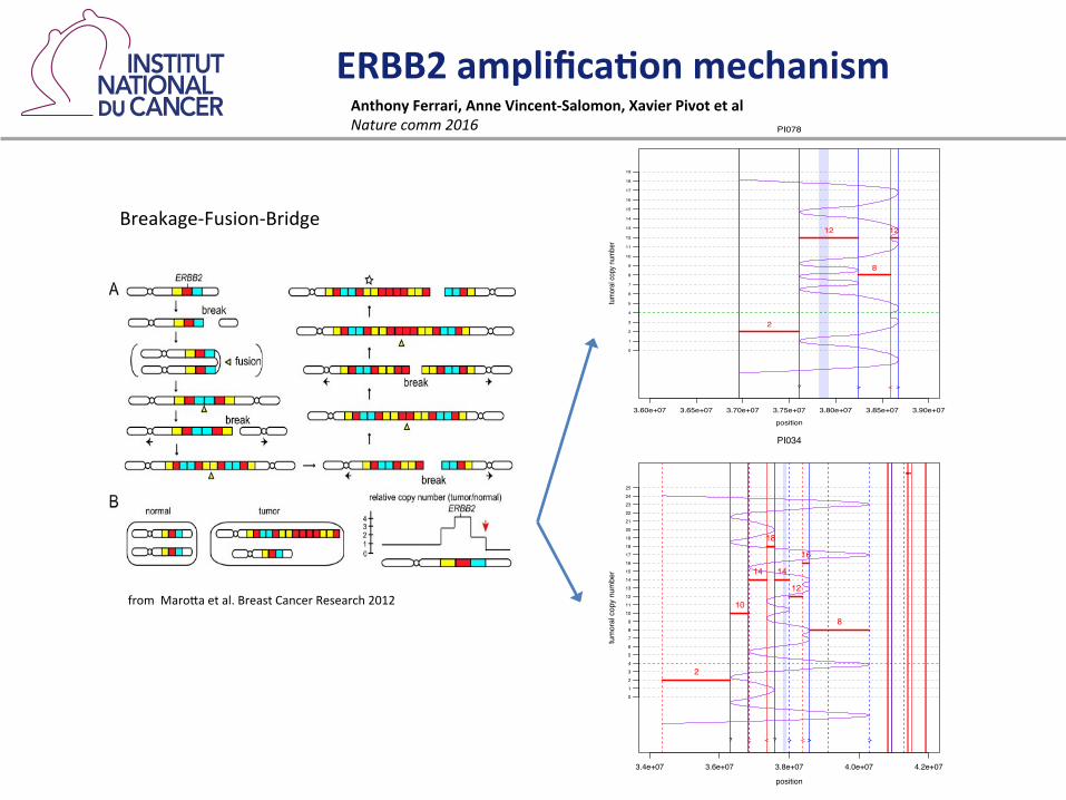
ERBB2 amplifica-on mechanism
from MaroZa et al. Breast Cancer Research 2012
3.60e+07 3.65e+07 3.70e+07 3.75e+07 3.80e+07 3.85e+07 3.90e+07
position
0
1
2
3
4
5
6
7
8
9
10
11
12
13
14
15
16
17
18
19
tumo
ral c
opy n
umbe
r
!!
PI078
? > < >
2
12
8
12
3.4e+07 3.6e+07 3.8e+07 4.0e+07 4.2e+07
position
0
1
2
3
4
5
6
7
8
9
10
11
12
13
14
15
16
17
18
19
20
21
22
23
24
25
tum
oral
cop
y nu
mbe
r
!!!!!! !
PI034
? < < ? > < > >
2
10
14
18
14
12
16
8
Breakage-‐Fusion-‐Bridge
Anthony Ferrari, Anne Vincent-‐Salomon, Xavier Pivot et al Nature comm 2016
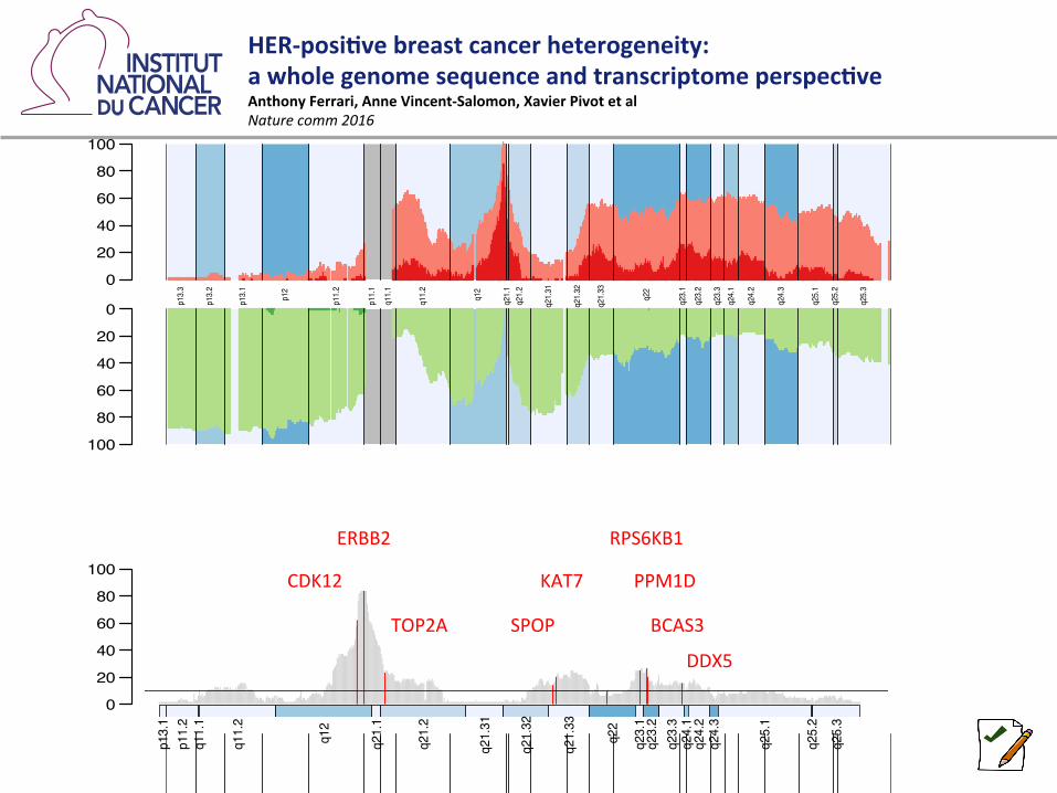
0
20
40
60
80
100
100
80
60
40
20
0 p13.3
p13.2
p13.1
p12
p11.2
p11.1
q11.1
q11.2
q12
q21.1
q21.2
q21.31
q21.32
q21.33
q22
q23.1
q23.2
q23.3
q24.1
q24.2
q24.3
q25.1
q25.2
q25.3
amplifications
frequency
0
20
40
60
80
100
p13.1
p11.2
q11.1
q11.2
q12
q21.1
q21.2
q21.31
q21.32
q21.33
q22
q23.1
q23.2
q23.3
q24.1
q24.2
q24.3
q25.1
q25.2
q25.3
CDK12
ERBB2
TOP2A SPOP
KAT7
RPS6KB1
BCAS3
PPM1D
DDX5
0
20
40
60
80
100
100
80
60
40
20
0 p13.3
p13.2
p13.1
p12
p11.2
p11.1
q11.1
q11.2
q12
q21.1
q21.2
q21.31
q21.32
q21.33
q22
q23.1
q23.2
q23.3
q24.1
q24.2
q24.3
q25.1
q25.2
q25.3
HER-‐posi-ve breast cancer heterogeneity: a whole genome sequence and transcriptome perspec-ve Anthony Ferrari, Anne Vincent-‐Salomon, Xavier Pivot et al Nature comm 2016
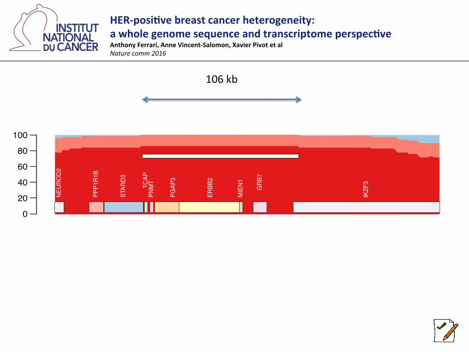
0
20
40
60
80
100
NEUR
OD2
PPP1R1
B
STAR
D3
TCAP
PNMT
PGAP
3
ERBB
2
MIEN1 GRB
7
IKZF3
106 kb
HER-‐posi-ve breast cancer heterogeneity: a whole genome sequence and transcriptome perspec-ve Anthony Ferrari, Anne Vincent-‐Salomon, Xavier Pivot et al Nature comm 2016
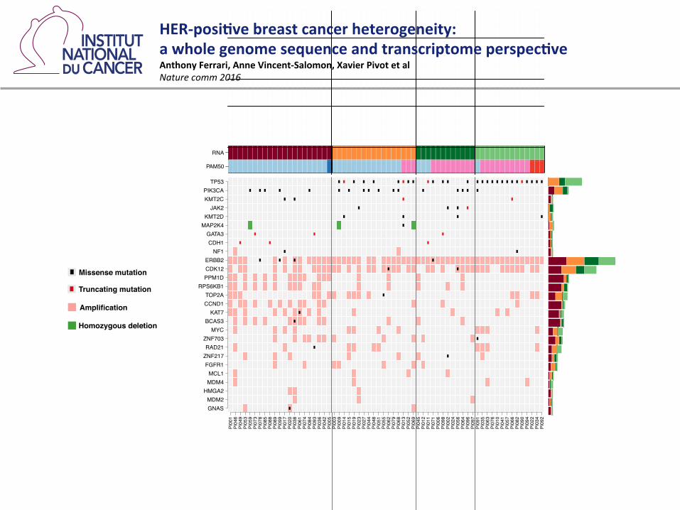
Missense mutation!
Truncating mutation!
Amplification!
Homozygous deletion!
SV17
mLum
pLum
LST
FGA
PLOIDY
HER2.CNV
HER2
PR
ER
PAM50
RNA
PI001
PI046
PI049
PI053
PI059
PI073
PI078
PI085
PI088
PI089
PI099
PI017
PI020
PI038
PI061
PI074
PI084
PI093
PI036
PI042
PI005
PI003
PI009
PI014
PI015
PI019
PI023
PI027
PI044
PI048
PI051
PI055
PI062
PI079
PI068
PI013
PI052
PI069
PI045
PI012
PI011
PI071
PI006
PI098
PI002
PI024
PI056
PI064
PI096
PI097
PI091
PI035
PI063
PI076
PI010
PI041
PI057
PI066
PI082
PI090
PI094
PI022
PI034
PI092
GNASMDM2HMGA2MDM4MCL1FGFR1ZNF217RAD21ZNF703
MYCBCAS3KAT7
CCND1TOP2A
RPS6KB1PPM1DCDK12ERBB2
NF1CDH1GATA3
MAP2K4KMT2DJAK2
KMT2CPIK3CATP53
0 10 20 30 40 50
HER-‐posi-ve breast cancer heterogeneity: a whole genome sequence and transcriptome perspec-ve Anthony Ferrari, Anne Vincent-‐Salomon, Xavier Pivot et al Nature comm 2016

SIGNAL study : GWAS findings
No associaGons between SNPs and HER2 status
X Pivot et al Nature Journal Breast Cancer 2017


![Guia de Nutrição Enteral Ambulatorial e Domiciliar...Guia de nutrição enteral ambulatorial e domiciliar [recurso eletrônico] / Lúcia Leite Lais e Sancha Helena de Lima Vale (organizadoras)](https://img.pdfslide.us/doc/110x75/60ca641fe2ac2f2be7117816/guia-de-nutrio-enteral-ambulatorial-e-domiciliar-guia-de-nutrio-enteral.jpg)


![[Laurence J. Peter] the Peter Principle(BookFi.org)](https://img.pdfslide.us/doc/110x75/55cf985c550346d0339732fb/laurence-j-peter-the-peter-principlebookfiorg.jpg)
