Embed Size (px)
Citation preview
125 International Journal of Scientific Study | September 2014 | Vol 2 | Issue 6
Herpes Zoster Leading to Viral Osteomyelitis or Neuralgia Inducing Cavitational Osteonecrosis? – A Case Report and Review of Literature
M L Asha1, Ingita Chatterjee2,
Preeti Patil3, Anu Vijayan2
1Professor, Department of Oral Medicine and Radiology, Sri Hasanamba Dental College and Hospital, Hassan, Karnataka, India, 2Post-graduate Student, Department of Oral Medicine and Radiology, Sri Hasanamba Dental College and Hospital, Hassan, Karnataka, India, 3Senior Lecturer, Department of Oral Medicine and Radiology, Sri Hasanamba Dental College and Hospital, Hassan, Karnataka, India
Corresponding Author: Dr. Ingita Chatterjee, Department of Oral Medicine and Radiology, Sri Hasanamba Dental College and Hospital, Hassan, Karnataka, India. Phone: +91-8884973049. E-mail: [email protected]
creates an anaerobic atmosphere that antibiotics cannot penetrate and effectively reduces the bactericidal activities of polymorpholeukocytes. The antibiotics fail to reach the organisms as the lowered vascularity reduces the rate of diffusion, despite therapeutic concentrations.5
CASE REPORT
A 75-year-old patient complained of pain in his lower right back tooth region since 3 months. Patient gave history of vesicular eruptions 3 months back which turned into ulcerations and healed with hyperpigmentation in the right side of his face, as well as the right ear. He also gave a history of hearing impairment since 15 days. Patient gave history of exfoliation of teeth in right mandibular region 2 months back. No relevant medical history was present. Patient revealed history of smoking bidi since 30-35 years, one-pack bidi per day.
Extra-oral ExaminationOn an inspection: Multiple areas of hyperpigmentation seen in the right middle and lower one-third of the face along the
INTRODUCTION
The scope of diagnosis of herpes zoster of the trigeminal nerve falls under all dental specialists. A thorough knowledge of this disease will prevent unnecessary and delayed treatment for the patient. During the prodromal stage of this disease in particular, the only presenting symptom may be odontalgia which may prove challenging to a clinician who is not familiar with herpes zoster of the trigeminal nerve.1 Postherpetic neuralgia (PHN); the most common complication of herpes zoster infection is easily recognized. Other developmental anomalies such as irregular short roots and missing teeth, periodontitis and calcified and devitalized pulps are difficult to diagnose.2 Herpes zoster has also been found associated with peri-apical lesions and resorption of roots.3 The term ‘osteomyelitis,’ which was introduced by Nelaton4 in 1844, implies an infection of the bone and marrow. Osteomyelitis most commonly results from bacterial infections, although fungi, parasites, and viruses can affect the bone and marrow. The chronicity of osteomyelitis is multifactorial. The lowered oxygen tension produced due to ischemia of the infected area and sequestrum
Case Report
AbstractHerpes zoster infection is considered to be a reactivation of varicella zoster infection. Herpes zoster involving the trigeminal branch leads to various complications like post herpetic neuralgia, osteomyelitis, tooth exfoliation, pulp necrosis etc. The purpose of this paper is to highlight the case of a 75-year-old patient suffering from herpes zoster with exposed necrotic mandibular alveolar bone. Based on the history and clinical examination a provisional diagnosis of herpes zoster of right V3 division of the trigeminal nerve with viral osteomyelitis of right mandible and a differential diagnosis of neuralgia inducing cavitational osteonecrosis was given.
Keywords: Herpes zoster, Neuralgia inducing cavitational osteonecrosis, Viral osteomyelitis
Asha, et al.: Viral Osteomyelitis
126International Journal of Scientific Study | September 2014 | Vol 2 | Issue 6
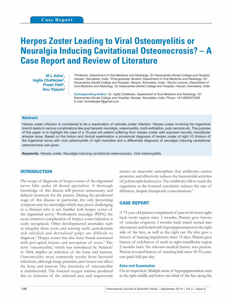
distribution of the mandibular division of the trigeminal nerve. Evidence of pus discharge from the right ear was present. Area of hypopigmentation was seen in the right half of lower lip extending into the chin. Area of hypopigmentation was also seen over the right side of the scalp (Figures 1 and 2).
On palpation: Lesional area was tender present. Pus discharge was seen from his left ear. No evidence of altered sensation was present.
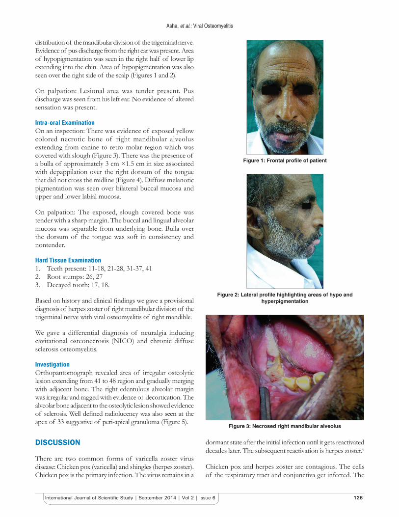
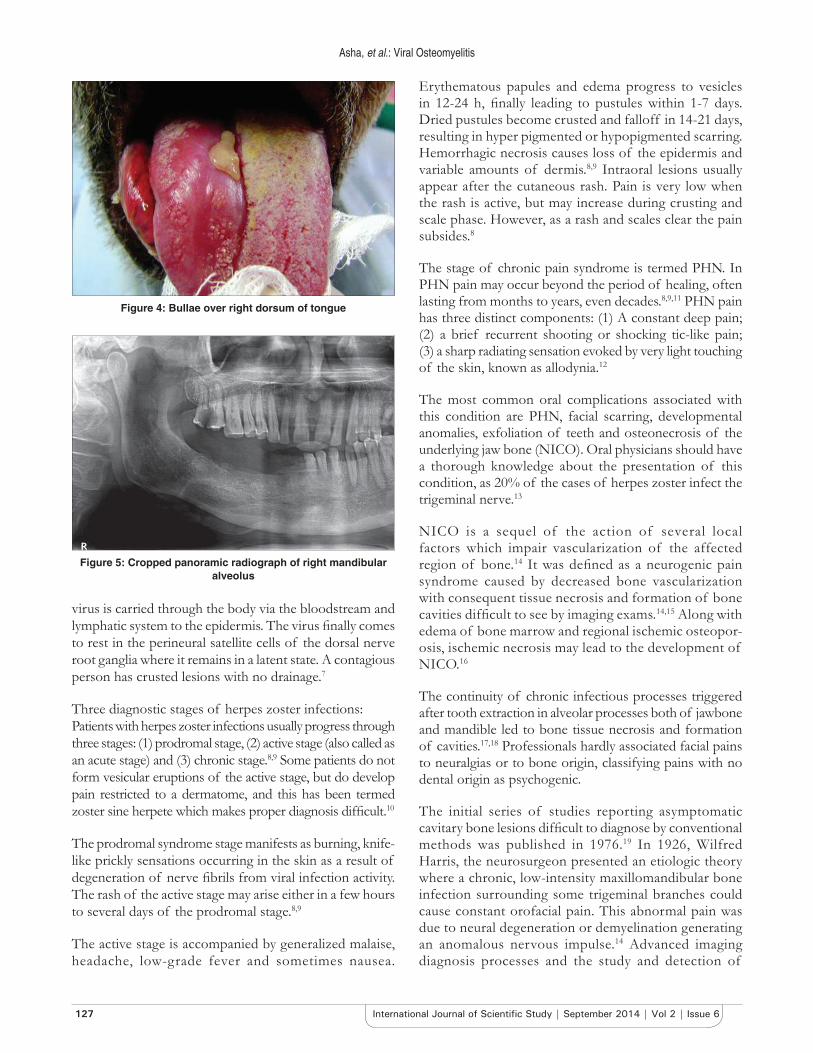
Intra-oral ExaminationOn an inspection: There was evidence of exposed yellow colored necrotic bone of right mandibular alveolus extending from canine to retro molar region which was covered with slough (Figure 3). There was the presence of a bulla of approximately 3 cm ×1.5 cm in size associated with depappilation over the right dorsum of the tongue that did not cross the midline (Figure 4). Diffuse melanotic pigmentation was seen over bilateral buccal mucosa and upper and lower labial mucosa.
On palpation: The exposed, slough covered bone was tender with a sharp margin. The buccal and lingual alveolar mucosa was separable from underlying bone. Bulla over the dorsum of the tongue was soft in consistency and nontender.
Hard Tissue Examination1. Teeth present: 11-18, 21-28, 31-37, 412. Root stumps: 26, 273. Decayed tooth: 17, 18.
Based on history and clinical findings we gave a provisional diagnosis of herpes zoster of right mandibular division of the trigeminal nerve with viral osteomyelitis of right mandible.
We gave a differential diagnosis of neuralgia inducing cavitational osteonecrosis (NICO) and chronic diffuse sclerosis osteomyelitis.
InvestigationOrthopantomograph revealed area of irregular osteolytic lesion extending from 41 to 48 region and gradually merging with adjacent bone. The right edentulous alveolar margin was irregular and ragged with evidence of decortication. The alveolar bone adjacent to the osteolytic lesion showed evidence of sclerosis. Well defined radiolucency was also seen at the apex of 33 suggestive of peri-apical granuloma (Figure 5).
DISCUSSION
There are two common forms of varicella zoster virus disease: Chicken pox (varicella) and shingles (herpes zoster). Chicken pox is the primary infection. The virus remains in a
dormant state after the initial infection until it gets reactivated decades later. The subsequent reactivation is herpes zoster.6
Chicken pox and herpes zoster are contagious. The cells of the respiratory tract and conjunctiva get infected. The
Figure 1: Frontal profile of patient
Figure 2: Lateral profile highlighting areas of hypo and hyperpigmentation
Figure 3: Necrosed right mandibular alveolus
Asha, et al.: Viral Osteomyelitis
127 International Journal of Scientific Study | September 2014 | Vol 2 | Issue 6
virus is carried through the body via the bloodstream and lymphatic system to the epidermis. The virus finally comes to rest in the perineural satellite cells of the dorsal nerve root ganglia where it remains in a latent state. A contagious person has crusted lesions with no drainage.7
Three diagnostic stages of herpes zoster infections:Patients with herpes zoster infections usually progress through three stages: (1) prodromal stage, (2) active stage (also called as an acute stage) and (3) chronic stage.8,9 Some patients do not form vesicular eruptions of the active stage, but do develop pain restricted to a dermatome, and this has been termed zoster sine herpete which makes proper diagnosis difficult.10
The prodromal syndrome stage manifests as burning, knife-like prickly sensations occurring in the skin as a result of degeneration of nerve fibrils from viral infection activity. The rash of the active stage may arise either in a few hours to several days of the prodromal stage.8,9
The active stage is accompanied by generalized malaise, headache, low-grade fever and sometimes nausea.
Erythematous papules and edema progress to vesicles in 12-24 h, finally leading to pustules within 1-7 days. Dried pustules become crusted and falloff in 14-21 days, resulting in hyper pigmented or hypopigmented scarring. Hemorrhagic necrosis causes loss of the epidermis and variable amounts of dermis.8,9 Intraoral lesions usually appear after the cutaneous rash. Pain is very low when the rash is active, but may increase during crusting and scale phase. However, as a rash and scales clear the pain subsides.8
The stage of chronic pain syndrome is termed PHN. In PHN pain may occur beyond the period of healing, often lasting from months to years, even decades.8,9,11 PHN pain has three distinct components: (1) A constant deep pain; (2) a brief recurrent shooting or shocking tic-like pain; (3) a sharp radiating sensation evoked by very light touching of the skin, known as allodynia.12
The most common oral complications associated with this condition are PHN, facial scarring, developmental anomalies, exfoliation of teeth and osteonecrosis of the underlying jaw bone (NICO). Oral physicians should have a thorough knowledge about the presentation of this condition, as 20% of the cases of herpes zoster infect the trigeminal nerve.13
NICO is a sequel of the action of several local factors which impair vascularization of the affected region of bone.14 It was defined as a neurogenic pain syndrome caused by decreased bone vascularization with consequent tissue necrosis and formation of bone cavities difficult to see by imaging exams.14,15 Along with edema of bone marrow and regional ischemic osteopor-osis, ischemic necrosis may lead to the development of NICO.16
The continuity of chronic infectious processes triggered after tooth extraction in alveolar processes both of jawbone and mandible led to bone tissue necrosis and formation of cavities.17,18 Professionals hardly associated facial pains to neuralgias or to bone origin, classifying pains with no dental origin as psychogenic.
The initial series of studies reporting asymptomatic cavitary bone lesions difficult to diagnose by conventional methods was published in 1976.19 In 1926, Wilfred Harris, the neurosurgeon presented an etiologic theory where a chronic, low-intensity maxillomandibular bone infection surrounding some trigeminal branches could cause constant orofacial pain. This abnormal pain was due to neural degeneration or demyelination generating an anomalous nervous impulse.14 Advanced imaging diagnosis processes and the study and detection of
Figure 4: Bullae over right dorsum of tongue
Figure 5: Cropped panoramic radiograph of right mandibular alveolus
Asha, et al.: Viral Osteomyelitis
128International Journal of Scientific Study | September 2014 | Vol 2 | Issue 6
genetic changes showed that decreased bone marrow blood flow causing bone cavities also include as a cause of NICO. Genetic mutations associated with NICO would predispose patients to thrombophilia and hypofibrinolysis.20,21
Two major theories for NICO etiopathogenesis have been suggested by some authors: One is infectious, considering bacteria as major disease-causing agents; the other is ischemic which has as its major cause bone tissue infarction due to lack of blood irrigation.14,22
Development Mechanism and EtiologyThe mechanism of development of NICO is under study. However, the five triggering factors associated with NICO14 are described below:1. Local or systemic immunodeficiency which impairs
local infection elimination2. Presence of specific pathogenic bacteria impairing
vascularization leading to infarction and necrosis3. Lack of tissue vascularization leads to bone marrow
in farction making odontogenic infections easier4. Lack of neutrophils and/or macrophages would lead
to decreased chemotaxis and phagocytosis, promoting infections
5. Lack or decrease of bone growth factors and a change in tissue pH, decreasing osteoinducing potential.
Researchers have concluded that hereditary factors are also associated with NICO as is seen in hereditary forms of thrombophilia and hypofibrinolysis.13,15,16 71% of NICO cases are caused by various factors like trauma, alcohol abuse, estrogen, prednisone, pregnancy, lupus erythematosus, sickle cell anemia, and use of chemotherapy for malignant neoplasias. Smoking and atherosclerosis are less commonly associated.23
Osteomyelitis and inanition are rarely associated with NICO.24 Bisphosphonates are potential risk factors especially for patients undergoing tooth exractions, as they induce osteoclast apoptosis and inhibit the release of growth factors and other bone matrix factors.25,26
Histological ExamThe histopathologic picture depends on the duration and the intensity of blood flow decrease in the medullary bone. Dilated and sinusoidal medullary capillaries depict features of bone marrow edema, blood vessels with serous exudates, adipocytes, ischemic myelofibrosis in between fat cells and a slight dispersion of chronic inflammatory cells in regions of myelofibros trabecular bones appear viable, however inactive, thin and largely spaced. Some researchers consider presence of microinfarctions as hallmark signs of osteonecrosis.16
Masses of stained globular calcified necrotic debris similar to those generated by rotary tools may be seen. However, these are more peripheral in tissue fragments, which allows for their differentiation.14,15,22
Sometimes periapical and panoramic X-rays are not enough for an accurate NICO diagnosis.14
Studies14,15 publishing NICO radiographic findings report the presence of light and discrete radiolucent areas, just like soap bubbles, and the radiopaque areas appear in the shape of cotton. It is possible to radiographically observe the lack of normal bone tissue healing, with the presence of lamina dura close to the alveolus, in the regions corresponding to the zones where teeth had been extracted.
It was reported by the same authors14,15 that ischemic osteonecrosis is difficult to see in conventional X-rays, however when present it appears as a radiolucent area and there may be a weak oval central sclerosis surrounded by a thick radiolucent circle which is also surrounded by a thick sclerotic ring however poorly distinct, being also described as “bull’s eye lesion.”
Bone scintigraphy using technetium 99 is the golden standard for a bone marrow ischemia diagnosis. This technique is still being used although being expensive and needing intravenous contrast injection, in addition to resulting in 30% of false-negatives.14,27,28
An ultrasonography bone densitometer purportedly detects and precisely images porosity of the bone to aid medical professionals in diagnosing bone marrow edema syndrome, NICO, osteomyelitis and periodontal pockets of the buccal bone. However, there are no articles on the effectiveness of the device published in peer-reviewed medical journals.29
TreatmentRecommended therapy for herpes zoster should include (i) patient isolation, (ii) local management of skin lesions, (iii) control and elimination of pain, (iv) limitation of the extent, duration and severity of the disease with antiviral agents and (v) treatment of PHN.8,9
Isolation: Patients with herpes zoster infections are contagious to persons at high-risk. The contagion is transmitted as varicella zoster virus (chicken pox). Herpes zoster patients remain contagious until scaling and crusting have taken place.8,9
The skin: Management includes the application of open wet dressings followed by lotions. Gauze or a cloth soaked in cool water is applied to the rash area for 30 min, 3-6 times a day. However, occlusive ointments should be avoided.
Asha, et al.: Viral Osteomyelitis
129 International Journal of Scientific Study | September 2014 | Vol 2 | Issue 6
Creams and lotions containing corticosteroids are not recommended.8
Pain: The acute pain of herpes zoster infection can be reduced during the prodromal phases by analgesics, such as acetaminophen, codeine and nonsteroidal anti-inflammatory agents. However, these analgesics are notoriously ineffective for the chronic PHN phase.8,9
Anti-viral drug therapy: Once a diagnosis of herpes zoster infection has been determined, anti-viral must be swift and precise. Acyclovir has been proven to decrease the duration and severity of the herpes zoster infection in the acute phase drastically, if treatment is started within 48 h of the onset of the rash.8,9,1
Acyclovir is beneficial in treating zoster infections due to its much higher rate of phosphorylation in herpes infected cells. Acyclovir also proves to be less toxic than other anti-viral drugs.8 Dosage of 800 mg 4 times a day for 10 days remains the standard of care. Research has shown that dosages of up to 800 mg 5 times have been given with even more promising results.30
Specifically to address the acute stage of herpes zoster (famciclovir), for immunocompromised patients (valaciclovir) are used. In the former, the dosage is 500 mg every 8 h for 7 days; for the later it is 1 g 3 times daily for 7 days.30-32
PHN: Standard analgesic narcotic combinations are not effective in patients with PHN. The treatment for PHN pain includes the topical use of capsaicin cream (zostrix), transcutaneous nerve stimulation, topical anesthetics, injected local anesthetics, and low-dose amitryptiline.9 Treatment is decided based on clinical and imaging exams. Complete resection of the painful bone tissue or bone marrow curettage is done followed by placing a sponge impregnated with antibiotics.14,15,17,18 Local infiltration with tetracycline alone or combined with cephalexin has shown 90% improvement.17
The effects of warfarin and stanozolol have been studied in patients with NICO and have been proved to be effective. However, these drugs have relieved pain in just 5% of cases. Various authors presented cases on herpes zoster with osteomyelitis of bone, which are being summarized in Table 1 below for highlighting the untoward complication of herpes zoster.
CONCLUSION
Herpes zoster infection clinically presents as vesicular eruption along a specific dermatome. Various complications of herpes zoster include bone osteomyelitis and NICO. A case of herpes zoster of right mandibular nerve with viral osteomyelitis of right mandible as its complication is highlighted in this paper. Follow-up of such patients is very crucial due to its long-term complication.
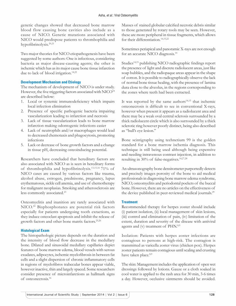
Table 1: Various case reports on herpes zoster leading to alveolar necrosis highlighting old age and male predilection33-39
Authors Age/sex of patient Clinical featuresKim et al. 1st case - 78 years/male Skin lesion along right maxillary and mandibular division pain and mobility of the mandibular right
canine impaired healing and osteonecrosis of mandible after extraction of canine2nd case - 77 years/male Skin lesion along the right mandibular division followed by sore gingiva in right mandibular canine
premolar region with exposed cortical bone beneath it3rd case - 74 years/male Lesions along the maxillary distribution followed by multiple mucosal vesicles and ulcer formation
on the left palate together with mobility of the remaining teeth and alveolar bone resorption in edentulous state except the maxillary right and left central incisor, lateral incisor, and canine
Arikawa et al. 74 years/male Vesicles and pustules on the right side of the lower lip, chin, cheek and external ear. Ulceration on the right side of the soft palate, buccal mucosa, and tongue. 44 days after the onset, the mandibular right lateral incisor, canine, and left lateral incisor were markedly loose and periodontal attachment tissue was necrotic
Onem et al. 76 years/male Hyperesthesia over the vesico-bullous lesions on the left trigeminal nerve. The alveolar process became exposed in the premolar area of left mandibulary bone
Mendieta et al. 63 years/female Redness of the alveolar mucosa and gingiva of the lower right quadrant with multiple well-delimited and painful erosive lesions affecting the attached gingiva around the teeth. 2 weeks later, lower right canine and lower right first premolar had class III mobility, flow of purulent exudate from the gingival sulcus,. Due to extensive necrosis there was no interdental alveolar bone
Kashinath and Shekar
58 years/male Vesicular eruptions along the right maxillary division followed with open tooth socket with respect to 13 tooth region and exposed alveolar bone with respect to 14, 15, 16 region along with receded palatal gingival margin with respect to 14, 15, 16
Sharma et al. 44 years/male Vesicular lesions along left maxillary and mandibular division followed by Odontalgia, alveolar bone necrosis and the spontaneous exfoliation of multiple teeth in left mandible and maxilla
Kwamin et al. 26 years/woman Painful skin eruptions on the right face 4 months earlier. These progressed into the right cheek and then to the jaw causing severe toothache in the lower anterior teeth which became increasingly painful and mobile. The periodontium covering 41-44 was necrotic with roots exposed. The involved teeth were grossly mobile and held in place by the lingual alveolar mucosa and fibrous strands
Asha, et al.: Viral Osteomyelitis
130International Journal of Scientific Study | September 2014 | Vol 2 | Issue 6
REFERENCES
1. Millar EP, Troulis MJ. Herpes zoster of the trigeminal nerve: The dentist’s role in diagnosis and management. J Can Dent Assoc 1994;60:450-3.
2. Bajwa ZH, Ho CC. Herpetic neuralgia. Use of combination therapy for pain relief in acute and chronic herpes zoster. Geriatrics 2001;56:18-24.
3. SolomonCS,CoffinerMO,ChalfinHE.Herpeszosterrevisited:Implicatedin root resorption. J Endod 1986;12:210-3.
4. Nelaton A. Elements de Pathologie Chirurgicale. Paris: Germer-Bailliere; 1859.
5. Eckardt JJ, Wirganowicz PZ, Mar T. An aggressive surgical approach to the managementofchronicosteomyelitis.ClinOrthopRelatRes1994:229-39.
6. Tidwell E, Hutson B, Burkhart N, Gutmann JL, Ellis CD. Herpes zoster of the trigeminal nerve third branch: A case report and review of the literature. IntEndodJ1999;32:61-6.
7. RubinE,FarberJL.EssentialPathology.Philadelphia,PA,USA:LippincottCompany; 1995.
8. StrommenGI, Pucino F, Tight RR, Beck CL. Human infectionwith H.Zoster, etiology, pathophysiology, diagnosis, clinical course and treatment. Pharmacotherapy 1998;8:52-68.
9. CarmichaelJK.TreatmentofH.Zosterandpostherpeticneuralgia.AmFamPract 1991;44:203-10.
10. Barrett AP, Katelaris CH, Morris JG, Schifter M. Zoster sine herpete of the trigeminalnerve.OralSurgOralMedOralPathol1993;75:173-5.
11. RowbothamMC,FieldsHL.Topicallidocainereducespaininpost-herpeticneuralgia. Pain 1989;38:297-301.
12. RowbothamMC,FieldsHL.Post-herpeticneuralgia: the relationofpaincomplaint, sensory disturbance, and skin temperature. Pain 1989;39:129-44.
13. BandralMR,ChidambarYS,TelkarS,JapattiS,ChoudaryL,DodamaniA.Oralcomplicationsofherpeszosterinfection-Reportof3cases.IntJDentClin 2010;2:70-3.
14. Bouquot JE, Roberts AM, Person P, Christian J. Neuralgia-inducingcavitationalosteonecrosis(NICO).Osteomyelitisin224jawbonesamplesfrom patients with facial neuralgia. Oral Surg Oral Med Oral Pathol1992;73:307-19.
15. Bouquot JE, Christian J. Long-term effects of jawbone curettage on the pain offacialneuralgia.JOralMaxillofacSurg1995;53:387-97.
16. Grossman E, Cousen T, Grossmann TK, Bérzin F. Neuralgia inducingcavitationalosteonecrosis.RevDor2012;13:000-0.
17. Ratner EJ, Person P, Kleinman DJ, Shklar G, Socransky SS. Jawbonecavitiesandtrigeminalandatypicalfacialneuralgias.OralSurgOralMedOralPathol1979;48:3-20.
18. Ratner EJ, Langer B, Evins ML. Alveolar cavitational osteopathosis.Manifestations of an infectious process and its implication in the causation of chronic pain. J Periodontol 1986;57:593-603.
19. Sardella A, Demarosi F, Barbieri C, Lodi G. An up-to-date view onpersistent idiopathic facial pain. Minerva Stomatol 2009;58:289-99.
20. GlueckCJ,McMahonRE,BouquotJ,StroopD,TracyT,WangP,et al. Thrombophilia, hypofibrinolysis, and alveolar osteonecrosis of the jaws.OralSurgOralMedOralPatholOralRadiolEndod1996;81:557-66.
21. GruppoR,GlueckCJ,McMahonRE,BouquotJ,RabinovichBA,BeckerA, et al. The pathophysiology of alveolar osteonecrosis of the jaw: anticardiolipinantibodies,thrombophilia,andhypofibrinolysis.JLabClinMed 1996;127:481-8.
22. Adams WR, Spolnik KJ, Bouquot JE. Maxillofacial osteonecrosis ina patient with multiple “idiopathic” facial pains. J Oral Pathol Med1999;28:423-32.
23. Bouquot JE, McMahon RE. Ischemic alveolar osteonecrosis in2.023patientswithchronicfacialpain.JOrofacPain1997;11:180-6.
24. Glueck CJ,McMahon RE, Bouquot JE, Triplett D, Gruppo R,Wang P.Heterozygosity for the Leiden mutation of the factor V gene, a common pathoetiology for osteonecrosis of the jaw, with thrombophilia augmented byexogenousestrogens.JLabClinMed1997;130:540-3.
25. Gegler A, Cherubini K, Figueiredo MA, Yurge LS, Azambuja AA.Bisfosfonatoseosteonecrosemaxilar:Revisãodaliteraturaerelatodedoiscasos.RevBrasCancerol2006;52:25-31.
26. RuggieroSL,DodsonTB,AssaelLA,LandesbergR,MarxRE,MehrotraB, et al.AmericanAssociationofOralandMaxillofacialSurgeonspositionpaper on bisphosphonate-related osteonecrosis of the jaw - 2009 update. Aust Endod J 2009;35:119-30.
27. DeNucci DJ, Chen CC, Sobiski C, Meehan S. The use of SPECT bone scans to evaluate patientswith idiopathic jawpain.Oral SurgOralMedOralPatholOralRadiolEndod2000;90:750-7.
28. FeinbergLS,StephanRB,FogartyKP,VoortmanL,TillerWA,Cassiani-IngoniR.ResolutionofcavitationalosteonecrosisthroughNeuroModulationTechnique, a novel form of intention-based therapy: a clinical case study. J Altern Complement Med 2009;15:25-33.
29. NeuralgiaInducingCavitationalOsteonecrosis(NICO)andUltrasonographBoneDensitometertoDetectNICO.ClinicalPolicyBulletin.
30. WoodMJ, Kay R, Dworkin RH, Soong SJ,Whitley RJ. Oral acyclovirtherapy accelerates pain resolution in patients with herpes zoster: A meta-analysisofplacebo-controlledtrials.ClinInfectDis1996;22:341-7.
31. Gill KS, Wood MJ. The clinical pharmacokinetics of famciclovir. Clin Pharmacokinet 1996;31:1-8.
32. GlueckCJ,McMahonRE,BouquotJE,TracyT,Sieve-SmithL,WangP.Apreliminarypilotstudyoftreatmentofthrombophiliaandhypofibrinolysisandameliorationofthepainofosteonecrosisofthejaws.OralSurgOralMedOralPatholOralRadiolEndod1998;85:64-73.
33. Kim NK, Kim BC, Nam JW, Kim HJ. Alveolar bone necrosis and spontaneous toothexfoliationassociatedwithtrigeminalherpeszoster:Areportofthreecases.JKoreanAssocOralMaxillofacSurg2012;38:177-83.
34. ArikawaJ,MizushimaJ,HigakiY,HoshinoJ,KawashimaM.Mandibularalveolar bone necrosis after trigeminal herpes zoster. Int J Dermatol2004;43:136-7.
35. Onem E, Alpoz E, Kandemir S, Akay C. Mandibular osteomyelitisfollowing trigeminal herpes zoster infection. Hacettepe Diş HekimliğiFakültesiDergisi2009;33:31-5.
36. Mendieta C,Miranda J, Brunet LI, Gargallo J, Berini L.Alveolar bonenecrosisandtoothexfoliationfollowingherpeszosterinfection:Areviewof the literature and case report. J Periodontol 2005;76:148-53.
37. Kashinath KR, Shekar LC. Herpes zoster along maxillary nerve withosteonecrosis.JDentSciRes2011;2:12-7.
38. SharmaD,JhingtaP,SinghM,BhardwajVK,VaidS,NegiN.Odontalgia,alveolar bone necrosis and spontaneous exfoliation of multiple teethfollowingherpeszoster infectionof trigeminalnerve.JCranio-MaxillaryDis 2012;1:27-32.
39. Kwamin F, Parkins G, Nyako E. Herpes zoster infection in humanimmunodeficiencyviruswithmaxillaryandmandibularosteonecrosiswithteethexfoliation.IntJDentCaseRep2012;2:7-10.
How to cite this article: Asha ML, Chatterjee I, Patil P, Vijayan A. Herpes Zoster Leading to Viral Osteomyelitis or Neuralgia Inducing Cavitational Osteonecrosis? – A Case Report and Review of the Literature. Int J Sci Stud 2014;2(6):125-130.
Source of Support: Nil, Conflict of Interest: None declared.