Embed Size (px)
Citation preview

NOVEMBER 1989Volume 75, No 11
I
I herapy
The Journal of the
Manipulation
The Concept of AdverseMechanical Tension in theNervous System
DAVID BUTLER Bph,y MAPA MMTAASenior Physiotherapist. West Hill Hospital. Dantbrd
LOUIS GIFFORDBScMcspPrivate Practitioner. Falmouth
Chartered Society of Physiotherapy

The Concept of AdverseMechanical Tension in the Nervous SystemPart 1: Testing for 'Dural tension'
DAVID BUTLER BPhtyMAPAMMTAA
Senior Physiotherapist. West Hill Hospital. Darttbrd
LOUIS GIFFORDBScMcspPrivate Practitioner. Falmouth
Key words: Nerve, meninges. biomechanics. examination, tension tests,straight leg raise.
Summary: The concept of using tests such as the straight leg raise andprone knee bend to stress 'dura' or what Maitland (1978) has'termed "painsensitive structures within the vertebral canal' is believed to be- an over-simplification of the true anatomical and biomechanical facts. This paperexpands this concept to embrace the nervous system as a whole. It introducesthe term 'adverse mechanical tension of the nervous system' to explain howpathology affects the normal movement and biomechanics of the nervoussystem and its surrounding tissues. Mechanisms of sign and symptomproduction based on current literature are also put forward.
The standard or 'base' tension tests and relevant biomechanics are reviewedand some new variations/combinations described. The key feature of thisapproach is to understand basic principles of tension testing and to be ableto apply them to a wide variety of clinical presentations.
Biography: David Butler graduated-from the University of Queenslandwith a Bachelor of Physiotherapy in 1978. He then worked in private practicein Brisbane and in 1985 completed the Graduate Diploma in AdvancedManipulative Therapy in Adelaide, South Australia. He is currently a seniorphysiotherapist at the West Hill Hospital, Dartford, Kent and involved inteaching clinical applications of altered nervous system mechanics.
Louis Gifford trained at Sheffield City Polytechnic where his interest inmanipulative therapy was first fostered. After working in Walton Hospital,Liverpool, and St Stephen's Hospital, Chelsea, he went on to complete theone-year postgraduate Diploma in Advanced Manipulative Therapy inAdelaide, South Australia, in 1985. He then spent two years working andteaching in Adelaide before returning in 1988 to private practice in Falmouth.His interest in manual treatment of mechanical disorders of the nervous systemhas been the result of a close friendship and working relationship withMr David Butler.
Figures 1, 2, 5 and 8 are reproduced with permission from Proceedings ofthe Fifth Biennial Conference of the Manipulative Therapists Associationof Australia, Melbourne, 1987.
Figure 9 is adapted from The upper limb tension test by Kenneallyet al in Physical Therapy of the Cervical and Thoracic Spines, edited byR Grant, published by Churchill Livingstone, 1988.
IN recent years manipulative therapy has broadened itshorizons and taken in a concern for what Maitland (1978)has called 'movement of pain sensitive structures within thevertebral canal' — notably the 'neuromeningeal tissues'.Many therapists are laudably attending to abnormalities inneural tissue tension/mechanics as well as mobilising jointsand muscles in the quest for more consistent results.So-called 'tension tests' are not new; what is new is therefining of the old standard tests — straight leg raise (SLR),prone knee bend (PKB), passive neck flexion (PNF), thecombination of some of these tests (slump) and thedevelopment of a test that stresses neural tissue in the upper
limb — the upper limb tension test (ULTT) (Elvey, 1979),These tests have superseded their original purpose of beingof purely diagnostic importance and are now fully integratedinto passive mobilising treatment techniques (Maitland,1985; Elvey, 1986; Butler, 1987; Kenneally et al, 1988).
Rather than limit our thinking about these tests asstressing 'dura' they should now be considered in the farbroader context of the nervous system a.s a whole. A SLRmoves and tensions all components of the sciatic nerve fromits terminations in the foot to its origins in the spinal canaland beyond. Structures that must be considered include:
• The nerve as a whole and its surrounding tissues (muscle,bone, fibrous tissue, etc).
• The connective tissue components within the nerve(epineurium, perineurium and endoneurium) which have theirown intrinsic nerve supply (Hromada, 1963) and aretherefore capable of producing symptoms.
• The connective tissues withjn the spinal canal — namelythe meninges (dura, arachnoid, pia) plus intra-'cord'connective tissues.
• The conducting elements of the nervous system (theneurones).
• The intrinsic blood supply of the nervous system.
It is important to emphasise the close mechanicalrelationship between nerves on one side of the body or ina single limb to other nerves in the same limb or other limbsand also to the autonomic trunks and ganglia. Changes oftension in lumbar nerve roots have been clearly demonstratedduring PNF (Brieg and Marions, 1963; Tencer et al, 1985),and instant alteration in neck and arm pain by the additionof ankle dorsiflexion to a SLR is a frequent clinicaloccurrence, as is the alteration in the intensity of headacheswith a similar manoeuvre.
Thinking of the nervous system as an organ stresses itscontinuity and emphasises that any interference with partof the system may have implications for the whole. Whileit is primarily considered in an impulse-conducting role, italso has to be capable of adapting to body movement andin positions such as the slump test it can easily be shownto limit the ranges of movement of the test (see sectionon the slump test). Without interference to impulseconduction, the nervous system adapts to endlesscombinations, ranges and speeds of movement of the body.For instance, it has to cater for a spinal canal that is up to9 cm longer in flexion than extension (Louis, 1981) andperipheral nerves that are located on the opposite side ofjoint axes such as the femoral and sciatic nerves at the hip.
In order to avoid confusion, Butler (1987) has introducedthe term 'adverse mechanical tension in the nervous system'(AMT) after Brieg (1978) rather than using 'neural tension'which implies that conducting elements of the nervoussystem are abnormal; or 'dural tension' which limits ourthoughts to the spinal canal.
Tension tests', and for that matter body movements, notonly produce an increase in tension within the nerve but also
622 Physiotherapy, November 1989, vol 75, no 11

move the nerve in relation to its .surrounding tissues. Thesesurrounding tissues have been referred to as 'mechanicalinterface' tissues (Butler, 1987). The mechanical interface(Ml) should be regarded as the tissue most anatomicallyadjacent to. the nervous system that can move independentlyof the nervous system, for example, the supinator muscleis a Ml to the radial nerve as it passes through the radialtunnel.
Thus pathology at the Ml anywhere along the length ofa nerve can give rise to abnormalities in nerve movementand cause increases in tension within the nerve that mayhave far-reaching effects. A common example of Mlpathology is lumbar disc protrusion or osteophyte impinge-ment on a nerve root in an irrtervertebral foramen.
However, there are many vulnerable sites in the bodywhere lesions that affect the elasticity and movement of thenervous system are known to begin (Sunderland, 1978;Dawson et al, 1983). Soft tissue or fibro-osseous tunnelssuch as the carpal tunnel are frequently encountered siteswhere Ml pathology begins. This 'pathology' can be thoughtof in a purely mechanical sense termed 'pathomechanics'or in an 'inflammatory' or 'chemical' sense termed'pathophysiology' (Elvey, 1987). Sunderland (1976) hasestablished how inflammatory changes occurring around anerve can lead to changes in the connective tissues withinnerves leading to 'intraneural fibrosis' with consequentalterations in conduction. Fibrotic nerves lose elasticity andmay therefore influence the tested extensibility exhibitedwhen performing standard 'tension tests'. Pathophysiologicalchanges can therefore lead to pathomechanics.
Butler (1987, 1989) has attempted to categorise move-ment disorders of the nervous system into those which affectthe movement and tension of nerves from outside the nerve,termed 'extraneural', and those which affect it from withinthe nerve, termed 'intraneural'. The two can obviously occurtogether. As discussed above, extraneural pathology can leadto intraneural pathology such as fibrosis and it seemsreasonable to assume that primary changes within the nerve(intraneural) could bring about a sequence of pathologicalevents operating in the opposite direction resulting inextraneural pathology. Sunderland (1976) refers to fibrosednerve setting up a 'friction fibrosis elsewhere'.
From clinical observation of patients with positive tensiontests yet negative electrodiagnostic tests it appears thatintraneural pathology of the connective tissues may occurwithout measurable detriment to the conducting elements.Even if nerve fibres are injured, and responsible forsymptoms, an uninjured funiculus may still account for thenormal electrodiagnostic test (MacKinnon and Dellon, 1986).
It should be clear that-a,positive 'tension test' is not anemphatic indicator of a spinal disorder, it is — more vaguely— an indicator of adverse mechanical tension somewherein the nervous system. It is up to the skilled therapist toisolate the sources or site of the AMT.
A mechanism whereby a primary and often long-standingdisorder, perhaps originating in the spine, can give rise tosecondary and 'remote' disorders in the periphery (or thereverse) has been postulated (Butler, 1987, 1989). Referenceto the 'double crush' phenomenon supports the theory whichneatly fits some commonly found clinical situations wheresymptoms 'spread' or 'jump' to different areas over thecourse of time. The double crush phenomenon is a termintroduced by Upton and McComas (1973). These authorsexamined 115 patients with either carpal tunnel syndromeor lesions of the ulnar nerve at the elbow and found :that81 (70%) had electrophysiological and clinical evidence of
neural lesions at the neck. Dyro (1983) noted and found ithard to explain why 27% of a group of 50 young people heexamined with brachial plexus lesions developed carpaltunnel syndrome. Crymble (1968) noted a similar occurrence.Lundborg (1988) has referred to a 'reversed double crush'referring to patients who present with an ulnar nerveentrapment at the wrist who then go on to develop ulnarnerve entrapment at the elbow. Another example, stressingnerve interaction in one limb, is the higher incidence of lateralepicondylitis in patients with carpal tunnel syndrome(Murray-Leslie and Wright, 1976).
Upton and McCdmas (1973) believe altered 'axoplasmicflow' to be the underlying cause of the observedphenomenon. However, prolonged AMT in one area of anerve (or within the spinal canal) is bound to have mechanicalrepercussions further along the tract. Sunderland (personalcommunication, 1988) considers such, an explanationfeasible. Figure 1 diagrammatically illustrates a theoreticalmodel of the way this may occur.
T1 T2
L1 i
( b )
(0
T1, T2 = tunnel sites
L1, L2 = position that N can reach during normal movement withT1 and T2 constant
The stippled area represents the surface of N that can be in contactwith T1 or T2 during normal movement
(a) Neutral(b) To reach L2, N has moved in relation to T1 and T2 and increasedintra N tension(c) With extraneural pathology (EP) and/or intraneural pathology (IP)at T1, for N to reach L2 requires Increased intra N tension andincreased friction as the stippled area can now never be free of T2during movement
Fig 1: Possible effect of extraneural and intraneural pathology atone site on other sites along the nerve trunk
Basic Principles of Tension Testing
Tension tests affect a lot of other structures as well asmoving and tensioning nerves. Normal neural tissue whichis being moved by testing may come into contact withsensitive interface structures and elicit pain. Although in thissituation the tension test.can be 'proved' by adding distalor proximal sensitising manoeuvres, the culpable tissuemay still be extraneural in origin. An example may be azygapophyseal joint in the lumbar spine. Here, a SLR maybring the adjacent nerve root into contact with the anterioraspect of the joint and adding dorsiflexion would increasepressure and consequently exacerbate symptoms. Often,treating one or other of the tissues is the only way to drawa retrospective diagnostic conclusion.
When performing a tension test the operator must:
1. Be aware of expected normal responses.
2. Know all details of all the symptoms.
3. Know the symptoms in the starting position.
Physiotherapy, November 1989, vol 75, no 17. 623

4. Carefully monitor symptoms throughout the procedureand be able to make the patient clear about the symptomscomplained of as compared to any pain or discomfort causedby the test. The patient'has to be made to concentrate.
Reproduction of the patient's symptoms during testing isvery useful and can be conclusive but so often many tissuesare moved and stressed by a single manoeuvre. A uniqueaspect of tension testing is that symptoms can be changedby the addition of remote testing procedures known as'sensitising additions'. The addition of ankle dorsifiexionto a SLR causing an increase in lumbar symptoms is anexample. In-order to be conclusive, the emphasis must beon precise handling and communication with the patient.The slightest movement of the patient's leg/back/head/trunkwhile a SLR is held and while the operator fumbles to adddorsifiexion to the test is bad handling and gives misleadingresults. All tension tests have sensitising additions whichare used to add weight to and confirm a 'diagnosis' of AMT.These are dealt with under each test.
5. Also to be noted during testing are:• The range of movement; where pain starts (P1) and if non-irritable, a predetermined acceptable pain limit (P2) (Maitland,1986).• The type/area of pain and other symptoms.• The end feel (ie R1-R2 curve) (Maitland, 1986).• The effect of sensitising additions/subtractions on thesymptoms produced.
Always compare with the contralateral limb if possibleand/or with what is known to be normal (see individualtests).
Positive Tension Tests — Guide lines
A tension test is deemed positive if the patient'ssymptoms are reproduced by the procedure and if they thencan be changed by adding or subtracting sensitisingmanoeuvres.
Frequently the exact symptoms complained of cannot beproduced, but the test may still be seen as relevant andpositive if the symptoms produced are different from whatis known to be normal or different from the response of theopposite limb.
The range of movement achieved is often a strong guideto test relevance. Altering sensitising additions can causechanges in the range of test movements. Small differencesin range of movement when compared to non-symptomaticcontralateral limbs can be relevant, for example in disorderswhen symptoms are manifest only after extreme and lengthyactivity. Restoring normal range by treatment may be theonly way of clarifying the relevance of minor asymmetricalrange discrepancies.
As pointed out earlier, a positive tension test does notnecessarily indicate that there is a mechanical disorder ofthe nervous system. The tension tests may be placing a forceon a surrounding interface structure. Another considerationis that part of the nervous system may be irritated andsymptom-provocative, but the mechanics are normal.Irritation of nerves rather than compression has beensuggested as a potent and underestimated symptomproducer (Triano and Luttges, 1982). Elvey (1986, 1987)makes the suggestion that nervous system mobilisation mayhave a beneficial physiological effect by causing pressurechanges within the system.
A positive tension test gives the therapist a valid reasonto examine away from the symptom area and known referralsources. For example, with a slump test .including kneeextension positive for headache, the entire spine may need
examination. Sources of altered nervous system movementand tension may be a considerable distance from the areaof symptoms. Butler (1987, 1989) advocates that areaswhere the nervous system moves little relative to itssurrounding interface during movement or where the systemis relatively fixed are likely focal points of AMT and shouldbe strongly considered in the objective examination. Theseareas have been termed 'tension points' (Butler, 1987) andare dealt with during discussion of the tests.
Nervous system mechanics cannot be adequatelyexamined by one traditional tension test. For example, it isnot enough to examine the SLR, find it negative and declarethat AMT has no part in the patient's symptoms. Differingspinal, hip, knee and ankle positions may be required.Subjective clues and a knowledge of nerve biomechanicsmust be used to make up new tension tests in order to fitthe test to the patient's complaint. It should be regarded asa concept of testing, not just a series of 'base tests'. Justas present-day manual therapy encourages the examinationof a joint in many directions (Maitland, 1986) this also appliesto the nervous system. It is the aim ef these two articlesto promote the exploration of tension testing combinationsoften "unique to individual patients.
The Base TestsThe Straight Leg Raise
It is well known that on performing a SLR that there isconsiderable caudad movement of lumbosacral nerve rootsin relation to interfacing tissue such as the intervertebralforamen (Goddard and Reid, 1965; Breig, 1978), yet littleconsideration has been given to the rest of the sciatic tract.Smith (1956) has shown that the tibial nerve proximal to theknee also moves caudad in relation to its mechanicalinterface. Distal to the knee, the tibial nerve moves craniallyin relation to its mechanical interface (fig 2). Thus there isa point posterior to the knee where the nerve/mechanicalinterface relationship is constant during SLR, an area referredto as a 'tension point'. The common peroneal nerve is quitefirmly attached at the head of the fibula, making this itstension point..
Fig 2: From position A to position B, movement of the tibial nervein relation to the tibia and femur is in the direction of the arrows.There is no movement posterior to the knee (adapted from Smith,1956)
624 Physiotherapy, November 1989, vol 75, no 7 7

The SLR should be examined routinely .in all vertebraldisorders, all lower limb disorders, and some upper limbdisorders. The protocol suggested^by.Breig and Troup (1979)is followed. The leg is simply raised in the sagittal plane withthe knee extension maintained.
Fig 3: Plantarflexion/inversion may be added to the SLR while theheel rests on the operator's shoulder
The sensitising additions are:• Ankle dorsiflexion (Breig and Troup, 1979). This stressesmore the tibial component of the sciatic nerve.• Ankle plantarflexion with inversion (fig 3). This stressesthe common peroneal nerve and is especially useful in theassessment of anterior shin-and dorsal foot symptoms.• Passive neck flexion. '• Increase medial hip rotation (Breig and Troup, 1979).• Increase hip adduction (Sutton, 1979).• Alter spine position. For example, a left SLR will besensitised by spinal lateral flexion to the right.
Take care with the interpretation of results and rememberto consider all joints, muscles and soft tissues that are alsostressed with these manoeuvres.
The following example illustrates how useful logicaljuggling about with the test components can be in drawingconclusions about the contribution of AMT to a disorder.A patient'complains of anterolateral shin pain whichcan be reproduced by inverting the foot and then addingplantarfiexion. On the basis of this positive findingalone, a conclusion that joints and tissues of the footor anterior compartment musculature and fascia are culpabletissues would seem reasonable. Testing standard SLR andcomparing to the opposite leg may reveal slightdifferences in range of movement and end feel. Howeverthe most logical way to approach this with AMT thinkingwould be to bring on the pain (invert and plantarflex),maintain the exact position and amount of pain and thenperform" a SLR. A further increase in symptoms wouldjustify a conclusion that AMT is a component of thedisorder and may need treatment. The examination can befurther sensitised by adding hip adduction, medial rotationor even passive neck flexion. -i .
Prone Knee Bend ;
This test moves and tensions the nerves and roots relatedto the L2, 3, and 4 spinal segments and in particular thefemoral nerve and its branches (O'C'onnell, 1951; Estridgeet al, 1982). The test is performed by merely flexing the kneeof the prone subject while attempting to stabilise hip andthigh. The pain response must be interpreted with cautionas the PKB stretches rectus femoris, tends to rotate thepelvis forwards and thereby extends the lumbar spine. Aswell as spinal extension being possibly pain provocative, italso lessens the tension that can be placed on the femoralnerve, also hindering effective interpretation.
PKB with the hip in extension is no more sensitive thanPKB with the hip-in neutral (fci'avi^dson, 1987). However, ithas been noted clinically that the symptoms of meralgiaparaesthetica (entrapped lateral femoral, cutaneous nerve)are better reproduced, in PKB/hip extension.
Interpretation can be very difficult and will rely onsubjective information unless sensitising informationclearly alters the test response. These additions are:• Cervical flexion.• Slump in side lying (Davidson, 1987 — fig 4).• Varying ranges of hip abduction, adduction and rotations.
Fig 4: Slump with prone knee bend. An assistant is necessary tocontrol the many components
Slump Test
The slump test is perhaps the most important tension testlinking neural and connective tissue components of thenervous system from the pons to the terminations of thesciatic nerve in the foot (Maitland, 1978, 1985; Massey,1985).
It is a very sensitive test and must be carried out with agreat deal of handling and interpretative skill. One study(Massey, 1985) showed that the- slump was the mostsensitive test out of all the tension tests for reproductionof back pain in 50 symptomatic subjects. It should beexamined routinelyin all spinal disorders (cervical to lumbarto c6ccygeal,);.most — possibly all — lower limb disorders,some upper limb disorders and certainly those which indicatethe possibility of 'nervous system involvement.." The standard test consists of thoracic and lumbar flexion,followed by.,cervical flexion, knee extension, and ankle
Physiotherapy,-November 1989, vol 75, no 11 625
dorsiflexion and if necessary, hip flexion produced by eitherbringing the trunk forward on the hips or by increasing theSLR. The test is well described by Maitland (1986).
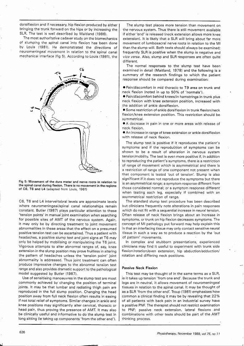
The most authoritative cadaver study on the biomechanicsof slumping the spinal canal into flexion has been doneby Louis (1981). He demonstrated the directions ofneuromeningeal movement in relation to the spinal canalmechanical interface (fig 5). According to-Louis (1981), the
Fig 5: Movement of the dura mater and nerve roots in relation tothe spinal canal during flexion. There is no movement in the regionsof C6, T6 and L4 (adapted from Louis, 1981)
C6, T6 and L4 intervertebral levels are approximate levelswhere neuromeningeal/spinal canal relationships remainconstant. Butler (1987) plays particular attention to these'tension points' in manual joint examination when searchingfor possible sites of AMT of the nervous system. Again,it may only be by directing treatment to joint movementabnormalities in these areas that the effect on a presumedpositive tension test can be ascertained. Thus a patient withheadaches, a positive slump test and joint signs at T6 mayonly be helped by mobilising or manipulating the T6 joint.Vigorous attempts to alter abnormal ranges of, say, kneeextension in the slump position may prove fruitless in alteringthe pattern of headaches unless the 'tension point' jointabnormality is addressed. Thus joint treatment can oftenproduce impressive changes to the abnormal tension testrange and also provides dramatic support to the pathologicalmodel suggested.by Butler (1987).
Use of sensitising manoeuvres in the slump test are mostcommonly achieved by changing the position of terminaljoints. It may be that lumbar and radiating thigh pain arereproduced in the full slump position. Changing the headposition away from full neck flexion often results in easingif not total relief of symptoms. Similar changes in ankle andknee positions may significantly alter cervical, thoracic orhead pain, thus proving the presence of AMT. It may alsobe clinically useful and informative to do the slump test inlong sitting (ie taking up components 'from the other end').
The slump test places more tension than movement onthe nervous system. Thus there is still movement availableif either 'end' is released (neck extension allows more kneeextension). It is likely that a SLR will bring about far moremovement of lumbosacral nerve roots in relation to the Mlthan the slump will. Both tests should always be examined;frequently SLR is positive when the slump is negative andvice versa. Also, slump and SLR responses are often quitedifferent.
The normal responses to the slump test have beenexamined in detail (Maitland, 1978) and the following is asummary of the research findings to which the patientresponse should be compared during examination:
• Pain/discomfort in mid thoracic to T9 area on trunk andneck flexion (noted in up to 50% of 'normals').• Pain/discomfort behind knees/in hamstrings in trunk plusneck flexion with knee extension position, increased withthe addition of ankle dorsiflexion.• Some restriction of-ankle dorsiflexion in trunk flexion/neckflexion/knee extension position. This restriction should besymmetrical.• A decrease in pain in one or more areas with release ofneck flexion.• An increase in range of knee extension or ankle dorsiflexionwith release of neck flexion.
The slump test is positive if it reproduces the patient'ssymptoms and if the reproduction of symptoms can beshown to be a result of alteration in nervous systemtension/mobility. The test is even more positive if, in additionto reproducing the patient's symptoms, there is a restrictionof range of movement which is asymmetrical and there isa restriction of range of one component not present whenthat component is tested 'out of tension'. Slump is alsosignificant if it does not reproduce the symptoms but thereis an asymmetrical range; a symptom response different fromthose considered normal; or a symptom response differentwhen testing each leg, especially if combined with anasymmetrical restriction of range.
The standard slump test procedure has been describedbut clinicians frequently note alterations in pain responseswhich do not fit with a sequential increase in neural tension.Often release of neck flexio.n brings about an increase insymptoms, or trunk on hip flexion decreases symptoms. Theconcept of Ml pathology put forward may help explain thisin that an interfacing tissue may only contact sensitive neuraltissue in such a way as to produce a reaction by the 'outof pattern' movements.
In complex and stubborn presentations, experiencedclinicians may find it useful to experiment with trunk sideflexion/rotation/even extension, hip abduction/adduction/rotation and differing neck positions.
Passive Neck Flexion
This test may be thought of in the same terms as a SLR,ie it takes up tension 'from one end'. Because the trunk andlegs are in neutral, it allows movement of neuromeningealtissues in relation to the spinal canal. It may be thought ofas a SLR 'from the other end'. Troup (1981) emphasises howcommon a clinical finding it may be by revealing that 22%of all patients with back pain in an industrial survey havea positive PNF. The therapist should not restrict examinationto PNF; passive neck extension, lateral flexions andcombinations with other tests should be part of the AMTthinking process.
626 Physiotherapy, November 1989, vol 75, no 11

Upper Limb Tension Tests' ; '
The upper limb tension test (ULTT) has been termed the'SLR of the arm' (Kenneally ef a/, 1988} with somejustification. Credit should be given to Australianphysiotherapist Robert Elvey for researching, developing andpopularising this most useful and sensitive test (Elvey, 1979,1986).
The standard ULTT (referred to as ULTT1) consists of threebasic movements detailed by Kenneally ef al (1988):
Position 7. Abduction,-extension and lateral rotation of theglenohumeral joint. • • ' • • 'Position 2. Forearm supination and elbow extension.Position'3: Wrist and finger extension (figure 6).
Fig 6: The ULTT1. With some shoulder girdle depression maintained,the glenohumeral joint is extended, abducted and laterally rotated,the elbow is extended, the wrist and elbow are supi.nated and thewrist, fingers and thumb are extended
With this position held, sensitising additions are classicallycervical lateral flexion away, the addition of ULTT1 on thecontralateral arm, and the addition of bi-lateral or uni-lateralSLR (figure 7).
Fig 7: The addition of SLR to the ULTTl
J
Starting position: The patientlies to the right side of the bedwith the scapula free of thebed. The legs and trunk areangled to the left of the bed sothat the patient feels relaxedand supported. A pillow underthe head is not necessary. Theexaminer's left thigh restsagainst the patient's shoulderand his hands support thepatient's arm at the elbow andwrist
Using his-thigh, the examinerdepr.sss.es the patient'sshoulder girdle. Protractionmay be examined independ-ently or in combination withdepression by the examinerslightly squatting and then.'picking up', the shouldergirdle with his thigh. The"shoulder girdle may also beretracted using the examiner'sthigh. -After every individualmovement, pain and range ofmovement responses shouldbe assessed
The selected shoulder girdleposition is maintained and theexaminer leans towards thepatient's feet and changes hisgrip, his right hand grasps thepatient's right wrist arid hisleft, the patient's right elbow.Movements now availableto the examiner are elbowflexion and extension, shoulderinternal and external rotation,and forearm supination andpronation. This figure shows .shoulder internal rotation,elbow extension and forearmpronatioh. This is often themost sensitive position. Thisposition allows a good viewof the patient's face duringtesting
The selected arm position ismaintained and the examiner'sright hand slides down.into thepatient's right hand, his thumbbetween the patient's thumband index finger. This allows'good control of the thumb,wrist and fingers, and willalso allow further supination/pronation, and radial andulnar deviations of the wrist.In this position, the cervicalspine position may be alteredand the shoulder extended orabducted if needed.
Cervical lateral flexion away from test side increases arm symptoms(93%)Cervical lateral flexion towards test side decreases arm symptoms(70%)
Fig 8: The ULTT2 — taken to the end of range
The relevant anatomy and biomechanics of the ULTT1,including sensitising additions, have been the focus of muchrecent attention and are comprehensively covered elsewhere(Kenneally ef a/, 1988). • , . . . . •
During arm movements, there is a remarkable'amount of
Physiotherapy, November 1989, vol 75, no 11 627

nerve movement. MacLelian and Swash (1976) observed themedian nerve sliding up to 2 cm in relation to interfacingtissue in the upper, limb of cadaver material during wrist andneck movements. Shaw Wilgis and Murphy (1986) notedsimilar nerve excursions. Arm movements also create largefluctuations in jntrane.ural tension. Pechan and Julis (1975)recorded intraulnar nervepressure quadrupling at the cubitaltunnel in a 'manoeuvre designed to tension the ulnar nerve'.Although not studied in any detail, it is very likely that nerve'tension points'' occur at the elbow and shoulder during neckand arm movement combinations. Rubenach (1987) notedvery little movement of the median nerve at the elbow duringULLT manoeuvres in a cadaver arid Sunderland (1978) hassuggested that where nerves branch or enter a muscle atan abrupt angle, movement of nerve is likely to be less. Justas in the spine, it is recommended that special attention isgiven to 'tension point' tissues and joints in examination andtreatment.
A second ULTT (ULJT2) matching the work posture usedin many upper limb repetition disorders has been developed(Butler, 1987). Clinically it may be more sensitive than theULTT1 and it has frequently been observed that a patientmay have one test positive but not the other. The shouldergirdle components of the test are crucial, especiallydepression as Smith (1956) has demonstrated on monkeysand humans and as the anatomy suggests it should be. Thereare no normative studies of the ULTT2 and comparison withthe other arm is essential. Both ULTT1 and ULTT2 must beconsidered as 'base tests' for to examine pain responsesand movement limitations fully, different combinationsmust be sought out. For example, pronation rather thansupination in ULTT1 is often positive and internal rotationof. the shoulder rather than external rotation may bepositive in ULTT2. A patient will often demonstrate anaggravating movement or position and in examination,a'-combination of known tension changing movementscan be added to test for the presence of AMT. The ULTT2is described in figure 8.
Upper limb tension tests are worth examining-in allpatients with thoracic,.cervical and upper limb symptomseven to the point of local finger pain. It is especially worthexamining if the origin of symptoms is not clear fromroutine examination and/or the symptoms have notresponded to traditional treatments.
The normal responses to the ULTT1 have been investigatedin detail (Kenneally et al, 1988) and are summarised infigure 9.
Anterior shoulderstretch (10%)
Deep painful stretchsensation extendingto hand (80%)
Deep painful stretch.sensation(99%)
Slight tinglingDefinite tingling(77%) .
Fig 9: Normal responses to the ULTTI. Percentages are the expectedincidences in normals . :
Precautions and Centra-indications to.NervousSystem Assessment and Mobilisation
Care is needed. This must be clinically expressed by clear.subjective and physical assessments and- continualreassessment. Existing handling skills may need develop-ment. These lists are from Butler (1989).
Precautions
1. Other structures involved in testing. For example, lumbardiscs during the spinal flexion phase of the slump test,symptomatic zygapophyseal joints during the cervical spinephase of the ULTT.2. Irritability related to the nervous system. The inherentmechanosensitivity of the nervous system needs consid-eration. Clinically, it appears easier to aggravate armsymptoms than leg. Irritable disorders may demonstratelatency.3. Neurological signs. In chronic, stable disorders wherenervous system ^mobilisation is possible, the neurologicalsigns must be continually monitored. A neurologicalexamination is required before any nervous system treatmentis begun.4. General health problems. Pathologies that affect thenervous system, for example, diabetes, multiple sclerosis,Guillaine Barre. Recent surgery and medical considerations.5. Dizziness in cervical spine problems.6. Circulatory disturbances. (In many areas of the body, thenervous system is connected to the adjacent artery.)
Centra-indications
1. Recent onset of, or worsening neurological signs.2. Cauda equina lesions.3. Cord signs. Physiotherapists treating via tension testsshould be aware of tethered cord syndrome (Pang andWilberger, 1982).
REFERENCES
Brieg, A (1978). Adverse Mechanical Tension in the Central NervousSystem, Almqvist and Wiksell, Stockholm.
Brieg, A and Marions, 0 (1963). 'Biomechanics of the lumbosacralnerve roots', Acta Radiologica, 1, 1141-61.
Brieg, A and Troup, J D G (1979). 'Biomechanical considerations• in the straight-leg-raising test, cadaveric and clinical studies of
the effects of medial hip rotation', Spine, 4, 242-250.Butler, D S (1987). 'Adverse mechanical tensions in the nervous
system: Application to repetition strain injury,' Proceedings of theManipulative Therapists Association of Australia, Fifth BiennialConference, Melbourne.
Butler, D S (1989). 'Adverse mechanical tension in the nervoussystem, a model for assessment and treatment', Australian Journalof Physiotherapy, in press.
Crymble, B (1968). 'Brachial neuralgia and the carpal tunnelsyndrome', British Medical Journal, 3, 470-471.
Davidson, S (1987) 'Prone knee bend — A normative study andinvestigation into the effect of cervical flexion and extension',Proceedings of the Manipulative Therapists Association ofAustralia, 5th Biennial Conference, Melbourne.
Dawson, D M, Hallet, M and Millender, L H (1983). EntrapmentNeuropathies, Little, Brown and Co, Boston.
Dyro, F M (1983). 'Peripheral entrapments following brachial plexuslesions', Electromyography and Clinical Neurophysiology, 23,251-256.
Elvey, R L (1979). 'Painful restriction of shoulder movement — Aclinical observational study', Proceedings, Disorders of the Knee,Ankle and Shoulder, Western Australian Institute of Technology,Perth.
Elvey, R L (1986), Treatment of arm pain associated with abnormalbrachial plexus tension', Australian Journal of Physiotherapy, 32,224-229. • •
Physiotherapy, November 1989, vol 75, no 11

Elvey, R L (1987), 'Pathophysiology of radiculopathy', in: Proceedingsof the Manipulative Therapists Association of Australia, FourthBiennial Conference, Brisbane. • •
Estridge, M, N, Stanley, M D, Rouhe, A and Johnson, N (1982). Thefemoral stretching test', Journal of Neurosurgery, 57, 813 — 817.
Goddard, M D and Reid, J D (1965). 'Movements induced by straightleg raising in the lumbosacral roots, nerves and plexus and in theintrapelvic section of the sciatic nerve', Journal of Neurology,Neurosurgery and Psychiatry, 28, :12—17. .'.
Hromada, J (1963). 'On the nerve supply of the connective tissueof some peripheral nervous system components,' Acta Anatomica,55, 343-351.
Kenneally, M (1985). 'The upper limb tension test' in: Proceedingsof the Manipulative Therapists Association of Australia, FourthBiennial Conference, Brisbane.
Kenneally, M, Rubenach, H and Efvey, R (1988). The upper limbtension test: The SLR test of the arm', in: Grant, R (ed) PhysicalTherapy of the Cervical and Thoracic Spine, Clinics in PhysicalTherapy 77, Churchill Livingstone, Edinburgh.
Louis, R (1981). 'Vertebroradicular and vertebromedullar dynamics','Anatomica Clin!ca,3, 1-11.
Lundborg, T (1988). Nerve Injury and Repair, Churchill Livingstone,Edinburgh.
MacKinnon, S E and Dellon, A L (1986), 'Experimental study ofchronic nerve compression', Hand Clinics, 2, 639 — 650.
Maitland, G D (1978). 'Movement of pain sensitive structures inthe vertebral canal in a group of physiotherapy students',Proceedings, Inaugural Congress of Manipulative TherapistsAssociation of Australia, Sydney.
Maitland, G D (1985). The slump test: Examination and treatment',Australian Journal of Physiotherapy, 31, 215-219.
Maitland, G D (1986). Vertebral Manipulation, Butterworth, London,5th edn.
McLellan, D C and Swash, M (1976). 'Longitudinal sliding of themedian nerve during movements of the upper limb', Journal ofNeurology, Neurosurgery and Psychiatry, 39, 556-570.
Massey, A E (1985). 'Movement of pain sensitive structures in the
neural canal', in: Grieve, G P (ed) Modern Manual Therapy,Churchill Livingstone, Edinburgh. : .
Murray-Leslie, F and Wright, V (-1976). 'Carpal tunnel syndrome,humeral epicondylitis and the cervical spiheTA study of clinicaland dimensional relations', British Medical Journal, 5, 1439.
O'Connell, J E A (1951). 'Protrusions of the lumbar intervertebraldiscs', Journal of Bone and Joint Surgery, 33B, 8 — 30.
Pang, D and Wilberger, J E (1982)/'Tethered cord syndrome inadults', Journal of Neurosurgery, 57, 32-47.
Pechan, J and Julis, F (1975). The pressure, measurement in theulnar nerve: A contribution to the pathophysiology of the cubitaltunnel syndrome',. Journal of Blomechanics, 8, 75-79.
Rubenach, H (1987). The upper limb tension test', in: Proceedingsof World Congress of Physiotherapy, Sydney.
Shaw Wilgis, E F and Murphy, R D (1986), The significance oflongitudinal excursion in peripheral nerves', Hand Clinics, 2,761-766.
Smith, C G (1956). 'Changes in length and posture of the segmentsof the spinal cord with changes in posture in the monkey',Radiology, 66, 259-265.
Sunderland, S (1976). The nerve lesion in carpal tunnal syndrome',Journal of Neurology, Neurosurgery and Psychiatry, 39, 615 — 626.
Sunderland, S (1978). Nerves and Nerve Injuries, ChurchillLivingstone, Edinburgh.
Sutton, J L (1979). The straight leg raising test', unpublished thesis,South Australian Institute of Technology, Adelaide.
Triano, J J and Luttges, M W (1982). 'Nerve irritation: A possiblemodel of sciatic neuritis', Spine, 7, 129-136.
Tencer, A N, Allen, B L and Ferguson R L (1985). 'A biomechanicalstudy of thoraco-lumbar spine fractures with bone in the canal,Part 111, Mechanical properties of the dura and its tetheringligaments', Spine, 10, 741-747.
Troup, J D G (1981). 'Straight leg raising (SLR) and the qualifyingtests for increased root tension', Spine, 6, 526 — 527.
Upton, A R M and McComas, A J (1973). The double crush in nerveentrapment syndromes', Lancet, 2, 359.
The Concept of AdverseMechanical Tension in the Nervous SystemPart 2: Examination and Treatment
DAVID BUTLER BPhty MAPA MMTAASenior Physiotherapist, West Hill Hospital, Dartford
LOUIS GIFFORD BSC MCSPPrivate Practitioner. Falmouth
Key words: Nerve, tension tests, mobilisation, manipulation, treatment,examination, straight leg raise.
Summary: Key features identifying typical signs, symptoms and historyof disorders demonstrating adverse mechanical tension of the nervous systemare addressed. An approach to 'musculoskeletal pain' embracing a broaderoutlook to all possible sources of presenting symptoms is highlighted as the'component concept'. The treatment principles of presentations showing signsof adverse mechanical tension of the nervous system are outlined, and clarifiedby many clinical examples.
Figure 4 is reproduced with permission from Proceedings of the Fifth BiennialConference of the Manipulative -Tlierupistx Association of - Australia-,Melbourne, 1987.
BEFORE embarking on a detailed method of tackling aproblem using nervous system mobilisation it should beemphasised that the therapist must have a high degree ofskill in musculoskeletal examination, assessment andtreatment as advocated by Maitland (1986), This is centralto the understanding of the approach put forward in thesepapers.
Treatment of neat diagnostic packages, such as tennis orgolfer's elbow, does not allow thoughts of more remoteculpable sources entering the treatment picture andapproach.
The Component Concept
The term 'component concept' is introduced to help guidethe examiner's thinking towards all tissues that could beresponsible for a patient's symptoms. For example, medialelbow pain may have components arising from the medialjoint capsule, the flexor tendon attachments or from as farafield as the C6 to T1 zygapophyseal joints. Componentsshould be considered 'locally' (eg joint, muscle, etc) as wellas 'remoteJy' (neck, shoulder, wrist joints; nerve root, etc)— see figure 1.
Physiotherapy, November 1989, vol 75, no 11 629

Remote components Local components
Fig 1: Possible components of medial elbow pain
The main features of the 'component' approach are:
1. Recognise and be aware of all components that arepossible sources of the symptoms. The idea of thinking alongnerve tracts will inevitably include more tissues from a widercatchment area than previously considered.
2. The examiner must have the skills to be able to establishwhether these 'possible' sources present any abnormalsigns. .
3. There should be clear objective signs that can bedemonstrated which are relevant to the patient's disorderand which can be re-tested following the application oftreatment directed to a specific component. Re-testingproves the validity of a technique (Maitland, 1986)!
Nervous System Components
Nervous system components can be considered locallyand remotely. Local components may involve intraneuralpathology (from within the nerve) or extraneural pathologyaffecting the mechanical interface. Remote sources must beconsidered either proximal or distal to the site of symptoms(fig 2).
Remote
\eelbpa
/
/
dialowin
\l
muscle,ligament,
fascia
Fig 2: Nervous system components of medial elbow pain
The presence of nervous system components becomesapparent when it is established that adverse mechanicaltension (AMT) is present; ie the patient has signs of anabnormal tension test (see part 1).
Identification of AMT as aDisorder Component
Subjective Examination
Symptom Area and NatureLines of pain may be present, often.over peripheral nerves
— for example, along the ulnar nerve in the upper arm, oralong the sciatic nerve in the buttocks. Butler (1989),supported by Sunderland (personal communication), has
suggested that pain indicated by the patient as running inlines may be peripheral nerve referred pain.
'Clumps' of pain (Butler, 1989), indicated by the patientplacing his whole hand over an area, are thought pertinentto pain originating from the nervous system. Clumps of painare often -over known 'tension point' areas.
Whole limb ache, catching pains, burning pains and evenvery localised 'spot'.pains can be shown to have all or partof their origins in the nervous system'.
The patient may use quite odd terms to describe .thesymptoms, eg burning, crawling, strings pulling, strangling,dragging. . .. . . . -
Sensations of swelling, especially in the web spaces ofthe hand, the m'etacarpophalangeal row and the feet, arecommon complaints.
Behaviour of SymptomsSymptoms may increase with known tension altering
positions (usually nervous system lengthening), such asgetting into a car, reaching up to a clothes line, kicking afootball, blow drying hair.
The patient may demonstrate activity specific mechano-sensitivity — for example, typing, repetitive sports or musicalinstrument action. It is suggested in these cases that asmall segment of the nervous system, has a symptomaticrelationship in one distinct part of the total nerve/interfacingtissue relationship.
Pain may increase at night.Symptoms may exhibit latency.Symptoms may relate to interfacing tissue — eg cervical
zygapophyseal closing may aggravate arm symptoms; flexedlumbar spine may increase disc symptoms.
Pain may be postural, when the patient cannot settleinto a static position and continually wriggles, or sustainedposture may aggravate.
HistoryThe patient may have suffered severe trauma, such as'a
motor vehicle accident or falling on the buttocks. Clinicallyit appears that an initial tension-creating incident may haveoccurred years previously. Pre-tensioning of the nervoussystem may predispose a person to symptoms.
There may be a history of repetitious use.Symptoms may be chronic.The patient may have undergone previous surgery or
neuromeningeal invasion, eg vascular surgery, laminectomy,myelogram, or lumbar puncture.
Other treatments may have been tried and failed.Traditional joint-based treatments may not 'get at' a nervoussystem component effectively.
The pain may have moved from place to place.
Objective Examination
PostureThe patient may prefer to adopt out-of-tension postures,
such as slight hip flexion, abduction and lateral rotation, kneeflexion and plantarflexion; cervical lateral flexion towardsthe site of pain; arm above the head or held in a sling.
Active MovementNervous system lengthening movements aggravate the
pain (though shortening can also). 'Irregular patterns' ofactive movement can suggest that structures other thanjoints are involved (Edwards, 1985).
Intervertebral MovementPassive'accessory intervertebral movement (PAIVM) and
passive physiological intervertebral movement (PPIVM) may
630 Physiotherapy, November 1989, vol 75, no 7 7

have a different 'feel' in the presence of local AMT whencompared with typical joint restriction. Butler (1989) notesthat this feel does not change during oscillation, has through-range resistance, and presents with a 'rubbery' kind ofresistance.
Anatomical ChangesCertain anatomical features are more likely to display
changes owing to their proximity to the nervous system.For example, the intervertebral joints, the scalene muscles,the first rib, the supinator, the radio-humeral and superiortibio-fibular joints, and the transverse carpal ligament.
A neurological examination may reveal changes althoughnot in specific dermatological or myotomal distributions.Often the whole limb can be affected.
It is often possible, using the interpretation of subjectiveand objective examination, to identify extraneural orintraneural components of a disorder (see table).
Link between pathology, biomechanics and treatment
Nervous systemadaptation tomovement
Locationof pathology
Passive movementtreatment
Tension (pressure)
Movement inrelation tointerface
Intraneural
Extraneural
End range tensiongeneration (IV)
Through range (III)Attention tomechanical interface
Treatment
The idea of passive mobilisation of the nervous systemis not new. 'Nerve stretching' for a variety of complaints wasa common treatment at the end of the last century. Englishsurgeons stretched the sciatic nerve by placing a hookaround the nerve at the gluteal fold and then attachingweights of around 30 Ib or more for periods of up to fiveminutes. It was considered better to 'angle the pulldownwards for neuralgia and upwards for tabes' (Symington,1882; Marshall, 1883). The French preferred a non-surgicalapproach employing strong straight leg raises. The straightleg raise was applied using a 'nerve stretching' boxcontraption to elevate the legs (Symington, 1882; Marshall,1883).
Besides being of historical interest, these examples do givesome idea of the strength of the sciatic nerve. In the lightof recent research into the somewhat complex biomechanicsof the nervous system (Breig, 1978; Sunderland, 1978;Lundborg, 1988) and the important clinical focusing onnervous system biomechanics by some medical specialists,especially hand surgeons, it seems vital for physiotherapiststo examine and address this aspect of 'pathology' inthe presentation of day-to-day musculoskeletal .problems.Physiotherapists trained in 'joint'-dominant manual therapymodels such as Cyriax, Kaltenborn, early Maitland andMcKenzie; or muscle-dominant approaches, such as Jandaor Lewit, may need to revise their interpretations of manydisorders, learn new handling skills and new concepts ofprogression and prognosis.
Many disorders that exhibit.a significant neural tensioncomponent will also have signs of other tissue (component)involvement, for example, tight muscles or joint stiffness.It is rare to encounter a problem where the nervous systemalone is implicated. It is suggested that those learning skillsof nervous system mobilisation begin by focusing treatment
on these non-neural components first while observing theeffect on the 'neural tension' components. It should be clearthat there are hazards in placing excessive stress on neuralstructures expecially those which are •mechanically orpathologically compromised in some way. Thus, the Maitlandapproach using continuous monitoring of signs andsymptoms with the application of each technique has toapply. Also, the use of the minimal amount of forceconcomitant with improvement in signs and symptoms isa pre-requisite if provocation and exacerbation are to beavoided.
The key to successful treatment is to consider mobilisingthe nervous system rather than just stretching it. There arethree ways to consider the treatment of symptoms whollyor partially arising from the nervous system:
1. Direct treatment of the nervous system via tension tests,either 'base' tests, or tests contrived to suit the presentation.
2. Treatment via surrounding mechanical interface (Ml) andrelated tissues. For example, the mobilising or manipulatingof a cervical zygapophyseal joint relevant to symptoms inthe arm that also have proved tension signs.
3. Attention to posture and ergonomic design.
Nervous system mobilising is applicable for symptomsand signs whose origins may be as a result of biomechanicalcompromise (pathomechanics), or irritation of someinflammatory (pathophysiological) irritation of the nervoussystem. It is most likely that the two exist together in themajority of cases we see (fig 3).
(acute)irritabledisorder
(sub-acute)moderately
irritable
(chronic)non-irritable
^^^^^^ Decreasing dominance of^^^^^ pathophysiological response
^^^^_ Increasing dominance of^^^^^ pathomechanical response
Fig 3: Relationship between irritability and pathology
One tends to envisage a situation whereby .an initialmechanical insult, such as a disc protruding on to a nerveroot, results in an acute inflammatory reaction and then therefollows a progressive regression of the inflammation leavingextraneural fibrosis (see part 1), tethering, and a disorder ofa pathomechanical nature. As well as pain, other commonmanifestations of neural injury, such as paraesthesia,heaviness, coldness and weakness may be successfullyaddressed using nervous system techniques.
It may be worth briefly clarifying the available ways inwhich forces can be brought to bear on nervous tissue(fig 4). Examples are given and it is suggested that this typeof thinking should be going on when noting the variousresponses to different tension tests during examination.It attempts to bracket disorders into either 'extraneural' or'intraneural' categories. It should be apparent that symptomswhose origins are more extraneural will be provoked moreby testing procedures which produce movement than thoseproducing tension. This has an important bearing ontechnique choice and mode of application (see table).
Nervous system mobilisation fits perfectly into theMaitland (1986) concept. Just as for 'joint', knowledgeof the irritability, severity and nature of the presentingsymptoms is essential to the initial treatment decision. Theall-encompassing term 'mobilisation' encourages considera-tion of gentle and.strong treatment, through-range andend-range techniques, movements in relation to pain andresistance, and continual re-assessment.
Physiotherapy, November 1989, vol 75, no 11 631

' - '•—mechanical interface
—extraneural connective tissue
_£— nerve
/ | ) ) f ( ) \l connective tissue
n M,
' - O o L o
1. Increased tension in the intraneural component, ie 'tensionfrom both ends' as in slump
\ o OoUoO U
2. Increased tension in the extraneural component, iemaximal movement of the nerve in relation to the mechanicalinterface, as in SLR
oo c ?3. Movement in another plane, ie tension in part of theextraneural component
Fig A-: Diagrammatic representation of passive movements of thenervous system available to examination and treatment
Treatment of the Irritable Disorder
Some.thoughts on the nature of the irritable disorder havealready arisen and it is regarded here as a situation in whichsome sort of acute inflammatory response is the primaryunderlying factor. The symptoms are.usually pain-dominant,of a severe continuous quality, easily provoked, and take along time to settle down once provoked. The very nature ofthis type of disorder, whatever the original cause, dictatesa cautious and gentle approach from the start.
Sorting out 'components' is often difficult as continuoustesting stirs things up, and most tests provoke the painanyway because even small amounts of stress are easilyregistered by mechanically and chemically sensitised tissues.
The beauty of examining 'nerve' components is thatalterations of tension can be achieved without handling ormoving the affected areas. Thus medial elbow pain may beprovoked with neck side flexion away or the contralateralupper limb tension test (ULTT) being carefully applied. Thecombination of neck positioning and single or double straightleg raising can often provoke distal arm symptoms providingunequivocal evidence that nervous tissue components playa part in the symptom picture. Having once established thepresence of AMT the therapist really has a profuse numberof starting points, techniques and progressions at hisdisposal. . : '
The Starting Point Technique
Whatever the starting point used in the irritable disorder, thefollowing should apply, during the first technique application:
• Tests should be welt away from the symptomarea.
• The technique should be non-provoking initially.
• A large amplitude technique (grade II or III) should be .usedif possible (it nearly always is). .
• Since most interfacing tissue is muscle, maximal relaxationof the patient, and the painful areas; will allow better nervemovement. - . • • - • . ,
• Monitor symptoms just as for a 'joint' treatment (Maitland,1986). If the technique starts to irritate the" pain, eitherreduce the amplitude/range/speed, etc, of the technique; or'regress' the technique by re-positioning in less nervoussystem tension. For instance, if grade III cervical side flexion•to the left was being used for right medial elbow pain; thetechniques could be regressed by positioning the arm so thatthe shoulder girdle was in a degree of elevation and thencontinuing the same neck technique (fig 5).
Fig 5: IN: scapular evalation (right). DO: cervical side flexion left
Choice of the starting point technique may appear difficultat first as there is such a vast choice if one 'thinks alongthe tract'. The following example may help to clarify thethinking processes involved. It takes again right medialelbow pain which is found to be provoked by positioningthe right arm in neutral with slight scapula depressionand then fully side flexing the neck to the left. The startingpoint technique now depends on how irritable the disorderis deemed to be.
Highly IrritableTechnique would be further along the tract (away from the
neck and right arm) either across the midline — use oppositeULTT positions, or towards the lower quarter and legs — usefor instance straight leg raise. Quite large amplitudetechniques into resistance can be used, eg SLR III + , orperform left elbow extension III+ in ULTT 1 with wristextension (fig 6).
IrritableIt may suffice here merely to release some of the tension
from the examining position and perform one of the proximalcomponents of the test in a very gentle manner. Thus, thetechnique mentioned earlier (fig 5) may be appropriate.
The use of these 'indirect components' away from the
632 Physiotherapy, November 1989, vol 75, no 11
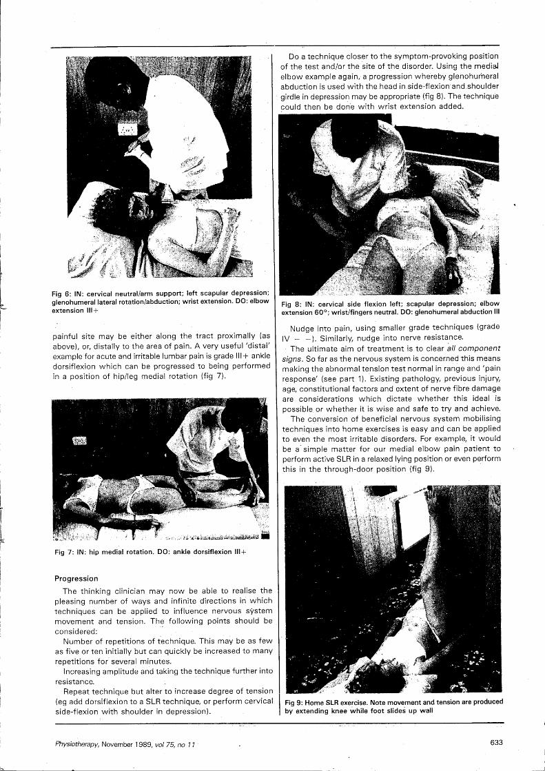
Fig 6: IN: cervical neutral/arm support; left scapular depression;glenohumeral lateral rotation/abduction; wrist extension. DO: elbowextension 111 +
painful site may be either along the tract proximally (asabove), or, distally to the area of pain. A very useful 'distal'example for acute and irritable lumbar pain is grade III + ankledorsiflexion which can be progressed to being performedin a position of hip/leg medial rotation (fig 7).
Fig 7: IN: hip medial rotation. DO: ankle dorsiflexion 111 +
Progression
The thinking clinician may now be able to realise thepleasing number of ways and infinite directions in whichtechniques can be applied to influence nervous systemmovement and tension. The following points should beconsidered:
Number of repetitions of technique. This may be as fewas five or ten initially but can quickly be increased to manyrepetitions for several minutes.
Increasing amplitude and taking the technique further intoresistance.
Repeat technique but alter to increase degree of tension(eg add dorsiflexion to a SLR technique, or perform cervicalside-flexion with shoulder in depression).
Do a technique closer to the symptom-provoking positionof the test and/or the site of the disorder. Using the mediaJelbow example again, a progression whereby glenohumeralabduction is used with the head in side-flexion'and-shouldergirdle in depression may be appropriate (fig 8). The techniquecould then be done with wrist extension added.
Fig 8: IN: cervical side flexion left; scapular depression; elbowextension 60°; wrist/fingers neutral. DO: glenohumeral abduction III
Nudge into pain, using smaller grade techniques (gradeIV - -). Similarly, nudge into nerve resistance.
The ultimate aim of treatment is to clear all componentsigns. So far as the nervous system is concerned this meansmaking the abnormal tension test normal in range and 'painresponse' (see part 1). Existing pathology, previous injury,age, constitutional factors and extent of nerve fibre damageare considerations which dictate whether this ideal ispossible or whether it is wise and safe to try and achieve.
The conversion of beneficial nervous system mobilisingtechniques into home exercises is easy and can be appliedto even the most irritable disorders. For example, it wouldbe a simple matter for our medial elbow pain patient toperform active SLR in a relaxed lying position or even performthis in the through-door position (fig 9).
Fig 9: Home SLR exercise. Note movement and tension are producedby extending knee while foot slides up wall
Physiotherapy, November 1989, vol 75, no 7 7 633

• Appropriate postural advice is an important consideration.The pathophysiological .and pathomechanical effects of
mobilising the nervous system in the irritable disorder canonly be assumed. Elvey (1987) has postulated a physiologicaleffect involving pressure changes within and around thenerve which may help to disperse unwanted inflammatoryby-products. Salter (1989) and others have shown that earlypassive mobilisation of injured joint tissue is beneficial forclearing oedema and haemarthrosis, promoting healing andpreventing adhesion formation. This is likely to-be significantin relation to the nervous system.
Treatment of the Won-irritable Disorder
The presumed underlying features of the non-irritabledisorder are that it is more of a pathomechanical nature thanpathophysiological. It is also likely to be longer standing.Beware of the acute disorder which appears to be non-irritable as strong techniques frequently aggravate.
It follows that the only way to tackle' a pathomechanicalproblem is to use techniques that address 'mechanics'. Bedrest, drug therapy and electrotherapy are unlikely to solvethe problem completely. Surgery may have to be resorted to.
The treatment rationale suggested here is really acombination of 'Maitland's' signs and symptoms approachcombined with thoughts of nerve mechanics and pathology(see part 1). It is important that the clinician has clear active,passive and subjective signs that can be monitored aftereach technique and between treatments. At this stage ofour knowledge every 'technique' should be regarded as ahypothesis which has to be proven. It is simple logic.
The following patient example will be used throughout thissection in order to illustrate the treatment approach:
A 33-year-old shop assistant has had a nagging kneeproblem present for eight months with no obvious reasonfor its onset. The pain is localised over the lateral aspect ofthe right knee and occasionally spreads down the lateral calfto the ankle. She is generally fit, but when questionedmentioned having been involved in a rear-end vehiclecollision as a teenager but with no serious or long lastingconsequences.
The pain is worse at the end of a working day (standing),and immediately provoked going up stairs and sitting withlegs up on a sofa and out to the left (fig 10). Her knee hasbeen treated extensively with electrotherapy and manualtherapy with only temporary improvement.
Fig 10: Patient sitting. Note lumbar left side flexion, right hipflexion/lateral rotation, right knee flexion, left footplantarflexion/inversion
Analysis ,
There narg.three different key aggravating^ factors. Twopositional (standing and sitting on the sofa) and. one active(stairs). It seems that standing takes a good while to provokethe pain;.it is therefore a useful indicator of 'subjective'progress from treatment session to treatment session. Goingup stairs makes a useful active re-test following'anytechnique.
Sitting,on the sofa is useful as it involves a combinationof positions of many joints and tissues and can be analysedas follows for nervous system tension components:
• Knee flexion (plus slight adduction?) = moves/tensionsfemoral nerve (prone knee bend — PKB), decreases .sciatictension.• Foot plantarflexion plus slight inversion=moves/tensionsperoneal nerve.• Hip slight flexion = moves/tensions sciatic nerve. Hip lateralrotation = decreases sciatic tension.• Spine flexion=tension increasing (part of slump). Sideflexion left=moves/tensions right nerve roots/nerves oflumbar and sacral plexus.
Examination
Other Components FoundKnee: lateral ligament and. head of fibula tender to
palpation. Strong adductjondn 10° flexion reproduced thepain. . -
Hip:V/Ankle: //Spine: Active tests normal. Palpation of L3, 4, 5 to postero-
anterior pressures were markedly tender and relativelyimmobile. T3-6 were also stiff.
Muscle: All relevant muscle tests
Nervous System Components FoundLoss of about 10° right SLR compared to left. AN
sensitising additions increased the existing end-rangediscomfort.
" PKB, like SLR, was tighter right than left.Standard slump: Neck plus spine flexion plus right knee
extension = -20° compared to left. Provokes pain over areaof .pain complained of but slightly different quality.
Left knee extension provokes posterior knee pain only(remember, think 'tension point'). The .addition of rightankle plantarflexion inversion increases existing discomfort.Release of neck flexion by only 5° or so abolishes all thepain and full knee extension is possible. Finally the additionof left lumbar side flexion in the full slump position intensifiedthe pain considerably and reproduced the exact pain.
So far the example underlines the need for detailedanalysis of subjective information, particularly keyaggravating postures/activities, and that deviations fromstandard tests are often necessary to make a tension testrelevant to the disorder. The response and range of standardtension tests compared to the known 'normal' (kneeextension -20°, pain in lower leg — slump) make useful re-assessments after a technique application. It is frequentlythe case that standard tension tests are unrevealing. Theclinician must improvise tests to fit the key features of thedisorder. Sometimes it is necessary to take tension Up fromthe 'other end' in order to hunt out a valid response. Forinstance in slump: Patient sits erect, head up — do ankle.
634 Physiotherapy,-November 1989, vol 75, no 7 7

dorsiflexion plus knee extension plus hip flexion plus spineflexion and finally neck flexion. Any component of a tensiontest can be added in any sequence for examination and/ortreatment purposes.
Starting Point Technique
In the passive movement assessment of abnormal jointand/or muscle, a close and directly proportional relationshipbetween resistance perceived and the onset and increaseof pain is frequently encountered. Final choice of techniquegrade is often made in terms of this relationship (seeMagarey, 198'5; Maitland, 1986). In nervous systemdisorders, abnormal resistance relevant to the disorder butwhich does not provoke the pain complained of isencountered, eg the SLR. of -10 ° or the PKB restriction inthe example.
Thus, the starting point technique is often into resistancebut short of provoking the pain. The ideal aim of treatmentis to try to establish a normal range of tension test movementcombined with a normal pain response.
A grade III mobilisation provides plenty of movement witha short or low dose of tension at the end of range for a givenperiod. Conversely, a grade IV, by definition, maintainstension at the end of range with very little movement. Farmore tension than movement is employed by a IV than agrade III for the same period.
In pathological terms (see table): through-range largeamplitude techniques (grade III) should be employed whereabnormalities of mechanical interface biomechanics of thenerve (extraneural) are thought to be responsible forsymptoms; and small-amplitude end-range techniques (IVs)where an intraneural disorder is thought likely. From the'signs and symptoms' viewpoint Ills are less provokingand IVs more provoking. Guide lines as stated by Maitland(1986) can be used.
Examples of mechanical interface pathology may befibrous tethering of a peripheral nerve in its bed. Oedemain the nerve bed in carpal-tunnel syndrome, or blood in thenerve bed following Colles fracture, are further exampleswhich could lead on to later fibrous tethering. Epineuralfibrosis or oedema that has gained access inside the
perineurium are considered examples of intraneuralpathology.
To go back to the example; our most likely first aim is totackle the abnormal SLR and to then progress to clearingall other components if necessary. As pain is reproducedduring tension testing by a manoeuvre providing moretension to the nervous system than movement (slump inleft side flexion), intraneural pathology is most suspect.But, as the symptoms are provoked by relatively out oftension postures (standing) and movements (stairs) anextraneural component more than likely needs addressingas well. The possibility of a nervous system insult in thespinal canal, following the road traffic accident which hasled to abnormal peripheral nerve mechanics in the region ofthe knee has to be considered as an underlying causalfeature.
The two areas may need attention for the best result. Itis possible that the clinician need go no further than usingtechniques that revolve around a standard SLR in order toclear this problem:
The most obvious starting point technique is to use theSLR employing tension development (IV), or if not toleratedfor long, a quickly-on quickly-off technique. Monitor allcomponent signs after each technique and the keyaggravating activity (stairs — do a step-up).
Strength and dose of technique in all situationsdepends on:
1. Nature of underlying pathology.
2. Whether or not improvements are being gained.
3. How much the patient can tolerate.
Always start with minimal force for a short period or fewrepetitions and progress from there. It is vftal that anydiscomfort or symptoms produced during a technique shouldsubside completely, immediately the technique is stopped.Neurological signs should be continually monitored.
ProgressionIt is worth exploring techniques using .SLR as a base in
order to demonstrate the many available avenues to take intackling the problem.
It appears that tension and movement are greatest at the
Fig 11: IN: hip flexion 90°. DO: kneeextension III. Movement is produced by theoperator side flexing his trunk
Fig 12: IN: cervical flexion, lumbar sideflexion left; hip flexion 90° with medialrotation and adduction. DO: Knee extension
Fig 13: IN: slump; cervical flexion, lumbarflexion and left side flexion; hip adductionand medial rotation; foot plantarflexioninversion. DO: knee extension IV+
Physiotherapy, November 1989r vol 75, no 11 635
component being moved (eg in the ULTT, if elbow extensionis the selected technique, more tension and movement willbe happening in the nerves around the elbow than at othersites). Hence, it could be said that as a progression thecomponent used should be as close to the origin ofsymptoms as possible. In our example, this could be the knee:
In hip flexion 90°, do knee extension (fig 11). Progress by:
• Add neck flexion to technique.• Add left side flexion• Add hip medial rotation.• Add hip adduction.• Add plantarflexion/inversion of the ankle (fig 12).
Further progression would be to use spinal flexion (ie slump)with the ultimate technique following this logic being shownin figure 13.
This 'nice' progression does not always provide the desiredresult. The following points should be considered:
Nervous system biomechanics dictate that abnormalitiesin one area of the nervous system will affect other areas.Sometimes it is necessary to clear one 'tension test' beforethe one being concentrated on can be cleared. Thus,attention to the abnormal PKB in the example, may benecessary before being able to achieve anything with theSLR or slump-based techniques. At other times, a slumpmay need to be treated before ULTT related disorders canbe helped.
Attention to abnormal mechanical interface tissues — anytissues or structures which are adjacent to nervous tissueand which exhibit abnormal signs often need treating. Goodexamples of muscle are the scalene! and piriformis. Spinal,rib or peripheral joints may need appropriate mobilisation.A knowledge of 'tension points' (see part 1) helps theclinician to examine for relevant abnormal signs at remotesites. In the example given relevant tension points andvulnerable mechanical interface sites are:
Superior tibio-fibular joint.Knee joint.L4 and joints above and below.Mid thoracic joints.The opposite SLR produced posterior knee pain whichis considered an abnormal 'tension point' response(Butler, 1988, 1989) and may need attention.
Abnormal joint/muscle components may require treatmentin positions of tension. In the example, knee adductionperformed in slump with knee extension is a possible andappropriate technique (fig 14). Local tender areas can be
Fig 14: IN: slump knee extension -20°; foot plantarflexion/inversion.DO: knee adduction IV+
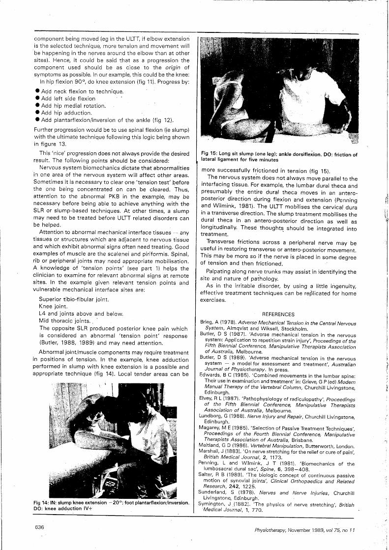
Fig 15: Long sit slump (one leg); ankle dorsiflexion. DO: friction oflateral ligament for five minutes
more successfully frictioned in tension (fig 15).The nervous system does not always move parallel to the
interfacing tissue. For example, the lumbar dural theca andpresumably the entire dural theca moves in an antero-posterior direction during flexion and extension (Penningand Wilmink, 1981). The ULTT mobilises the cervical durain a transverse direction. The slump treatment mobilises thedural theca in an antero-posterior direction as well aslongitudinally. These thoughts should be integrated intotreatment.
Transverse frictions across a peripheral nerve may beuseful in restoring transverse or antero-posterior movement.This may be more so if the nerve is placed in some degreeof tension and then frictioned.
Palpating along nerve trunks may assist in identifying thesite and nature of pathology.
As in the irritable disorder, by using a little ingenuity,effective treatment techniques can be replicated for homeexercises.
Brieg, A (1978). Adverse Mechanical Tension in the Central NervousSystem, Almqvist and Wiksell, Stockholm.
Butler, D S (1987). 'Adverse mechanical tension in the nervoussystem: Application to repetition strain injury', Proceedings of theFifth Biennial Conference, Manipulative Therapists Associationof Australia, Melbourne.
Butler, D S (1989). 'Adverse mechanical tension in the nervoussystem — a model for assessment and treatment', AustralianJournal of Physiotherapy, in press.
Edwards, B C (1985). 'Combined movements in the lumbar spine:Their use in examination and treatment' in: Grieve, G P (ed) ModernManual Therapy of the Vertebral Column, Churchill Livingstone,Edinburgh.
Elvey, R L (1987). 'Pathophysiology of radiculopathy', Proceedingsof the Fifth Biennial Conference, Manipulative TherapistsAssociation of Australia, Melbourne.
Lundborg, G (1988). Nerve Injury and Repair, Churchill Livingstone,Edinburgh.
Magarey, M E (1985). 'Selection of Passive Treatment Techniques',Proceedings of the Fourth Biennial Conference, ManipulativeTherapists Association of Australia, Brisbane.
Maitland, G D (1986). Vertebral Manipulation, Butterworth, London.Marshall, J (1883). 'On nerve stretching for the relief or cure of pain',
British Medical Journal, 2, 1173.Penning, L and Wilmink, J T (1981). 'Biomechanics of the
lumbosacral dural sac', Spine, 6, 398-408.Salter, R B (1989). The biologic concept of continuous passive
motion of synovial joints', Clinical Orthopaedics and RelatedResearch, 242, 1225.
Sunderland, S (1978). Nerves and Nerve Injuries, ChurchillLivingstone, Edinburgh.
Symington, J (1882). 'The physics of nerve stretching', British'Medical Journal, 1, 770.
636 Physiotherapy, November 1989, vol 75, no