Embed Size (px)
Citation preview
HER2 change between primary and metastatic breast cancer 1
HER2 PROTEIN AND GENE VARIATION BETWEEN PRIMARY AND METASTATIC
BREAST CANCER: SIGNIFICANCE AND IMPACT ON PATIENT CARE
Alessandra Fabi1*, Anna Di Benedetto2*, Giulio Metro1, Letizia Perracchio2, Cecilia Nisticò1,
Franco Di Filippo3, Cristiana Ercolani2, Gianluigi Ferretti1, Elisa Melucci2, Simonetta Buglioni2,
Isabella Sperduti4, Paola Papaldo1, Francesco Cognetti1 and Marcella Mottolese2
Medical Oncology1, Department of Pathology2, Department of Surgery3, Biostatistics4, Regina
Elena Cancer Institute, Rome, Italy.
Keywords: breast cancer, gene amplification, HER2, silver in situ hybridization (SISH), metastatic disease.
TRANSLATIONAL RELEVANCE
The introduction of trastuzumab, a monoclonal antibody against HER2, into metastatic and
(neo)adjuvant settings has completely changed the natural history of HER2-positive breast cancer
patients. Although HER2 is usually evaluated in primary tumor, knowledge of the HER2 status in
metastases may be of potential value for therapeutic decision making. In this study, the extent of
HER2 changes between primary and metastatic breast cancer was investigated by
immunohistochemistry and silver in situ hybridization. We show that HER2 status changes in 10%
of metastases and that the increase of HER2 gene copy number, together with chromosome 17
centromere gain, is a frequent event during progression. Our results were confirmed by Multiplex
Ligation-dependent Probe Amplification, a quantitative PCR-based test. Patients who changed
HER2 status from negative to positive presented longer time to progression when treated with
trastuzumab. According to our findings, HER2 measurement in metastatic lesions seems advisable,
especially in primary tumors with positive hormonal receptors.
* Alessandra Fabi and Anna Di Benedetto contributed equally to this work.
Corresponding Author:
Marcella Mottolese, PhD Pathology Department, Regina Elena Cancer Institute Via Elio Chianesi 53, 00144, Rome, Italy. Tel: +39-06-52666139 Fax: +39-06-52662920 e-mail: [email protected]
HER2 change between primary and metastatic breast cancer 2
ABSTRACT
Purpose: To analyze HER2 status in primary breast cancer (BC) compared with correspondent
metachronous metastases and to investigate whether BC phenotype may be predictive of change in
HER2 expression.
Experimental Design: HER2 was investigated by immunohistochemistry, silver (SISH) and
fluorescent in situ hybridization (FISH), in a series of 137 tumors, building up a tissue microarray
to concurrently analyze each single primary and metastatic BC on the same slide.
Results: HER2 status was discordant in 14 cases (10%): 12 negative in primary BC and positive in
metastases and 2 positive in primary BC and negative in metastases (p=0.04). These findings were
confirmed by a PCR based test termed Multiplex Ligation-dependent Probe Amplification (MLPA).
HER2 status changed in hormone receptor-positive BC more frequently than in negative ones (p=
0.002). In addition, we evaluated HER2 gene and chromosome 17 copy number by SISH in the 123
cases with unchanged HER2 status during progression. We found consistent HER2 gene copy
number stability in the 100 non-amplified cases. Conversely, of the 23 amplified primary BC, 13
(57%) demonstrated a significant increase in the HER2 gene and chromosome 17 copy number in
their paired metastases (p=0.01), as defined by SISH (k= 0.54, p <0.0001) and MLPA. Patients who
changed HER2 status from negative to positive, presented significant longer time to progression
when treated with trastuzumab compared to those who were untreated (p=0.04).
Conclusions: When feasible, HER2 reassessment in metastatic lesions should be carefully taken
into account, especially for metastases coming from primary hormone receptor-positive BC.
HER2 change between primary and metastatic breast cancer 3
INTRODUCTION
HER2 is one of the most important therapeutic targets in breast cancer (BC) and its overexpression,
in the majority of cases, is due to the amplification of the HER2 oncogene. The introduction of
trastuzumab (Herceptin®, Genentech, San Francisco, CA, USA), the humanized monoclonal
antibody (MoAb) against HER2 into the metastatic setting and, more recently, also in the
(neo)adjuvant setting (1, 2) has completely changed the natural history of HER2 positive BC
patients both in terms of time to recurrence and survival. Despite the benefits shown by
trastuzumab, a percentage of these patients have demonstrated clinical resistance. In metastatic
breast cancer (MBC), 44% to 64% of patients show upfront resistance to trastuzumab as a single-
agent therapy (2, 3), whereas 12% to 22% of patients are primarily resistant to trastuzumab when
given in combination with various cytotoxic drugs (2). Even though the efficacy of trastuzumab is
firstly dependent on the accuracy in assessing HER2 status, various mechanisms are also involved
in the resistance to the MoAb. Nagata et al. (4) identified PTEN as a key modulator of trastuzumab
sensitivity and Berns et al. demonstrated that the concomitant loss of PTEN and oncogenic mutation
in PIK3CA can significantly contribute to resistance mechanisms (5). Although HER2 is usually
evaluated in primary BC (PBC), knowledge of the HER2 status in metachronous metastatic
dissemination could be of potential value for therapeutic decision making. It has recently been
reported that HER2 status is mostly unchanged between primary tumors and their synchronous
lymph node metastases (6), but may be discordant in 6% to 48% of metachronous metastases (7-
14). This discordance may be due to the increasing level of genetic instability occurring throughout
disease progression that can significantly influence the alterations of the HER2 gene as well as
chromosome 17 (Chr17) (7). In particular, chromosomal rearrangements occurring during the
metastatization process may substantially determined the clinical management of MBC patients. In
fact, some recent studies (15-18) demonstrated that true Chr17 polysomy is a rare event in BC and
that an increase of centromere17 copy number is mostly related to gain or amplification of the
centromeric region (15-19). These findings provided evidence that correcting the HER2 gene copy
number with centromere17 enumeration probe (CEP17) might induce misleading results in HER2
amplification.
The primary aim of our study was to assess the extent of HER2 changes in a series of 137 PBC and
their correspondent metachronous metastases paired on the same tissue microarray (TMA). In our
series, we studied HER2 expression by immunohistochemistry (IHC) and gene amplification
together with CEP17 polisomy by silver in situ hybridization (SISH) in each single case. To verify
HER2 variation and Chr17 alterations during progression, we tested selected paired cases by the
Multiplex Ligation-dependent Probe Amplification (MLPA), a novel molecular assay which allows
HER2 change between primary and metastatic breast cancer 4
the concomitant analysis of a set of genes along Chr17. Furthermore, we investigated whether the
PBC phenotype could be predictive of change in HER2 during neoplastic progression, evaluating
the impact of trastuzumab treatment on the outcome of HER2 positive metastatic BC patients who
were previously diagnosed as a HER2 negative PBC.
MATERIALS AND METHODS
Case selection and tissue microarray construction
One hundred and thirty seven patients diagnosed with invasive BC between 1999 and 2007,
underwent biopsies to pathologically confirm the presence of a metastasis during follow up, were
selected from the surgical pathology files of the Regina Elena National Cancer Institute, Rome,
Italy. In all the 137 PBC, which were all trastuzumab untreated, the HER2 status had already been
assessed at the time of surgery. To concomitantly evaluate HER2 protein overexpression and/or
gene amplification in PBC and metachronous MBC, a TMA was constructed from the original
formalin fixed paraffin embedded (FFPE) blocks. To this end, two representative tumor areas were
carefully selected on routine haematoxylin/eosin-stained sections. Two core cylinders (1 mm
diameter) were taken from each PBC and MBC and deposited into two separate recipient paraffin
blocks using a specific arraying device (Alphelys, Euroclone, Milan, Italy). In cases where
informative results on TMA were absent due to missing tissue, no tumor tissue, or unsuccessful
staining or hybridization, we re-analyzed the correspondent routine tissue section. In addition to
tumor tissues, the recipient block also received normal breast tissue and cell line pellets as negative
and positive controls. Three-μ sections of the resulting microarray block were made and used for
IHC or gene amplification analysis after transferring them to SuperFrost Plus slides (Menzel-
Gläser, Braunschweig, Germany).
Immunohistochemistry
HER2 immunostaining on TMA was performed by using the polyclonal antibody A0485 (Dako,
Milan, Italy) whereas estrogen (ER) and progesterone (PgR) receptors were analyzed by using the
MoAbs 6F11 and 1A6, respectively (Novocastra, Menarini, Florence, Italy). Immunoreactions were
revealed by a streptavidin-biotin enhanced immunoperoxidase technique (Super Sensitive
MultiLink, Menarini) in an automated autostainer. Diaminobenzidine was used as chromogenic
substrate.
TMA immunostaining was evaluated by two expert pathologists (LP, MM). Discordant cases were
independently reviewed by another pathologist who was blinded to the previous results.
Silver in situ hybridization and fluorescent in situ hybridization
HER2 change between primary and metastatic breast cancer 5
To assess HER2 gene and Chr17 polisomy on TMA we used a fully automated single color in situ
hybridization assay based on the use of a validated silver deposition technology (SISH, Inform
HER2 DNA Probe; Inform Chr17 probe, Ventana, Roche Diagnostic, Milan, Italy) to detect HER2
gene and Chr17 status (20). The silver precipitation was visualized as a black dot in cell nuclei.
Fluorescent in situ hybridization (FISH) (pharmDX, Dako, Milan, Italy) was performed using a
HER2 DNA probe directly labeled with Texas Red fluorochrome targeting the HER2 amplicon (red
signals) and a CEN-17 PNA probe directly labeled with fluorescein (FITC) targeting the
centromeric region of the chromosome (green signals). The assay was performed according to the
manufacturer’s instructions.
The 100x oil immersion objective was used to score signals in all the neoplastic cells present in
each duplicate TMA cores both for SISH and FISH.
SISH results were analyzed by using a light microscope (Nikon, Eclipse 55i) equipped with a
software able to capture images (Eureka Interface System, Menarini, Firenze, Italy) and the FISH
results were assessed with an epi-fluorescence microscope (Zeiss, Axioscope 40) equipped with
Image Processing analysis software (Media Cybernetics) able to DAPI/specific Texas Red and
FITC single filters.
Multiplex Ligation-dependent Probe Amplification
50–100 μl of the genomic DNA solution, extracted from two whole 4 m� paraffin BC sections
using the QIAamp Mini kit (Qiagen, Medicalproducts, Rome Italy), was used in the MLPA analysis
following the manufacturers’ instructions. The kit (P004-B1 kit , MRC Holland, Resnova, Italy).
contains 3 probes for the HER2 gene, and 21 probes for other genes on Chr17 and 6 control probes
located on other chromosomes. All tests were performed in duplicate in an ABI 9700 PCR machine.
PCR products were analyzed on an ABI3130 capillary sequencer (Applied Biosystems, Monza,
Italy). Gene copy numbers were analyzed using Genemapper 4.0 and Coffalyser (version 7.0)
software. For genes with more than one probe present in the kit, the mean of all the probe peaks of
the gene was calculated in duplicate. A mean value below 1.5 was defined as normal, between 1.5–
2.0 as low level amplification (LA) and a value >2.0 as high level amplification (HA), according to
the definitions in the Coffalyser software (21).
Scoring criteria
Immunohistochemistry
HER2 IHC positivity was determined according to ASCO-CAP guidelines (22) and was scored as
follows: 0 and 1+ negative, 2+ equivocal and 3+ positive. ER and PgR were considered positive
when >10% of the neoplastic cells showed distinct nuclear immunoreactivity.
HER2 change between primary and metastatic breast cancer 6
SISH and FISH
Following the manufacturer’s guidelines, scoring of SISH results was carried out assuming that a
single signal was counted as 1 gene copy, a small cluster as 8 gene copies, a large cluster as 16 gene
copies. According to the ASCO-CAP guidelines (22), PBC and MBC were defined as “non-
amplified” (NA) by SISH when a HER2 gene copy number <4 was observed and by FISH when a
HER2/CEP17 ratio <1.8 was detected. Cases were defined as “amplified” (A) when SISH displayed
a gene copy number >6 or when the FISH ratio was >2.2. Polysomy 17 - intended as an increased
CEP17 copy number (CEP17CN) – is considered to be present in BC when a mean number of �3
signals is shown.
For the purpose of our study, we defined “low amplification” by SISH when BC presented >6
signals/nucleus, “moderate amplification” when BC presented >10 signals/nucleus and “high
amplification” when BC presented >20 signals/nucleus. Furthermore, “low polysomy” by SISH
was a CEP17CN �3 and “high polysomy” a CEP17CN >4. Lymphocytes and normal breast
glandular epithelial cells served as an internal control.
Statistical analysis
Descriptive statistics were used to describe the patient’s characteristics. The proportions are
presented as numbers and percentages. For the statistical analysis, HER2 negative cases are defined
those with an IHC score of 0, 1+ and 2+ lacking gene amplification and positive cases those with
IHC score 3+ and 2+ displaying gene amplification. The Mc Nemar paired test was performed to
evaluate statistical significant differences in HER2 status between PBC and MBC. The rate of
concordance between HER2 and CEP17 copy gains in primary and metastatic BC was analyzed
with the k test. Significance was assessed at a level of 5%. The statistical software package used for
this analysis was SPSS for Windows version 17.0 (Inc, Chicago, IL, USA).
RESULTS
Patient characteristics
One hundred and fourteen out of the 137 PBC included in the analysis were infiltrating ductal
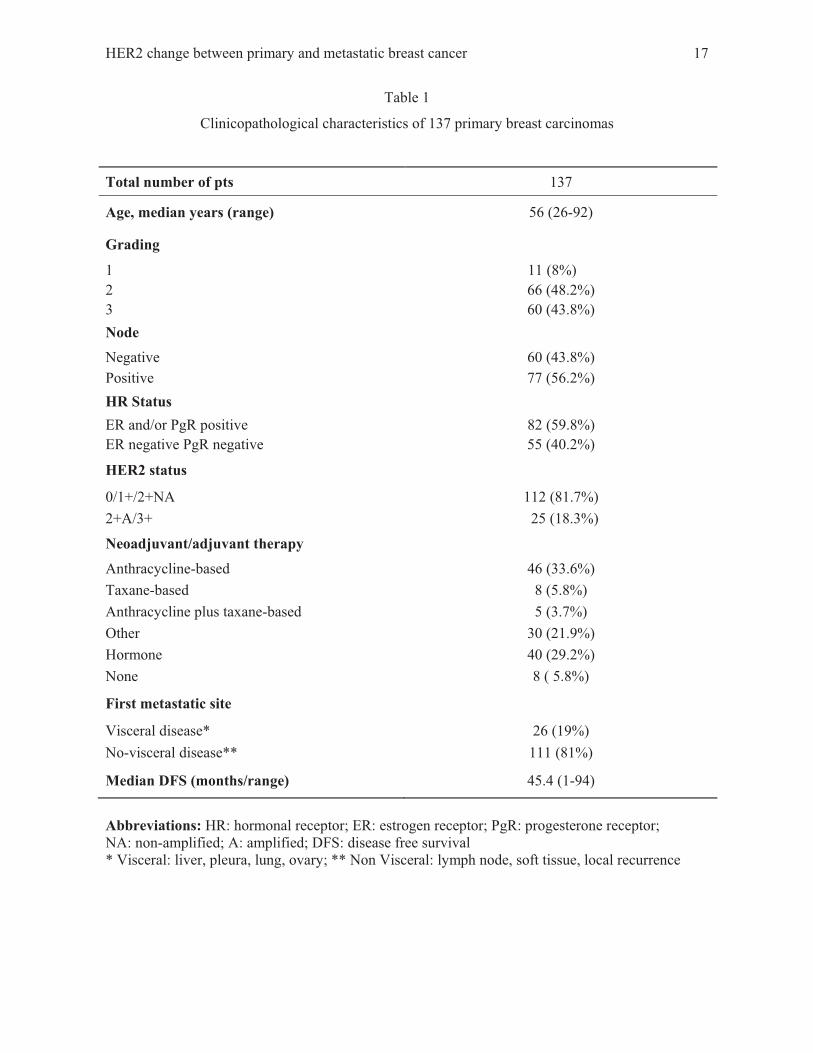
carcinomas, 14 invasive lobular carcinomas and 9 other histotypes. As summarized in Table 1, 11
(8%) tumors were graded, using the Bloom and Richardson scoring system, as well differentiated
(G1), 66 (48.2%) and 60 (43.8%) as moderately (G2) and poorly differentiated (G3) carcinomas,
respectively. Furthermore, 60 (43.8%) patients were node negative and 77 (56.2%) node positive.
ER and PgR were positive in 82 (59.8%) BC and HER2 was positive in 25 (18.3%) cases.
Staging was performed by following the Unione Internationale Contre le Cancer tumor-node-
metastasis (TNM) system criteria (23).
HER2 change between primary and metastatic breast cancer 7
In our series, 80 (58%) patients were administered (neo)adjuvant chemotherapy. In particular, 46
patients were given an anthracycline–based therapy, 8 and 5 taxane or anthracycline plus taxane
regimens respectively, 30 other chemotherapies and 40 hormone therapy alone. Only 8 women did
not undergo any treatment. None of the HER2 positive patients received anti HER2 therapy alone or
in combination as (neo)adjuvant treatment. Twenty-six (19%) patients developed visceral
metastases (11 in the liver, 6 in the pleura, 8 in the lung and 1 in the ovary) and 111 (81%) non-
visceral metastases (25 in the lymph nodes, 26 in the soft tissue and 60 experienced a local
recurrence). The median interval between the PBC and the first recurrence of the disease (disease
free progression) was 45.4 months (range 1-94 months).
The study was reviewed and approved by the Local Ethical Committee at the Regina Elena National
Cancer Institute, and a written informed consent was obtained from all patients.
HER2 status in primary and paired metastatic breast carcinomas by SISH and MLPA
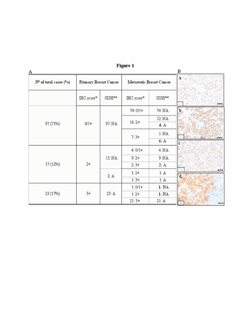
Table in Figure 1A summarizes IHC and SISH results obtained in the 137 PBC and MBC paired on
the same TMA. HER2 immunoreactivity was scored as follows: 97 cases (71%) as 0/1+, 17 (12 %)
as 2+ and 23 (17 %) as 3+. The 97 0/1+ score PBC were also non-amplified by SISH. Of the 17
scoring 2+ PBC, only 2 (12%) were amplified. All the 23 cases with a score of 3+ resulted
amplified.
When we concurrently analyze each single primary tumor in comparison with the metachronous
metastases, we found that of the 97 HER2 negative PBC (score 0/1+ by IHC, NA by SISH), 74
(76%) maintained a concordant score in the matched metastases, whereas 23 (16+7, 24%) displayed
an increased IHC score (Figure 1B, panel a-b). Among the 17 PBC scoring 2+ by IHC, in 4 cases
(23%) there was a decrease of the IHC score (0/1+) during progression, in 10 cases (59%) the same
score was maintained and in 3 cases (18%) an increase of the IHC score (3+), associated with gene
amplification, was registered (Figure 1B, panel c-d). Two (8.6%) of the 23 PBC scoring 3+ by IHC
and amplified by SISH, showed a decrease in the score (0/1+) and a change in the HER2 gene CN
in the correspondent metastasis.
Overall, a significant change of HER2 immunoreactivity and gene amplification in metachronous
metastases was observed as compared with the primary tumors (Mc Nemar test, IHC: p<0.0001,
SISH: p= 0.01). Among the 14 patients who changed HER2 status, 7 (50%) and 4 (28.5%) had
received anthracycline or no anthracycline-based therapy in (neo)adjuvant setting, respectively.
We summarized our findings in a flow chart by taking into account both the IHC and SISH
findings. Of the 137 PBC, 123 (90%) maintained a concordant HER2 status during disease
progression whereas 14 cases (10%) changed HER2 status (12 from negative to positive and 2 from
positive to negative, Figure 2A, grey boxes; Figure 2B, panel a-b). These findings were further
HER2 change between primary and metastatic breast cancer 8
confirmed by MLPA assay (Supplementary Table S1, panel a) in 7 out of 8 cases (88%; cases 1-
6,8,13 illustrated in Table 2) that had previously been evaluated by SISH. In Figure 2C a descriptive
case clearly highlighted that HER2 gene, as determined by MLPA, was non-amplified in the PBC
(ratio 1.36, panel a) and highly amplified in the paired metastasis (ratio 8.02, panel b).
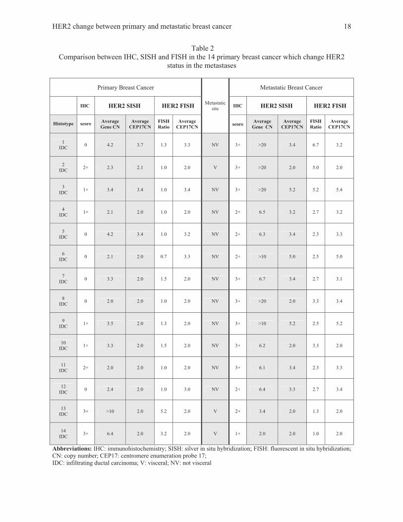
Comparison between IHC, SISH, and FISH in the 14 primary breast cancer changing HER2
status during breast cancer progression
We correlated HER2 protein overexpression with gene amplification detected by SISH and FISH in
14 cases all presenting a variation in HER2 status between primary and metastatic BC. As
summarized in Table 2, the agreement between IHC and HER2 gene amplification, evaluated either
by SISH or FISH, was 100% in all the 12 cases displaying an increase and in the 2 cases displaying
a decrease of HER2 overexpression during disease progression. In regards to CEP17CN, we found
that 2 non-amplified PBC (cases n. 6 and n. 12) were disomic by SISH and low polysomic by FISH.
The latter 2 cases were amplified and polysomic in the autologous MBC with both SISH and FISH.
Furthermore, only 1 case (case n. 8) was found disomic by SISH and polysomic by FISH in MBC.
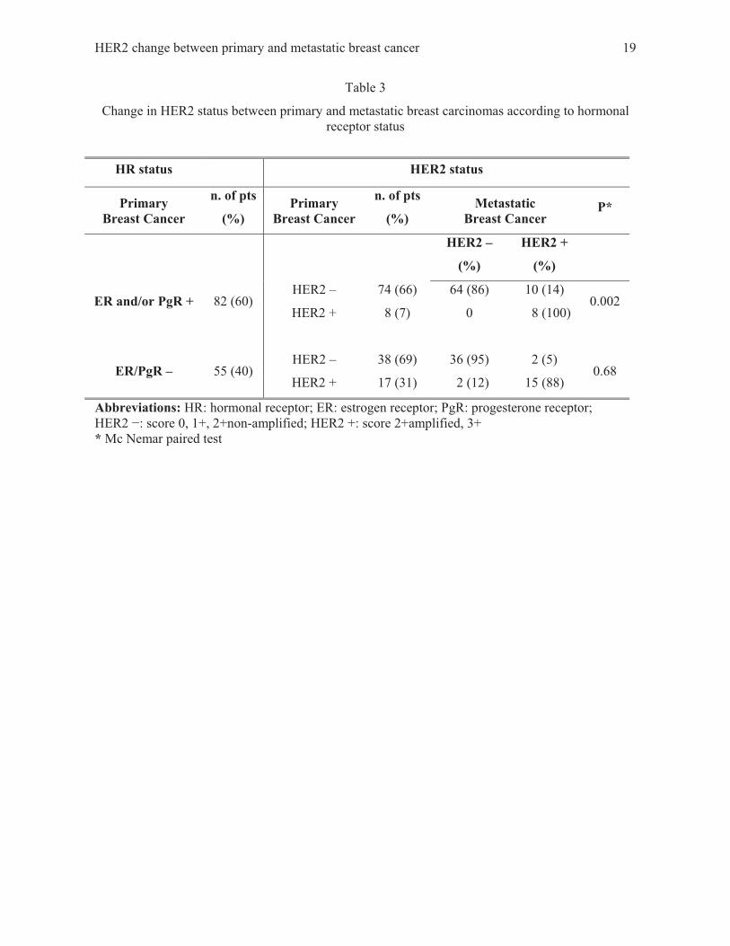
HER2 change according to biological and clinical features
As summarized in Table 3, in the group of 82 HR positive PBC, 74 (66%) were HER2 negative and
8 (7%) HER2 positive. Among the 74 HR positive/HER2 negative PBC, 10 (13.5%) changed HER2
status from negative to positive, whereas none of the 8 (100%) HR positive/HER2 positive patients
showed HER2 change (p=0.002). No statistically significant differences were seen in the group of
patients with negative HR.
HER2 variation was not significantly related to tumor size (p=0.11), node status (p=0.48), grading
(p=0.41), site of metastasis (p=0.41), previous anthracycline- and/or taxane-based adjuvant therapy
(0.12) and disease free progression (0.14) (data not shown).
HER2 gene and chromosome 17 copy gains in primary and metastatic breast cancer with
unchanged HER2 status
We analyzed HER2 gene CN and CEP17CN by SISH in the 123 PBC that maintained a concordant
HER2 status in the paired metastases.
The 100 non-amplified PBC (≤6 signals/nucleus) maintained the same HER2 gene CN in their
paired metastases (Figure 3A), whereas 13 of 23 (57%) cases, amplified both in primary and in
metastatic BC, showed an increased HER2 gene CN in the paired metastases (Figure 3C, panel a-b).
Concomitantly (Figure 3B), an increased CEP17CN was also detected in MBC. In detail, of 108
disomic PBC, 8 became polysomic in the autologous metastases (5 low polysomic and 3 high
polysomic; Figure 3C, panel c-d). Moreover, of 7 low polysomic PBC, 6 (86%) displayed high
HER2 change between primary and metastatic breast cancer 9
polysomy in their paired metastases. The k test indicated a significant concordance between HER2
gene CN and CEP17CN gain during BC progression (k=0.54, p<0.0001).
These results were subsequently supported by MLPA assay (Supplementary Table S1, panel b) in
representative 4 cases that displayed concordant HER2 status in primary and metastatic BC. Figure
3D, panel a-b shows an representative case: HER2 ratio by MLPA in PBC was 3.72 and in MBC
10.02. Moreover, WSB1 and NOS2A ratio in PBC was 1.45 and 1.25 and 3.13 and 2.39 in MBC
respectively.
Clinical outcome of HER2 positive metastatic breast cancer patients
In our series, of 18 patients who underwent trastuzumab based therapy at the appearance of the first
progression, 12 were HER2 positive both in PBC and in MBC (HER2 +/+) and 6 changed HER2
status (HER2 �/+) in the metastasis. These 18 patients had a median time to progression (TTP) of
10.3 months whereas the TTP of the remaining 17 HER2 positive BC patients (11 HER2 +/+, 6
HER2 �/+) not administered the monoclonal antibody therapy, was 5.2 months (p=0.04.
Supplementary Figure S1). In detail, in the 12 trastuzumab treated patients with HER2 +/+, the TTP
was 11 months and in the 6 patients with HER2 �/+ 8 months. Furthermore, in the 11 trastuzumab
untreated patients with HER2 +/+ the TTP was 2 months and in the 6 with HER2 �/+ was 5 months.
DISCUSSION
HER2 overexpression/amplification in BC is of particular clinical relevance when selecting patients
eligible for anti HER2 based therapy. In a metastatic setting, the evaluation of HER2 status is
mostly performed on the primary tumor based on the notion that the HER2 status does not undergo
significant change during disease progression (24, 25).
In the last few years, several studies delved deeper into the matter by reporting a significant
discordance between PBC and paired asynchronous metastases ranging between 6% to 48% (7-14).
Due to this wide variability of results, the present study analyzed HER2 status in 137 PBC and
autologous metachronous metastases from trastuzumab-näive patients using IHC and SISH on
paired TMA. Additionally, we evaluated the amplification of HER2 gene and of genes
(WSB1/NOS2A) located very close to the centromeric region by the means of MLPA in a selected
group of primary and metastatic BC. To our knowledge, this is the first study where HER2 variation
was investigated, case-by-case, in a large series of trastuzumab untreated 137 patients. We aimed to
concurrently analyze primary and metastatic lesions, paired on the same TMA, by IHC and SISH
(26). Since one the major limit of TMA is the reduced amount of tissue analyzed which may be not
representative of the phenotypic and genotypic patterns of the tumor, we supported the
morphological-based assays using MLPA, a molecular technique (16) able to determine relative
HER2 change between primary and metastatic breast cancer 10
gene copy numbers in a quantitative way. Our results showed that HER2 status significantly
changed in 10% of cases. In particular, 11% of the HER2 negative PBC expressed HER2 in their
metastatic sites while 8% of HER2 positive PBC became negative in their paired asynchronous
metastases. In addition, in the group of the 14 cases undergoing HER2 variation, the SISH results
were further confirmed by FISH and, in a subset of available cases, by MLPA. Interestingly, we
found that HER2 status changed more frequently in HR positive PBC patients than in the negative
counterpart. These findings might reflect acquired resistance to tamoxifen treatment in the adjuvant
setting of HR positive BC patients. It has been recently reported that acquired endocrine resistance
in positive ER/negative HER2 BC may be associated with an adaptive increase in HER2, although
exactly how aberrant HER2 signalling affects the ER� pathway is poorly understood (27).
The discordance between PBC and MBC reported in our series resembles other retrospective and
prospective studies (9, 12, 14, 28). Several authors demonstrated primary intratumoral
heterogeneity for both HER2 overexpression and gene amplification (10, 11, 29). This
heterogeneity may arise via random genetic alterations with clonal progression, likely resulting in
genetic subclones of cells within the PBC. Consequently, one may hypothesize that metastatic cells
enhanced HER2 alterations in MBC as compared with the PBC.
In the study by Lower (10), which included 382 BC and is the largest report up to now, 23.6% of
cases, evaluated by IHC, changed from HER2 positive to HER2 negative and only 9.6% from
negative to positive. The authors suggested that the decrease in immunoreactivity may be a possible
misclassification of the IHC 2+ score patients not confirmed by FISH. In addition, Lorincz et al
(30) found that half of PBC with HER2 amplification lost this genotype in the correspondent bone
metastases. Furthermore, even though HER2 amplification was retained in the MBC, the copy
number decreased compared to the primary tumor. These findings may be explained by technical
limitations of the FISH analysis known not to be consistently successful on decalcified bone
metastasis due to DNA breakdown (11). For this reason, we excluded bone metastases from the
IHC and SISH analyses in our study. In addition, some of the causes of the discordant results in the
above cited studies may be due to heterogeneity in case selection, tissue sampling and processing
procedures. Furthermore, unlike our study, the other reports rarely provided case-by-case results.
Some authors reported that the specimens were handled as part of routine clinical care and were not
re-stained, but rather re-read by at least two pathologists according to the initial procedures (either
IHC or FISH). Retesting was done in cases considered unsuitable for re-evaluation (9, 10, 30). In
other studies HER2 status was assayed on histological samples in PBC and on cytological samples
in MBC (13, 28, 29). Zidan et al and Gancberg et al (8, 14) retested their series of paired PBC and
HER2 change between primary and metastatic breast cancer 11
MBC through the use of IHC whereas FISH was performed exclusively on cases presenting a 2+/3+
score by IHC.
Only Regitning et al (11) have analyzed, through the use of IHC and FISH, a case-by-case TMA
which included PBC and paired MBC similar to our study. The authors demonstrated that, even in a
very small group of 31 cases, HER2 IHC expression changed at a high percentage rate (48.4%) in
distant metastases. Nevertheless, HER2 immunoreactivity is attributable to gene amplification in
only 14.3% of the MBC tested. In our series HER2 protein expression increased in 26 (23%) MBC
as compared with 114 HER2 negative PBC whereas gene amplification occurred only in 11% of
metastases. All the authors agreed on the concept that the HER2 status may be different in the
metastasis in comparison with the primary tumor and stressed the need to verify these results with a
larger number of patients in order to apply these findings to clinical practice.
Unlike other authors, we took our analyses one step further by evaluating HER2 gene and Chr17
status by SISH in the 123 cases showing a concordant HER2 status (100 NA and 23 A). We
demonstrated that MBC derived from the 100 PBC with a gene CN ranging between 2 to 6 is
consistent with HER2 gene stability whereas MBC derived from the 23 amplified PBC (gene CN
>6) had a significant increase in HER2 gene CN as well in CEP17CN during metastatization. These
findings were further supported and quantitatively confirmed by MLPA analysis in a small group of
unchanged 4 primary and metastatic paired cases. An increase in CEP17CN, detected by fluorescent
or chromogenic in situ hybridization, raises the question whether it could reflect true “polysomy”
17 or rather is related to unbalanced chromosomal rearrangements. Recent studies analyzed HER2
status by comparative genomic hybridization (CGH) or MLPA methods, both in BC diagnosed as
polysomic by routine FISH (15, 17) and in randomly selected BC (16, 18). These authors reported
that true Chr17 polysomy is a very rare event in BC and that CEP17CN >3, detected by FISH or
CISH assay, is most often related to gain or amplification of the centromeric region. So far, non-
amplified polysomic BC, presenting a HER2:CEP17 ratio <2 by in situ hybridization, are not
eligible for trastuzumab therapy. Conversely, based on CGH or MLPA data, we may have
misinterpreted HER2 amplification. As previously discussed, in the group of our series of BC
patients with unchanged HER2 status we found a 11.4% increase in CEP17CN during
metastatization. Some of these cases were considered amplified, but about 5% displayed a high
CEP17 polisomy and were thus considered non-amplified. Since abnormal CEP17CN might arise
from high level gains or amplification of CEP17, correcting CEP17 probes may provide misleading
HER2 status assessment lowering the number of cases in which a change in HER2 status may occur
during metastatization.
HER2 change between primary and metastatic breast cancer 12
Focusing on the outcome of HER2 positive MBC patients both in changing (negative/positive) and
non-changing tumors (positive/positive), we observed significant longer time to progression (10
months vs 4 months) in patients treated with trastuzumab compared with those who were not treated
with the MoAb. Despite the limited number of cases, these data not only underline the importance
of testing HER2 status in metastases, possibly using alternative molecular techniques, but also open
up the possibility of significantly improving the prognosis of these subsets of patients. In the era of
targeted therapy, an accurate definition of the metastatic disease in patients who can experience
great benefit by trastuzumab or any novel anti HER2 molecule, represents a pivotal commitment in
the clinical management of BC patients.
HER2 change between primary and metastatic breast cancer 13
Reference List
1. Madarnas Y, Trudeau M, Franek JA, et al. Adjuvant/neoadjuvant trastuzumab therapy in women with HER-2/neu-overexpressing breast cancer: a systematic review. Cancer Treat Rev 2008;34:539-57.
2. Metro G, Mottolese M, Fabi A. HER-2-positive metastatic breast cancer: trastuzumab and beyond. Expert Opin Pharmacother 2008;9:2583-601.
3. Baselga J, Carbonell X, Castaneda-Soto NJ, et al. Phase II study of efficacy, safety, and pharmacokinetics of trastuzumab monotherapy administered on a 3-weekly schedule. J Clin Oncol 2005;23:2162-71.
4. Nagata Y, Lan KH, Zhou X, et al. PTEN activation contributes to tumor inhibition by trastuzumab, and loss of PTEN predicts trastuzumab resistance in patients. Cancer Cell 2004;6:117-27.
5. Berns K, Horlings HM, Hennessy BT, et al. A functional genetic approach identifies the PI3K pathway as a major determinant of trastuzumab resistance in breast cancer. Cancer Cell 2007;12:395-402.
6. Carlsson J, Nordgren H, Sjostrom J, et al. HER2 expression in breast cancer primary tumours and corresponding metastases. Original data and literature review. Br J Cancer 2004;90:2344-8.
7. Edgerton SM, Moore D, Merkel D, et al. erbB-2 (HER-2) and breast cancer progression. Appl Immunohistochem Mol Morphol 2003;11:214-21.
8. Gancberg D, di LA, Cardoso F, et al. Comparison of HER-2 status between primary breast cancer and corresponding distant metastatic sites. Ann Oncol 2002;13:1036-43.
9. Guarneri V, Giovannelli S, Ficarra G, et al. Comparison of HER-2 and hormone receptor expression in primary breast cancers and asynchronous paired metastases: impact on patient management. Oncologist 2008;13:838-44.
10. Lower EE, Glass E, Blau R, et al. HER-2/neu expression in primary and metastatic breast cancer. Breast Cancer Res Treat 2009;113:301-6.
11. Regitnig P, Schippinger W, Lindbauer M, et al. Change of HER-2/neu status in a subset of distant metastases from breast carcinomas. J Pathol 2004;203:918-26.
12. Santinelli A, Pisa E, Stramazzotti D, et al. HER-2 status discrepancy between primary breast cancer and metastatic sites. Impact on target therapy. Int J Cancer 2008;122:999-1004.
13. Tapia C, Savic S, Wagner U, et al. HER2 gene status in primary breast cancers and matched distant metastases. Breast Cancer Res 2007;9:R31.
14. Zidan J, Dashkovsky I, Stayerman C, et al. Comparison of HER-2 overexpression in primary breast cancer and metastatic sites and its effect on biological targeting therapy of metastatic disease. Br J Cancer 2005;93:552-6.
HER2 change between primary and metastatic breast cancer 14
15. Marchio C, Lambros MB, Gugliotta P, et al. Does chromosome 17 centromere copy number predict polysomy in breast cancer? A fluorescence in situ hybridization and microarray-based CGH analysis. J Pathol 2009;219:16-24.
16. Moelans CB, de Weger RA, van Diest PJ. Absence of chromosome 17 polysomy in breast cancer: analysis by CEP17 chromogenic in situ hybridization and multiplex ligation-dependent probe amplification. Breast Cancer Res Treat 2010;120:1-7.
17. Troxell ML, Bangs CD, Lawce HJ, et al. Evaluation of Her-2/neu status in carcinomas with amplified chromosome 17 centromere locus. Am J Clin Pathol 2006;126:709-16.
18. Yeh IT, Martin MA, Robetorye RS, et al. Clinical validation of an array CGH test for HER2 status in breast cancer reveals that polysomy 17 is a rare event. Mod Pathol 2009;22:1169-75.
19. Viale G. Be precise! The need to consider the mechanisms for CEP17 copy number changes in breast cancer. J Pathol 2009;219:1-2.
20. Dietel M, Ellis IO, Hofler H, et al. Comparison of automated silver enhanced in situ hybridisation (SISH) and fluorescence ISH (FISH) for the validation of HER2 gene status in breast carcinoma according to the guidelines of the American Society of Clinical Oncology and the College of American Pathologists. Virchows Arch 2007;451:19-25.
21. Coffa J, van de Wiel MA, Diosdado B, et al. MLPAnalyzer: data analysis tool for reliable automated normalization of MLPA fragment data. Cell Oncol 2008;30:323-35.
22. Wolff AC, Hammond ME, Schwartz JN, et al. American Society of Clinical Oncology/College of American Pathologists guideline recommendations for human epidermal growth factor receptor 2 testing in breast cancer. Arch Pathol Lab Med 2007;131:18-43.
23. Tavassoli F, Devilee P. Pathology and genetics. Tumours of the breast and female genital organs. Lyon (France): IARC Press., ed. Tumors of the breast.1.: 9-112.2003.
24. Niehans GA, Singleton TP, Dykoski D, et al. Stability of HER-2/neu expression over time and at multiple metastatic sites. J Natl Cancer Inst 1993;85:1230-5.
25. Vincent-Salomon A, Jouve M, Genin P, et al. HER2 status in patients with breast carcinoma is not modified selectively by preoperative chemotherapy and is stable during the metastatic process. Cancer 2002;94:2169-73.
26. Carbone A, Botti G, Gloghini A, et al. Delineation of HER2 gene status in breast carcinoma by silver in situ hybridization is reproducible among laboratories and pathologists. J Mol Diagn 2008;10:527-36.
27. Leary AF, Drury S, Detre S, et al. Lapatinib restores hormone sensitivity with differential effects on estrogen receptor signaling in cell models of human epidermal growth factor receptor 2-negative breast cancer with acquired endocrine resistance. Clin Cancer Res 2010;16:1486-97.
28. Simmons C, Miller N, Geddie W, et al. Does confirmatory tumor biopsy alter the management of breast cancer patients with distant metastases? Ann Oncol 2009;20:1499-504.
29. Gong Y, Booser DJ, Sneige N. Comparison of HER-2 status determined by fluorescence in situ hybridization in primary and metastatic breast carcinoma. Cancer 2005;103:1763-9.

HER2 change between primary and metastatic breast cancer 15
30. Lorincz T, Toth J, Badalian G, et al. HER-2/neu genotype of breast cancer may change in bone metastasis. Pathol Oncol Res 2006;12:149-52.

HER2 change between primary and metastatic breast cancer 16
Competing interests
The authors declare that they have no competing interests.
Acknowledgements
This work was supported by AIRC (Italian Association for Cancer Research, project n. 8706),
Italian Ministry of Health (Alliance against Cancer – Project RIBBO). We would like to thank
Salvatore Conti for his precious advice in evaluating MLPA results, Maria Assunta Fonsi for her
secretarial assistance and Tania Merlino for the English language editing in the manuscript.
HER2 change between primary and metastatic breast cancer 17
Table 1
Clinicopathological characteristics of 137 primary breast carcinomas
Total number of pts 137
Age, median years (range) 56 (26-92)
Grading
1 11 (8%) 2 66 (48.2%) 3 60 (43.8%) Node
Negative 60 (43.8%) Positive 77 (56.2%) HR Status ER and/or PgR positive 82 (59.8%) ER negative PgR negative 55 (40.2%)
HER2 status
0/1+/2+NA 112 (81.7%) 2+A/3+ 25 (18.3%)
Neoadjuvant/adjuvant therapy
Anthracycline-based 46 (33.6%) Taxane-based 8 (5.8%) Anthracycline plus taxane-based 5 (3.7%) Other 30 (21.9%) Hormone 40 (29.2%) None 8 ( 5.8%)
First metastatic site
Visceral disease* 26 (19%) No-visceral disease** 111 (81%)
Median DFS (months/range) 45.4 (1-94)
Abbreviations: HR: hormonal receptor; ER: estrogen receptor; PgR: progesterone receptor; NA: non-amplified; A: amplified; DFS: disease free survival * Visceral: liver, pleura, lung, ovary; ** Non Visceral: lymph node, soft tissue, local recurrence
HER2 change between primary and metastatic breast cancer 18
Table 2 Comparison between IHC, SISH and FISH in the 14 primary breast cancer which change HER2
status in the metastases
Primary Breast Cancer
Metastatic site
Metastatic Breast Cancer
IHC HER2 SISH HER2 FISH IHC HER2 SISH HER2 FISH Histotype
score Average
Gene CN Average
CEP17CN FISH Ratio
Average CEP17CN score Average
Gene CN Average
CEP17CN FISH Ratio
Average CEP17CN
1 IDC 0 4.2 3.7 1.3 3.3 NV 3+ >20 3.4 6.7 3.2
2 IDC 2+ 2.3 2.1 1.0 2.0 V 3+ >20 2.0 5.0 2.0
3 IDC 1+ 3.4 3.4 1.0 3.4 NV 3+ >20 5.2 5.2 5.4
4 IDC 1+ 2.1 2.0 1.0 2.0 NV 2+ 6.5 3.2 2.7 3.2
5 IDC 0 4.2 3.4 1.0 3.2 NV 2+ 6.3 3.4 2.3 3.3
6 IDC 0 2.1 2.0 0.7 3.3 NV 2+ >10 5.0 2.5 5.0
7 IDC 0 3.3 2.0 1.5 2.0 NV 3+ 6.7 3.4 2.7 3.1
8 IDC 0 2.0 2.0 1.0 2.0 NV 3+ >20 2.0 3.3 3.4
9 IDC 1+ 3.5 2.0 1.3 2.0 NV 3+ >10 5.2 2.5 5.2
10 IDC 1+ 3.3 2.0 1.5 2.0 NV 3+ 6.2 2.0 3.3 2.0
11 IDC 2+ 2.0 2.0 1.0 2.0 NV 3+ 6.1 3.4 2.3 3.3
12 IDC 0 2.4 2.0 1.0 3.0 NV 2+ 6.4 3.3 2.7 3.4
13 IDC 3+ >10 2.0 5.2 2.0 V 2+ 3.4 2.0 1.3 2.0
14 IDC 3+ 6.4 2.0 3.2 2.0 V 1+ 2.0 2.0 1.0 2.0
Abbreviations: IHC: immunohistochemistry; SISH: silver in situ hybridization; FISH: fluorescent in situ hybridization; CN: copy number; CEP17: centromere enumeration probe 17; IDC: infiltrating ductal carcinoma; V: visceral; NV: not visceral
HER2 change between primary and metastatic breast cancer 19
Table 3
Change in HER2 status between primary and metastatic breast carcinomas according to hormonal receptor status
HR status HER2 status
Primary Breast Cancer
n. of pts
(%) Primary
Breast Cancer
n. of pts
(%) Metastatic
Breast Cancer P*
HER2 –
(%)
HER2 +
(%)
ER and/or PgR + 82 (60) HER2 – 74 (66) 64 (86) 10 (14)
0.002 HER2 + 8 (7) 0 8 (100)
ER/PgR –
55 (40)
HER2 –
38 (69)
36 (95)
2 (5)
0.68 HER2 + 17 (31) 2 (12) 15 (88)
Abbreviations: HR: hormonal receptor; ER: estrogen receptor; PgR: progesterone receptor; HER2 �: score 0, 1+, 2+non-amplified; HER2 +: score 2+amplified, 3+ * Mc Nemar paired test
HER2 change between primary and metastatic breast cancer 20
Legends to figures
Figure 1:
HER2 status in 137 primary and paired metastatic breast cancer Table A: Comparison of HER2 status in 137 primary and paired metastatic breast cancer as
determined by immunohistochemistry and SISH.
Panel B: Two representative examples of� �HER2 immunohistochemical variation between primary
and metastatic breast cancer: (a) a primary breast cancer with HER2 score 0 which becomes (b)
score 3+ in the metachronous liver metastasis; (c) a primary breast cancer with HER2 score 2+
which becomes (d) score 3+ in the metachronous supraclavear metastatic lymphnode.
Scale bar = 30�m.
Abbreviations: IHC: immunohistochemistry, NA: Non-amplified; A: Amplified;
*p<0.0001; **p=0.01.
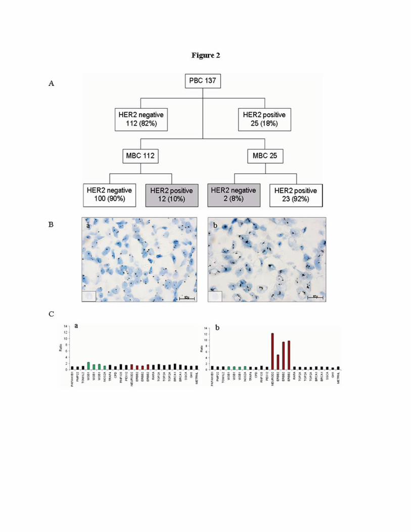
Figure 2:
HER2 variation in primary and metastatic breast cancer
Panel A: The flow chart summarizes HER2 change during disease progression in the entire series
of 137 primary and metastatic breast cancers. One hundred and twenty three cases (100 HER2
negative and 23 HER2 positive) maintained the same HER2 status (90%) (white boxes) whereas 14
cases (10%) changed HER2 status (grey boxes). p= 0.04.
Panel B: SISH images demonstrate: (a) no HER2 amplification in a primary breast cancer (gene
copy numbers �6) and (b) HER2 moderate gene amplification in the metachronous liver metastasis
(gene copy numbers >10); Scale bar = 10�m.
Panel C: the BC illustrated in panel b has been also analyzed by MLPA showing: (a) a normal
HER2 status in PBC and (b) an amplified HER2 status in the paired metastasis (HER2 ratio 1.36 vs
8.02).
Abbreviations: PBC: Primary Breast Cancer; MBC: Metastatic Breast Cancer; HER2 negative:
score 0, 1+, 2+ non-amplified; HER2 positive: score 2+ amplified, 3+.
HER2 change between primary and metastatic breast cancer 21
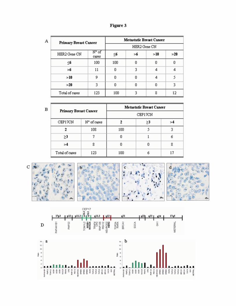
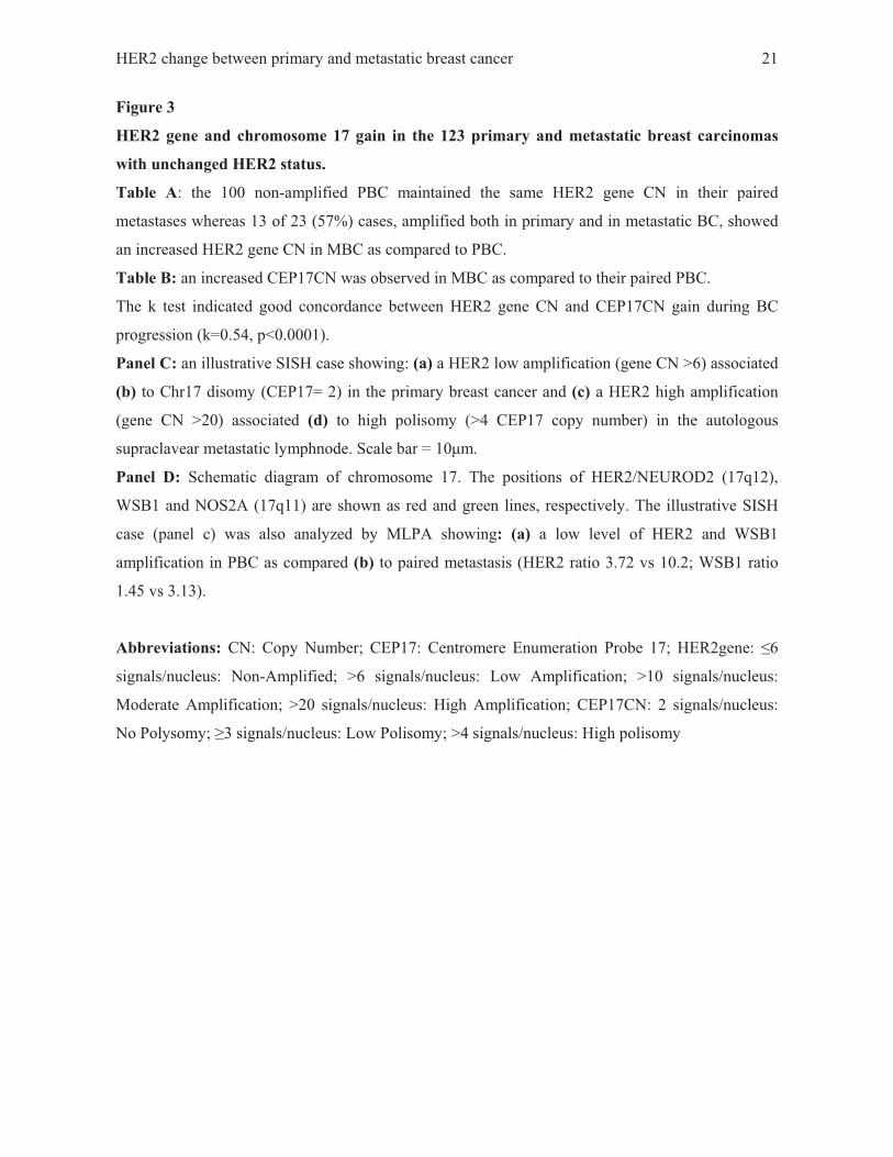
Figure 3
HER2 gene and chromosome 17 gain in the 123 primary and metastatic breast carcinomas
with unchanged HER2 status.
Table A: the 100 non-amplified PBC maintained the same HER2 gene CN in their paired
metastases whereas 13 of 23 (57%) cases, amplified both in primary and in metastatic BC, showed
an increased HER2 gene CN in MBC as compared to PBC.
Table B: an increased CEP17CN was observed in MBC as compared to their paired PBC.
The k test indicated good concordance between HER2 gene CN and CEP17CN gain during BC
progression (k=0.54, p<0.0001).
Panel C: an illustrative SISH case showing: (a) a HER2 low amplification (gene CN >6) associated
(b) to Chr17 disomy (CEP17= 2) in the primary breast cancer and (c) a HER2 high amplification
(gene CN >20) associated (d) to high polisomy (>4 CEP17 copy number) in the autologous
supraclavear metastatic lymphnode. Scale bar = 10�m.
Panel D: Schematic diagram of chromosome 17. The positions of HER2/NEUROD2 (17q12),
WSB1 and NOS2A (17q11) are shown as red and green lines, respectively. The illustrative SISH
case (panel c) was also analyzed by MLPA showing: (a) a low level of HER2 and WSB1
amplification in PBC as compared (b) to paired metastasis (HER2 ratio 3.72 vs 10.2; WSB1 ratio
1.45 vs 3.13).
Abbreviations: CN: Copy Number; CEP17: Centromere Enumeration Probe 17; HER2gene: �6
signals/nucleus: Non-Amplified; >6 signals/nucleus: Low Amplification; >10 signals/nucleus:
Moderate Amplification; >20 signals/nucleus: High Amplification; CEP17CN: 2 signals/nucleus:
No Polysomy; �3 signals/nucleus: Low Polisomy; >4 signals/nucleus: High polisomy