Embed Size (px)
Citation preview
MEDICINE
Hepatosplenic Cat Scratch Disease: Description of Two CasesUndergoing Contrast-Enhanced Ultrasound for Diagnosisand Follow-Up and Systematic Literature Review
Daniela Tirotta1 & Vincenzo Mazzeo1& Maurizio Nizzoli1
Accepted: 26 April 2021# The Author(s), under exclusive licence to Springer Nature Switzerland AG 2021
AbstractCat scratch disease (CSD) is a disease usually characterized by self-limited lymphadenopathy of the young man. Rarely CSD,however, can manifest itself as an unusual hepatosplenic form (HS-CSD) in immunocompetent patients. HS-CSD diagnosis isgenerally based on clinical features, imaging, and serologies, but sensitivity of serologies is very variable, like that of otherdiagnostic methods, as Warthin-Starry silver stain and isthology. Also there are no specific markers for the follow-up. The use ofthe CEUS (abdominal contrast-enhanced ultrasound) in HS-CSD is not previously described in literature examined, but we thinkthat CEUS can be of help to diagnosis and follow-up of these patients, even after an initial CT scan, because it is a sensitivemethod, as seen in other diseases associated with granulomas, such as sarcoidosis. We describe 2 new cases of HS-CSD, and weperformed a systematic review of the clinical cases reported in the past 10 years in the literature associated to an analysis ofclinical, diagnostic, and therapeutic aspects of the disease.
Keywords Cat scratch disease . Immunocompetent patient . Hepatosplenic cat scratch disease . Abdominal contrast-enhancedultrasound . Diagnosis . Follow-up
Introduction
Cat scratch disease (CSD) is a disease transmitted from ascratch or bite of an infected cat (rarely dog), as well as fromcat flea or contact with cat saliva through broken skin or mu-cosal surfaces. CSD is usually characterized by self-limitedlymphadenopathy of the young man. In these patients, theuse of antimicrobials is rarely necessary [1, 2].
Rarely CSD, however, can include visceral, neurologic,and ocular involvement (in 5–20% of patients), and, in partic-ular, an unusual hepatosplenic form (HS-CSD) is described in2.3% of patients.
Bartonella henselae (B. henselae) can cause intraerythrocyticbacteremia of the cat, for 1 year or longer [3]. In humans, generallyB. henselae causes a local infection, but it can also invade
endothelial cells, and it can cause an acute inflammatory reactionassociated to activation of a proinflammatory cascade [4]. Whysome patients have a local self-limited lymphadenopathy andothers a disseminated disease (sometimes associated to life-threatening complications) is unclear.
Patients and Methods
We present 2 cases of HS-CSD observed in immunocompetentpatients and a subsequent systematic review of CSD cases pub-lished in the literature in the last 10 years. We performed a sys-tematic research on PubMed for cases of Bartonella henselaeinfection with liver and spleen involvement (Mesh term “CatScratch Disease” AND “liver” AND/OR “spleen”, with detailedcontrol of references).We have extended the search toweb searchengine (Google), using the following keywords: “Cat ScratchDisease,” “hepatic and spleen involvement,” “Bartonellahenselae”, “immunocompetent adults,” and “Cat scratch disease.”
We obtained 42 cases; we excluded 27 for temporallimits (temporal limits: 2010–2020), age limit (adult:19+ years), and language limit (English language) withtotal of 15 cases (Table 1).
This article is part of the Topical Collection onMedicine
* Daniela [email protected]
1 Morgagni-Pierantoni Hospital, Forli’ (AUSL Romagna), InternalMedicine Unit, Via Carlo Forlanini, 47121, Forlì, Italy
https://doi.org/10.1007/s42399-021-00940-1
/ Published online: 15 June 2021
SN Comprehensive Clinical Medicine (2021) 3:2154–2166

Table1
Our
Cases
andCases
resultedfrom
literaturereview
Epidemiology
Clin
icalmanifestatio
nsCom
orbidity
Diagnosis
Case,year
ofpublication
(reference)
Age
(years)
Sex
System
icsymptom
sOrgan
involvem
ent
Immunodeficiency
Laboratorytests(W
BC,C
RP,
liver
enzymes)
Case1,2019
37F
Fever
Bilateralcervicaland
axillarylymphadenopathy,
multip
lehypodensehepatic
andsplenic
lesions;thelargest2
cmin
diam
eter
NO
WBCelevated,C
RPelevated,
liver
enzymes
norm
al
Case2,2020
51M
Fever,headaches
Multiplehypodensehepatic
andspleniclesions
NO
WBCelevated,C
RPelevated,
liverenzymes
elevated
Case3,2017
[5]
28M
Fevers,chills,abdom
inalpain,nightsw
eats,and
weightlossforthepast3weeks,cramping
abdominalpain
notassociatedwith
eatin
g
Severalh
epaticandsplenicmasseswith
central
necrosisin
abdomen
CT
NO
WBC,C
RP,
liver
enzymes
norm
al
Case4[6]2015
18M
Fever,headaches,nightsweats,diffuse
arthralgia
Bilateralcervicallym
phadenopathy,m
ultip
lehypodensehepatic
andspleniclesions;the
largest2
cmin
diam
eter,subchondral
sclerosisanderosionof
theventralthoracic
spinewereshow
nwith
maxim
alaffectionof
theseventhvertebralb
ody.
NO
WBCnorm
al,C
RPelevated,
liver
enzymes
norm
al
Case5[7]2015
63M
Recurrent
interm
ittentfeversandfatig
ue,w
eight
loss,m
ildheadaches,andarthralgias
Multiple,hypodense
hepatic,and
spleniclesions.
The
largestm
easured1.3cm
.Latenttuberculosis,which
hadbeen
treated30
yearsearlierwith
a9-month
course
ofisoniazid
WBCelevated,C
RPelevated,
liver
enzymes
norm
al
Case6[8]2014
50F
Fever,epigastriaandleftflankpain,rigors,
vomiting,and
malaise
Enlargedspleen
(16.5cm
)with
multiple
hypoechoiclesionsup
to1.5cm
,small
amount
offree
fluidin
theabdomen,m
ultip
lehypodenseroundnon-enhancinglesionsinthe
spleen
(upto2cm
),andfewintheliver(up
to0.5cm
)
NO
WBCelevated,C
RPNA,liver
enzymes
norm
al
Case7,2014
[9]
61M
Fever
Solitaryipodense,liver,and
spleen
lesions,
cervicalandgastrohepatic
ligam
ent
lymphadenopathy
NO
WBCnorm
al,C
RP,
andliver
enzymes
elevated
Case8,2014
[9]
41F
Fever
Adenopathy(right
axillaryandmultip
leabdominal),multip
leipodense
leverlesions
NO
WBCnorm
al,C
RP,
andliver
enzymes
elevated
Case9,2014
[9]
86F
Feverandabdominalpain
Multipleipodense
leverlesions,abdominal
lymphadenopathy
NO
WBCnorm
al,C
RPandliv
erenzymes
elevated
Case10,2013[10]
28M
Abdom
inalpain,fever
Spleen
abscess,skin
lesion,lym
phadenopathy
NO
Leukocytosis,CRPelevated,
liver
enzymes
norm
alCase11
,2013[11]
34F
Abdom
inalpain,fever
Multip
lespleen
abscess,skin
lesion,lym
phade
NO
WBCnorm
al,C
RP,
andliver
enzymes
NA
Case12
[12],2012
36F
Abdom
inalpain,fever
5cm
righth
epaticlobe
mass
NO
WBCnorm
al,C
RPNA,liver
enzymes
elevated
Case13
[13],2011
47M
Fever
C7cervicalosteom
yelitis,m
ultiplespleen
abscess,lymphadenopathy
NO
WBCnorm
al,C
RPelevated,
liverenzymes
elevated
Case14
[14],2011
45F
Abdom
inalpain,fever
Multip
lehypodensespleen
andliv
erlesions
NO
WBCnorm
al,C
RPelevated,
liverenzymes
elevated
Case15
[14],2011
27M
Feverandconstitutional
Multiplehypodenseliv
erlesions,celiac
lymphadenopathy
NO
WBCnorm
al,C
RPelevated,
liver
enzymes
norm
alCase16
[14],2011
33M
Abdom
inalpain,fever
NO
2155SN Compr. Clin. Med. (2021) 3:2154–2166

Tab
le1
(contin
ued)
Multiplehypodensespleen
lesions,celiac
lymphadenopathy
WBCnorm
al,C
RPelevated,
liver
enzymes
norm
alCase17
[14],2011
71M
Feverandconstitutional
Multip
lehypodensespleen
andleverlesions
NO
WBCnorm
al,C
RPelevated,
liver
enzymes
norm
al
Diagnosis
Evolutio
nTreatment
Case,year
ofpublication
(reference)
Diagnostic
imaging
US,CEUS,CT,M
RI,laparotomy,
others)
Microbiology
Histopathology
Clin
icalresolutio
n/tim
eto
cure
Treatment
Case1,2019
US,
CEUS,
TCabdomen
CultureNA.P
CRNA.S
erology
positive
NA
2weeks/3
months
DOXcp
3months
Case2,2020
US,
CEUS,
TCabdomen,P
ETFP
DG,
transoesophagealechocardiography,
spineMRI,lymph
node,and
liver
biopsy
CultureNA.P
CRnegativ
e.Serology
positive
Axillary
lymph
node
cytology:small
size
lymphocytes
mixed
with
mononuclear
largesize
and
histoepithelioid
microgranulom
as
1month/6
months
DOXcp
2months(associated
with
RIF
2days
(stopped
due
toabnorm
alLE)andDOX
for2months,2-weekcourses
ofGENcombinedwith
DOX,
AZM
3months
Case3,2017
[5]
TCabdomen,liver
biopsy
Culture
negativ
e.PC
RNA.S
erology
positive
Liver
biopsy
with
hematoxylin-eosin
staining
show
edabscesswith
numerousneutrophils,histio
cytes,
andlymphocytes,surrounding
fibroblastproliferationwith
background
lymphocytes,
histiocytes,andeosinophils
7weeks/7
weeks
and4
months
RIF
andAZM
2week,DOXand
RIF
5months
Case4[6]2015
CTbody
scan,boneMRI
Culture
NA.P
CRNA.S
erology
positive
NA
2weeks
and1month/NA
(>1mo)
IvAZM
(1.5
gtotald
ose)
and
RIF300mgtwicedaily
for3
weeks
Case5[7]2015
Transthoracicechocardiogram
was
unremarkable
Com
putedtomography(CT)of
the
chest,abdomen,and
pelvis,liver
biopsy
CultureNA.P
CRNA.S
erology
positive
Liver
biopsy
demonstratedamixed
inflam
matoryinfiltratewith
giant
cellhistiocytesbutn
owell-form
edgranulom
as
4weeks/NA
4-month
course
ofDOXand
RIF
Case6[8]2014
USabdomen,C
Tabdomen,
echocardio,T
EE,biopsyof
spleen
with
intraabdom
inalhemorrhage
originatingfrom
thespleen
andan
urgent
splenectom
yperformed
Emocolture
negativ
e,serology
positive.PC
Rpositiv
e.transesophagealechocardiogram
revealed
prolapse
oftheposterior
leafleto
fthemitralvalvewith
two
jetsof
mild
regurgitationandasm
all
mobile
echogenicmass(2·4mm)
attached
toit
Pathologicalexam
inationofthespleen:
superficialm
ultip
lewhitenodules
(upto
1cm
).Microscopic
exam
ination:
necrotizing
granulom
aswith
several
multin
ucleated
giantcells.
Ziel–Nielsen,silv
er,and
Gram
stains
werealln
egative.A
Whartin–S
tarrystainwas
equivocal.
Bacterial,fungal,andmycobacterial
cultu
resfrom
thespleen
were
negative
NA/5
months
AMPandGEN2weeks,
intravenousgentam
icin
for2
weeks
andoralDOXfor6
weeks
Case7,2014
[9]
CTbody
scan
Culturenegativ
e.PC
Rpositive.
Serology
positive
Necrotizinggranulom
atous
lymphadenitis.Warthin-Starrystain:
negative
3weeks/5
months
AZM
5days
2156 SN Compr. Clin. Med. (2021) 3:2154–2166

Tab
le1
(contin
ued)
Case8,2014
[9]
CTbody
scan
Culturenegativ
e.PC
Rpositive.
Serology
positive
Necrotizinggranulom
atous
lymphadenitis.Warthin-Starrystain:
negative
4weeks/8
months
DOX+RIF
8d,AZT5d,
Case9,2014
[9]
CTbody
scan
ND
ND
4week/4months
CRO+VAN+GEN1w,A
ZM
5d,
Case10,2013[10]
Abdom
enCT
Culture
positiv
e.PC
RND.S
erology
positive
ND
2weeks/NA
Ciprofloxacine2week,
CT-guideddrainage,
splenectom
yCase11
,2013[11]
Abdom
enMRI
PCRandserology
positive.Culture
ND
ND
NA/2.5months
AZT+DOX5d,CLR+RIF
5weeks
Case12
[12],2012
Abdom
enCT
Culture
positiv
e.PC
Rpositiv
e.Serology
negativ
eNecrotizinggranulom
atoushepatitis.
Steinerstainnegativ
e,ICHpositiv
e12
weeks/NA
Partialh
epatectomy,AZM
2weeks,C
LR9week,CIP
6week,RIF+DOX8week
Case13
[13],2011
Thoraxandabdomen
CT,technetium
bone
scan.V
ertebralmagnetic
resonance
CultureNA.P
CRpositive.Serology
negativ
eNecrotizinggranulom
atous
lymphadenitis.Warthin-Starrystain:
ND
12weeks/NA
DOX+EIF
6week
Case14
[14],2011
CTabdomen
Culture
NA,P
CRNA,and
serology
positive
ND
2weeks/4
months
CIP+GENiv
10d,CIP+DOC3
weeks
Case15
[14],2011
CTabdomen
Culture
NA,P
CRNA,and
serology
positive
ND
2weeks/4
months
CIP+GENIV
10d,Cip+ERY3
weeks
Case16
[14],2011
CTabdomen
Culture
NA,P
CRNA,and
serology
positive
ND
NA/3
months
DOX3weeks
Case17
[14],2011
CTabdomen
Culture
NA,P
CRNA,and
serology
positive
ND
NA/NA
DOX+RIF
3weeks
Clin
icalResolution:
norm
alizationof
Clin
icalsignsandsymptom
sandlaboratory
findings,T
imeto
cure:tim
eto
clinicalandradiologicalresolutio
n
AMKam
ikacine,AMPam
picilline,A
ZMazith
romycin,C
ROceftriaxone,CIP
ciprofloxacin,CLR
clarith
romycin,C
RPC-reactiveprotein,DOXdoxycycline,Ery
erythrom
ycine,FLQ
fluoroquinolone,
GENgentam
icine,IH
Cim
munohistochem
icalstain,IV
intravenous,LE
liverenzymes,N
Anotavailable,NDnotd
etermined,P
DNprednisone,R
IFrifampicin,RXM
roxithromycin,U
Sultrasound
scan,
VANvancom
ycin,V
oviaoral,W
BCwhitebloodcount
2157SN Compr. Clin. Med. (2021) 3:2154–2166

Demographic data (sex, age), epidemiologic data, clinicalpresentation, possible immunodeppression status, laboratoryfindings, radiologic image, surgery and histologic findings,clinical course, and treatments were reported and discussed.
Results
Case Series
Case 1
A 33-year-old woman was admitted in 2019 in the InternalMedicine Unit due to continuous fever for 1 week andsplenomegaly.
Her medical history was negative. She denied alcohol ordrug use. She did not report cat scratch or bite in the weeksbefore admission, but she had a cat.
Our abdomen ultrasound (US) showed multiplehypoechoic splenic lesions and splenomegaly. Chest-abdomen CT confirmed multiple nodular lesions compatiblewith splenic abscess. CEUS (contrast-enhanced ultrasound ),after administration of Sonovue 1 f ev, showed several nodu-lar splenic lesions (the max diameter was 1.5 cm) withoutarterial wash-in and without venous wash-out (Fig. 1).
Laboratory data revealed neutrophilic leukocytosis (WBC8000/mmc) and biological inflammation syndrome (CRP 100mg/l >> 10 mg/l); liver and kidney tests were normal.Bartonella IgM was positive, repeated at 5 days (IgM 100,IgG positive, IFA test), and blood cultures and electrophoresiswere negatives. We performed also serology for Toxoplasma,Borrelia, Cytomegalovirus, and Epstein-Barr that resultednegative. Defervescence occurred after 1 week of anti-biotic therapy (doxycycline). The patient was dischargedwith doxycycline 100 mg 1 cp × 2. After 3 months, the
lesions disappeared on abdomen US and CEUS, whichwe used for follow-up of patient.
Case 2
A 51-year-old man with no past medical history presented toour InternalMedicine Unit in 2020with fever (up to 39C for 6days), headache, and superficial lymphadenomegaly.
He reported a cat scratch in the right hand in the weeksbefore admission, but no bite. He denied alcohol or drug use.
Laboratory studies showed neutrophilic leukocytosis(WBC 10400> 6000/mmc) and biological inflammation syn-drome (CRP 116 >> 21 mg/l), and liver and kidney tests andelectrophoresis were normal.
We performed in our Internal Medical Unit an abdomenUS (which showed splenomegaly with multiple hypoechoiclesions of maximum 14 mm) and a CEUS: after administra-tion of Sonovue 1 f ev, we did not observed in the arterialphase wash-in of the described focal lesions of the spleen andthe liver (1.56 cm maximum diameter), except weakly in theperiphery of the nodular lesions of the spleen, and we did notobserve wash-out in the venous phase (Fig. 2.1, 2.2 and 2.3).
An immunofluorescence assay (IFA) detected the presenceof IgM antibodies against B. henselaewith a titer of IgG < 64.After 5 days, titer of igM was 128, and IgG was positive.Blood cultures were negative.
We performed also two COVID-19 swabs and serology forToxoplasma, Borrelia, andMycoplasma that resulted negativeand Legionella and Pneumococcus antigens and quantiferonthat resulted negative.
The following instrumental tests were performed:
– Chest CT: some lymphadenomegalies in the right axillaryarea, with a reactive appearance (maximum diameter27×14 mm).
Fig. 1 CEUS of patient 1. Spleen,arterial phase: nodular spleniclesions without arterial wash-inand without venous wash-out
2158 SN Compr. Clin. Med. (2021) 3:2154–2166

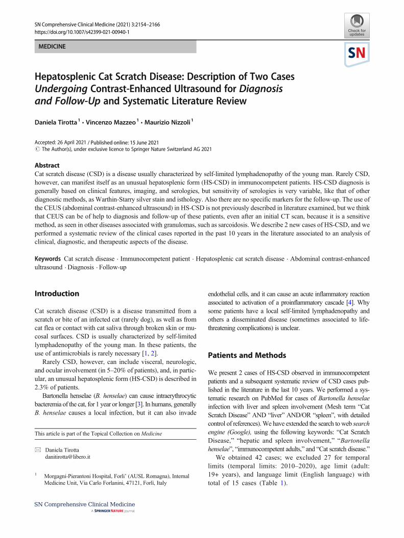
– Abdomen CT: hepatomegaly associated to somesubcentimetric and hypodense nodular lesions, someof which after administration of contrast agentshowed peripheral rim enhancement (microabscesses?Not characterized because of small size); splenomeg-aly (diameter 15 cm) with hypodense nodular forma-tions (greater than 10 mm ), some of which associatedto peripheral rim enhancement, periaortic andinteraortocaval lymphadenomegalies (max diameter17 mm), and lymphadenopathy in hepatic hilum (28x15 mm) (Fig. 3).
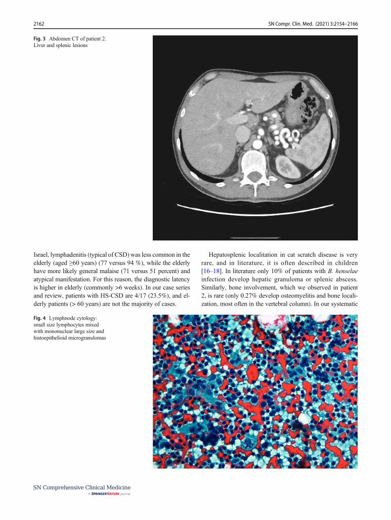
– Axillary lymph node cytology: small size lymphocytesmixed with mononuclear large size and histoepithelioidmicrogranulomas (Fig. 4).
– Brain CT: negative.– Lumbar puncture for CSF analysis (performed for neck
rigidity and headache): clear liquid, meningitis was ex-cluded through chemical and physical examination.
– 18F-FDG PET scans: accumulation of agent contrast inthe axillary lymph node (SUV 13.44), lymph nodes of thehepatic hilum, some hepatic and splenic inhomogeneity,vertebral D11 hyperaccumulation.
– Transparietal echocardium: thickening of the non-coronary aortic cusp, non-specific, associated to mildregurgitation.
– Transesophageal echocardium: no suspicious images forvegetations (performed 4 days later).
– Eye examination: no uveitis.– Spine MRI: inhomogeneity in the posteroinferior edge of
the soma of D11.
We prescribed antibiotic therapy with doxycycline and ri-fampicin, but because of the onset of a cholestatic hepatitis,then we administered only doxycycline.
The patient became apyretic and asymptomatic after 10 days.He was discharged with diagnosis of Bartonella en-
docarditis in the early phase (3 minor criteria of Dukesand one major) in course of HS-CSD, complicated bybone localization of the posteroinferior edge of D11 andintercurrent iatrogenic cholestatic liver disease.
One month later, he performed abdomen US, whichshowed at least five hypoechoic images of the liver (maximumdiameter 1.8 cm) and of the spleen (maximum diameter 1.7cm). CEUS, used for follow-up, showed a nodular lesion notpreviously present in the VII segment (Fig. 2.3).
Laboratory tests showed decreased CRP (8.2mm/l) andnormal liver tests.
Recent medical history was positive for photosensitivityreaction in the hands and occasional lower back pain.
The patient was hospitalized again. Blood tests showedBartonella seroconversion (negative IGM, IgG 512, IFA test),procalcitonin, complement, lymphocyte typing, blood cul-tures were normal.
We repeated echocardium (negative), lumbosacralMRI: minimal L1 inhomogeneity, and PET positive forpersistent hepatosplenic uptake with no accumulation ofthe contrast in L1 and SUV attenuation in abdominallymph nodes.
He performed a double liver biopsy: we sent a sam-ple to S Matteo in Pavia for PCR Bartonella test(negative) and one to the Anatomy Pathology of Forli' (liver parenchyma was normal).
Fig. 2.1 CEUS patient 2. Left liver, arterial phase: nodular liver lesions with peripheral arterial wash-in and without venous wash-out
2159SN Compr. Clin. Med. (2021) 3:2154–2166

Fig. 2.2 One month later. CEUSof patient. Liver and spleen,arterial phase. New nodular lesionwithout arterial wash-in andwithout venous wash-out in VIIsegment
2160 SN Compr. Clin. Med. (2021) 3:2154–2166

After the biopsy, a new therapeutic scheme began: genta-micin in single administration (3 mg/kg) and azithromycinintravenous 2 weeks and then azithromycin orally.
Six months later, abdomen US combined with CEUSshowed resolution of liver and splenic lesions (Fig. 2.3).
Table 1 summarizes the characteristics of the other 15 pa-tients described in the literature.
Discussion
Our case series and cases of literature review stress how HSCSD has multisystem involvement and how there is a lack of
definitive diagnostic tests, so diagnosis and management ofHS-CSD is often difficult.
Epidemiology CSD is commonly a disease of children andyoung adults, but a surveillance study in Israel showed that6 % of immunocompetent patients with CSD was ≥ 60 yearsold [15]. The mean age of patients observed is 44.47 years andmedian 41 age (range 28–86 years). In total 7/17% of patientsare women.
Atipical CSD In the literature, older CSD patients have morefrequently atypical locations (Table 2 showed majormanifestation of atypical SCD): in a surveillance study in
Fig. 2.3 CEUS of patient, sixmonths later. Liver and spleen,arterial phase. Resolution ofnodular lesion
2161SN Compr. Clin. Med. (2021) 3:2154–2166
Israel, lymphadenitis (typical of CSD)was less common in theelderly (aged ≥60 years) (77 versus 94 %), while the elderlyhave more likely general malaise (71 versus 51 percent) andatypical manifestation. For this reason, the diagnostic latencyis higher in elderly (commonly >6 weeks). In our case seriesand review, patients with HS-CSD are 4/17 (23.5%), and el-derly patients (> 60 years) are not the majority of cases.
Hepatosplenic localitation in cat scratch disease is veryrare, and in literature, it is often described in children[16–18]. In literature only 10% of patients with B. henselaeinfection develop hepatic granuloma or splenic abscess.Similarly, bone involvement, which we observed in patient2, is rare (only 0.27% develop osteomyelitis and bone locali-zation, most often in the vertebral column). In our systematic
Fig. 3 Abdomen CT of patient 2.Liver and splenic lesions
Fig. 4 Lymphnode cytology:small size lymphocytes mixedwith mononuclear large size andhistoepithelioid microgranulomas
2162 SN Compr. Clin. Med. (2021) 3:2154–2166
review, we have identified only 17 cases of HS-CSD in thepast 10 years in adult patients.
Symptoms and Organ InvolvementVisceral CSD can involvethe liver, spleen, or both. Patients with visceral CSD maymanifest fever of unknown origin (FUO), abdominalgia,weight loss, and hepatomegaly or splenomegaly on physicalexamination [19]. Many patients do not have peripheraladenopathy. In our case series, the most frequent clinical man-ifestation is fever in 100% of patients with or without head-ache and abdominal pain in 8/17 patients (47%). Liver and/orsplenic involvement is often not associated with superficiallymphadenomegaly.
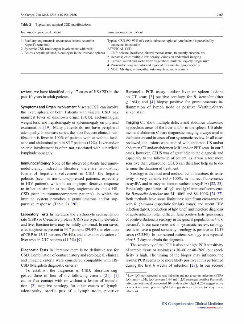
Immunodeficiency None of the observed patients had immu-nodeficiency. Indeed in literature, there are two distinctforms of hepatic involvement in CSD: the hepaticpeliosis (seen in immunosuppressed patients, especiallyin HIV patient), which is an angioproliferative responseto infection similar to bacillary angiomatosis and a HS-CSD (seen in immunocompetent patients), in which theimmune system provokes a granulomatous and/or sup-purative response (Table 2) [20].
Laboratory Tests In literature the erythrocyte sedimentationrate (ESR) or C-reactive protein (CRP) are typically elevated,and liver function tests may be mildly abnormal. In our cases,a leukocytosis is present in 5/17 patients (29.4%), an elevationof CRP in 13/17 patients (76.4%), and alteration elevation ofliver tests in 7/17 patients (41.2%) [9].
Diagnostic Tests In literature there is no definitive test forCSD. Combination of contact history and serological, clinical,and imaging criteria were considered compatible with HS-CSD (Margileth diagnostic criteria).
To establish the diagnosis of CSD, literature sug-gested three of four of the following criteria [21]: [1]cat or flea contact with or without a lesion of inocula-tion; [2] negative serology for other causes of lymph-adenopathy, sterile pus of a lymph node, positive
Bartonella PCR assay, and/or liver or spleen lesionson CT scan; [3] positive serology for B. henselae (titer≥ 1:64); and [4] biopsy positive for granulomatous in-flammation of lymph node or positive Warthin-Starrysilver stain.
Imaging CT show multiple defects and abdomen ultrasoundhypoechoic areas of the liver and/or in the spleen. US abdo-men and abdomen CT are diagnostic imaging always used inthe literature and in cases of our systematic review. In all casesreviewed, the lesions were studied with abdomen US and/orabdomen CT and/or abdomen MRI and/or PET scan. In our 2cases, however, CEUS was of great help to the diagnosis andespecially to the follow-up of patient, as it was a test moresensitive than ultrasound. CEUS can therefore help us to de-termine the duration of treatment.
Serology is the most used method, but in literature, its sensi-tivity is very variable (<30–100%, in indirect fluorescenceassay:IFA and in enzyme immunosorbent assay:EIA) [22, 23].Particularly specificities of IgG and IgM immunofluorescencefor Bartonella henselae are 34–100% and 86–100% [24–27].Both methods have some limitations: significant cross-reactionwith B. Quintana (especially for IgG assays) and recent EBVinfection (IgM), production of IgM brief, and therefore diagnosisof acute infection often difficult, false positive tests (prevalenceof positive Bartonella serology in the general population is 4 to 6percent)1. In our case series and in systematic review, serologyseems to have a good sensitivity: serology is positive in 14/17cases (82.35%). In our second patient, serology was repeatedafter 5–7 days to obtain the diagnosis.
The sensitivity of the PCR is also not high: PCR sensitivityof sample tissue or aspirates is 30–60 or 40–76%, but speci-ficity is high. The timing of the biopsy may influence theresults: PCR seems to be more likely positive if it is performedduring the first 6 weeks of infection [28]. In our second
1 Low IgG may represent a past infection and not a current infection (if IFAIgG titers <1:64), IgG between 1:64 and 1:256 represent possible Bartonellainfection (test should be repeated 10–14 days after), IgG>1:256 suggest activeor recent infection; positive IgM test suggests acute disease (or very recentinfection).
Table 2 Typical and atypical CSD manifestations
Immunocompromised patient Immunocompetent patient
1. Bacillary angiomatosis (cutaneous lesions resembleKaposi’s sarcoma)
2. Systemic CSD (multiorgan involvement with rash)3. Peliosis hepatis (dilated, blood cysts in the liver and spleen)
Typical CSD (90–95% of cases): subacute regional lymphadenitis preceded bycutaneous inoculation
ATYPICAL CSD1. CNS: seizure, headache, altered mental status, frequently encephalitis2. Hepatosplenic: multiple low density lesions on abdominal imaging3. Cardiac: mitral and aortic valve vegetations multiple, rapidly progressive4. Parinaud’s: conjunctivitis and regional preauricular lymphadenitis5. MSK: Myalgia, arthropathy, osteomyelitis, and tendinitis
2163SN Compr. Clin. Med. (2021) 3:2154–2166

patient, PCR was required for the presence of new liver le-sions, despite ongoing antibiotic therapy, but it was negative.In our systematic review (limitations are the small number ofpatients), the sensitivity of the CRP is low: B. henselae PCRwas positive only in 7/17 cases (41 %).
Bartonella grows very slowly, and it is hardly detectable inblood cultures. Culture is difficult even after 2–6 weeks ofincubation: B. henselae is gram-negative, characterized by avery slow growth, and so it requires specific conditions: it isimportant to inform the laboratory of microbiology thatBartonella is a potential diagnosis (to optimize culture tech-niques and extend the incubation period for a minimum of 21days). In our systematic review, the culture test was positiveonly in 2/17 cases (11.76%).
Histopathology shows granulomatous infection associatedto acellular, necrotic center. B. henselae can be identified withWarthin-Starry silver stain (46% sensitive, rarely positive inthe later stages). Histopathology of lymph node is oftenaspecific and variable with the stage of the disease: beforelymphoid hyperplasia, then granulomas (centers acellularand necrotic; histiocytes and peripheral lymphocytes, similarto findings in skin samples), and at the end microabscessesconfluent. Actually biopsies are rarely performed, and itshould include histology, Warthin-Starry staining, PCR test,and tests to investigate other disorders. That is because clinicaland imaging pictures are generally typical. A biopsy (lymphnode or tissue) is indicated only in certain circumstances, asdelayed resolution of systemic symptoms (>5–7 days) andsuspicion of other diagnosis.
In our case series and in our systematic review, a histopath-ological examination was obtained only in 9/17 patients(52.9%), and it showed sometimes hepatic granulomas, some-times hepatic abscesses, sometimes splenic granulomas, andsometimes granulomas or microgranulomas of lymph node.
Time for Clinical Resolution The mean to clinical remission is4.9 weeks (range 2–12 weeks), and the mean time to cure is3.8 months (range 3–6 months).
Therapy Different therapy schemes were performed and withdifferent duration (see Table 1 for details).
Conclusions
With the limitations of the low number of patients analyzed,the data would suggest the following:
& HS-CSD diagnosis is generally based on clinical features,imaging, and serologies, but sensitivity of serologies isvery variable: negative serologic test not rule out diagno-sis, if the clinical probability of patient is high. If there is ahigh clinical suspicion, the serology should be repeated.
& Histopathology is aspecific and shows granulomatous infec-tion associated to acellular, necrotic center. B. henselae can beidentified, but Warthin-Starry silver stain sensitivity (46%,rarely positive in the later stages) and PCR sensitivity (40%)are small. So a biopsy (lymph node or tissue) is indicated onlyin certain circumstances, as delayed resolution of systemicsymptoms (>5–7 days) and suspicion of other diagnosis.
& In our case series, we have identified CEUS as a promis-ing method for diagnosis and follow-up of HS-CSD.
US B mode accuracy for focal splenic lesions is 50%and Doppler US not add substantial help. The sensitivityof US is inferior to that of CT or MR and improves afterthe injection of US contrast agents: in literature CEUSsensitivity and specificity, positive and negative predictivevalues reached 100%, 83.8%, 87.8%, and 100% for dif-ferentiating benign and malignant lesions [29] in liver andsplenic nodules [30, 31]. Indeed small ipoechoic/isoechoic splenic or liver lesions may be difficult to iden-tify with conventional US, but CEUS typically demon-strate no enhancement in any phase or rapid enhancementfollowed by persistent late-phase enhancement of benignsplenic/liver lesions (Strong Consensus) [29]. Splenic ab-scesses typically appear as non-enhancing areas, or anenhanced border may be seen , especia l ly inmicroabscesses. Instead malignant lesions demonstratelow early diffuse or peripheral enhancement followed bywashout of microbubbles in the late phases. [32–34]. Soevidences suggested that CEUS can be a helpful tool forhealth care professionals to confirm the diagnosis of solidlesions of liver and spleen [34].
However, the use of the CEUS in HS-CSD is not previ-ously described in any of the cases of HS-CSD of theliterature examined. Also in literature, there are few studiesfocused on the ability of CEUS to detect granulomatouslesions, such as in sarcoidosis. In observational studies,CEUS has the potential to become a valid and safe screen-ing tool for systemic infiltration, as in sarcoidosis, and animportant method of monitoring the effects of therapy [31,35]. CT and MRI scans show granulomas as multiple, non-enhancing subcentimetric nodules, but these tools are as-sociated with minor safety (contrast allergy, MRI—claus-trophobia, pacemakers, metallic foreign bodies). CEUSallowed us to distinguish healthy (hyperechoic) liver andspleen parenchyma from small, avascular granulomas, andit is associated with increased security. Splenic and hepaticgranulomas (or microabscess) are prevalent in HS CSD;instead we think that CEUS can be of great help in thediagnosis and the follow-up of these patients, even afteran initial CT scan, because it is a sensitive method.
& A presumption diagnosis of HS-CSD generally involvesempirical therapy. Within the limits of the few cases
2164 SN Compr. Clin. Med. (2021) 3:2154–2166

observed in the immunocompetent adult, a prolongedcombination therapy may be beneficial in patients withsevere disease or unresponsive to single therapy.
Combination therapy may reduce the possibility ofB. henselae becoming sequestered in the bone marrow,lymph nodes, and thymus. For patients with disseminateddisease or HS-CSD combination therapy (e.g.,azithromycin plus rifampin or rifampin plus gentamicinor higher dose of azithromycin alone) can be prescribed.Rarely adjunctive corticosteroid therapy can also beadministered.
& There are no specific markers for the follow-up. It may bereasonable to monitor the clinical response and C-reactiveprotein levels. Some believes that an imaging technique isrequired approximately 6 months after treatment, to doc-ument the lesion regression and others imaging tests arenot necessary if the patient has complete clinical resolu-tion.
In patients with HS-CSD, we believe that is it isuseful to perform a follow-up with abdominal USand CEUS monthly; also after an initial CT scan,CEUS can be used for follow-up, to evaluate reduc-tion in size of lesions, with no additional radiationexposure, as in patients with splenic trauma, ab-scesses, and infarcts [29].
& Several areas need to be explored by research: therole of CEUS in the diagnosis and follow-up ofhepatosplenic abscesses and the correct modality offollow-up, antibiotic choice, combination treatment,treatment timing, and why some patients with HS-HSD respond poorly to antibiotic therapy, evenwithout an immunosuppression.
Acknowledgements The authors acknowledge Dr Michele Gaudio ofPathological Anatomy Institute, Morgagni Pierantoni Hospital, Forli’.
Code Availability Not applicable.
Author Contribution Each authors contributed equally to the writing ofthe article.
Data Availability Not applicable.
Declarations
Ethics Approval Not applicable.
Consent to Participate Not applicable.
Consent for Publication Patients gave written consent to thepublication.
Conflict of Interest The authors declare no competing interests.
References
1. Bass JW, Vincent JM, Person DA. The expanding spectrum ofBartonella infections: II. Cat-scratch disease. Pediatr Infect Dis J.1997;16:163.
2. Spach DH, Koehler JE. Bartonella-associated infections. Infect DisClin North Am. 1998;12:137.
3. Jacomo V, Kelly PJ, Raoult D. Natural history of Bartonella infec-tions (an exception to Koch's postulate). Clin Diagn Lab Immunol.2002;9:8.
4. Dehio C. Molecular and cellular basis of Bartonella pathogenesis.Annu Rev Microbiol. 2004;58:365.
5. Verma SK, Martin A, Montero J. Atypical cat scratch disease withhepatosplenic involvement. Clin Gastroenterol Hepatol. 2017Jan;15(1):e5–6.
6. Knafl D, Lötsch F, Burgmann H. Hepatosplenic abscesses and os-teomyelitis of the spine in an immunocompetent adult with catscratch disease. Case Rep Infect Dis. 2015;2015:317260.
7. Bieraugel K, Oehler D, NeSmith M. Cat got your spleen?Hepatosplenic Bartonella infection Am J Med. 2015 Mar;128(3):246–9.
8. Shasha D, Gilon D, Vernea F. Visceral cat scratch disease withendocarditis in an immunocompetent adult: a case report and re-view of the literature. VECTOR-BORNE AND ZOONOTICDISEASES. 2014;14(3):p175–18.
9. García JC, MD NMJ, Castro B, et al. Hepatosplenic cat scratchdisease in immunocompetent adults. Report of 3 Cases andReview of the Literature. Medicine (Baltimore). 2014 Oct;93(17):267–79.
10. Anyfantakis D, Kastanakis M, Papadomichelakis A, et al. Cat–scratch disease presenting as a solitary splenic abscess in an immu-nocompetent adult: case report and literature review. Le Infezioni inMedicina, n. 2013;2:130–3.
11. Liberto MC, Matera G, Lamberti AG, et al. Diagnosis and follow-up of Bartonella henselae infection in the spleen of an immuno-competent patient by real-time quantitative PCR. J Med Microbiol.2013;62:1081–5.
12. VanderHeyden TR, Yong SL, Breitschwerdt EB, et al.Granulomatous hepatitis due to Bartonella henselae infection inan immunocompetent patient. BMC Infect Dis. 2012;12:17.
13. Graveleau J, Grossi O, Lefebvre M, et al. Vertebral osteomyelitis:an unusual presentation of Bartonella henselae infection. Semin.Arthritis Rheum. 2011;41:511–6.
14. Zenone T. Systemic Bartonella henselae infection in immunocom-petent adults presenting as fever of unknown origin. Case ReportMed. 2011;2011:183397.
15. Ben-Ami R, Ephros M, Avidor B, et al. Cat-scratch disease inelderly patients. Clin Infect Dis. 2005;41:969.
16. Lenoir AA, Storch GA, DeSchryver-Kecskemeti K, et al.Granulomatous hepatitis associated with cat scratch disease.Lancet. 1988;1:1132.
17. Delahoussaye PM, Osborne BM. Cat-scratch disease presenting asabdominal visceral granulomas. J Infect Dis. 1990;161:71.
18. Fretzayas A, Papadopoulos NG, Moustaki M, et al. Unsuspectedextralymphocutaneous dissemination in febrile cat scratch disease.Scand J Infect Dis. 2001;33:599.
19. Arisoy ES, CorreaAG,WagnerML, Kaplan SL. Hepatosplenic cat-scratch disease in children: selected clinical features and treatment.Clin Infect Dis. 1999;28:778.
20. Tappero JW, Koehler JE, Berger TG, et al. Bacillary angiomatosisand bacillary splenitis in immunocompetent adults. Ann InternMed. 1993;118:363.
21. Margileth AM. Recent advances in diagnosis and treatment of catscratch disease. Current Infectious Disease Reports. 2000;2(2):141–6.
2165SN Compr. Clin. Med. (2021) 3:2154–2166

22. English CK, Wear DJ, Margileth AM, et al. Cat-scratch disease.Isolation and culture of the bacterial agent. JAMA. 1988;259:1347.
23. Brenner DJ, Hollis DG, Moss CW, et al. Proposal of Afipia gen.nov., with Afipia felis sp. nov. (formerly the cat scratch diseasebacillus), Afipia clevelandensis sp. nov. (formerly the ClevelandClinic Foundation strain), Afipia broomeae sp. nov., and three un-named genospecies. J Clin Microbiol. 1991;29:2450.
24. Bergmans AM, Peeters MF, Schellekens JF, et al. Pitfalls and fal-lacies of cat scratch disease serology: evaluation of Bartonellahenselae-based indirect fluorescence assay and enzyme-linked im-munoassay. J Clin Microbiol. 1997;35:1931.
25. Dupon M, Savin De Larclause AM, Brouqui P, et al. Evaluation ofserological response to Bartonella henselae, Bartonella quintanaand Afipia felis antigens in 64 patients with suspected cat-scratchdisease. Scand J Infect Dis. 1996;28:361.
26. Sander A, Posselt M, Oberle K, Bredt W. Seroprevalence of anti-bodies to Bartonella henselae in patients with cat scratch diseaseand in healthy controls: evaluation and comparison of two commer-cial serological tests. Clin Diagn Lab Immunol. 1998;5:486.
27. Zbinden R,Michael N, Sekulovski M, et al. Evaluation of commer-cial slides for detection of immunoglobulin G against Bartonellahenselae by indirect immunofluorescence. Eur J Clin MicrobiolInfect Dis. 1997;16:648.
28. Ridder GJ, Boedeker CC, Technau-Ihling K, et al. Role of cat-scratch disease in lymphadenopathy in the head and neck. ClinInfect Dis. 2002;35:643.
29. Hopkins A, Cokkinos DD. Contrast enhanced sonographic study ofthe spleen. HJR. 2(1):49–65.
30. Berzigotti A, Ferraioli G, Bota S, Gilja OH, Dietrich CF. Novelultrasound-based methods to assess liver disease: the game has justbegun. Dig Liver Dis. 2018;50:107–12.
31. Tana C, Schiavone C, Ticinesi A, Ricci F, Giamberardino MA,Cipollone F, et al. Ultrasound imaging of abdominal sarcoidosis:state of the art. World J Clin Cases. 2019;7(7):809–18.
32. Omar A, Freeman S. Contrast-enhanced ultrasound of the spleen.Ultrasound. 2016;24(1):41–9.
33. GORG C. The forgotten organ: contrast enhanced sonography ofthe spleen. Eur. J. Radiol. 2007;64(2):189–201.
34. Sidhu PS, Cantisani V, Dietrich CF, Gilja OH, Saftoiu A, Bartels E,et al. The EFSUMB guidelines and recommendations for the clin-ical practice of contrast-enhanced ultrasound (CEUS) in non-hepatic applications: update 2017 (long version). Ultraschall Med.2018;39:e2–e44.
35. Grzelak P, Augsburg L, Majos A, Stefańczyk L, Górski P,Piotrowski W, et al. Diagnostic potential of contrast-enhanced ul-trasound (CEUS) in the assessment of spleen and liver granulomasin the course of sarcoidosis. Pneumonol Alergol Pol. 2013;81(5):424–8.
Publisher’s Note Springer Nature remains neutral with regard to jurisdic-tional claims in published maps and institutional affiliations.
2166 SN Compr. Clin. Med. (2021) 3:2154–2166


![Cat Scratch (Bartonella henselae) - Biocare Medical · Cat Scratch (Bartonella henselae) [H2A10] is a mouse monoclonal antibody that is intended for laboratory use in the qualitative](https://img.pdfslide.us/doc/110x75/606375af7209417ea40878e9/cat-scratch-bartonella-henselae-biocare-medical-cat-scratch-bartonella-henselae.jpg)