Embed Size (px)
Citation preview

JOURNAL OF VIROLOGY, Sept. 2011, p. 9359–9368 Vol. 85, No. 180022-538X/11/$12.00 doi:10.1128/JVI.00682-11Copyright © 2011, American Society for Microbiology. All Rights Reserved.
Hepatitis C Virus Infection Is Blocked by HMGB1Released from Virus-Infected Cells�
Jong Ha Jung,1 Ji Hoon Park,1 Min Hyeok Jee,1 Sun Ju Keum,1 Min-Sun Cho,2Seung Kew Yoon,3 and Sung Key Jang1,4,5*
PBC, Department of Life Science,1 Division of Integrative Biosciences and Biotechnology, WCU Program,4 andBiotechnology Research Center,5 Pohang University of Science and Technology, Pohang, Kyungbuk, Republic ofKorea; Department of Pathology, Ewha Womans University School of Medicine, Seoul, Republic of Korea2;
and Department of Internal Medicine & WHO Collaborating Center of Viral Hepatitis, Catholic University,Seoul, Republic of Korea3
Received 5 April 2011/Accepted 25 June 2011
High-mobility group box 1 (HMGB1), an abundant nuclear protein that triggers host immune responses, isan endogenous danger signal involved in the pathogenesis of various infectious agents. However, its role inhepatitis C virus (HCV) infection is not known. Here, we show that HMGB1 protein is translocated from thenucleus to cytoplasm and subsequently is released into the extracellular milieu by HCV infection. SecretedHMGB1 triggers antiviral responses and blocks HCV infection, a mechanism that may limit HCV propagationin HCV patients. Secreted HMGB1 also may have a role in liver cirrhosis, which is a common comorbidity inHCV patients. Further investigations into the roles of HMGB1 in the diseases caused by HCV infection willshed light on and potentially help prevent these serious and prevalent HCV-related diseases.
Hepatitis C virus (HCV) is one of the major causative agentsof hepatitis, liver cirrhosis, and hepatocellular carcinoma(HCC) (17, 30). More than 170 million people are estimated tosuffer from HCV infection worldwide (17). Chronic and per-sistent infection is a characteristic feature of HCV pathogen-esis (30). During chronic infection, the production of virusparticles is limited, and a restricted number of liver cells areinfected. As a result, the viral dose in patients’ blood generallyis lower than that of other hepatitis-causing viruses, such ashepatitis B virus (HBV) (4). Moreover, a large portion ofhepatocytes often remains uninfected by the virus even afterlong-term infection (28). These phenomena indicate the exis-tence of balance between the HCV infection process and hostmechanisms that protect against HCV infection. We speculatethat innate and adaptive immunities contribute to the balancebetween infection and protection.
High-mobility group box 1 (HMGB1) protein is a highlyconserved nuclear protein that participates in DNA organiza-tion and the regulation of transcription. In addition to itsnuclear function, HMGB1 plays an important role as a cyto-kine, mediating the responses to infection, injury, and inflam-mation (1, 2, 29, 42). HMGB1 is released passively from ne-crotic cells and is actively secreted from activated immunecells, such as macrophages, natural killer cells, and maturedendritic cells (2). The functionality of actively secretedHMGB1 is known to be modulated by posttranslational mod-ifications, such as oxidation (2, 36). Extracellular HMGB1 canfunction by itself and/or in association with other molecules,including CpG DNA, lipopolysaccharide (LPS), and interleu-
kin-1 (IL-1) (5). HMGB1 induces a variety of cellular re-sponses that contribute to innate immunity, tissue repair, andcell migration through interactions with various receptors thatactivate multiple signal transduction responses. The Toll-likereceptors (e.g., TLR2, TLR4, and TLR9) and the receptor foradvanced glycation end products (RAGE) are known recep-tors for the cytokine functions of HMGB1 (2). TLR4, themajor component of the LPS recognition receptor complex,engages in downstream signaling through MyD88 and the Toll-like adapter protein TRIF to produce proinflammatory cyto-kines and type I interferons (IFNs), which potentially partici-pate in blocking virus infections (34).
No role of HMGB1 in HCV infection has been demon-strated yet. However, HMGB1 is known to be an indicatorof human liver injury (19), and HMGB1 levels in the sera ofpatients with chronic hepatitis and cirrhosis are significantlyelevated (8). The source of this elevated serum HMGB1 andthe molecular mechanism responsible for the secretion ofHMGB1 from cells are not known. One possible mechanismunderlying elevated HMGB1 secretion is increased reactiveoxygen species (ROS) in HCV-infected cells. Two HCV-encoded proteins, core and NS5A, induce oxidative stress ininfected cells (15, 25–27), and it has been reported that ROSinduces nuclear-to-cytoplasmic translocation and the subse-quent secretion of HMGB1 from cells (36). However, thelocalization of HMGB1 in HCV-infected cells and the effectof HMGB1 on HCV infectivity remain to be elucidated.
Here, we investigated the localization and secretion ofHMGB1 upon HCV infection. We found that some HMGB1protein was translocated from the nucleus to the cytoplasm andsecreted into the medium before virus production was ob-served. This suggests that HCV-infected cells sense the HCVinfection and propagate a warning signal to uninfected cells.We tested the potential role of secreted HMGB1 in HCVinfection using an antibody against HMGB1 and purified
* Corresponding author. Mailing address: PBC, Department of LifeScience, Pohang University of Science and Technology, San 31, Hyoja-Dong, Pohang 790-784, Republic of Korea. Phone: 82-54-279-2298.Fax: 82-54-279-8009. E-mail: [email protected].
� Published ahead of print on 13 July 2011.
9359
Dow
nloa
ded
from
http
s://j
ourn
als.
asm
.org
/jour
nal/j
vi o
n 24
Jan
uary
202
2 by
191
.53.
249.
168.
HMGB1 protein. The treatment of HCV culture medium withthe anti-HMGB1 antibody increased the infectivity of HCV.Conversely, the pretreatment of cells with purified HMGB1protein reduced the infectivity of HCV, indicating thatHMGB1 secreted from infected cells blocked the infection ofneighboring cells by HCV. Our investigation of the molecularbasis of this protection further revealed that TLR4, a potentialHMGB1 receptor, plays an important role in the antiviraleffect via the activation of interferon signaling.
MATERIALS AND METHODS
Cell culture and HCV production. The human hepatoma cell line Huh7.5.1was kindly provided by Francis V. Chisari (Scripps Research Institute). Huh7.5.1and HEK293FT (human kidney carcinoma) cells were grown in Dulbecco’smodified Eagle’s medium (DMEM; Invitrogen) supplemented with 10% fetalbovine serum (FBS; HyClone), penicillin, and streptomycin. The in vitro synthe-sis of HCV RNA (derived from JFH1 and JFH5a-Rluc vectors) and electropo-ration were performed as described previously (22). Infectious HCV in cellculture (HCVcc) was produced by the transfection of Huh7.5.1 cells with invitro-transcribed HCV RNA. The virus titer was determined as described else-where (11) and stored at �80°C.
Reagents. The antibodies against HMGB1, TLR4, and isotype control IgGwere purchased from Abcam. The antibody against HCV core for immunocyto-chemistry was obtained from Affinity Bioreagents. The antibodies to PARP[poly(ADP-ribose) polymerase], �-actin, and GAPDH (glyceraldehyde 3-phos-phate dehydrogenase) were obtained from Santa Cruz Biotechnology, Sigma,and AbD Serotec, respectively. The secondary antibodies for immunostainingwere purchased from Invitrogen and Jackson ImmunoResearch. The antibodyagainst HCV core for Western blotting was a gift from Ralf Bartenschlager(University of Heidelberg). LPS and Hoechst 33258 were obtained from Sigma.
Patient tissue. Informed consent to use tissue specimens for research purposeswas obtained from all patients, and the utilization of specimens for this researchwas authorized by the Institutional Review Boards of the College of Medicine,Ewha Womans University (case numbers S10-6410 and S09-2974), Yonsei Uni-versity (case number T6), and Catholic University (case number 14266130).
RT-PCR. Total RNA was purified with TRI reagent, and cDNA was synthe-sized using reverse transcription reagents (Promega). Reverse transcription-PCR(RT-PCR) was performed using a LightCycler 480 SYBR green I master system(Roche). Primer sequences for reverse transcription, PCR, and real-time PCRwere the following: IFN�1, 5�-CAG CAA TTT TCA GTG TCA GAA GC-3� and5�-TCA TCC TGT CCT TGA GGC AGT-3�; interferon-stimulated gene 15(ISG15), 5�-ACT CAT CTT TGC CAG TAC AGG AG-3� and 5�-CAG CATCTT CAC CGT CAG GTC-3�; GAPDH, 5�-TGC ACC ACC AAC TGC TTAG-3� and 5�-GAG GCA GGG ATG ATG TTC-3�; and �-actin, 5�-CTG GAACGG TGA AGG TGACA-3� and 5�-AAG GGA CTT CCT GTA ACA ATGCA-3�.
Plasmid constructs and mutagenesis. The plasmid pHMGB1-eGFPN1 wasprovided by J. S. Shin (Yonsei University, South Korea). To create pcDNA3.1-Flag-HMGB1, we first constructed the plasmid pcDNA3.1-Flag by insertingannealed oligonucleotides encoding Flag sequences (5�-CTA GCC GCC ACCATG GAC TAC AAA GAC GAT GAC GGT GAT TAT AAA GAT GATGAC ATC GAT TAC AAG GAT GAC GAT GAC A-3� and 5�-AGC TTG TCATCG TCA TCC TTG TAA TCG ATG TCA TCA TCT TTA TAA TCA CCGTCA TCG TCT TTG TAG TCC ATG GTG GCG G-3�) into pcDNA3.1(�)-Hyg (Invitrogen) treated with NheI and HindIII. A pHMGB1-eGFPN1 PCRproduct amplified using the primers 5�-CCC AAG CTT GGC AAA GGA GATCCT AAG AAG C-3� and 5�-GCT GAT ATC TTA TTC ATC ATC ATC ATCTTC TTC TTC ATC-3� was digested with HindIII and EcoRV and then sub-cloned into pcDNA3.1-Flag to generate pcDNA3.1-Flag-HMGB1. The mutantconstruct pcDNA3.1-Flag-HMGB1-C106S was generated by DpnI-mediatedsite-directed mutagenesis (14) using the primers 5�-GCC TTC TTC CTC TTCTCC TCT GAG TAT CGC-3� and 5�-GCG ATA CTC AGA GGA GAA GAGGAA GAA GGC-3�. All clones were confirmed by sequencing. The plasmidpcDNA3/myc-hTLR4 was provided by Yoomi Kim (Postech, South Korea).
Purification of HMGB1 and HMGB1-C106S proteins. Flag-HMGB1 andFlag-HMGB1-C106S proteins were purified using an anti-FLAG M2 affinity gel(Sigma) according to the manufacturer’s instructions, with minor modifications.Briefly, HEK293FT cells, grown on 30 150-mm plates, were transfected with 8�g/plate of pcDNA3.1-Flag-HMGB1 or pcDNA3.1-Flag-HMGB1-C106S usingthe calcium phosphate transfection method and further cultivated for 3 days. The
cells were washed twice with ice-cold phosphate-buffered saline (PBS) and thenharvested. The cell pellets were resuspended in 45 ml of lysis buffer (50 mMTris-Cl, pH 7.4, 1% Triton X-100, 350 mM NaCl, 10 mM mercaptoethanol, 1mM EDTA, and 1 mM phenylmethylsulfonyl fluoride), sonicated, and thencentrifuged at 12,000 � g for 15 min at 4°C. The supernatant was collected, mixedwith anti-FLAG M2 affinity gel (200 �l), gently rotated for 2 h at 4°C, and washedfive times with lysis buffer. FLAG-HMGB1 was eluted using lysis buffer (100 �l)containing the FLAG peptide (30 �g/ml) and dialyzed with H100 buffer (20 mMHEPES-KOH, pH 7.5, 100 mM KCl, 1 mM MgCl2, 1 mM mercaptoethanol, and0.1 mM EDTA) at 4°C overnight. Protein concentration was determined bycomparing the intensity of Coomassie-stained purified protein bands with thoseof standard bovine serum albumin (BSA) protein (Sigma).
Cytoplasmic/nuclear protein fractionation. Cytoplasmic/nuclear protein frac-tionation was performed as described by Kim et al. (24).
Immunocytochemistry and immunohistochemistry. Immunocytochemistrywas performed as previously described (22). For immunohistochemistry, paraf-fin-embedded liver tissues from HCV patients were deparaffinized and rehy-drated with xylene and ethanol. Antigenic epitopes were exposed by treatingsamples with 10 mM citrate buffer and heating in a microwave oven. Sampleswere sliced and incubated in blocking solution (5% horse serum and 0.02%Triton X-100 in PBS) at room temperature for 4 h, followed by an additionalovernight incubation with primary antibodies. After washing with blocking buf-fer, the samples were incubated with secondary antibodies. The subcellularlocalization of HMGB1 and core proteins was observed and analyzed using anOlympus inverted confocal microscope (FV1000) and accompanying software.Quantitative analyses of fluorescence intensities were performed using Meta-morph and ImageJ softwares.
Transfection of DNA and siRNA. Cells seeded on 12-well plates (4 � 104
cells/well) were transfected with siRNAs (20 nM final concentration) and DNAs(1 �g/well) using Oligofectamine (Invitrogen) and Fugene HD (Roche), respec-tively, according to the manufacturer’s instructions. The small interfering RNA(siRNA) targeting human TLR4 mRNA (5�-CUG CAU AAA GUA UGG UAGA-3�) and a control siRNA were purchased from Bioneer Inc. (South Korea).
Luciferase assay. Luciferase assays were performed using a luciferase assay kit(Promega) according to the manufacturer’s instructions. Luciferase activity incells was normalized to protein concentrations determined by Bradford assays.
Statistical analysis. Results are expressed as means � standard deviations.Comparisons between two groups were performed using Student’s t test. Differ-ences with P values of �0.05 were considered significant.
RESULTS
HCV infection promotes nuclear-to-cytoplasmic transloca-tion of HMGB1 in virus-infected cells. The subcellular local-ization of HMGB1 changes depending on environmental con-ditions. For instance, oxidative stress induces HMGB1translocation from the nucleus to the cytoplasm (37, 38). Be-cause HCV infection induces ROS production (9, 40), weassessed HMGB1 dynamics during HCV infection using animmunocytochemical method with confocal immunofluores-cence microscopy detection. Huh7.5.1 cells were mock infectedor infected with HCV (JFH; multiplicity of infection of 1),grown on glass coverslips, fixed with paraformaldehyde, andprocessed for indirect immunofluorescence with anti-core andanti-HMGB1 antibodies. One day postinfection (1 dpi), HCVcore proteins were observed mainly around lipid droplets (Fig.1A). Interestingly, a portion of HMGB1 protein was translo-cated from the nucleus to the cytoplasm in HCV-infected cellswithin 1 dpi, when the viral protein core could first be detectedby Western blotting, although a majority of the protein re-mained in the nucleus (Fig. 1A and B). Consistently with this,the amount of HMGB1 in the cytoplasm increased more than3-fold at 1 and 2 dpi, as monitored by the fractionation of cellextracts and Western blotting (Fig. 1B). Moreover, the secre-tion of HMGB1 into the medium after HCV infection wasdetected at 1 and 2 dpi by Western blotting, even though nocytopathic effects were observed at these time points (Fig. 1C
9360 JUNG ET AL. J. VIROL.
Dow
nloa
ded
from
http
s://j
ourn
als.
asm
.org
/jour
nal/j
vi o
n 24
Jan
uary
202
2 by
191
.53.
249.
168.

FIG. 1. Translocation of HMGB1 in HCV-infected cells. (A) Huh7.5.1 cells infected with JFH or mock infected were analyzed immunocyto-chemically 1 day (1d) or 2 days (2d) after infection using an anti-HCV core (green) or anti-HMGB1 (red) antibody. Representative fluorescencemicrographic images (HMGB1 and Core) and merged images (Merged) are shown. Combined merged and differential interference contrastmicroscopic (DIC) images (Merged � DIC) also are shown. Scale bar, 5 �m. (B) Cell extracts from Huh7.5.1 cells, mock infected or HCV infectedfor 1 or 2 days, were divided into cytosolic (Cyt) and nuclear (Nu) fractions. The fractionation efficiency was confirmed by Western blotting forPARP and GAPDH, which are representative nuclear and cytoplasmic proteins, respectively. The infection of HCV was confirmed by theproduction of HCV core protein (Core). The levels of nuclear and cytoplasmic HMGB1 protein were analyzed by Western blotting (HMGB1).The intensities of HMGB1 protein bands were quantified densitometrically using ImageJ software. Band intensities of nuclear and cytoplasmicHMGB1 were normalized to those of GAPDH and PARP, respectively. Band intensity values relative to those in mock-infected cells are depictedin the bottom panel. The mean values and standard deviations are depicted as numbers and bars, respectively (�, P � 0.05 versus the mock-infectedgroup). (C) The amount of HMGB1 protein secreted by HCV-infected and uninfected cells was analyzed by Western blotting with an anti-HMGB1antibody. (D) The viability of Huh7.5.1 cells before and 1 and 2 days after HCV infection was determined by measuring the release of adenylatekinase (AK) from damaged cells into the culture medium using the ToxiLight BioAssay kit (Cambrex) according to the manufacturer’s instructions.The means and standard deviations are depicted as numbers and bars, respectively.
VOL. 85, 2011 INHIBITION OF HCV INFECTION BY SECRETED HMGB1 9361
Dow
nloa
ded
from
http
s://j
ourn
als.
asm
.org
/jour
nal/j
vi o
n 24
Jan
uary
202
2 by
191
.53.
249.
168.
and D). These results indicate that HCV infection triggers thetranslocation and secretion of HMGB1 proteins in virus-in-fected cells.
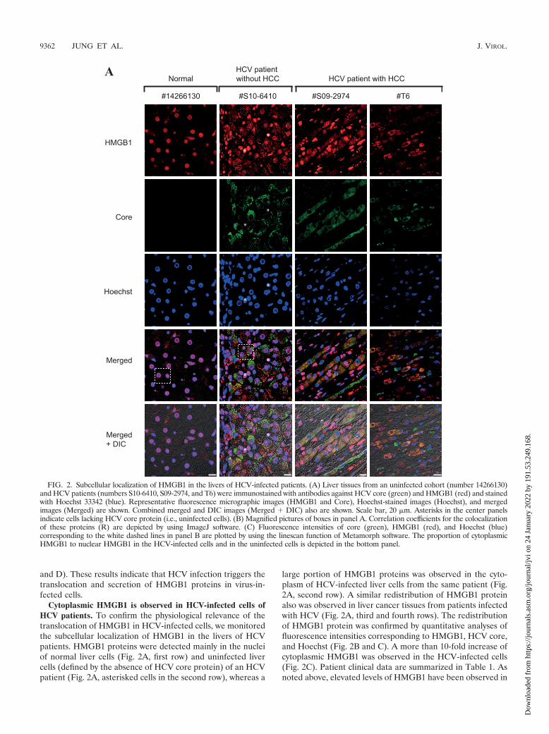
Cytoplasmic HMGB1 is observed in HCV-infected cells ofHCV patients. To confirm the physiological relevance of thetranslocation of HMGB1 in HCV-infected cells, we monitoredthe subcellular localization of HMGB1 in the livers of HCVpatients. HMGB1 proteins were detected mainly in the nucleiof normal liver cells (Fig. 2A, first row) and uninfected livercells (defined by the absence of HCV core protein) of an HCVpatient (Fig. 2A, asterisked cells in the second row), whereas a
large portion of HMGB1 proteins was observed in the cyto-plasm of HCV-infected liver cells from the same patient (Fig.2A, second row). A similar redistribution of HMGB1 proteinalso was observed in liver cancer tissues from patients infectedwith HCV (Fig. 2A, third and fourth rows). The redistributionof HMGB1 protein was confirmed by quantitative analyses offluorescence intensities corresponding to HMGB1, HCV core,and Hoechst (Fig. 2B and C). A more than 10-fold increase ofcytoplasmic HMGB1 was observed in the HCV-infected cells(Fig. 2C). Patient clinical data are summarized in Table 1. Asnoted above, elevated levels of HMGB1 have been observed in
FIG. 2. Subcellular localization of HMGB1 in the livers of HCV-infected patients. (A) Liver tissues from an uninfected cohort (number 14266130)and HCV patients (numbers S10-6410, S09-2974, and T6) were immunostained with antibodies against HCV core (green) and HMGB1 (red) and stainedwith Hoechst 33342 (blue). Representative fluorescence micrographic images (HMGB1 and Core), Hoechst-stained images (Hoechst), and mergedimages (Merged) are shown. Combined merged and DIC images (Merged � DIC) also are shown. Scale bar, 20 �m. Asterisks in the center panelsindicate cells lacking HCV core protein (i.e., uninfected cells). (B) Magnified pictures of boxes in panel A. Correlation coefficients for the colocalizationof these proteins (R) are depicted by using ImageJ software. (C) Fluorescence intensities of core (green), HMGB1 (red), and Hoechst (blue)corresponding to the white dashed lines in panel B are plotted by using the linescan function of Metamorph software. The proportion of cytoplasmicHMGB1 to nuclear HMGB1 in the HCV-infected cells and in the uninfected cells is depicted in the bottom panel.
9362 JUNG ET AL. J. VIROL.
Dow
nloa
ded
from
http
s://j
ourn
als.
asm
.org
/jour
nal/j
vi o
n 24
Jan
uary
202
2 by
191
.53.
249.
168.
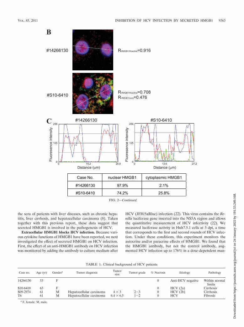
the sera of patients with liver diseases, such as chronic hepa-titis, liver cirrhosis, and hepatocellular carcinoma (8). Takentogether with this previous report, these data suggest thatsecreted HMGB1 is involved in the pathogenesis of HCV.
Extracellular HMGB1 blocks HCV infection. Because vari-ous cytokine functions of HMGB1 have been reported, we nextinvestigated the effect of secreted HMGB1 on HCV infection.First, the effect of an anti-HMGB1 antibody on HCV infectionwas monitored by adding the antibody to culture medium after
HCV (JFH/5aRluc) infection (22). This virus contains the Re-nilla luciferase gene inserted into the NS5A region and allowsthe quantitative measurement of HCV infectivity (22). Wemeasured luciferase activity in Huh7.5.1 cells at 3 dpi, a timethat corresponds to the first and second rounds of HCV infec-tion. Under these conditions, this experiment monitors theautocrine and/or paracrine effects of HMGB1. We found thatthe HMGB1 antibody, but not the control antibody, aug-mented HCV infection up to 176% in a dose-dependent man-
FIG. 2—Continued.
TABLE 1. Clinical background of HCV patients
Case no. Age (yr) Gendera Tumor diagnosis Tumorsize Tumor grade % Necrosis Etiology Pathology
14266130 53 F 0 Anti-HCV negative Within normallimits
S10-6410 63 F 0 HCV (2a) CirrhosisS09-2974 61 M Hepatocellular carcinoma 4 � 3 23 0 HCV (2b) CirrhosisT6 61 M Hepatocellular carcinoma 6.4 � 6.5 12 0 HCV Fibrosis
a F, female; M, male.
VOL. 85, 2011 INHIBITION OF HCV INFECTION BY SECRETED HMGB1 9363
Dow
nloa
ded
from
http
s://j
ourn
als.
asm
.org
/jour
nal/j
vi o
n 24
Jan
uary
202
2 by
191
.53.
249.
168.
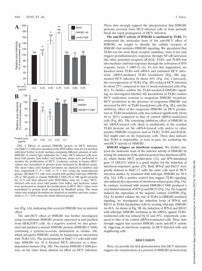
ner (Fig. 3A), indicating that secreted HMGB1 has an antiviraleffect.
The anti-HCV effect of HMGB1 was further investigatedusing recombinant HMGB1 protein expressed in and purifiedfrom HEK293FT cells. As a negative control, we also gener-ated and purified a mutant HMGB1 protein (HMGB1-C106S)containing a cysteine-to-serine substitution at residue 106,which abrogates HMGB1 activity by hampering its interactionwith TLR4 (41). The pretreatment of Huh7.5.1 cells with wild-type HMGB1 for 18 h blocked HCV infection in a dose-dependent manner (Fig. 3B). The mutant HMGB1-C106S pro-tein, on the other hand, showed no effect on HCV infection.
These data strongly support the interpretation that HMGB1proteins secreted from HCV-infected cells at least partiallyblock the rapid propagation of HCV infection.
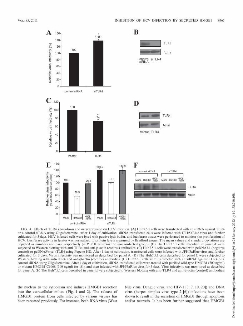
The anti-HCV activity of HMGB1 is mediated by TLR4. Tounderstand the molecular basis of the anti-HCV effect ofHMGB1, we sought to identify the cellular receptor ofHMGB1 that mediates HMGB1 signaling. We speculated thatTLR4 was the most likely receptor candidate, since it not onlytriggers proinflammatory responses through NF-B activationlike other potential receptors (RAGE, TLR2, and TLR9) butalso mediates antiviral responses through the activation of IFNresponse factor 3 (IRF-3) (2). To test this supposition, weknocked down TLR4 with siRNA and examined HCV infec-tivity. siRNA-mediated TLR4 knockdown (Fig. 4B) aug-mented HCV infection by about 40% (Fig. 4A). Conversely,the overexpression of TLR4 (Fig. 4D) reduced HCV infectionby about 25% compared to that of mock-transfected cells (Fig.4C). To further confirm the TLR4-mediated HMGB1 signal-ing, we investigated whether the knockdown of TLR4 rendersHCV replication resistant to exogenous HMGB1 treatment.HCV production in the presence of exogenous HMGB1 wasincreased by 80% in TLR4 knockdown cells (Fig. 4E), and theinhibitory effect of the exogenous HMGB1 on HCV produc-tion in TLR4 knockdown cells was reduced significantly (from44 to 28%) compared to that of control siRNA-transfectedcells (Fig. 4E). The remaining inhibitory effect of HMGB1 inthe siRNA-treated cells likely is attributable to the residualTLR4 proteins on the siRNA-treated cells and/or to otherputative HMGB1 receptors, such as TLR2, TLR9, and RAGE,that might exist on the hepatocyte cells. These data indicatethat TLR4 is responsible, at least in part, for mediating theanti-HCV activity of HMGB1.
HMGB1 triggers an interferon response. We further ana-lyzed the molecular basis of the antiviral activity of HMGB1 bytesting the induction of the antiviral protein beta interferon (IFN-�), which blocks HCV proliferation (13), and IFN-stimulatedgene 15 (ISG15), which is a good marker for the induction ofinterferon-responsive genes (33). Both IFN-� and ISG15 weregreatly induced in Huh7.5.1 cells, the same cells used in HCVinfection studies, by treatment with wild-type HMGB1 for 48 h(Fig. 5A). LPS, a positive control that triggers TLR4 signaling,also induced the expression of interferon-related genes (Fig. 5A).In contrast, treatment with mutant HMGB1-C106S produced avery limited induction of IFN-� and ISG15 (Fig. 5A). No reagentsinduced the expression of the negative control, GAPDH (Fig.5A). To further validate the role of TLR4 in HMGB1-mediatedsignaling, we investigated the induction levels of IFN-� andISG15 in TLR4 knockdown cells by treating wild-type HMGB1for 48 h. As shown in Fig. 5B, the induction of IFN-� and ISG15with wild-type HMGB1 treatment in the siRNA against TLR4-transfected cells was reduced by 63 and 39%, respectively, com-pared to that of the control siRNA-transfected cells. These datastrongly suggest that secreted HMGB1 exerts anti-HCV activityby triggering an interferon response in HCV-infected cells andneighboring cells.
DISCUSSION
Here, we provide the first demonstration that HCV infectiontriggers the translocation of a portion of HMGB1 protein from
FIG. 3. Effects of secreted HMGB1 protein on HCV infection.(A) Huh7.5.1 cells were inoculated with JFH/5aRluc virus for 4 h and thencultivated further in fresh medium containing different amounts of anti-HMGB1 or control IgG antibodies for 3 days. HCV-infected cells werelysed with passive lysis buffer, and luciferase assays were performed tomonitor the proliferation of HCV. Luciferase activity in lysates (RLUvalues) was normalized to protein levels measured by Bradford assays.The mean values and standard deviations are depicted as numbers andbars, respectively (�, P � 0.05; ��, P � 0.01 versus the mock-infectedgroup). (B) Huh7.5.1 cells were treated with purified wild-type HMGB1(30 or 300 ng/ml) or mutant HMGB1-C106S (30 or 300 ng/ml) proteinsfor 18 h and then infected with JFH/5aRluc virus for 3 days. HCV-infected cells were lysed with passive lysis buffer, and luciferase assayswere performed to monitor the proliferation of HCV. RLU values werenormalized to protein levels measured by Bradford assays. The meanvalues and standard deviations are depicted as numbers and bars, respec-tively (�, P � 0.05 versus the mock-infected group).
9364 JUNG ET AL. J. VIROL.
Dow
nloa
ded
from
http
s://j
ourn
als.
asm
.org
/jour
nal/j
vi o
n 24
Jan
uary
202
2 by
191
.53.
249.
168.
the nucleus to the cytoplasm and induces HMGB1 secretioninto the extracellular milieu (Fig. 1 and 2). The release ofHMGB1 protein from cells infected by various viruses hasbeen reported previously. For instance, both RNA virus (West
Nile virus, Dengue virus, and HIV-1 [3, 7, 10, 20]) and DNAvirus (herpes simplex virus type 2 [6]) infections have beenshown to result in the secretion of HMGB1 through apoptosisand/or necrosis. It has been further suggested that HMGB1
FIG. 4. Effects of TLR4 knockdown and overexpression on HCV infection. (A) Huh7.5.1 cells were transfected with an siRNA against TLR4or a control siRNA using Oligofectamine. After 1 day of cultivation, siRNA-transfected cells were infected with JFH/5aRluc virus and furthercultivated for 3 days. HCV-infected cells were lysed with passive lysis buffer, and luciferase assays were performed to monitor the proliferation ofHCV. Luciferase activity in lysates was normalized to protein levels measured by Bradford assays. The mean values and standard deviations aredepicted as numbers and bars, respectively (�, P � 0.05 versus the mock-infected group). (B) The Huh7.5.1 cells described in panel A weresubjected to Western blotting with anti-TLR4 and anti-�-actin (control) antibodies. (C) Huh7.5.1 cells were transfected with pcDNA3.1 (negativecontrol) or pcDNA3/myc-hTLR4 using Fugene HD. After 1 day of cultivation, transfected cells were infected with JFH/5aRluc virus and furthercultivated for 3 days. Virus infectivity was monitored as described for panel A. (D) The Huh7.5.1 cells described for panel C were subjected toWestern blotting with anti-TLR4 and anti-�-actin (control) antibodies. (E) Huh7.5.1 cells were transfected with an siRNA against TLR4 or acontrol siRNA using Oligofectamine. After 1 day of cultivation, siRNA-transfected cells were treated with purified wild-type HMGB1 (300 ng/ml)or mutant HMGB1-C106S (300 ng/ml) for 18 h and then infected with JFH/5aRluc virus for 3 days. Virus infectivity was monitored as describedfor panel A. (F) The Huh7.5.1 cells described in panel E were subjected to Western blotting with anti-TLR4 and anti-�-actin (control) antibodies.
VOL. 85, 2011 INHIBITION OF HCV INFECTION BY SECRETED HMGB1 9365
Dow
nloa
ded
from
http
s://j
ourn
als.
asm
.org
/jour
nal/j
vi o
n 24
Jan
uary
202
2 by
191
.53.
249.
168.

protein secreted in response to viral infections contributes tothe pathogenesis of these infections (39). The elevation ofHMGB1 levels in the plasma of HCV patients with chronichepatitis, liver cirrhosis, and HCC (8) likely is attributable toboth the active secretion of HMGB1 and cytopathic effects ofHCV infection, even though we showed that the secretion ofHMGB1 can occur without killing the HCV-infected cells froman in vitro HCV culture system (Fig. 1D).
In the current study, we focused on the function of secretedHMGB1 protein. Using an anti-HMGB1 antibody and purifiedHMGB1 protein, we showed that secreted HMGB1 partiallyblocks HCV infection (Fig. 3). This is the first report demon-strating an anti-HCV effect of HMGB1. By monitoring theeffects of TLR4 knockdown and overexpression on HCV in-fection, we also showed that the anti-HCV effect of HMGB1 ismediated, at least in part, by TLR4 (Fig. 4). Moreover, weshowed that HMGB1 induces the expression of interferon-related genes in the hepatocellular carcinoma cell line(Huh7.5.1) used in HCV infection studies (Fig. 5). These datalead us to conclude that HCV infection induces the secretionof HMGB1 protein into the extracellular milieu, and secreted
HMGB1 triggers the production of antiviral proteins throughTLR4-mediated interferon responses. We speculate that theanti-HCV effect of HMGB1 at least partly contributes to lim-iting the propagation of HCV in HCV patients. Moreover,secreted HMGB1 is likely to trigger inflammatory responses(16) by activating neighboring immune cells, such as Kupffercells and hepatic dendritic cells, to release more proinflamma-tory cytokines. Therefore, HMGB1 by itself or in combinationwith other proinflammatory cytokines also may contribute tothe chronic inflammation of liver cells (hepatitis) in HCV pa-tients. Because TLR4 enhances the signaling of TGF-�, whichis the cytokine best known to trigger liver cirrhosis and pro-mote hepatic fibrosis in quiescent hepatic stellate cells (31),secreted HMGB1 also may be associated with liver cirrhosis,another pathological symptom caused by HCV infection. Theinvolvement of TLR4 in liver cirrhosis also was strongly sup-ported by a recent study indicating that a single-nucleotidepolymorphism of TLR4 is the key factor in significantly reduc-ing the risk of developing cirrhosis in patients with chronicHCV infection (18). These observations support the view thatsecreted HMGB1 functions as a double-edged sword: it pro-
FIG. 5. Induction of interferon-related genes by HMGB1 protein. (A) The mRNA levels of the interferon-related genes, IFN-� and ISG15, andthe control gene, GAPDH, in Huh7.5.1 cells were monitored by quantitative RT-PCR after treatment with HMGB1 (300 ng/ml), HMGB1-C106S(300 ng/ml), or LPS (2 �g/ml) for the indicated times. The changes in mRNA concentration, normalized to the level of control mRNA (�-actin)3 h after treatment, are depicted. The values presented in the graphs are means and standard deviations. (B) Huh7.5.1 cells were transfected withan siRNA against TLR4 or a control siRNA using Oligofectamine. After 1 day of cultivation, siRNA-transfected cells were treated with purifiedwild-type HMGB1 (300 ng/ml) or mutant HMGB1-C106S (300 ng/ml) for 48 h. The mRNA levels of IFN-�, ISG15, and �-actin were measuredas described for panel A (�, P � 0.05 versus the control siRNA-transfected group).
9366 JUNG ET AL. J. VIROL.
Dow
nloa
ded
from
http
s://j
ourn
als.
asm
.org
/jour
nal/j
vi o
n 24
Jan
uary
202
2 by
191
.53.
249.
168.

tects hepatocytes from infection while at the same time con-tributing to triggering and maintaining a pathological status(e.g., in hepatitis and liver cirrhosis). Confirming the roles ofHMGB1 in HCV pathology and the physiological activities ofHMGB1 during HCV infection will require further investiga-tion using appropriate animal model systems.
The molecular basis of the translocation of HMGB1 duringHCV infection is not fully understood. However, the mostlikely mechanism involves ROS generated in HCV-infectedcells. HCV proteins are known to cause endoplasmic reticulum(ER) stress, which results in the release of calcium from theER. The released calcium, in turn, reduces mitochondrialmembrane potential and increases ROS production (27). Forexample, HCV core and NS5A proteins induce oxidative stressin HCV-infected cells (15, 25, 26). In addition, it is known thatHMGB1 proteins are translocated from the nucleus to cyto-plasm or even secreted into the extracellular milieu underconditions in which intracellular ROS levels are elevated (36).We confirmed that ROS was elevated in HCV-infected cells(data not shown) and demonstrated that HMGB1 was secretedfrom cells treated with H2O2, which increased intracellularROS levels (data not shown). These observations suggest thatthe elevated levels of ROS in HCV-infected cells contribute, atleast in part, to the translocation of HMGB1.
Although this study did not focus on the role of cytoplasmicHMGB1, it is worth emphasizing the potential significance ofthe translocation of HMGB1 from the nucleus to the cyto-plasm (Fig. 1A and B and 2). One possibility is that cytoplasmicHMGB1 is involved in the induction of autophagy in HCV-infected cells. Autophagy is known to play an important role inhost immunity against virus infections (23). On the other hand,autophagy is known to be required for HCV replication (12),and the knockdown of proteins involved in autophagy en-hances the innate immune response in HCV-infected hepato-cytes (21, 32). A relationship between autophagy and HMGB1has been reported recently (35). The authors of this study showedthat cytoplasmic HMGB1 enhances autophagic flux by directlyinteracting with the autophagy protein Beclin1 and displacingBcl-2 (35). Studies on the role of cytoplasmic HMGB1 in HCVpathogenesis are under way in our laboratory.
In conclusion, HMGB1 protein mobilized by HCV infectionappears to play an important role in the pathogenesis of HCV-related diseases. Further investigations into the roles ofHMGB1 in the diseases caused by HCV infection will shedlight on and potentially help prevent these serious and preva-lent HCV-related diseases.
ACKNOWLEDGMENTS
We are grateful to Ralf Bartenschlager (University of Heidelberg)for the core antibodies and to Francis Chisari (Scripps Research In-stitute) for the Huh7.5.1 cell line.
This work was supported in part by grants from the 21C FrontierFunctional Proteomics Project (FPR08B1-220), Bio R&D Program(2010-0018167), World Class University Program (R31-10105), NCRC(2010-0028447), and BRL (2010-0019706) through the National Re-search Foundation funded by the Ministry of Education, Science andTechnology.
REFERENCES
1. Andersson, U., H. Erlandsson-Harris, H. Yang, and K. J. Tracey. 2002.HMGB1 as a DNA-binding cytokine. J. Leukoc. Biol. 72:1084–1091.
2. Andersson, U., and K. J. Tracey. 2010. HMGB1 is a therapeutic target forsterile inflammation and infection. Annu. Rev. Immunol. 29:139–162.
3. Barqasho, B., P. Nowak, S. Abdurahman, L. Walther-Jallow, and A. Son-nerborg. 2010. Implications of the release of high-mobility group box 1protein from dying cells during human immunodeficiency virus type 1 infec-tion in vitro. J. Gen. Virol. 91:1800–1809.
4. Bertoletti, A., and C. Ferrari. 2003. Kinetics of the immune response duringHBV and HCV infection. Hepatology 38:4–13.
5. Bianchi, M. E. 2009. HMGB1 loves company. J. Leukoc. Biol. 86:573–576.6. Borde, C., et al. 2011. Stepwise release of biologically active HMGB1 during
HSV-2 infection. PLoS One 6:e16145.7. Chen, L. C., T. M. Yeh, H. N. Wu, Y. Y. Lin, and H. W. Shyu. 2008. Dengue
virus infection induces passive release of high mobility group box 1 proteinby epithelial cells. J. Infect. 56:143–150.
8. Cheng, B. Q., et al. 2008. Serum high mobility group box chromosomalprotein 1 is associated with clinicopathologic features in patients with hep-atocellular carcinoma. Dig. Liver Dis. 40:446–452.
9. Choi, J., and J. H. Ou. 2006. Mechanisms of liver injury. III. Oxidative stressin the pathogenesis of hepatitis C virus. Am. J. Physiol. Gastrointest. LiverPhysiol. 290:G847–G851.
10. Chu, J. J., and M. L. Ng. 2003. The mechanism of cell death during WestNile virus infection is dependent on initial infectious dose. J. Gen. Virol.84:3305–3314.
11. Diamond, D. L., et al. 2010. Temporal proteome and lipidome profiles revealhepatitis C virus-associated reprogramming of hepatocellular metabolismand bioenergetics. PLoS Pathog. 6:e1000719.
12. Dreux, M., P. Gastaminza, S. F. Wieland, and F. V. Chisari. 2009. Theautophagy machinery is required to initiate hepatitis C virus replication.Proc. Natl. Acad. Sci. U. S. A. 106:14046–14051.
13. Festi, D., et al. 2004. Safety of interferon beta treatment for chronic HCVhepatitis. World J. Gastroenterol. 10:12–16.
14. Fisher, C. L., and G. K. Pei. 1997. Modification of a PCR-based site-directedmutagenesis method. Biotechniques 23:570–574.
15. Gong, G., G. Waris, R. Tanveer, and A. Siddiqui. 2001. Human hepatitis Cvirus NS5A protein alters intracellular calcium levels, induces oxidativestress, and activates STAT-3 and NF-kappa B. Proc. Natl. Acad. Sci. U. S. A.98:9599–9604.
16. Gremion, C., and A. Cerny. 2005. Hepatitis C virus and the immune system:a concise review. Rev. Med. Virol. 15:235–268.
17. Hoofnagle, J. H. 2002. Course and outcome of hepatitis C. Hepatology36:S21–S29.
18. Huang, H., et al. 2007. A 7 gene signature identifies the risk of developingcirrhosis in patients with chronic hepatitis C. Hepatology 46:297–306.
19. Ilmakunnas, M., et al. 2008. High mobility group box 1 protein as a markerof hepatocellular injury in human liver transplantation. Liver Transpl. 14:1517–1525.
20. Kamau, E., et al. 2009. Dengue virus infection promotes translocation ofhigh mobility group box 1 protein from the nucleus to the cytosol in dendriticcells, upregulates cytokine production and modulates virus replication.J. Gen. Virol. 90:1827–1835.
21. Ke, P. Y., and S. S. Chen. 2010. Activation of the unfolded protein responseand autophagy after hepatitis C virus infection suppresses innate antiviralimmunity in vitro. J. Clin. Investig. 121:37–56.
22. Kim, C. S., J. H. Jung, T. Wakita, S. K. Yoon, and S. K. Jang. 2007.Monitoring the antiviral effect of alpha interferon on individual cells. J. Vi-rol. 81:8814–8820.
23. Kim, H. J., S. Lee, and J. U. Jung. 2010. When autophagy meets viruses: adouble-edged sword with functions in defense and offense. Semin. Immuno-pathol. 32:323–341.
24. Kim, J. H., et al. 2003. Heterogeneous nuclear ribonucleoprotein C modu-lates translation of c-myc mRNA in a cell cycle phase-dependent manner.Mol. Cell. Biol. 23:708–720.
25. Korenaga, M., et al. 2005. Hepatitis C virus core protein inhibits mitochon-drial electron transport and increases reactive oxygen species (ROS) pro-duction. J. Biol. Chem. 280:37481–37488.
26. Okuda, M., et al. 2002. Mitochondrial injury, oxidative stress, and antioxi-dant gene expression are induced by hepatitis C virus core protein. Gastro-enterology 122:366–375.
27. Piccoli, C., et al. 2007. Hepatitis C virus protein expression causes calcium-mediated mitochondrial bioenergetic dysfunction and nitro-oxidative stress.Hepatology 46:58–65.
28. Rullier, A., et al. 2001. Immunohistochemical detection of HCV in cirrhosis,dysplastic nodules, and hepatocellular carcinomas with parallel-tissue quan-titative RT-PCR. Mod. Pathol. 14:496–505.
29. Scaffidi, P., T. Misteli, and M. E. Bianchi. 2002. Release of chromatin proteinHMGB1 by necrotic cells triggers inflammation. Nature 418:191–195.
30. Seeff, L. B. 2002. Natural history of chronic hepatitis C. Hepatology 36:S35–S46.31. Seki, E., et al. 2007. TLR4 enhances TGF-beta signaling and hepatic fibrosis.
Nat. Med. 13:1324–1332.32. Shrivastava, S., A. Raychoudhuri, R. Steele, R. Ray, and R. B. Ray. 2010.
Knockdown of autophagy enhances the innate immune response in hepatitisC virus-infected hepatocytes. Hepatology 53:406–414.
33. Skaug, B., and Z. J. Chen. 2010. Emerging role of ISG15 in antiviral immu-nity. Cell 143:187–190.
VOL. 85, 2011 INHIBITION OF HCV INFECTION BY SECRETED HMGB1 9367
Dow
nloa
ded
from
http
s://j
ourn
als.
asm
.org
/jour
nal/j
vi o
n 24
Jan
uary
202
2 by
191
.53.
249.
168.
34. Takeuchi, O., and S. Akira. 2010. Pattern recognition receptors and inflam-mation. Cell 140:805–820.
35. Tang, D., et al. 2010. Endogenous HMGB1 regulates autophagy. J. Cell Biol.190:881–892.
36. Tang, D., R. Kang, H. J. Zeh, and M. T. Lotze. 2010. HMGB1, oxidativestress, and disease. Antioxid. Redox Signal. 14:1315–1335.
37. Tang, D., et al. 2007. Hydrogen peroxide stimulates macrophages and mono-cytes to actively release HMGB1. J. Leukoc. Biol. 81:741–747.
38. Tsung, A., et al. 2007. HMGB1 release induced by liver ischemia involvesToll-like receptor 4 dependent reactive oxygen species production and cal-cium-mediated signaling. J. Exp. Med. 204:2913–2923.
39. Wang, H., M. F. Ward, X. G. Fan, A. E. Sama, and W. Li. 2006. Potential roleof high mobility group box 1 in viral infectious diseases. Viral Immunol.19:3–9.
40. Woodhouse, S. D., et al. 2010. Transcriptome sequencing, microarray, andproteomic analyses reveal cellular and metabolic impact of hepatitis C virusinfection in vitro. Hepatology 52:443–453.
41. Yang, H., et al. 2010. A critical cysteine is required for HMGB1 binding toToll-like receptor 4 and activation of macrophage cytokine release. Proc.Natl. Acad. Sci. U. S. A. 107:11942–11947.
42. Yang, H., H. Wang, and K. J. Tracey. 2001. HMG-1 rediscovered as acytokine. Shock 15:247–253.
9368 JUNG ET AL. J. VIROL.
Dow
nloa
ded
from
http
s://j
ourn
als.
asm
.org
/jour
nal/j
vi o
n 24
Jan
uary
202
2 by
191
.53.
249.
168.