Embed Size (px)
Citation preview
Hepatic HIF-2 down-regulates hepcidin expression in mice throughEPO-mediated increase in erythropoiesis
by Maria Mastrogiannaki, Pavle Matak, Jacques R.R. Mathieu, Stephanie Delga,Patrick Mayeux, Sophie Vaulont, and Carole Peyssonnaux
Haematologica 2011 [Epub ahead of print]
Citation: Mastrogiannaki M, Matak P, Mathieu JR, Delga S, Mayeux P, Vaulont S, and Peyssonnaux C. Hepatic HIF-2 down-regulates hepcidin expression in mice through EPO-mediated increase in erythropoiesis. Haematologica. 2011; 96:xxx doi:10.3324/haematol.2011.056119
Publisher's Disclaimer. E-publishing ahead of print is increasingly important for the rapid dissemination of science.Haematologica is, therefore, E-publishing PDF files of an early version of manuscripts thathave completed a regular peer review and have been accepted for publication. E-publishingof this PDF file has been approved by the authors. After having E-published Ahead of Print,manuscripts will then undergo technical and English editing, typesetting, proof correction andbe presented for the authors' final approval; the final version of the manuscript will thenappear in print on a regular issue of the journal. All legal disclaimers that apply to the journal also pertain to this production process.
Haematologica (pISSN: 0390-6078, eISSN: 1592-8721, NLM ID: 0417435, www.haemato-logica.org) publishes peer-reviewed papers across all areas of experimental and clinicalhematology. The journal is owned by the Ferrata Storti Foundation, a non-profit organiza-tion, and serves the scientific community with strict adherence to the principles of openaccess publishing (www.doaj.org). In addition, the journal makes every paper publishedimmediately available in PubMed Central (PMC), the US National Institutes of Health (NIH)free digital archive of biomedical and life sciences journal literature.
Official Organ of the European Hematology AssociationPublished by the Ferrata Storti Foundation, Pavia, Italy
www.haematologica.org
Early Release Paper
Support Haematologica and Open Access Publishing by becoming a member of the EuropeHematology Association (EHA) and enjoying the benefits of this membership, which inc
participation in the online CME?program
Copyright 2011 Ferrata Storti Foundation.Published Ahead of Print on December 29, 2011, as doi:10.3324/haematol.2011.056119.
1
Hepatic HIF-2 down-regulates hepcidin expression in mice through
epo-mediated increase in erythropoiesis
Maria Mastrogiannaki1,2,3, Pavle Matak1,2,3, Jacques R.R. Mathieu1,2,3, Stéphanie Delga1,2,3,
Patrick Mayeux1,2,3, Sophie Vaulont1,2,3 , and Carole Peyssonnaux1,2,3
1INSERM, U1016, Institut Cochin, Paris, France; 2CNRS, UMR8104, Paris, France, and
3Université Paris Descartes, Sorbonne Paris Cité, Paris, France
Correspondence Carole Peyssonnaux, PhD, Institut Cochin, Faculté de Médecine Cochin Port Royal, 24, rue du Faubourg Saint Jacques 75014 Paris, France. Phone: international +33.1.44412452. Fax: international +33.1.44412421. E-mail : [email protected]
DOI: 10.3324/haematol.2011.056119
2
ABSTRACT
Background
Iron metabolism, regulated by the iron hormone hepcidin, and oxygen homeostasis, dependent
on HIF-hypoxia inducible factors, are strongly interconnected. We previously reported that in
mice where both liver HIF-1 and HIF-2 are stabilized (the hepatocyte VHL knockout mouse
model), hepcidin expression was strongly repressed and we hypothesized that HIF-2 could be
the major regulatory component contributing to the hepcidin down-regulation.
Design and methods
We generated and analyzed hepatocyte-specific knockout mice harboring either HIF-2α
deficiency (Hif2a KO) or constitutive HIF-2α stabilization (Vhlh/Hif1a KO) and ex vivo
systems (primary hepatocyte cultures). Hif2a KO mice were fed an iron deficient diet for
two months and Vhlh/Hif1a KO treated with neutralizing EPO antibody.
Results
We demonstrate that HIF-2 is dispensable in hepcidin gene regulation in the context of an
adaptive response to iron deficiency anemia. However, its overexpression in the double
Vhlh/Hif1a hepatocyte-specific knockout mice, indirectly down-regulates hepcidin
expression through increased erythropoiesis and EPO production. Experiments in
primary hepatocytes confirmed the non-autonomous role of HIF-2 in hepcidin regulation.
Conclusion
While our results indicate that HIF-2 is not directly involved in hepcidin repression,
they highlight the contribution of hepatic HIF-2 to repress hepcidin through EPO-mediated
increased erythropoiesis, a result of potential clinical interest.
DOI: 10.3324/haematol.2011.056119
3
INTRODUCTION
Adequate supply of oxygen in tissues is necessary for survival and normal organ
function. The number of circulating red blood cells is the major determinant of tissue
oxygenation. As a consequence, lack of oxygen (hypoxia) triggers induction of erythropoietin
(EPO) to increase the production of new red blood cells. The liver is the primary source of
EPO during embryogenesis, however in adults, the site of EPO production switches from the
fetal liver to the kidney (1).
Hypoxia-Inducible Factor-1 (HIF-1) was initially identified in 1992 as a
transcriptional factor able to regulate EPO production (2). A number of laboratories have
since demonstrated HIF-1 to be implicated in most aspects of hypoxia-induced gene
expression, and to operate not only in kidneys but also in a wide range of organs and cell
types. HIF is a heterodimeric transcription factor stabilized by low oxygen concentrations.
HIF consists of two helix-loop-helix proteins: a α-regulatory subunit, which is the oxygen and
iron-responsive component, and the β-subunit, also known as the aryl hydrocarbon receptor
nuclear translocator (ARNT), which is constitutively expressed. Three regulatory HIF
subunits have been cloned and named HIF-1α, HIF-2α and HIF-3α. In the presence of
oxygen, the regulatory subunit is hydroxylated on two proline residues by the oxygen and
iron-dependent prolyl-hydroxylases (PHDs); the α subunit is then targeted for degradation to
the proteasome by interacting with the von Hippel-Lindau (VHL) E3 ubiquitin ligase tumor
suppressor protein. Under hypoxia, the activity of PHDs is inhibited, therefore permitting
HIF-α to escape degradation and translocate into the nucleus, where it binds to HIF-β/ARNT.
The HIF heterodimer binds to hypoxic response elements (HREs) of target gene regulatory
sequences, and recruits transcriptional cofactors such as CBP and p300 to induce the
DOI: 10.3324/haematol.2011.056119
4
expression of genes involved in the control of metabolism and angiogenesis, as well as
apoptosis, cellular stress and other critical processes. More than a decade after the discovery
of HIF which led in turn to the discovery of VHL and the PHDs, mutations in genes encoding
all three of these essential components of the oxygen-sensing axis have been identified in
humans with familial erythrocytosis (3).
To perform their duty as oxygen carriers, erythrocytes require iron and failure to
incorporate adequate iron into heme results in impaired erythrocyte maturation, leading to
microcytic, hypochromic anemia. The most important systemic factors that influence iron
availability is hepcidin, a circulating peptide that maintains iron homeostasis. Hepcidin is an
hyposideremic hormone made predominantly by the hepatocytes acting to down-regulate iron
absorption by the duodenal enterocytes and iron release by the macrophages (4).
Hepcidin expression is increased by iron loading, thus avoiding excess of free toxic iron, and
decreased in response to hypoxia, iron deficiency and increased erythropoiesis allowing iron
supply to match the erythropoietic demand (4).
Recently, the BMP6 (Bone Morphogenetic Protein 6) / HJV (hemojuvelin) signaling
cascade has emerged as the principal pathway to regulate hepcidin gene expression (4). BMP6
signals through a BMPR receptor complex requiring HJV as a co-receptor. In addition, type II
Transmembrane Serine Proteinase (TMPRSS6 encoding the liver matriptase 2) has recently
been identified as a repressor of hepcidin gene expression (5) able to antagonize hepcidin
induction by BMP6 by cleaving HJV from the cell membrane (6). Finally, HJV was shown to
be cleaved by the furin proconvertase, which releases a soluble form of HJV (sHJV) that
suppresses BMP signaling and hepcidin expression by acting as a decoy that competes with
membrane HJV for BMP ligands (7).
DOI: 10.3324/haematol.2011.056119
5
We previously reported that HIF factors control iron homeostasis by repressing
hepcidin synthesis in the liver (8) and VHLR200W mutation has been shown to be associated
with downregulation of hepcidin expression in chuvash polycythemia patients (9). A marked
down-regulation of hepcidin was observed in conditional knockout of Vhlh in the liver, in
which both HIF-1 and HIF-2 were stabilized (8). However, deletion of Hif1a alone in the liver
of adult mice accounted only for a small fraction of hepcidin repression in response to an iron
deficient diet, thus possibly suggesting that HIF-2 may be a putative candidate contributing to
the observed hepcidin down-regulation.
To answer this question and to further determine the molecular mechanisms,
direct or indirect, of HIF factors in the regulation of hepcidin gene expression, we
developed and analyzed hepcidin expression by a combination of in vivo (hepatocyte-
specific knockout mice harboring either HIF-2α deficiency or constitutive HIF-2α
stabilization) and ex vivo systems (primary hepatocyte cultures). In this study, we
demonstrate that HIF-2 is not involved in the repression of hepcidin in the setting of
iron deficiency. However, we show that its overexpression in the double Vhlh/Hif1a
hepatocyte-specific knockout mice, indirectly down-regulates hepcidin expression
through increased erythropoiesis and EPO production, and not through transcriptional
activation of TMPRSS6, the negative regulator of the BMP/HJV pathway, as recently
suggested in vitro (10).
DESIGN AND METHODS
Animals.
All mice used in the experiments were cared for according to criteria outlined by the
European Convention for the Protection of Laboratory Animals. Animal studies described
DOI: 10.3324/haematol.2011.056119
6
here were reviewed and approved (Agreement n° P2.CP.151.10.) by the “Président du Comité
d'Ethique pour l'Expérimentation Animale Paris Descartes”. Mice with hepatocyte-specific
inactivation of Hif2a (referred as Hif2a KO) were generated by cross-breeding Albumin-Cre
transgenic mice with Hif2alox/lox mice (provided by Celeste Simon, University of
Pennsylvania) and compared to wild type littermates (Hif2a WT). Mice with hepatocyte-
specific inactivation of both Hif1a and Vhlh (Vhlhlox/lox/ Hif1alox/lox/AlbuminCre, referred as
Vhlh/Hif1a KO) were generated by breeding Vhlhlox/lox/ Hif1alox/lox and Vhlhlox/lox/ Hif1alox/lox
AlbuminCre mice. 3 to 4 week-old Vhlh/ Hif1a KO males and females were used and
compared to littermates of all other genotypes. All mouse strains were on a C57BL/6
background. When indicated, 4 week-old male mice were fed an iron deficient diet for 2
months (3 ppm iron; Scientific Animal Food & Engineering).
Treatment with anti-EPO blocking serum.
3 week-old mice were injected with Anti-Erythropoietin (Anti-EPO) rabbit serum or
0,5M NaCl (placebo). Injections were performed for 5 consecutive days and mice were
sacrificed 18 hours after the last injection. The neutralizing capacity of the anti-Epo serum
being 50 ng of recombinant EPO (5 Epo units) for 100 µl of serum antibody and the estimated
amount of EPO in a normal 3-week-old mice being 0.25 ng, we injected 300 µl of a 1:60
NaCl dilution of the Anti-EPO serum/day, i.e. a dose able to neutralize a ten fold excess of
circulating EPO. Assessing that circulating EPO levels was at least 60 times increased in the
Vhlh/Hif1a KO mice, these mice received 300 µl of the original Anti-EPO serum/day.
Reticulocytes and red blood cells counts
Haematological parameters were measured using a Coulter MAXM automatic analyzer
(Beckman Coulter) as previously described (11). Reticulocytes counts were determined
DOI: 10.3324/haematol.2011.056119
7
according to Lee et al. (12). Briefly, 1 µl of total blood was incubated with 1 ml of thiazole
orange dye solution (100 ng/ml) in phosphate buffer saline (PBS) for 15 minutes at room
temperature prior analysis. Unstained blood sample was used as negative control. All samples
were analyzed on a FACSCanto II (BD Biosciences).
EPO ELISA
EPO protein levels in plasma were determined using the Quantikine Mouse EPO ELISA kit
(R&D Systems).
Isolation and culture of primary hepatocytes.
Hepatocytes were isolated from 2-4 month old Hif2a KO and Hif2a WT mice of matched sex.
Hepatocytes were seeded in 6-well plates at a density 300.000 cells/well and cultured at
standard conditions (5% CO2, 37°C) in M199 medium containing 2% Ultroser G, for 4 h
(adapted from (13)). After cell attachment, the medium was replaced by fresh M199 medium
supplemented with 10% calf serum (Invitrogen).
Generation of the HIF-2α adenovirus construct
Human HIF-2α adenovirus constructs used to transfect primary hepatocytes were generated
by subcloning the 2.6 kb human EPAS1 ORF into the pAd-Track-CMV vector, followed by
recombination with the pAdEasy-1 vector and transfection into the HEK293-AV packaging
cell line as outlined (14).
Reverse transcription and real-time quantitative PCR.
Total RNA was extracted from whole liver or primary hepatocytes and homogenized in 1 ml
of TRIZOL reagent (Invitrogen). Reverse transcription was done with 2-3 µg of total RNA.
DOI: 10.3324/haematol.2011.056119
8
Quantitative PCR was performed with 2 µl of a 1:10 dilution of reverse-transcribed total RNA
and 10 µM of each primer diluted in 1× LightCycler DNA Master SYBR Green I mix using a
LightCycler apparatus (Roche Applied Science). All samples were normalized to the
threshold cycle value for 18S or Cyclophilin-A. The following primer sequences were used :
Hepcidin1 forward 5’-CCTATCTCCATCAACAGAT-3’; Hepcidin1 reverse 5’-
TGCAACAGATACCACACTG-3’; EPO forward 5’-CACAACCCATCGTGACATTTTC-3’;
EPO reverse 5’-CATCTGCGACAGTCGAGTTCTG-3’; Furin forward 5’-
CAGCCTCGGTACACACAGAT-3’; Furin reverse 5’-AGCTACACCTACGCCACAGA-3’;
Tmprss6 forward 5’-CCTGGTGAGTTCCTCTGCTC-3’; Tmprss6 reverse 5’-
CTTGGCACTGTTCTTCGTCA-3’; Hif1a forward 5’-TGAGCTTGCTCATCAGTTGC-3’;
Hif1a reverse 5’-CCATCTGTGCCTTCATCTCA-3’; Hif2a forward 5’-
TGAGTTGGCTCATGAGTTGC-3’; Hif2a reverse 5’-TTGCTGATGTTTTCCGACAG-3’;
BMP6 forward 5’-GTTCCGCGTCTACAAGGACT-3’; BMP6 reverse 5’-
CAGCCAACCTTCTTCTGAGG-3’; Hjv forward 5’-TCTGACCTGAGTGAGACTGC-3’;
Hjv reverse 5’-GATGATGAGCCTCCTACCTA-3’.
Statistical analysis.
All values in the figures are expressed as mean ± SEM. Statistical analysis was performed
using GraphPad Prism 4.0 and unpaired (2-tailed) Student’s t-test or one-way ANOVA
analysis followed by a Bonferroni post test when more than 2 groups were compared
respectively. Statistical significance is indicated by * symbols (* p< 0.05, ** p< 0.01, *** p<
0.001).
DOI: 10.3324/haematol.2011.056119
9
RESULTS
HIF2-α is not involved in the regulation of hepcidin gene expression in the
setting of iron deficiency anemia
To investigate the role of HIF-2α in hepcidin regulation, we have generated mice
deficient for Hif2a specifically in hepatocytes by breeding Hif2alox/lox mice with a transgenic
strain expressing the Cre recombinase under the control of the murine albumin promoter.
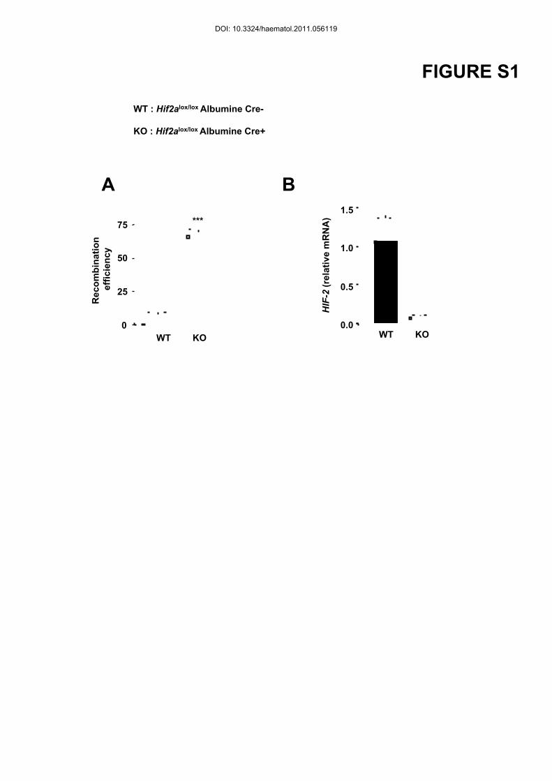
Deletion efficiency of Hif2a in the liver was ~66% as determined by quantitative PCR on
genomic DNA from liver. However, HIF-2α mRNA levels were decreased by 95% in primary
hepatocytes derived from Hif2a KO versus Hif2a WT littermates (Figure S1). In the liver of
Hif2a KO and WT littermates on a standard diet, hepcidin mRNA levels were similar.
Following an eight week iron deficient diet, livers from WT and Hif2a KO mice showed a
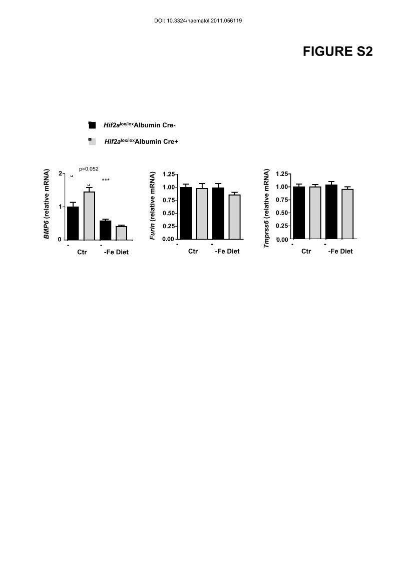
similar repression of hepcidin mRNA (Figure 1A). BMP6 mRNA levels were decreased in
both WT and Hif2a KO mice, under iron deficiency compared to controls (Figure S2).
Interestingly, plasma erythropoietin (EPO) was also found similarly upregulated in both Hif2a
KO and WT mice (Figure 1A). While EPO levels were undetectable in the liver (data not
shown), renal EPO was strongly induced in both WT and Hif2a KO mice under iron
deficiency indicating that the increase of systemic EPO in iron deficiency is not dependent
upon hepatic HIF-2α (Figure 1A). Haematological parameters, such as red blood cell content,
hematocrit and hemoglobin levels were also similarly decreased in the Hif2a KO and WT
littermates fed on the iron deficient diet compared to mice on a standard diet (Figure 1B). No
significant differences in plasma or liver iron were measured between WT and Hif2a KO
DOI: 10.3324/haematol.2011.056119
10
mice (Figure 1C). Our findings suggest that, while HIF-1α is a weak repressor of hepcidin
(8), HIF-2 is not involved in hepcidin gene regulation in the context of an adaptive response
to iron deficiency anemia.
Constitutive HIF-2 activation in hepatocyte-specific Vhlh/Hif1a null mice
represses hepcidin
To examine the effect of HIF-2 stabilization on hepcidin expression, we
generated, on a pure C57BL/6 background, the Vhlh/Hif1a KO murine model, which lacks
both VHL and HIF-1α in hepatocytes. These mice, similarly to the Vhlh KO mice - which
constitutively express both HIF-1α and HIF-2α (8, 15)- died between 3 and 5 weeks of age,
presented with alopecia, weight loss, severe hepatomegaly and splenomegaly as well as iron
deficiency (Figure 2A). We found that, as compared to control littermates, hepatic hepcidin
mRNA levels were repressed in the Vhlh/Hif1a KO (Figure 2B), as strongly as in the Vhlh
KO model previously described ((8) and Figure S3). Whether hepcidin is regulated directly or
through alternative pathways by HIF has been the subject of intense research in vitro (10, 16-
18). Our previous in vitro studies suggested a direct regulation of hepcidin by HIF (8).
However, our in vivo data could not exclude indirect alternative pathways. Furin gene
expression was demonstrated to be increased by hypoxia via HIF-1 (7, 19). It was therefore
proposed that this up regulation of furin could lead to the increase of sHJV production and, in
turn, hepcidin gene repression. However, we found that in the liver of Vhlh/Hif1a KO mice,
furin mRNA levels were decreased rather than increased compared with control littermates
(Figure 2B). More recently, it has been shown, in vitro, that TMPRSS6 expression was
upregulated by both HIF-1α and HIF-2α in hepatoma cell lines, leading to an increase in
membrane HJV shedding and a decrease in hepcidin promoter responsiveness (10). In vivo,
DOI: 10.3324/haematol.2011.056119
11
we found that TMPRSS6 mRNA levels were decreased in Vhlh/Hif1a KO (Figure 2B) as
compared to WT littermates suggesting that hepcidin repression by HIF-2 is not a
consequence of an increase in TMPRSS6 signaling.
The double Vhlh/Hif1a KO mutant mice showed a strong increase in erythropoiesis and an
upregulation of EPO mRNA in the liver and in the serum as compared to WT mice (Figure
2C), correlated to high levels of hemoglobin and reticulocyte counts (Figure 2D).
Interestingly, the increase of EPO mRNA in the kidney seen in the 3 week-old control mice
has been blunted in the double Vhlh/Hif1a KO mutant mice as if the forced dramatic increase
in liver EPO levels has altered the regulation of EPO mRNA in the kidney (Figure 2C).
Altogether, these results confirmed that HIF-2 but not HIF-1 is the main regulator of hepatic
EPO production (20) and hepcidin repression in the Vhlh-deficient background.
Hepcidin repression by HIF-2 is dependent on EPO-mediated increased
erythropoiesis
EPO and increased erythropoiesis have been reported to decrease hepcidin levels in
mice (21-23) and in humans (24). EPO has been shown to be a HIF-2 target gene in both liver
(20) and kidney (25). We therefore examined whether increased erythropoiesis as a
consequence of HIF-2 stabilization in the liver was the underlying cause of hepcidin
reduction. To this aim, inbred Vhlh/Hif1a KO mice and control littermates were injected with
neutralizing EPO antiserum for five consecutive days.
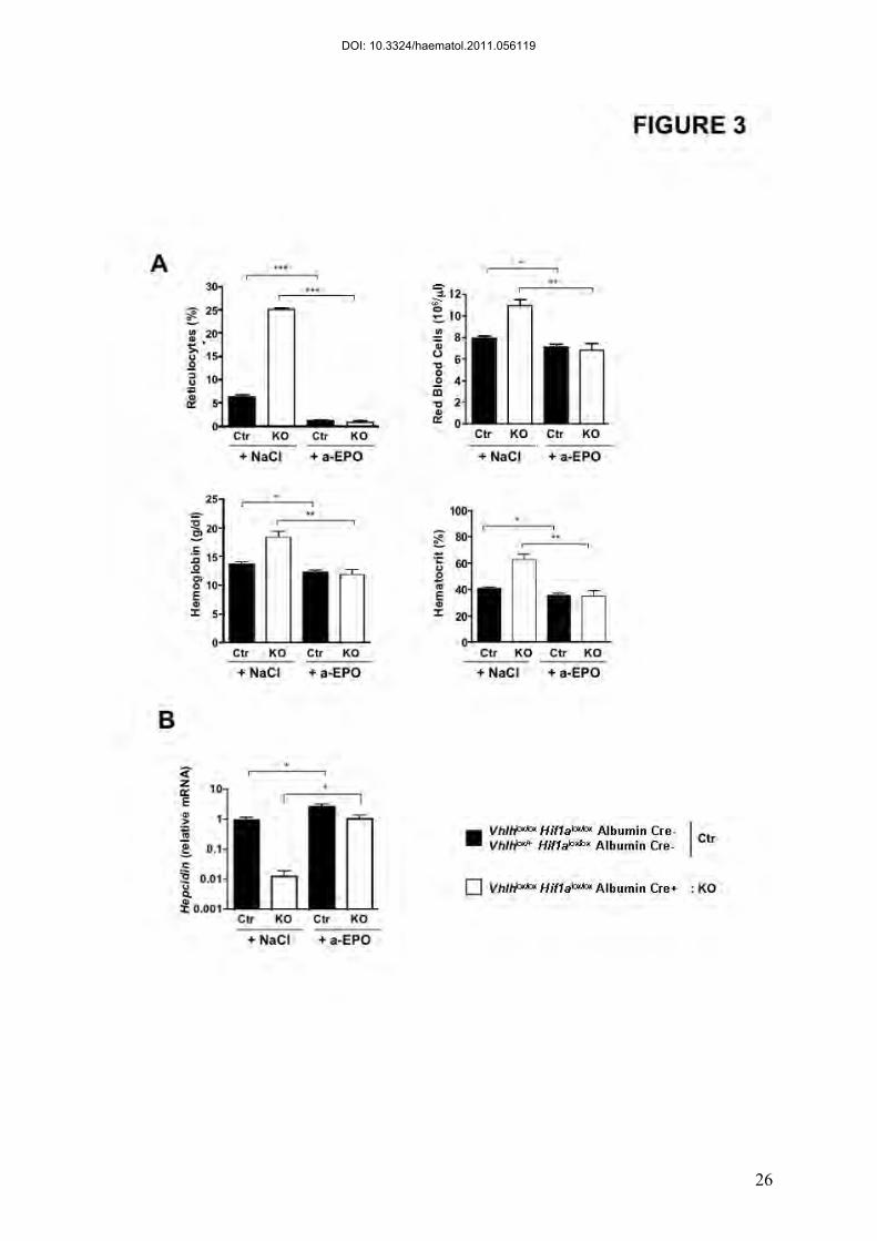
Upon anti-EPO injection, erythropoiesis was dramatically reduced in both control and
Vhlh/Hif1a KO mice, as measured by reticulocyte counts (Figure 3A). As expected, the blood
DOI: 10.3324/haematol.2011.056119
12
parameters (Red Blood Cells, Hemoglobin, Hematocrit) were consecutively decreased in both
control and Vhlh/Hif1a KO mice after neutralizing EPO antibody injection (Figure 3A).
Decrease of erythropoiesis increased hepcidin mRNA levels in control mice as
previously reported (23) (Figure 3B). Interestingly, hepcidin levels in Vhlh/Hif1a KO mice
injected with neutralizing EPO serum returned to values similar to NaCl-injected control mice
(Figure 3B). Noteworthy, in conditions of abolished erythropoiesis, hepcidin mRNA values in
Vhlh/Hif1a KO mice were still slightly lower than control mice.
Altogether, these results suggest that stabilized HIF-2α in the liver does not repress
hepcidin directly but through an erythropoietic drive due to EPO overexpression.
Cell-autonomous regulation of hepcidin expression is independent of HIF-2
To further ensure the absence of a direct effect of HIF-2 on hepcidin gene expression,
we sought to analyze hepcidin gene expression in primary hepatocytes, independently of the
systemic effect of erythropoiesis. We first validated our model of primary hepatocytes by
showing conditions (BMP2 treatment or infection by an adenovirus expressing TMPRSS6)
where hepcidin can be either up- or down-regulated (Figure S4). For the purpose of our study,
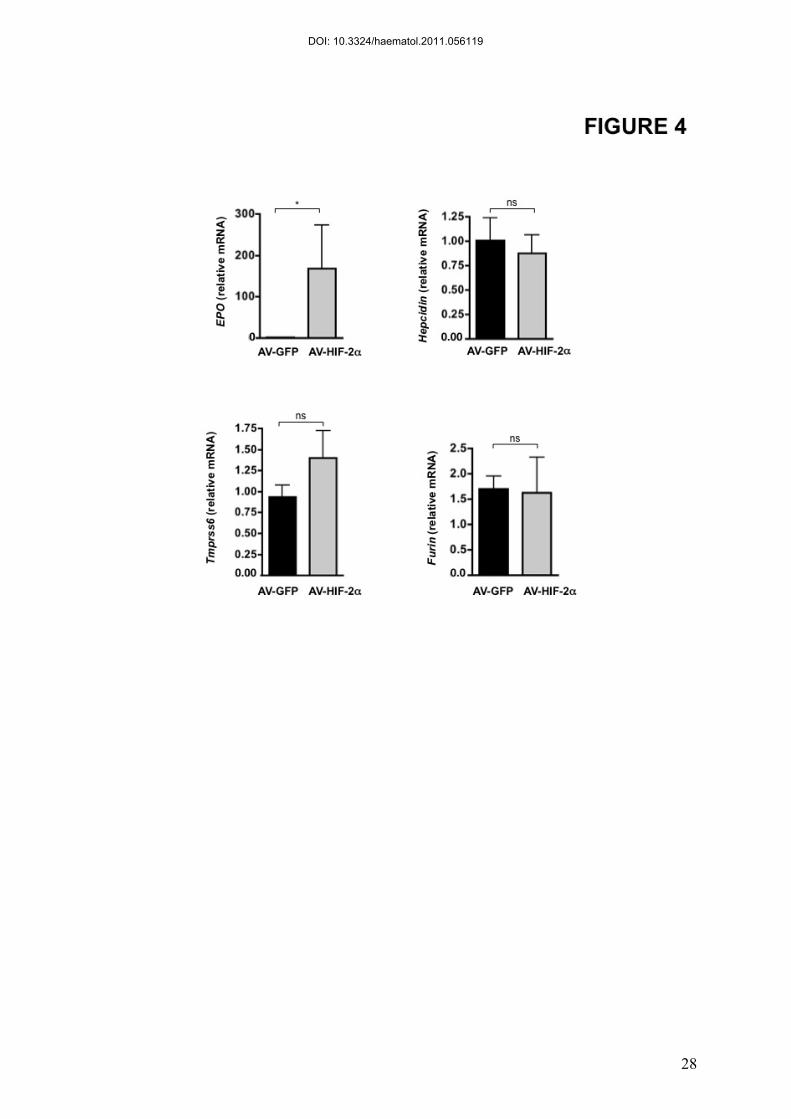
we generated HIF-2α overexpressing adenovirus (AV-HIF-2). Primary hepatocytes were
either infected with a GFP overexpressing adenovirus as a control or AV-HIF-2 at a
Multiplicity Of Infection (MOI) of 10. While AV-HIF-2 promotes a 160 fold induction of
EPO levels compared to AV-GFP infection, hepcidin levels were not affected by HIF-2
overexpression (Figure 4). In our model of primary hepatocytes, we also confirmed the
absence of regulation of TMPRSS6 and furin by HIF-2. Altogether, these data suggest that in
DOI: 10.3324/haematol.2011.056119
13
vitro stabilization of HIF-2α alone in primary mouse hepatocytes is not sufficient to repress
hepcidin expression.
DISCUSSION
The current study addresses the role of hepatic HIF-2 in the repression of hepcidin
expression by the use of complementary ex vivo and in vivo experimental models. Models of
dietary iron deficiency and of constitutive HIF-2 activation (in hepatocyte-specific VHL HIF-
1α KO mice and in primary hepatocytes) have been used.
Our previous work, using the iron chelator DFO as a HIF stabilizer, suggested a direct
repression of hepcidin by HIF-1 in vitro (8). However, other studies in HepG2 cells showed
that overexpression or knockdown of HIF-1 did not affect hepcidin expression (18) and have
suggested indirect pathways including 2-oxoglutarate (16) or ROS (17) dependent pathways.
Moreover, HIF-2 but not HIF-1 seems to play a major role in iron metabolism and
erythropoiesis (26). HIF-2 regulates EPO and key iron-related genes in the liver (25) and we
recently showed that HIF-2 but not HIF-1 regulates iron absorption in enterocytes (11).
In a model of constitutive HIF-2 activation in hepatocytes (Vhlh/Hif1a KO), we found
hepcidin to be strongly repressed. We aimed to determine by which pathway HIF-2 regulates
hepcidin in vivo in this model. HIF-2 may down-regulate hepcidin expression by affecting the
BMP/HJV pathway. BMP-6, recently shown to be an important hepcidin regulator, is
increased in models with hepatic iron overload, and decreased in response to iron deficiency
(27-29). However, BMP6 mRNA levels were found to be increased, rather than decreased, in
the Vhlh/Hif1a KO model and cannot account for the hepcidin diminution (Figure S3). HJV
DOI: 10.3324/haematol.2011.056119
14
mRNA levels were decreased in livers from Vhlh/Hif1a KO mice as compared to WT
littermates (Figure S3). Furin, which cleaves HJV and produce sHJV, has been identified as a
HIF-1 target gene and could also have affected hepcidin expression by decreasing the
BMP/HJV pathway (7, 19). However, furin mRNA levels were not increased in the liver of
mice overexpressing HIF-2 (Figure 2) as well as in VhlhKO overexpressing both HIF
isoforms (data not shown). Lakhal and colleagues recently demonstrated in vitro that
TMPRSS6 is a new HIF-1 and HIF-2 target gene. In hepatoma cells, both HIF stabilization by
hypoxia or chemical inducers induce an increase in TMPRSS6 mRNA, therefore decreasing
transcriptional activity of the hepcidin promoter by impairing BMP/ HJV signaling (10).
However, we found a decrease rather than an increase in TMPRSS6 mRNA levels in our HIF-
2 overexpression model in vivo. The decrease in HJV, furin and TMPRSS6 mRNA levels
observed in the liver of Vhlh/Hif1a KO mice may be due to an in vivo compensatory
mechanism as the levels of these genes were unchanged in primary hepatocytes upon AV-
HIF-2 infection. The expression of these genes were not changed neither in the Hif2a KO and
WT littermates fed on the iron deficient diet compared to mice on a standard diet (Figure S2).
Accordingly, Krijt et al. recently reported that tissue hypoxia, resulting from repeated
phlebotomies, does not transcriptionally regulate furin or TMPRSS6 (30).
We next addressed the putative role of EPO in hepcidin repression in our model since
EPO administration is known to result in a strong down-regulation of hepcidin expression
(21). Irradiation of the bone marrow or inhibition of erythropoiesis by chemical inhibitors in
mice abolished the effect of EPO treatment or phlebotomy on hepcidin expression (22, 23),
suggesting that the action of EPO in repressing hepcidin was not direct but relied on the
erythropoietic activity. However, Pinto et al. suggested a direct effect of EPO on hepcidin
expression through EPOR-mediated regulation of the transcription factor C/EBPα (31).
DOI: 10.3324/haematol.2011.056119
15
Nevertheless, we did not detect EPOR expression in liver cells using highly sensitive
detection methods (32). Since EPO is preferentially regulated by HIF-2 but not by HIF-1 in
the liver (20), we examined whether HIF-2 contributes to hepcidin repression through the
transcription of its target gene EPO and the subsequent increase of erythropoiesis. Our in vivo
data clearly demonstrated that hepcidin repression by HIF-2 was due to the increase in
erythropoiesis. However, while the neutralizing EPO antibody completely reversed the
increased erythropoiesis in Vhlh/Hif1a KO mice, hepcidin levels remain slightly lower in the
knockout compared to the control mice. This difference may be due to a dose or timing effect
of the neutralizing EPO serum.
Regulation of hepcidin by erythropoietic activity is of particular importance in iron
loading anemias such as β-thalessemias. Patients with thalassemia intermedia present very
low hepcidin levels, despite high serum iron, and develop lethal tissue iron overload (33).
However, so far, the underlying mechanism by which erythropoiesis decreases hepcidin levels
has not yet been demonstrated. A hypothesis has been put forward, that a plasma circulating
erythroid factor would be responsible for this regulation. GDF15 and TWSG1 have been
proposed as potential candidates (34, 35). However, administration of EPO to healthy
volunteers has been shown to suppress circulating hepcidin, independently of GDF15 (24).
The development of the Hif2a KO mice has allowed us to demonstrate that HIF-2 is
not involved in hepcidin repression triggered by iron deficiency anemia. Several HIF-2-
independent mechanisms could contribute to this process. As previously mentioned,
TMPRSS6 is required in the liver for sensing the iron deficiency and decrease hepcidin
expression (36). TMPRSS6 could therefore contribute to hepcidin down-regulation observed
in our models of iron deprivation. A decrease in the BMP signaling could also be considered,
as previously suggested (27).
DOI: 10.3324/haematol.2011.056119
16
Interestingly, it was recently proposed that hypoxia inhibits hepcidin expression in
hepatoma cells via a marked decrease in SMAD4 mRNA expression (37), indicating that the
involvement of the BMP/SMAD signaling in hypoxia deserves further investigations. In vivo,
hypoxia has been shown to decrease hepcidin expression in both acute and chronic
experimental models (21, 38, 39) as well as in humans (40). Hypoxia is also known to induce
the expression of renal EPO, therefore promoting increased erythropoiesis to compensate for
the decrease in ambient oxygen. Therefore, the delayed hepcidin decrease in response to
hypoxia in vivo could be explained by the time needed to mount an increase in erythropoietic
activity. In support to that, hepcidin was less repressed in experimental models of short-term
hypoxia in vivo than in response to EPO injection or experimental models of increased
erythropoiesis induced by phlebotomy (38).
It has recently been reported that hepatocyte-derived HIF-2 can substitute as the main
regulator of systemic EPO homeostasis when EPO production in the kidney is impaired (25).
Interestingly, in the Vhlh/Hif1a KO mouse model, we show that liver EPO is contributing to
plasma EPO. The lack of renal EPO production could arise by several mechanisms and awaits
further investigations.
Importantly, our study showing the ability of hepatic HIF-2 to repress hepcidin
through increased erythropoiesis in order to increase iron absorption may be clinically
interesting in the treatment of patients with chronic kidney disease, resulting in renal anemia.
DOI: 10.3324/haematol.2011.056119
17
ACKNOWLEDGMENTS
We are very grateful to Jean-Christophe Deschemin for helpful technical contribution. We
also thank the Immunobiology platform of the Cochin Institute for help with FACS analysis.
This study was supported by a funding from the Agence Nationale pour la Recherche (ANR-
08-JCJC-0123 and ANR-08-GENO) and the European Research Council under the European
Community’s Seventh Framework Program (FP7/2011-2015 Grant agreement no 261296).
M.M. is supported by a fellowship from the Association pour la Recherche sur le Cancer
(ARC).
Authorship and Disclosures
All the authors conceived, analyzed and interpreted the experiments. MM, PMatak, JRM SD
and CP performed experiments. PMayeux contributed reagents. MM, SV and CP wrote the
manuscript.
The authors have declared that no conflict of interest exists.
DOI: 10.3324/haematol.2011.056119
18
REFERENCES
1. Weidemann A, Johnson RS. Nonrenal regulation of EPO synthesis. Kidney Int. 2009;75(7):682-8. 2. Semenza GL, Wang GL. A nuclear factor induced by hypoxia via de novo protein synthesis binds to the human erythropoietin gene enhancer at a site required for transcriptional activation. Mol Cell Biol. 1992;12(12):5447-54. 3. Semenza GL. Involvement of oxygen-sensing pathways in physiologic and pathologic erythropoiesis. Blood. 2009;114(10):2015-9. 4. Nemeth E, Ganz T. The role of hepcidin in iron metabolism. Acta Haematol. 2009;122(2-3):78-86. 5. Du X, She E, Gelbart T, Truksa J, Lee P, Xia Y, et al. The serine protease TMPRSS6 is required to sense iron deficiency. Science. 2008;320(5879):1088-92. 6. Silvestri L, Pagani A, Nai A, De Domenico I, Kaplan J, Camaschella C. The serine protease matriptase-2 (TMPRSS6) inhibits hepcidin activation by cleaving membrane hemojuvelin. Cell Metab. 2008;8(6):502-11. 7. Silvestri L, Pagani A, Camaschella C. Furin-mediated release of soluble hemojuvelin: a new link between hypoxia and iron homeostasis. Blood. 2008 Jan 15;111(2):924-31. 8. Peyssonnaux C, Zinkernagel AS, Schuepbach RA, Rankin E, Vaulont S, Haase VH, et al. Regulation of iron homeostasis by the hypoxia-inducible transcription factors (HIFs). J Clin Invest. 2007;117(7):1926-32. 9. Gordeuk VR, Miasnikova GY, Sergueeva AI, Niu X, Nouraie M, Okhotin DJ, et al. Chuvash polycythemia VHLR200W mutation is associated with downregulation of hepcidin expression. Blood. 2011. 10. Lakhal S, Schodel J, Townsend AR, Pugh CW, Ratcliffe PJ, Mole DR. Regulation of type II transmembrane serine proteinase TMPRSS6 by hypoxia-inducible factors: new link between hypoxia signaling and iron homeostasis. J Biol Chem. 2011;286(6):4090-7. 11. Mastrogiannaki M, Matak P, Keith B, Simon MC, Vaulont S, Peyssonnaux C. HIF-2alpha, but not HIF-1alpha, promotes iron absorption in mice. J Clin Invest. 2009;119(5):1159-66. 12. Lee LG, Chen CH, Chiu LA. Thiazole orange: a new dye for reticulocyte analysis. Cytometry. 1986;7(6):508-17. 13. Ramey G, Deschemin JC, Vaulont S. Cross-talk between the mitogen activated protein kinase and bone morphogenetic protein/hemojuvelin pathways is required for the induction of hepcidin by holotransferrin in primary mouse hepatocytes. Haematologica. 2009;94(6):765-72. 14. He TC, Zhou S, da Costa LT, Yu J, Kinzler KW, Vogelstein B. A simplified system for generating recombinant adenoviruses. Proc Natl Acad Sci U S A. 1998;95(5):2509-14. 15. Haase VH, Glickman JN, Socolovsky M, Jaenisch R. Vascular tumors in livers with targeted inactivation of the von Hippel-Lindau tumor suppressor. Proc Natl Acad Sci U S A. 2001;98(4):1583-8.
DOI: 10.3324/haematol.2011.056119
19
16. Braliou GG, Verga Falzacappa MV, Chachami G, Casanovas G, Muckenthaler MU, Simos G. 2-Oxoglutarate-dependent oxygenases control hepcidin gene expression. J Hepatol. 2008;48(5):801-10. 17. Choi SO, Cho YS, Kim HL, Park JW. ROS mediate the hypoxic repression of the hepcidin gene by inhibiting C/EBPalpha and STAT-3. Biochem Biophys Res Commun. 2007;356(1):312-7. 18. Volke M, Gale DP, Maegdefrau U, Schley G, Klanke B, Bosserhoff AK, et al. Evidence for a lack of a direct transcriptional suppression of the iron regulatory peptide hepcidin by hypoxia-inducible factors. PLoS One. 2009;4(11):e7875. 19. McMahon S, Grondin F, McDonald PP, Richard DE, Dubois CM. Hypoxia-enhanced expression of the proprotein convertase furin is mediated by hypoxia-inducible factor-1: impact on the bioactivation of proproteins. J Biol Chem. 2005;280(8):6561-9. 20. Rankin EB, Biju MP, Liu Q, Unger TL, Rha J, Johnson RS, et al. Hypoxia-inducible factor-2 (HIF-2) regulates hepatic erythropoietin in vivo. J Clin Invest. 2007;117(4):1068-77. 21. Nicolas G, Chauvet C, Viatte L, Danan JL, Bigard X, Devaux I, et al. The gene encoding the iron regulatory peptide hepcidin is regulated by anemia, hypoxia, and inflammation. J Clin Invest. 2002;110(7):1037-44. 22. Pak M, Lopez MA, Gabayan V, Ganz T, Rivera S. Suppression of hepcidin during anemia requires erythropoietic activity. Blood. 2006;108(12):3730-5. 23. Vokurka M, Krijt J, Sulc K, Necas E. Hepcidin mRNA levels in mouse liver respond to inhibition of erythropoiesis. Physiol Res. 2006;55(6):667-74. 24. Ashby DR, Gale DP, Busbridge M, Murphy KG, Duncan ND, Cairns TD, et al. Erythropoietin administration in humans causes a marked and prolonged reduction in circulating hepcidin. Haematologica. 2010;95(3):505-8. 25. Kapitsinou PP, Liu Q, Unger TL, Rha J, Davidoff O, Keith B, et al. Hepatic HIF-2 regulates erythropoietic responses to hypoxia in renal anemia. Blood. 2010;116(16):3039-48. 26. Haase VH. Hypoxic regulation of erythropoiesis and iron metabolism. Am J Physiol Renal Physiol. 2010;299(1):F1-13. 27. Kautz L, Meynard D, Monnier A, Darnaud V, Bouvet R, Wang RH, et al. Iron regulates phosphorylation of Smad1/5/8 and gene expression of Bmp6, Smad7, Id1, and Atoh8 in the mouse liver. Blood. 2008;112(4):1503-9. 28. Meynard D, Vaja V, Sun CC, Corradini E, Chen S, Lopez-Otin C, et al. Regulation of TMPRSS6 by BMP6 and iron in human cells and mice. Blood. 2011;118(3):747-56. 29. Ramos E, Kautz L, Rodriguez R, Hansen M, Gabayan V, Ginzburg Y, et al. Evidence for distinct pathways of hepcidin regulation by acute and chronic iron loading in mice. Hepatology. 2011;53(4):1333-41. 30. Krijt J, Fujikura Y, Sefc L, Vokurka M, Hlobenova T, Necas E. Hepcidin downregulation by repeated bleeding is not mediated by soluble hemojuvelin. Physiol Res. 2010;59(1):53-9. 31. Pinto JP, Ribeiro S, Pontes H, Thowfeequ S, Tosh D, Carvalho F, et al. Erythropoietin mediates hepcidin expression in hepatocytes through EPOR signaling and regulation of C/EBPalpha. Blood. 2008;111(12):5727-33. 32. Forejtnikova H, Vieillevoye M, Zermati Y, Lambert M, Pellegrino RM, Guihard S, et al. Transferrin receptor 2 is a component of the erythropoietin receptor complex and is required for efficient erythropoiesis. Blood. 2010;116(24):5357-67. 33. Nemeth E. Hepcidin in beta-thalassemia. Ann N Y Acad Sci. 2010;1202:31-5.
DOI: 10.3324/haematol.2011.056119
20
34. Tanno T, Bhanu NV, Oneal PA, Goh SH, Staker P, Lee YT, et al. High levels of GDF15 in thalassemia suppress expression of the iron regulatory protein hepcidin. Nat Med. 2007;13(9):1096-101. 35. Tanno T, Porayette P, Sripichai O, Noh SJ, Byrnes C, Bhupatiraju A, et al. Identification of TWSG1 as a second novel erythroid regulator of hepcidin expression in murine and human cells. Blood. 2009;114(1):181-6. 36. Zhang AS, Anderson SA, Wang J, Yang F, DeMaster K, Ahmed R, et al. Suppression of hepatic hepcidin expression in response to acute iron deprivation is associated with an increase of matriptase-2 protein. Blood. 2011;117(5):1687-99. 37. Chaston TB, Matak P, Pourvali K, Srai SK, McKie AT, Sharp PA. Hypoxia inhibits hepcidin expression in HuH7 hepatoma cells via decreased SMAD4 signaling. Am J Physiol Cell Physiol. 2011;300(4):C888-95. 38. Huang H, Constante M, Layoun A, Santos MM. Contribution of STAT3 and SMAD4 pathways to the regulation of hepcidin by opposing stimuli. Blood. 2009;113(15):3593-9. 39. Leung PS, Srai SK, Mascarenhas M, Churchill LJ, Debnam ES. Increased duodenal iron uptake and transfer in a rat model of chronic hypoxia is accompanied by reduced hepcidin expression. Gut. 2005;54(10):1391-5. 40. Piperno A, Galimberti S, Mariani R, Pelucchi S, Ravasi G, Lombardi C, et al. Modulation of hepcidin production during hypoxia-induced erythropoiesis in humans in vivo: data from the HIGHCARE project. Blood. 2011;117(10):2953-9.
DOI: 10.3324/haematol.2011.056119
21
FIGURE LEGENDS
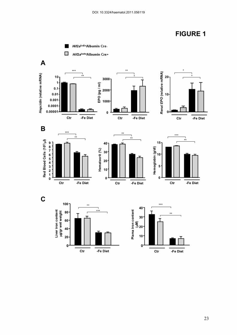
Figure 1. Mice with HIF2-α-deficient liver respond normally to diet-induced
iron deficiency.
Hif2alox/loxAlbumin Cre- and Hif2alox/loxAlbumin Cre+ littermates were fed an iron deficient (-
Fe diet) or control diet (Ctr) during 2 months after weaning. (A) Hepatic hepcidin and renal
EPO relative mRNA expression normalized to the Cyclophilin-A mRNA. Results are
expressed as a fold change compared with Hif2alox/loxAlbumin Cre- mice on a control diet.
EPO in plasma of WT and Hif2alox/loxAlbumin Cre+ mice fed an iron deficient or control diet.
(B) Haematological parameters. (C) Liver and plasma iron contents. n ≥ 4 for each group.
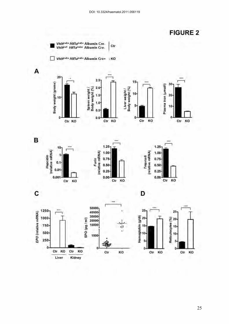
Figure 2. Characterization of Vhlhlox/loxHif1a lox/loxAlbumin Cre+ mice.
(A) Body, spleen and liver weight of Vhlhlox/lox Hif1alox/loxAlbumin Cre+ (KO) and control
mice (Ctr). n = 4. Plasma iron in Vhlhlox/lox Hif1alox/loxAlbumin Cre+ (KO) and control mice
(Ctr). n=5. (B) Relative mRNA expression normalized to the Cyclophilin-A mRNA in the
liver of Vhlhlox/lox Hif1a lox/loxAlbumin Cre+ compared to Ctr. Ctr : n=9; KO : n=8 . (C)
Hepatic and renal EPO mRNA (Ctr : n=11; KO : n=6) and plasma EPO levels in
Vhlhlox/loxHif1alox/loxAlbumin Cre+ (KO) compared with control littermates (Ctr). Ctr : n=31;
KO : n=14. (D) Hemoglobin and reticulocyte counts in Vhlhlox/lox Hif1alox/loxAlbumin Cre+
(KO) and control mice (Ctr). n ≥ 3 for each group.
DOI: 10.3324/haematol.2011.056119
22
Figure 3. Inhibition of erythropoietic activity in Vhlhlox/loxHif1alox/loxAlbumin
Cre+ mice normalizes liver hepcidin expression. Mice were injected every day with
300 µl NaCl or Rabbit anti-Erythropoietin serum (a-EPO) for 5 days and sacrificed 16h after
the last injection. (A) Reticulocytes, Red Blood Cells, Hemoglobin and Hematocrit counts (B)
Relative hepcidin mRNA expression in the liver expressed as a fold difference compared with
NaCl injected control (Ctr) mice. Ctr: NaCl (n=10), Ctr: Anti-EPO (n=9), KO: NaCl (n=3),
KO: Anti-EPO (n=5)
Figure 4. HIF-2α overexpression does not repress hepcidin expression in
primary hepatocytes
Infection of wild-type primary hepatocytes with a control GFP-adenovirus (AV-GFP) (black
bar) or with a HIF2-α-overexpressing adenovirus (AV-HIF-2α) (grey bar) at a Multiplicity Of
Infection (MOI) of 10. Relative mRNA expression normalized to Cyclophilin-A.
Representation of 3 independent experiments made in triplicate.
DOI: 10.3324/haematol.2011.056119
23
DOI: 10.3324/haematol.2011.056119
25
DOI: 10.3324/haematol.2011.056119
26
DOI: 10.3324/haematol.2011.056119
28
DOI: 10.3324/haematol.2011.056119
0
25
50
75 ***
Rec
ombi
natio
n e
ffici
ency
WT KO
FIGURE S1
WT : Hif2alox/lox Albumine Cre- KO : Hif2alox/lox Albumine Cre+
A B
WT KO 0.0
0.5
1.0
1.5
HIF
-2 (r
elat
ive
mR
NA
)
DOI: 10.3324/haematol.2011.056119
Furin
(rel
ativ
e m
RN
A)
Ctr -Fe Diet Ctr -Fe Diet
Tmpr
ss6
(rel
ativ
e m
RN
A)
FIGURE S2
Hif2alox/loxAlbumin Cre-
Hif2alox/loxAlbumin Cre+
0.00! 0.00!BM
P6 (r
elat
ive
mR
NA
)
Ctr -Fe Diet
0!
p=0,052
***
DOI: 10.3324/haematol.2011.056119
BM
P6 (r
elat
ive
mR
NA
)
Hjv
(rel
ativ
e m
RN
A)
FIGURE S3
Ctr KO Ctr KO vHL vHLHIF-1α
Ctr KO Ctr KO vHL vHLHIF-1α
0.0! 0.00!
***
***
** ***
Vhlhlox/lox Hif1a lox/loxAlbumin Cre- Vhlhlox/+ Hif1a lox/lox Albumin Cre-
Vhlhlox/lox Hif1a lox/loxAlbumin Cre+
Vhlhlox/lox Albumin Cre+
Vhlhlox/lox Albumin Cre -
ns
Hep
cidi
n
(rel
ativ
e m
RN
A)
Ctr KO Ctr KO vHL vHLHIF-1α
*** ***
Ctr
: KO
: Ctr
: KO
DOI: 10.3324/haematol.2011.056119
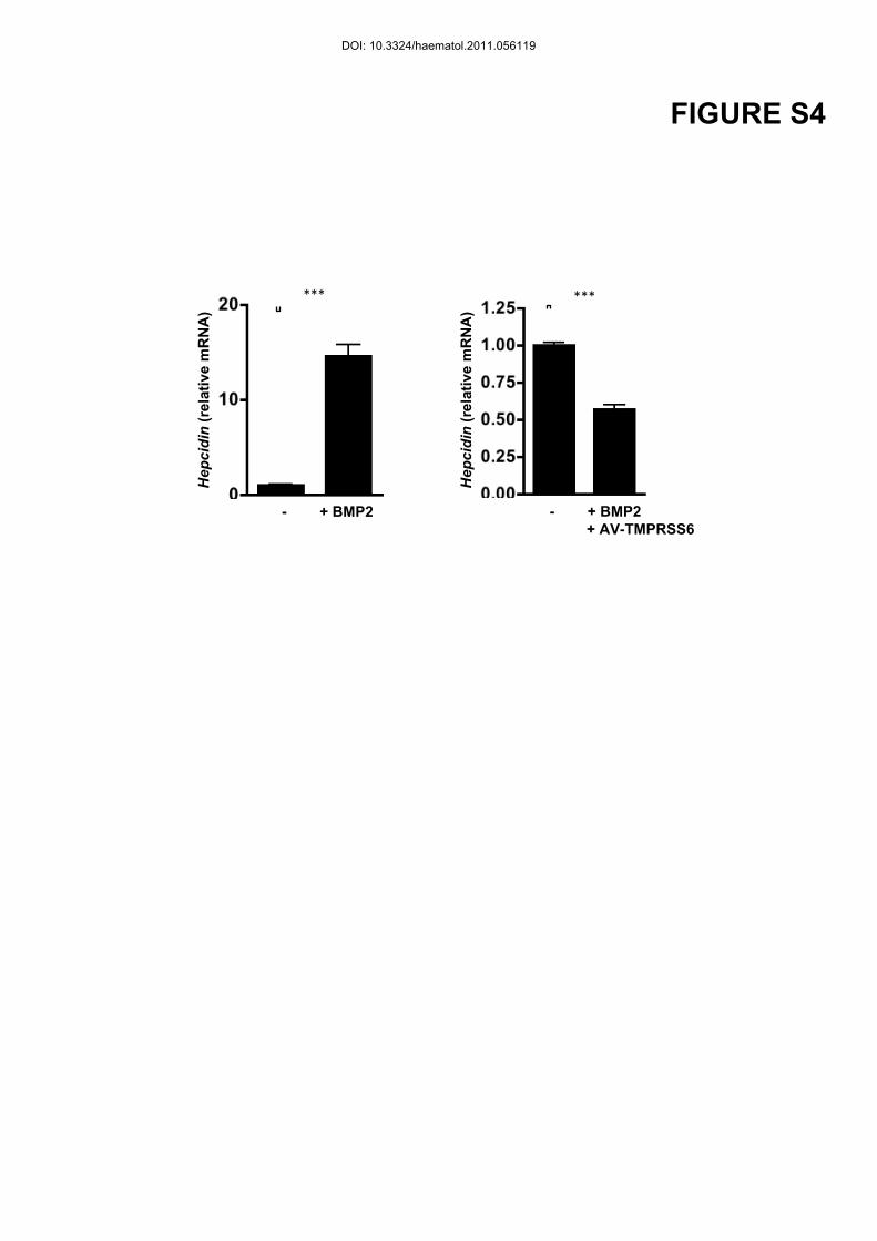
FIGURE S4
Hep
cidi
n (r
elat
ive
mR
NA
)
Hep
cidi
n (r
elat
ive
mR
NA
)
- + BMP2 - + BMP2 + AV-TMPRSS6
*** ***
DOI: 10.3324/haematol.2011.056119
Figure S1: Hepatocyte specific deletion of HIF-2α
(A) Recombination efficiency for Hif2lox/lox alleles quantified by real-time PCR of genomic
DNA isolated from the liver of Hif2a lox/loxAlbumine Cre- (WT) and Hif2a lox/loxAlbumine
Cre+ (KO) mice. (B) HIF-2α mRNA levels in primary hepatocytes of WT and KO mice as
determined by real-time PCR.
Figure S2: Iron deficiency does not transcriptionaly regulate furin nor TMPRSS6
Hif2alox/loxAlbumin Cre- and Hif2alox/loxAlbumin Cre+ littermates were fed an iron deficient (-
Fe diet) or control diet (Ctr) during 2 months after weaning. (A) BMP6, Furin and TMPRSS6
relative mRNA expression normalized to the 18S mRNA. Results expressed as a fold change
compared with Hif2alox/loxAlbumin Cre- mice on a control diet. n ≥ 4 for each group.
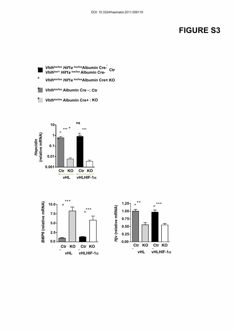
Figure S3: Trancriptional regulation of hepcpdin, BMP6 and Hjv in the liver of
vhlhlox/lox Albumin Cre+ and vhlhlox/lox Hif1a lox/loxAlbumin Cre+ mice.
Relative mRNA expression normalized to the 18S mRNA in the liver of vhlhlox/lox Albumin
Cre+ (KO grey bar) or vhlhlox/lox Hif1a lox/loxAlbumin Cre+ (KO white bars) compared to
vhlhlox/lox Albumin Cre- (dark grey bar) or Ctr (black bars). Ctr : n=9; KO : n=8
Figure S4: Trancriptional regulation of hepcidin in primary hepatocytes
Incubation of primary hepatocytes with BMP2 (20 ng/ml) for 24 hours, infected or not with a
TMPRSS6 adenovirus (AV-Tmprss6) at a Multiplicity Of Infection of 1. Hepcidin relative
mRNA expression normalized to Cyclophilin-A. n=3 per goup.
DOI: 10.3324/haematol.2011.056119

![haematologica - FIMMG MATERA homematera.fimmg.org/Linee guida/LineeGuidaTrombocitemia.pdf · haematologica vol. 88[supplemento 11]: maggio 2003 In occasione delle Giornate Ematologiche](https://img.pdfslide.us/doc/110x75/5c68ce4c09d3f206678c15d1/haematologica-fimmg-matera-guidalineeguidatrombocitemiapdf-haematologica.jpg)