Embed Size (px)
Citation preview
Hendry, K., Swann, G., Leng, M., Sloane, H., Goodwin, C., Berman, J., &Maldonado, M. (2015). Technical Note: Silica stable isotopes andsilicification in a carnivorous sponge Asbestopluma sp. Biogeosciences,3489-3498. DOI: 10.5194/bg-12-3489-2015
Peer reviewed version
License (if available):CC BY
Link to published version (if available):10.5194/bg-12-3489-2015
Link to publication record in Explore Bristol ResearchPDF-document
University of Bristol - Explore Bristol ResearchGeneral rights
This document is made available in accordance with publisher policies. Please cite only the publishedversion using the reference above. Full terms of use are available:http://www.bristol.ac.uk/pure/about/ebr-terms
Technical Note: Silica stable isotopes and silicification in a carnivorous sponge 1
Asbestopluma sp. 2
Katharine R Hendry1, George EA Swann2, Melanie J Leng3,4, Hilary J Sloane3, Claire Goodwin5, Jade 3
Berman6, and Manuel Maldonado7 4
1 School of Earth Sciences, University of Bristol, Wills Memorial Building, Queen’s Road, Bristol, BS8 5
1RJ, UK 6
2 School of Geography, University of Nottingham, University Park, Nottingham, NG7 2RD, UK 7
3 NERC Isotope Geosciences Facilities, British Geological Survey, Keyworth, Nottingham, NG12 5GG, 8
UK 9
4 Centre for Environmental Geochemistry, University of Nottingham, University Park, Nottingham, 10
NG7 2RD, UK 11
5 National Museums Northern Ireland, 153 Bangor Road, Cultra, Holywood, Co. Down, BT18 0EU, UK 12
6 Ulster Wildlife, 3 New Line, Crossgar, Co Down, Northern Ireland, BT30 9EP, UK 13
7 Centro de Estudios Avanzados de Blanes (CEAB-CSIC), Accés a la Cala St. Francesc, 14, Blanes 17300, 14
Girona, Spain 15
16
Keywords: 17
Silicon isotopes, oxygen isotopes, siliceous spicule, proxy 18
Abstract: 19
The stable isotope composition of benthic sponge spicule silica is a potential source of 20
palaeoceanographic information about past deep seawater chemistry. The silicon isotope 21
composition of spicules has been shown to relate to the silicic acid concentration of ambient 22
water, although existing calibrations do exhibit a degree of scatter in the relationship. Less is 23
known about how the oxygen isotope composition of sponge spicule silica relates to 24
environmental conditions during growth. Here, we investigate the vital effects on silica silicon and 25
oxygen isotope composition in a carnivorous sponge, Asbestopluma sp., from the Southern Ocean. 26
We find significant variations in silicon and oxygen isotopic composition within the specimen that 27
are related to unusual spicule silicification. The largest variation in both isotope systems was 28
associated to the differential distribution of an unconventional, hypersilicified spicule type 29
(desma) along the sponge body. The absence an internal canal in the desmas suggests an 30
unconventional silicification pattern leading to an unusually heavy isotope signature. Additional 31
internal variability derives from a systematic offset between the peripheral skeleton of the body 32
having systematically a higher isotopic composition than the internal skeleton. A simplified silicon 33
isotope fractionation model, in which desmas were excluded, suggests that the lack of a system 34
for seawater pumping in carnivorous sponges favours a low replenishment of dissolved silicon 35
within the internal tissues, causing kinetic fractionation during silicification that impacts the 36
isotope signature of the internal skeleton. Analysis of multiple spicules should be carried out to 37
“average out” any artefacts in order to produce more robust downcore measurements. 38
Introduction: 39
The formation of amorphous biogenic silica (or opal) by photosynthetic diatoms, which play a 40
major role in the export of organic matter to the seafloor, is a key part to both the cycling of silicon 41
and carbon in the Earth’s climate system (Tréguer and De La Rocha, 2013). Quantifying the dissolved 42
silicon, or silicic acid (Si(OH)4), concentration of upwelling waters is essential if we are to understand 43
the distribution and growth of diatoms in surface waters and so the drawdown on atmospheric carbon 44
dioxide (Hendry and Brzezinski, 2014). The silicon isotope (30Si) and oxygen isotope (18O) 45
compositions of biogenic silica have been used to infer modern nutrient cycling, past nutrient supply 46
and utilization, and hydrological cycling. Whilst the isotope composition of diatom opal has been used 47
widely to understand past surface conditions (Leng et al., 2009), the chemical composition of benthic 48
dwelling, deep-sea sponge opal holds the potential to reveal insights into bottom water conditions. 49
Both silicon and oxygen are present in three stable isotopes: 28Si (92.22%), 29Si (4.68%) and 50
30Si (3.08%); and 16O (~99.7%), 17O (~0.04%) and 18O (~0.2%) respectively 51
(http://www.nndc.bnl.gov/chart/). The per mille Si isotopic composition is expressed relative to the 52
NIST standard, NBS 28, according to Equation 1, and similarly the O isotopic composition is expressed 53
relative to VSMOW, according to Equation 2: 54
55
10001
2828
30
28
30
30
NBS
sample
SiSi
SiSi
Si (1) 56
10001
16
18
16
18
18
VSMOW
sample
OO
OO
O (2) 57
Recent work has shown that 30Si of a wide range of deep-sea sponges from different ocean 58
basins reflects the availability of dissolved silicon (silicic acid [Si(OH)4]) during growth, with minimal 59
impact from temperature, pH and (to date, and on few studies) no systematic species-dependent 60
fractionation (Hendry and Robinson, 2012; Wille et al., 2010). With sponge spicules ubiquitous in 61
sediments throughout the ocean and with degradation occurring at rates that are an orders of 62
magnitude slower than those for diatoms and other siliceous organisms (Maldonado et al. 2005, 63
Maldonado et al. 2012), there is significant potential for spicules to be used as a proxy for past ocean 64
conditions. Whilst a number of papers have explored the use of 30Si in sponges (e.g. Ellwood et al., 65
2010; Hendry et al., 2014), there is still scatter in the calibration of the 30Si-Si(OH)4 relationship, 66
with the sources of variability poorly understood. Likewise, little is known about the sponge spicule 67
silica 18O, although it is likely impacted by biological factors (Matteuzzo et al., 2013) that cause 68
systematic offsets when compared to diatom silica 18O (Snelling et al. 2014). Here, we investigate 69
the impact of derived biomineralisation mechanisms that could be responsible for variations in 70
silicon and oxygen isotope fractionation in sponges using a carnivorous sponge specimen from the 71
Southern Ocean as a case study. 72
73
Sponges and sponge biomineralisation: 74
Sponges (Porifera) are sessile filter-feeding animals. Their body plan has evolutionarily been 75
shaped to optimize the feeding function, evolving an architectural design that, in general, is shared 76
by the four major sponge lineages (Demospongiae, Hexactinellida, Homosclerophorida, and 77
Calcaraea). The anatomical archetype of a sponge is a vase-shaped or oblate body crossed by a 78
system of aquiferous canals that communicate to the outside at both ends, and through which a 79
current of environmental water flows, transporting bacteria and dissolved compounds that nourish 80
the sponge, oxygen and waste products. The histological archetype of a sponge consists of two 81
epithelial layers of flattened cells (pinacocytes), an external layer that forms the wall of the body, 82
and an internal layer that forms the wall of the aquiferous canals. Between the epithelium of the 83
canals and the external epithelium, there is a mesenchyme-like zone that is rich in collagen and is 84
populated by different groups of mobile amoeboid cells. The spicules (i.e., siliceous or calcareous 85
skeletal pieces that give structural support to these often soft-bodied organisms) are also produced 86
and assembled together by cells (i.e. sclerocytes) in the mesenchyme-like zone. The aquiferous 87
canals include chamber-shaped expansions, in which the walls are coated not by pinacocytes but 88
choanocytes that is pseudocylindrical cells possessing a flagellum surrounded by a collar of microvilli 89
at the distal pole. These cells phagocytose picoplankton from the water passing through the 90
chambers; they are the most distinctive feature of the phylum Porifera. 91
However, a group of demosponges, currently mainly classed in the family Cladorhizidae 92
(Order Poecilosclerida), have evolved a carnivorous habit (Vacelet, 2006), thought to be an 93
adaptation to the nutrient-poor environments in which they inhabit, where a ‘sit-and-wait’ 94
predatory strategy is beneficial because of the low energy expenditure between rare feeding 95
opportunities (Vacelet and Duport, 2004; Vacelet, 2007). Carnivorous sponges are usually associated 96
with low nutrient mid basin areas of the deep-sea (the deepest recorded at 8840m) but a few are 97
found around 100m depth in high latitudes and some species have also been found in shallow 98
sublittoral and littoral caves in the Mediterranean, where they are thought to have colonised from 99
deep-water populations (Aguilar et al., 2011; Bakran‐Petricioli et al., 2007; Chevaldonné et al., 2014; 100
Lopes, et al., 2012; Vacelet, 2006, 2007). These carnivorous sponges show not only an unusual 101
internal body organization lacking choanocytes and aquiferous canals, but also a convergence 102
towards characteristic morphological adaptations including an upright stalked body, with branches, 103
and feather-like or balloon-like lateral expansions to enhance encounter rates with prey. 104
Carnivorous sponges have developed either rhizoid-like or bulbous bases for holding their erect 105
bodies on muddy and hard substrates respectively (Vacelet, 2007). 106
The family Cladorhizidae, despite being relatively small (7 genera, 140 spp.; Porifera World 107
Database, September 2014), has a moderate diversity of spicules. In these sponges, the silica 108
spicules are needed not only to provide skeletal support to the body, but also to capture prey. Their 109
relatively small bodies (rarely taller than 10 cm) usually have an internal, central skeletal core (axial 110
skeleton) made by a bundle of highly-packed needle-like spicules, typically shorter than 700 µm 111
each, and with one or both ends being pointed (i.e., monactinal or diactinal megascleres). From this 112
axial skeleton radiating spicule tracts diverge (extra-axial skeleton) to core either the branches or 113
any of the other types of lateral processes occurring in the body, depending on the genera and 114
species. In addition to this main supportive skeleton, there are thousands of smaller (< 100 µm; 115
microscleres) hook-like spicules, being either simple hooks (sigmata) or tooth-bearing hooks 116
(chelae). These are scattered through the internal mesenchyme-like tissue and, more importantly, 117
also at the external epithelia, where they project part of their hooking structure out of the body to 118
capture small crustaceans that may contact the external sponge surface. Some of these sponge 119
species have additional microscleric spicules to reinforce the skeleton, but very few carnivorous 120
species - and in only the genera Asbestopluma (Family Cladorhizidae) , Euchelipluma (Family 121
Guitarridae) and Esperiopsis (Family Esperiopsidae) - have been described having hypersilicified 122
spicules (called desmas). Desmas are usually confined to the basal body region, probably to 123
strengthen the area through which the sponge attaches to the substrate (Vacelet, 2007). 124
Because carnivorous sponges lack the aquiferous system that conventionally transports 125
ambient seawater into the sponge body and because the isotope signal of their silica spicules has 126
never been assessed before, it is compelling to examine whether silicon fractionation values in 127
carnivorous sponges differ from those measured in the more conventional, filter-feeding sponges. As 128
carnivorous sponges are typically constrained to bathyal habitats (Vacelet, 2007), their skeletons 129
may turn into a good tool to infer traits of deep regional water masses. The recent collection of a 130
new species of desma-bearing cladorohizid to be formally described in the genus Asbestopluma 131
(Goodwin et al., in prep.) has provided an unparalleled opportunity to investigate δ30Si and δ18O of 132
its silica spicules. 133
134
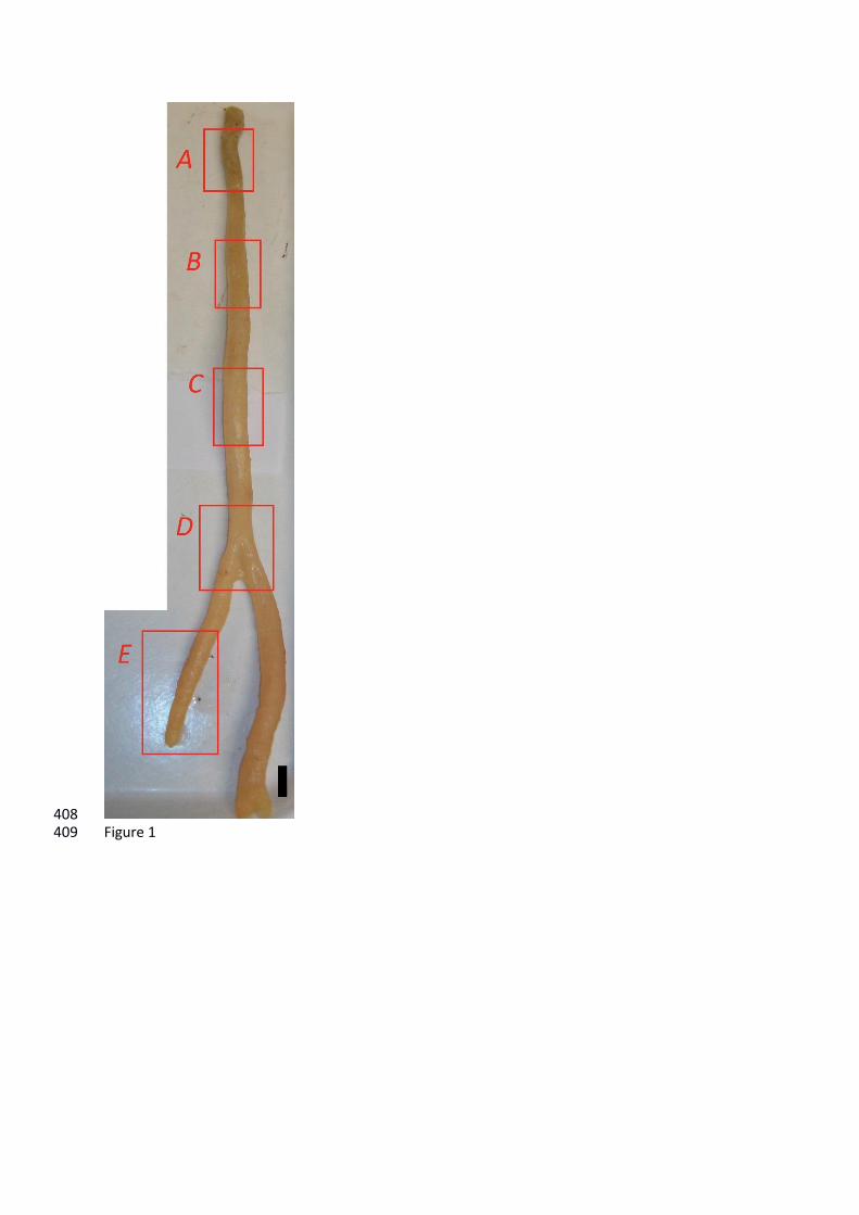
Methods: 135
Specimen: 136
Specimen DH19-2 (Asbestopluma sp.) was recovered by Hein Dredge from Burdwood Bank 137
(1500-1530 m water depth, 54° 45’S, 62° 16’W) in the Atlantic Sector of the Southern Ocean from 138
the R/V Nathaniel B. Palmer in 2011 (National Science Foundation NBP1103). The specimen was 139
photographed and dried for transportation. Temperature, salinity, and Si(OH)4 concentrations of the 140
ambient water are estimated as 2.5-3°C, 34.5, and 60 M, respectively (from on-board 141
measurements and literature data available at www.eWOCE.org). 142
The specimen has an upright, moderately branching form (Figure 1). The basal body portion 143
contains internally interlocked desmas (Figure 2A-B), externally surrounded by layer of microscleric 144
acanthotylostrongyles (Figure 2B) and scarce sigmas. It is worth noting that the abundance of 145
desmas decreases significantly from the basal body portion to the branch tips and that the 146
acanthotylostrongyles occur exclusively at basal portion of the sponge. Further up the axis, the stem 147
is cored by large smooth monoactines (styles), with smaller styles and diactines with rounded ends 148
(anisostrongyles) outside this core, sigmas and chelae microscleres are also present (Figure 2C). 149
Desmas become less frequent with increasing distance from the base (with desmas representing 150
approximately 90% of the spicules from A, 50% from B, 40% from C and less than 25% from D and E) 151
so that at the growth tips, there are only styles, sigma and chelae. 152
153
Sample preparation: 154
Five sponge tissue samples (A to E) were taken along the body length of the specimen, that 155
is, at increasing distance from the attachment point, covering from the base to the branch tip (Figure 156
1). Samples were cleaned for organic matter by heating to 80°C in 30% hydrogen peroxide for at 157
least an hour and rinsing thoroughly in deionised water at least three times. At this stage, for each 158
tissue sample, two skeletal subsamples were obtained from: 1) the spicules of the axial skeleton 159
(axial or “internal” samples), and 2) the spicules of the radiating skeleton and the external 160
epithelium (extra-axial or “external” samples). The subsamples were then heated to 80°C in trace 161
metal grade concentrated nitric acid for at least an hour, and rinsed thoroughly in 18 MΩ.cm Milli-Q 162
water at least three times. Standards and samples were prepared by alkaline fusion with sodium 163
hydroxide pellets, acidified with ultra-clean nitric acid (Optima), and purified using cation exchange 164
resin (Georg et al., 2006). Note that heating opal to 80°C during the organic matter removal process 165
does not result in additional fractionation of spicule silicon isotopes (Hendry et al., 2011). Although 166
there is no available information specifically about modern sponge spicules, heating samples of 167
phytolith opal to 80°C during cleaning does not result in additional fractionation of oxygen isotopes 168
(Crespin et al., 2008). One study suggests that heating diatom opal to over 60°C results in a potential 169
offset in 18O as a result of dissolution (Crespin et al., 2008). However, the 18O values from 70 and 170
90°C from this study were within analytical error of the values of diatoms treated at 60°C. 171
Furthermore, heating of diatom opal to 70°C using different cleaning methods does not result in 172
measurable changes in 18O (Tyler et al., 2007). Heating to higher temperatures of 80-90°C is routine 173
in downcore spicule opal 18O analyses (e.g. Snelling et al., 2014). 174
175
Silicon isotope analysis: 176
The samples were analysed for silicon isotope ratios (29Si/28Si, 30Si/28Si) using a Thermo 177
Neptune Multi-Collector Inductively Coupled Plasma Mass Spectrometer (MC-ICP-MS) at Bristol 178
University (Bristol Isotope Group). The isotope ratios were measured using 20 cycles per block. 179
Machine blanks were monitored, and were <1% of the signal on 28Si. Mass bias and matrix effects 180
were corrected using standard-sample bracketing, and internal Mg-doping (Cardinal et al., 2003; 181
Hendry and Robinson, 2012). Silicon and magnesium intensities were matched within 10% (typically 182
<5%). The results are reported as 30Si values relative to the standard NBS28 (RM8546). Analysis of 183
“diatomite” during the study yielded a mean 30Si value of –1.25‰ (± 0.18 2SD, n = 70); “big-batch” 184
yielded a mean 30Si value of –10.67‰ (± 0.08 2SD, n = 3) (Reynolds et al., 2007). Repeat analyses of 185
sponge standard LMG08 (Hendry and Robinson, 2012) during each run were used to assess long-186
term external reproducibility, and yielded a mean 30Si value of –3.41‰ over 6 months (± 0.16 2SD, 187
n = 31). 29Si/ 30Si for all samples and standards was ~0.51, consistent with mass-dependent 188
fractionation (Cardinal et al., 2003). 189
190
Oxygen isotope analysis: 191
Aliquots of spicule samples were analysed for oxygen isotope ratios (18O/16O) following a 192
step-wise fluorination procedure (Leng and Sloane, 2008) verified through an inter-laboratory 193
calibration exercise (Chapligin et al., 2011). Samples were outgassed in nickel reaction vessels and 194
reacted with BrF5 for 6 minutes at 250°C to remove all Si-OH bonds. Oxygen from Si-O-Si bonds was 195
subsequently released by reaction with further reagent overnight at 550°C before being converted 196
and collected as CO2. Oxygen isotope measurements were made on a Finnigan MAT 253 with values 197
converted to the VSMOW scale using the run laboratory diatom standard BFCmod calibrated against 198
NBS28. Repeat analysis of BFCmod indicates reproducibility is 0.6‰ (2SD) (Leng and Sloane, 2008). 199
200
Electron microscopy: 201
Scanning electron microscopy (SEM) was used to describe the siliceous skeleton at the various body 202
regions. An aliquot of spicules from each subsample was mounted onto an SEM aluminium stub, 203
coated by gold sputtering and imaged using a HITACHI S-3500N Scanning Electron Microscope. 204
To document the presence/absence of an axial canal at the core of the various spicules types, 1mm3 205
sponge tissue samples were collected, placed onto a glass cover slip and subsequently cleaved 206
multiple times to fracture the spicules using a scalpel blade under a dissecting scope. The cover slip 207
with the cleaved tissue was placed onto a glass slide and, to eliminate the organic matter from the 208
silica skeleton, three drops of concentrate nitric acid were added on the tissue sample while 209
maintaining the slide above the flame of an alcohol burner. After boiling and evaporation of acid, 210
new acid drops were added and the operation repeated several times until corroborating through a 211
light microscope that the silica spicules were externally cleaned from organic remains, before rinsing 212
three times in milli-Q water. The slip bearing the cleaned, fractured spicules mounted onto an SEM 213
aluminium stub and coated by gold sputtering for further observation of fracture planes and axial 214
canals using a HITACHI TM300 Scanning Electron Microscope. 215
216
Results: 217
Silicon isotopes: 218
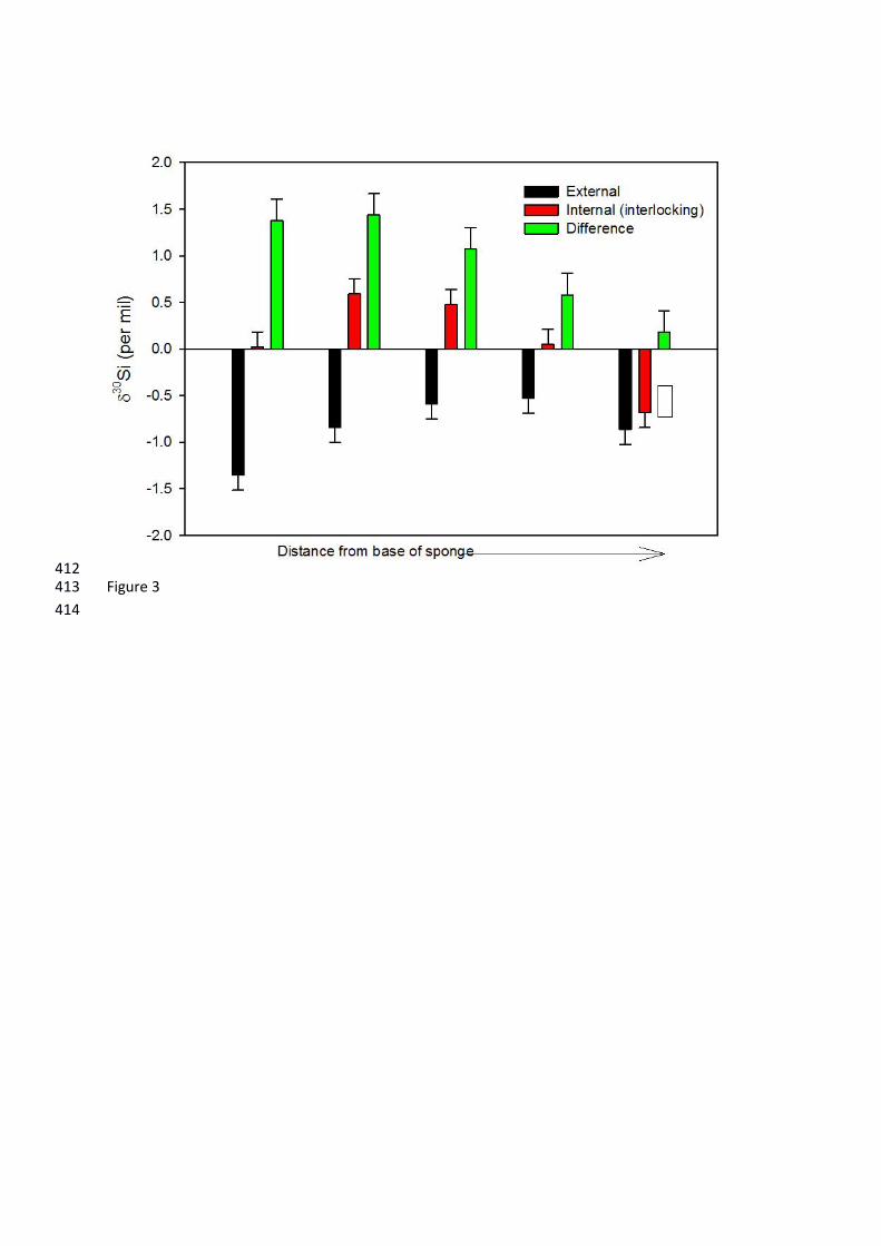
The average 30Si value for the cladorhizid DH19-2 was −0.37‰, but values ranged from –219
1.35 to +0.59‰, with an overall range of 1.94 ‰ (Figure 3). These values fall within the total range 220
of modern sponge 30Si measurements in the literature (e.g. Hendry & Robinson, 2012). Since 221
previous studies have found no discernible variation within an individual (Hendry et al., 2010; 222
Hendry et al., 2011), this is an unprecedented variability within a single specimen, and represents 223
approximately 40% of the total range of isotope values for existing calibrations (~5 ‰) (Hendry and 224
Robinson, 2012). The external spicules were significantly and consistently isotopically lighter than 225
the internal interlocking spicules. The external and internal spicules became isotopically heavier and 226
lighter respectively along the axis, such that the difference between the internal and external 227
spicules decreased away from the base of the specimen (from approximately 1.4‰ at A to 228
approximately 0.1‰ at E; Figure 3). 229
230
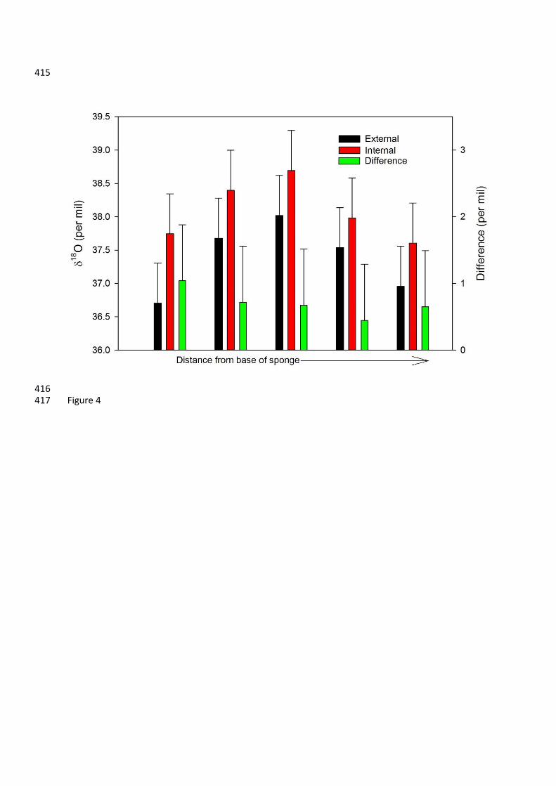
Oxygen isotopes: 231
The average 18O value for DH19-2 was +37.7‰, ranging from +36.7 to +38.7‰ (Figure 4), 232
giving a range of 2‰. The 18O of the marine specimen in this study is significantly heavier than 233
values obtained for freshwater sponge spicules (approximately +22 to +30‰). The fractionation 234
factor (18Osilica-seawater) for the marine sponge (+36 to +39‰) was greater than that of freshwater 235
sponges (+28‰) (Matteuzzo et al., 2013). The variation within the one individual from this study 236
compares to an entire range of 18Owater of less than 0.8‰ and potential temperature variations of 237
~5°C across the Drake Passage (Meredith et al., 1999), and represents nearly half of the 5‰ 238
variations found in a downcore sponge spicule 18O record from Pliocene sediments (Snelling et al., 239
2014). The external spicules were consistently isotopically lighter than the internal interlocking 240
spicules, although the difference between them (0.4 to 1‰) is approximately the same as the 241
analytical error (2SD of 0.6‰). The trend in 18O along the axis of the specimen is less clear than for 242
30Si: both the external and internal spicules because isotopically heavier from A to C, and then 243
isotopically lighter from C to E. There is also a positive correlation between 30Si and 18O (r=0.88, 244
p=0.001, n=10). 245
246
Discussion: 247
Silicon isotopes and internal fractionation: 248
The large variation in both isotope systems within the studied individual relates to a 249
differential distribution of the spicule types along the sponge body (i.e., distance from sponge base), 250
and also to differences in the abundance of given spicule types between the internal (axial) and 251
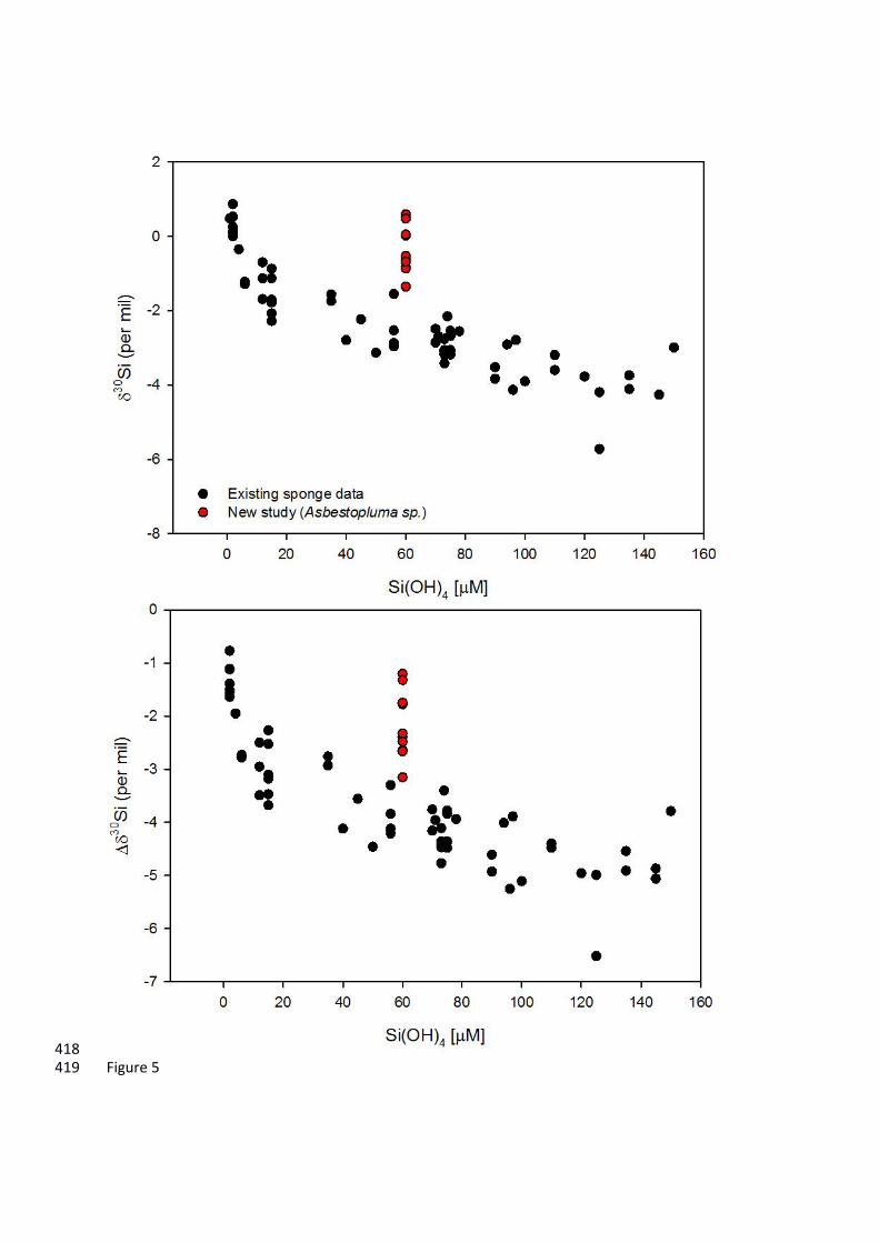
external (extra-axial) body regions. The external basal skeleton (i.e., mostly acanthotylostrongyles) 252
has the most isotopically light (negative) 30Si that lies close to the existing 30Si-Si(OH)4 calibration 253
curve (Figure 5). The internal basal skeleton (i.e., mostly desmas) has a very isotopically heavy 254
(positive) 30Si compared to that of the external spicules, which may relate to the presence of desma 255
spicules. 256
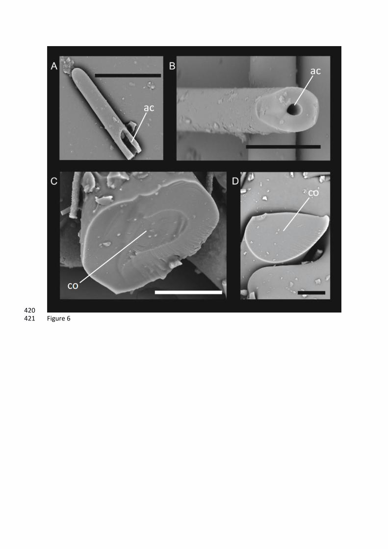
The desmas of this carnivorous sponge have an unusual formation mechanism compared to 257
other megascleric demosponge spicules. Nearly all types of megascleric spicule, including most 258
desmas, show an internal or "axial” canal (Fig. 6A). This canal originally harbours a filament of the 259
enzymatic protein silicatein (Shimizu et al., 1998), responsible for initiating the polymerization of 260
biogenic silica, the growth of which starts intracellularly through an enzymatically-guided 261
polycondensation of dissolved silicon. The term “desmas” represents a large variety of 262
phylogenetically unrelated spicule morphologies, which only share the feature of being massive, 263
relatively irregular skeletal pieces produced by hypersilicification, and may or may not possess an 264
axial canal. How and where the hypersilicification of desmas is achieved remains poorly understood. 265
In all cases described to date, the desmas in carnivorous sponges are anaxial, that is, lack axial canals 266
(Figure 6B). The absence of an axial canal indicates that their silicification does not involve an initial 267
intracellular, enzyme-guide silica polymerization. Consequently, these anaxial desmas must grow via 268
a mechanism different from that taking place in other demosponge spicules, which may account for 269
their distinctive silicon and oxygen isotopic composition. This idea is in agreement with previous 270
findings indicating that some cellular mechanisms for spicule silicification may have evolved 271
independently in different sponge lineages (Maldonado and Riesgo, 2007). The level at which the 272
secondary hypersilicification step of desmas could also contribute, if any, to their isotope signal 273
remains unknown, and further study into the potential differences in the isotopic signal between 274
desmas with and without axial canals is required. 275
The decreasing abundance of desmas with increasing distance from the sponge base is at 276
least one of the plausible factors responsible for the within-sponge variation in isotope compositions 277
observed in this study. We suggest that the likely extracellular silicification of these desmas could 278
result in kinetic fractionation of silicon isotopes. It should also be noted that the external basal 279
spicules (i.e., the acanthotylostrongyles), although forming the “best fit” to the existing 30Si-Si(OH)4 280
calibration, are still outside of analytical error of the calibration curve, and this offset could be 281
explained by some desma contamination (Figure 2b). 282
Further up the axis away from the base, the extra-axial styles have a higher 30Si, moving 283
further away from the existing 30Si-Si(OH)4 calibration curve (Figure 5), and then lower again 284
towards the growing tip. This does not reflect contamination from microscleres (i.e., sigmas and 285
chelae), as individually picked and cleaned styles are within analytical uncertainty (±0.15‰) of the 286
bulk measurement (see white box on Figure 3). Although the internal styles also become isotopically 287
enriched, the difference between the internal and external spicule 30Si declines up the axis, most 288
likely because of a decline in the number of desmas. 289
There are also two alternative, but probably less plausible explanations, for the large intra-290
individual silicon isotopic variation along the body axis. One is that this sponge grows extremely slowly 291
(over centuries) in the deep-sea environment. If so, it could be that during the first decades of its life, 292
what is now the basal body portion was exposed to a water mass with temperature and silicic acid 293
concentration different from present, progressively changing overtime towards the current values and 294
impacting accordingly the isotope signal during sponge growth. A second possibility is that this sponge 295
grows very rapidly. If so, the basal portion could have been formed during an episodic input of 296
seawater with abnormal silicic acid concentration and temperature, compared to the ambient 297
conditions during the subsequent growth. Because virtually nothing is known about the longevity and 298
growth rate of these sponges, these ideas remain mere speculation. 299
Could the heavy silicon isotope bias be a consequence of the absence of an aquiferous system 300
in the carnivorous sponge? Given that the aquiferous system usually allows the circulation of ambient 301
seawater throughout the body, the loss of this system could result in internal silicon isotope 302
fractionation as the isotopes in the aqueous component becomes progressively heavier due to 303
precipitation of silica in a closed system. This process would explain not only the offset between the 304
external and internal spicules but also the trends along the length of the sponge stem. Again, nothing 305
is known about how the dissolved silicon molecules are transported into the body by these sponges 306
or about the average replenishment rate for dissolved silicon within the internal tissues. Nevertheless, 307
if a simplified silicon isotope fractionation model is formulated, ignoring the impact of desmas and 308
assuming a variable silicon isotopic fractionation during sponge growth according to the core top 309
spicule calibration of Hendry and Robinson (2012), we can examine the impact of an isotopically closed 310
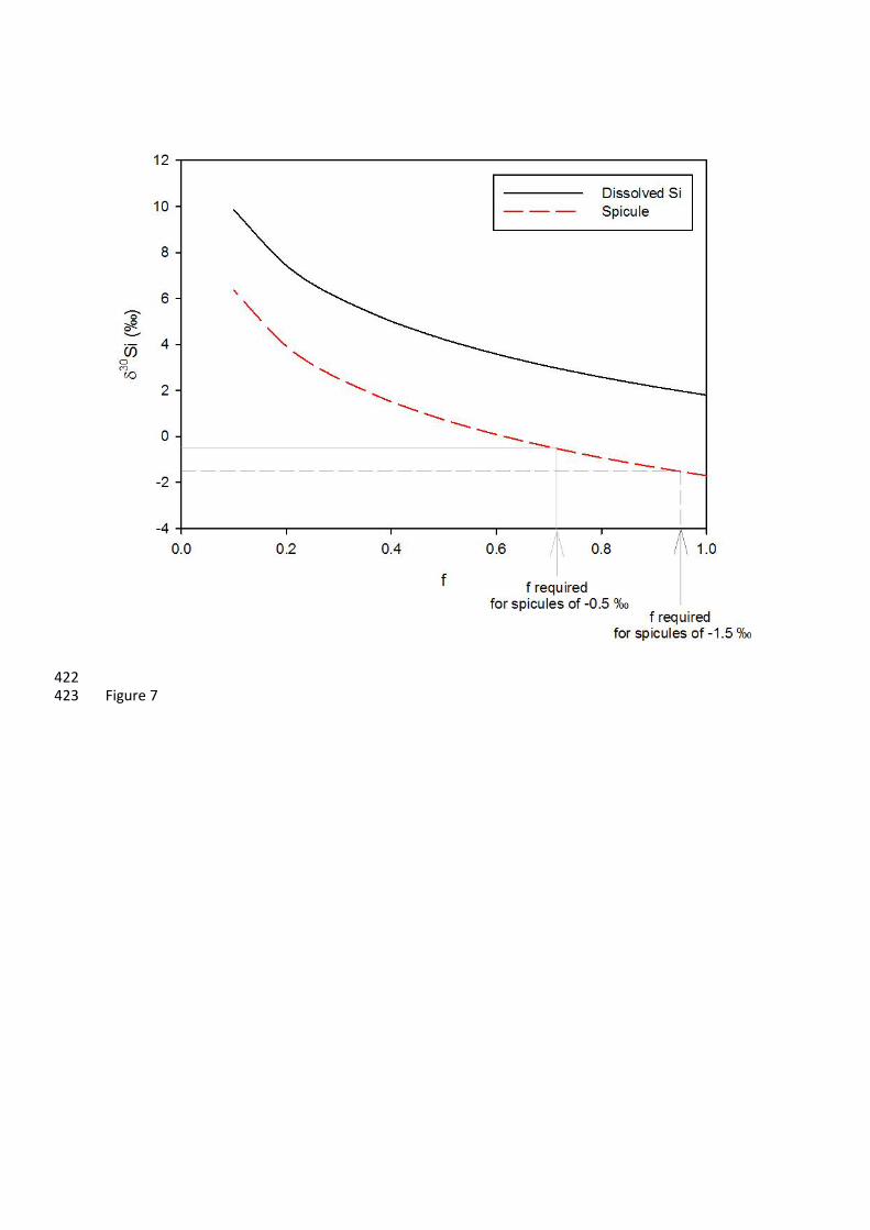
system on changes in spicule composition with cellular silicon utilisation (Figure 7). This simplified 311
model suggests that relatively small degrees of cellular silicon utilisation (less than 30%) could result 312
in heavier 30Si observed up the axis of the Asbestopluma sp. specimen. A higher rate of dissolved 313
silicon replenishment and a faster sponge growth rate could explain the return to lighter isotopic 314
compositions at the growing tips. 315
316
Oxygen isotopes and additional fractionation processes: 317
The positive correlation that we find within one individual between 30Si and 18O indicates 318
that there may be some shared mechanisms behind fractionation of the two isotope systems, at least 319
in Asbestopluma. There is a similar systematic offset between the external and internal spicules in 320
18O as for 30Si values, suggesting that the unusual silicification process that results in desma 321
formation may fractionate oxygen isotopes in a similar manner to silicon. However, the along-axis 322
trend is less clear in the 18O than 30Si, suggesting desmas cause a smaller bias in oxygen isotope 323
systematics than for silicon. Furthermore, there is no significant along-axis decrease in 18O in the 324
internal spicules, as observed for 30Si, suggesting that any internal fractionation of oxygen isotopes 325
is less pronounced and within the analytical uncertainty. 326
Additional processes active at the surface of the sponge spicule silica may also have an 327
influence on both the silicon and oxygen isotope values, including precipitation processes and 328
dissolution. There is some evidence from one laboratory study for a silicon isotope fractionation 329
during dissolution of diatom opal (Demarest et al., 2009), which is not supported by more recent 330
laboratory and field studies (Egan et al., 2012; Wetzel et al., 2014). Although there is potential for 331
kinetic fractionation of oxygen during dissolution and reprecipitation of silica (Crespin et al., 2008; 332
Dodd et al., 2012), further work is required to investigate whether fractionation of either silicon or 333
oxygen isotopes occurs during the dissolution of sponge spicules, or by any additional surface 334
precipitation processes. 335
336
Implications for palaeoclimate and outlook: 337
This first study of within-sponge differential fractionation has a number of implications for 338
biomineralisation and the use of isotope proxies for reconstructing past nutrient conditions. Firstly, 339
our findings suggest that internal non-equilibrium fractionation of silicon isotopes in sponges can 340
occur, depending on silicic acid replenishment rates in the internal tissues, which could explain some 341
of the scatter in the 30Si-Si(OH)4 calibration plot (Hendry and Robinson, 2012). Internal fractionation 342
could also impact sponge 18O, but less severely. We suggest that the anaxial desmas of this and 343
probably other carnivorous sponges have a different mode of silicification causing an unusual 344
isotopic signature in their biogenic silica. 345
Secondly, this study highlights the need for caution when preparing samples in order to compile 346
robust palaeoclimate archives. A large number of spicules should be picked for such archives in 347
order to “average” out variations caused by kinetic fractionation in Cladorhizid sponges, which 348
cannot be readily distinguished using light microscopy. Furthermore, desma formation may result in 349
very different fractionation behaviour. However, desmas are morphologically distinct, and should be 350
excluded from proxy measurements for palaeoclimate applications until further studies have been 351
completed to assess the level at which these spicules result in isotopic bias. Whether axial and 352
anaxial desmas can provide an independent complementary proxy to corroborated trends inferred 353
from the "conventional" silica spicules is a possibility that needs to be explored in future studies. 354
355
Author contributions: 356
KRH and GS/HS/ML carried out the isotope and SEM analyses, and CG/JB carried out the sponge 357
identification, MM carried out further SEM and spicule analyses. All authors contributed 358
significantly to discussions and the preparation of the manuscript. 359
360
Acknowledgements: 361
The authors would like to thank C. Coath (Bristol) for assistance with mass spectrometry, L. 362
Robinson (Bristol), R. Waller (Maine), the captain and crew of the R/V Nathaniel B. Palmer. Samples 363
were processed in the laboratories at Cardiff University, with thanks to R. Perkins. SEM images were 364
taken at the University of Bristol with the assistance of S. Kearns, B. Buse and A. Anton-Stephens, 365
and at CEAB-CSIC, Blanes. This work was funded by the National Science Foundation (grants 366
0944474, 0636787 and 1029986), The Leverhulme Trust (Research Grant RPG-2012-615); KH is 367
funded by the Royal Society. MM is funded by the Spanish Ministry of Innovation and 368
Competitiveness (CTM2012-37787). CG is a research associate at Queen’s University Marine 369
Laboratory, Portaferry. The authors would like to thank two anonymous reviewers for their 370
constructive comments. 371

372
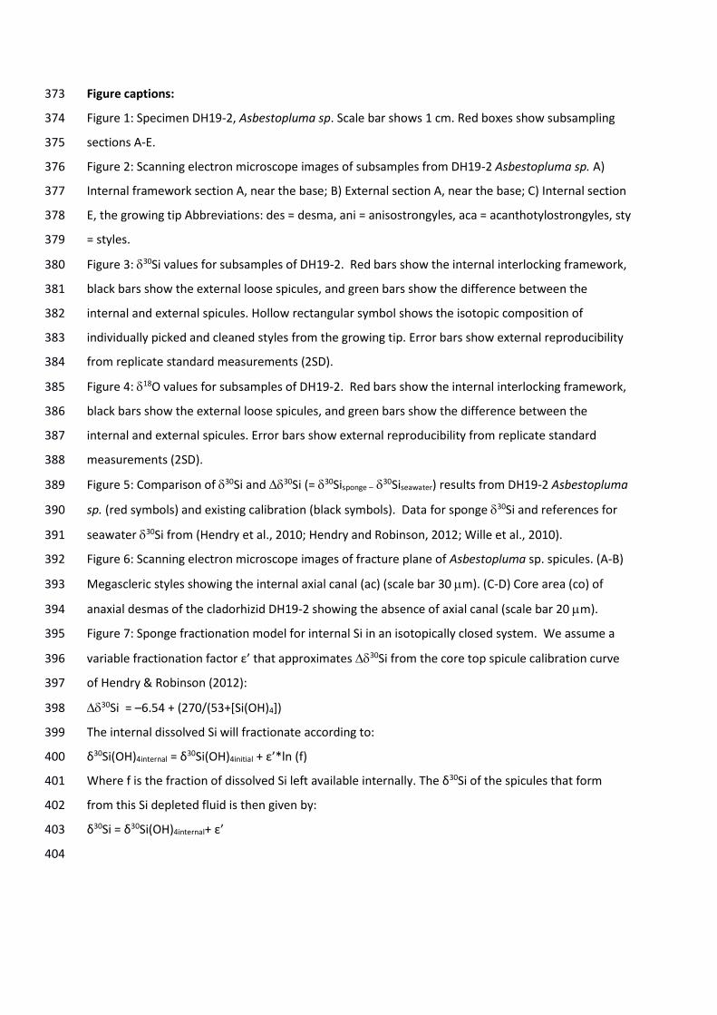
Figure captions: 373
Figure 1: Specimen DH19-2, Asbestopluma sp. Scale bar shows 1 cm. Red boxes show subsampling 374
sections A-E. 375
Figure 2: Scanning electron microscope images of subsamples from DH19-2 Asbestopluma sp. A) 376
Internal framework section A, near the base; B) External section A, near the base; C) Internal section 377
E, the growing tip Abbreviations: des = desma, ani = anisostrongyles, aca = acanthotylostrongyles, sty 378
= styles. 379
Figure 3: 30Si values for subsamples of DH19-2. Red bars show the internal interlocking framework, 380
black bars show the external loose spicules, and green bars show the difference between the 381
internal and external spicules. Hollow rectangular symbol shows the isotopic composition of 382
individually picked and cleaned styles from the growing tip. Error bars show external reproducibility 383
from replicate standard measurements (2SD). 384
Figure 4: 18O values for subsamples of DH19-2. Red bars show the internal interlocking framework, 385
black bars show the external loose spicules, and green bars show the difference between the 386
internal and external spicules. Error bars show external reproducibility from replicate standard 387
measurements (2SD). 388
Figure 5: Comparison of 30Si and 30Si (= 30Sisponge –30Siseawater) results from DH19-2 Asbestopluma 389
sp. (red symbols) and existing calibration (black symbols). Data for sponge 30Si and references for 390
seawater 30Si from (Hendry et al., 2010; Hendry and Robinson, 2012; Wille et al., 2010). 391
Figure 6: Scanning electron microscope images of fracture plane of Asbestopluma sp. spicules. (A-B) 392
Megascleric styles showing the internal axial canal (ac) (scale bar 30 m). (C-D) Core area (co) of 393
anaxial desmas of the cladorhizid DH19-2 showing the absence of axial canal (scale bar 20 m). 394
Figure 7: Sponge fractionation model for internal Si in an isotopically closed system. We assume a 395
variable fractionation factor ε’ that approximates 30Si from the core top spicule calibration curve 396
of Hendry & Robinson (2012): 397
30Si = –6.54 + (270/(53+[Si(OH)4]) 398
The internal dissolved Si will fractionate according to: 399
δ30Si(OH)4internal = δ30Si(OH)4initial + ε’*ln (f) 400
Where f is the fraction of dissolved Si left available internally. The δ30Si of the spicules that form 401
from this Si depleted fluid is then given by: 402
δ30Si = δ30Si(OH)4internal+ ε’ 403
404

Table 1: Stable isotope results for DH19-2 Asbestopluma sp. specimen 405
Internal External
Subsample 30Si (‰) 18O (‰) 30Si (‰) 18O (‰)
A +0.02 +37.74 −1.35 +36.71
B +0.59 +38.40 −0.84 +37.68
C +0.48 +38.69 −0.59 +38.02
D +0.05 +37.98 −0.53 +37.54
E −0.68 +37.61 −0.86 +36.96 406

407
408 Figure 1 409

410 Figure 2 411
412 Figure 3 413
414
415
416 Figure 4 417
418 Figure 5 419
420 Figure 6 421
422 Figure 7 423
References: 424 425 Aguilar, R., Correa, M. L., Calcinai, B., Pastor, X., & De la Torriente, A. First records of Asbestopluma 426
hypogea Vacelet and Boury-Esnault, 1996 (Porifera, Demospongiae Cladorhizidae) on 427 seamounts and in bathyal settings of the Mediterranean Sea. Zootaxa, 2925, 33-40, 2011. 428
Bakran‐Petricioli, T., Vacelet, J., Zibrowius, H., Petricioli, D., and Chevaldonné, P. New data on the 429 distribution of the ‘deep‐sea’sponges Asbestopluma hypogea and Oopsacas minuta in the 430 Mediterranean Sea. Marine Ecology, 28(s1), 10-23, 2007. 431
Cardinal, D., Alleman, L. Y., de Jong, J., Ziegler, K., and Andre, L. Isotopic composition of silicon 432 measured by multicollector plasma source mass spectrometry in dry plasma mode. Journal 433 of Analytical Atomic Spectrometry, 18, 213-218, 2003. 434
Chapligin, B., Leng, M. J., Webb, E., Alexandre, A., Dodd, J. P., Ijiri, A., Lücke, A., Shemesh, A., 435 Abelmann, A. and Herzschuh, U. Inter-laboratory comparison of oxygen isotope 436 compositions from biogenic silica. Geochimica et Cosmochimica Acta, 75(22), 7242-7256, 437 2011. 438
Chevaldonné, P., Pérez, T., Crouzet, J. M., Bay‐Nouailhat, W., Bay‐Nouailhat, A., Fourt, M., Almòn, b., 439 Pérez, J., Aguilar, R., and Vacelet, J. Unexpected records of ‘deep‐sea’carnivorous sponges 440 Asbestopluma hypogea in the shallow NE Atlantic shed light on new conservation issues. 441 Marine Ecology, doi:10.1111/maec.12155, 2014. 442
Crespin, J., Alexandre, A., Sylvestre, F., Sonzogni, C., Pailles, C., and Garreta, V. IR laser extraction 443 technique applied to oxygen isotope analysis of small biogenic silica samples, Analytical 444 Chemistry, 80(7), 2372-2378, 2008. 445
Demarest, M. S., Brzezinski, M. A. and Beucher, C. Fractionation of silicon isotopes during biogenic 446 silica dissolution, Geochimica et Cosmochimica Acta, 73, 5572-5583, 2009. 447
Dodd, J. P., and Sharp, Z.D. A laser fluorination method for oxygen isotope analysis of biogenic silica 448 and a new oxygen isotope calibration of modern diatoms in freshwater environments, 449 Geochimica et Cosmochimica Acta, 74(4), 1381-1390, 2010. 450
Egan, K., Rickaby, R. E. M., Leng, M. J., Hendry, K. R., Hermoso, M., Sloane, H. J., Bostock, H. and 451 Halliday, A.N. Diatom silicon isotopes as a proxy for silicic acid utilisation: A Southern Ocean 452 core top calibration, Geochimica et Cosmochimica Acta, 96, 174-192, 2012. 453
Ellwood, M. J., Wille, M., and Maher, W. Glacial silicic acid concentrations in the Southern Ocean. 454 Science, 330, 1088-1091, 2010. 455
Georg, R. B., Reynolds, B. C., Frank, M., and Halliday, A. N. New sample preparation techniques for 456 the determination of Si isotopic composition using MC-ICPMS. Chemical Geology, 235, 95-457 104, 2006. 458
Hendry, K. R., Georg, R. B., Rickaby, R. E. M., Robinson, L. F., and Halliday, A. N. Deep ocean nutrients 459 during the Last Glacial Maximum deduced from sponge silicon isotopic compositions. Earth 460 and Planetary Science Letters, 292, 290-300, 2010. 461
Hendry, K. R., Leng, M. J., Robinson, L. F., Sloane, H. J., Blusztjan, J., Rickaby, R. E. M., Georg, R.B., 462 and Halliday, A.N. Silicon isotopes in Antarctic sponges: an interlaboratory comparison. 463 Antarctic Science, 23, 34-42, 2011. 464
Hendry, K. R., and Robinson, L. F. The relationship between silicon isotope fractionation in sponges 465 and silicic acid concentration: modern and core-top studies of biogenic opal. Geochimica et 466 Cosmochimica Acta, 81, 1-12, 2012. 467
Hendry, K. R., Robinson, L. F., McManus, J. F., and Hays, J. D. Silicon isotopes indicate enhanced 468 carbon export efficiency in the North Atlantic during deglaciation. Nature Communications, 469 5., 05399, doi:10.1038/ncomms4107, 2014. 470
Hendry, K. R., and Brzezinski, M.A. Using silicon isotopes to understand the role of the Southern 471 Ocean in modern and ancient biogeochemistry and climate. Quaternary Science Reviews, 89, 472 13-26, 2014. 473
Leng, M. J., and Sloane, H. J. Combined oxygen and silicon isotope analysis of biogenic silica. Journal 474 of Quaternary Science, 23, 313-319, 2008. 475
Leng, M. J., Swann, G. E. A., Hodson, M. J., Tyler, J. J., Patwardhan, S. V., and Sloane, H. J. The 476 potential use of silicon isotope composition of biogenic silica as a proxy for environmental 477 change. SILICON, 1, 65-77, 2009. 478
Lopes, D. A., Bravo, A., and Hajdu, E. New carnivorous sponges (Cladorhizidae: Poecilosclerida: 479 Demospongiae) from off Diego Ramírez Archipelago (south Chile), with comments on 480 taxonomy and biogeography of the family. Invertebrate Systematics, 25(5), 407-443, 2012. 481
Maldonado, M., Carmona, M. C., Uriz, M. J., and Cruzado, A. Decline in Mesozoic reef-building 482 sponges explained by silicon limitation. Nature, 401, 785-788, 1999. 483
Maldonado, M., Carmona, M. C., Velásquez, Z., Puig, A., Cruzado, A., López, A., and Young, C. M.: 484 Siliceous sponges as a silicon sink: An overlooked aspect of the benthopelagic coupling in the 485 marine silicon cycle, Limnol. Oceanogr., 50, 799-809, 2005. 486
Maldonado, M., Hooper, J. N. A., and Van Soest, R. W. M.: Family Pachastrellidae Carter, 1875. In: 487 Systema Porifera: A guide to the classification of sponges, Kluwer Academic/Plenun Publisher, 488 New York, 141-162, 2002. 489
Maldonado, M., Ribes, M., and Van Duyl, F. C.: Nutrient fluxes through sponges: biology, budgets, an 490 ecological implications, Adv. Mar. Biol., 62, 114-182, 2012. 491
Maldonado, M. and Riesgo, A.: Intra-epithelial spicules in a homosclerophorid sponge, Cell Tissue 492 Research, 328, 639-650, 2007. 493
Matteuzzo, M., Alexandre, A., Varajão, A., Volkmer-Ribeiro, C., Almeida, A., Varajão, C., Vallet-494 Coulomb, C., Sonzogni, C., and Miche, H. Assessing the relationship between the δ 18 O 495 signatures of siliceous sponge spicules and water in a tropical lacustrine environment (Minas 496 Gerais, Brazil). Biogeosciences Discussions, 10(8), 12887-12918, 2013. 497
Meredith, M. P., Grose, K. E., McDonagh, E. L., Heywood, K. J., Frew, R. D., and Dennis, P. F. 498 Distribution of oxygen isotopes in the water masses of Drake Passage and the South Atlantic. 499 Journal of Geophysical Research, 104, 20949-20962, 1999. 500
Pike, J., Swann, G. E. A., Leng, M. J., and Snelling, A. M. Glacial discharge along the west Antarctic 501 Peninsula during the Holocene. Nature Geoscience, 6, 199-202, 2013. 502
Pisera, A. Some aspects of silica deposition in lithistid demosponge desmas. Microscopy research and 503 technique, 62(4), 312-326, 2003. 504
Reynolds, B. C., Aggarwal, J., Andre, L., Baxter, D., Beucher, C., Brzezinski, M. A., Engstrom, E., Georg, 505 R.B., Land, M., Leng, M.J., Opfergelt, S., Rodushkin, I., Sloane, H.J., van der Boorn, S.H.J.M., 506 Vroon, P.Z., and Cardinal, D. An inter-laboratory comparison of Si isotope reference 507 materials. Journal of Analytical Atomic Spectrometry, 22, 561-568, 2007. 508
Shimizu, K., Cha, J. N., Stucky, G. D., and Morse, D. E.: Silicatein alpha: cathepsin L-like protein in 509 sponge biosilica, Proc. Natl. Acad. Sci. USA, 95, 6234-6238, 1998. 510
Van Soest, R.W.M; Boury-Esnault, N.; Hooper, J.N.A.; Rützler, K.; de Voogd, N.J.; Alvarez de Glasby, 511 B.; Hajdu, E.; Pisera, A.B.; Manconi, R.; Schoenberg, C.; Janussen, D.; Tabachnick, K.R., 512 Klautau, M.; Picton, B.; Kelly, M.; Vacelet, J.; Dohrmann, M.; and Cristina Díaz, M.; Cárdenas, 513 P. World Porifera database. Accessed at http://www.marinespecies.org/porifera on 2014-514 09-16, 2014. 515
Snelling, A. M., Swann, G. E. A., Pike, J., and Leng, M. J. Pliocene diatom and sponge spicule oxygen 516 isotope ratios from the Bering Sea: isotopic offsets and future directions. Clim. Past Discuss., 517 10(3), 2087-2104, 2014. 518
Tréguer, P., and De la Rocha, C.L., The world ocean silica cycle, Annual Review of Marine Science, 5, 519 477-501, 2013. 520
Tyler, J. J., Leng, M.J. and Sloane, H.J. The effects of organic removal treatment on the integrity of 521 δ18O measurements from biogenic silica, Journal of Paleolimnology, 37(4), 491-497, 2007. 522
Uriz, M. J., Turon, X., Becerro, M. A., and Agell, G. Siliceous spicules and skeleton frameworks in 523 sponges: origin, diversity, ultrastructural patterns, and biological functions. Microscopy 524 Research and Technique, 62, 279-299, 2003. 525
Vacelet, J. New carnivorous sponges (Porifera, Poecilosclerida) collected from manned submersibles 526 in the deep Pacific. Zoological Journal of the Linnean Society, 148(4), 553-584, 2006. 527
Vacelet, J. Diversity and evolution of deep-sea carnivorous sponges. In: Porifera research: 528 biodiversity, innovation and sustainability (Custódio, M.R.; Lôbo-Hadju. G., Hadju, E., Muricy, 529 G eds) pp 107-115. Museu Macional, Rio de Janeiro, 2007. 530
Vacelet, J., and Duport, E. Prey capture and digestion in the carnivorous sponge Asbestopluma 531 hypogea (Porifera: Demospongiae). Zoomorphology, 123(4), 179-190, 2004. 532
Wetzel, F., de Souza, G., and Reynolds, B. What controls silicon isotope fractionation during 533 dissolution of diatom opal?, Geochimica et Cosmochimica Acta, 131, 128-137, 2014. 534
Wille, M., Sutton, J., Ellwood, M. J., Sambridge, M., Maher, W., Eggins, S., and Kelly, M. Silicon 535 isotopic fractionation in marine sponges: a new model for understanding silicon isotopic 536 fractionation in sponges. Earth and Planetary Science Letters, 292, 281-289, 537 doi:10.1016/j.epsl.2010.1001.1036, 2010. 538
539