Embed Size (px)
Citation preview
Citation: Dłuski DF, Sawicki M, Sagan D, Czekajska-Chehab E, Jargiełło T, Paszkowski T, et al. Hemoptysis in Pregnancy State of the Art-Clinical Case and Review of Literature. Austin J Womens Health. 2020; 7(1): 1038.
Austin J Womens Health - Volume 7 Issue 1 - 2020Submit your Manuscript | www.austinpublishinggroup.com Dłuski et al. © All rights are reserved
Austin Journal of Women’s HealthOpen Access
Abstract
The hemoptysis can be defined as the spitting of blood that originated in lungs or bronchi as a result of pulmonary or bronchial hemorrhage. Only a few cases of hemoptysis in pregnancy were reported. That is the first case with hemoptysis during pregnancy caused by additional vessel of right bronchial artery.
28 year-old patient in her 2nd pregnancy, 33 weeks of gestation with 1 miscarriage in history, was admitted to the 3rd Department of Gynecology with hemoptysis as an emergency case. 2 days earlier patient had been hospitalized in a regional hospital, due to nasal bleeding, the nasal tamponade was applied which resulted in the ceasing of bleeding. Due to the next episode of hemoptysis and lack of the equipment as well as the absence of the specialist care (Ear Nose and Throat (ENT) specialist) the patient was transferred to the hospital of tertiary level of reference.
After diagnostics, C-section was recommended, patient denied. When there appeared another episode of hemoptysis, the patient agreed for C-section. After C-section due to ARDS, the patient was transferred to Intensive Care Unit and the male newborn was transferred to Neonatal Intensive Care Unit. The woman went home on 12th day after delivery and the infant went home on 37th day after delivery.
Hemoptysis in the pregnant needs multidisciplinary team to undertake correct decisions and follow with appropriate medical procedures.
Keywords: Bleeding; Emergency case; Hemoptysis; Pregnancy; Case report
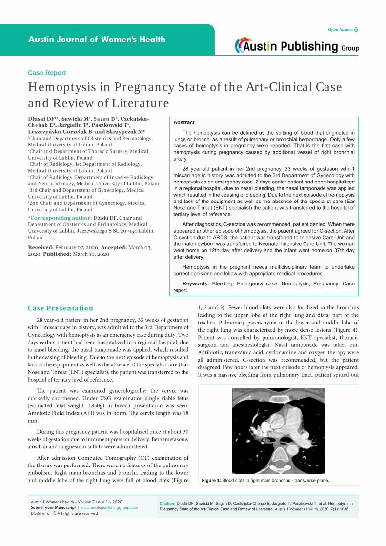
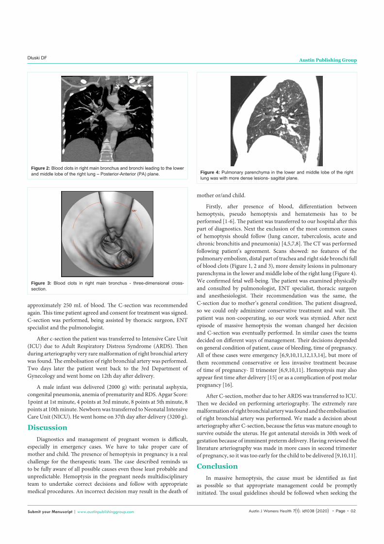
1, 2 and 3). Fewer blood clots were also localized in the bronchus leading to the upper lobe of the right lung and distal part of the trachea. Pulmonary parenchyma in the lower and middle lobe of the right lung was characterized by more dense lesions (Figure 4). Patient was consulted by pulmonologist, ENT specialist, thoracic surgeon and anesthesiologist. Nasal tamponade was taken out. Antibiotic, tranexamic acid, cyclonamine and oxygen therapy were all administered. C-section was recommended, but the patient disagreed. Few hours later the next episode of hemoptysis appeared. It was a massive bleeding from pulmonary tract, patient spitted out
Case Presentation28 year-old patient in her 2nd pregnancy, 33 weeks of gestation
with 1 miscarriage in history, was admitted to the 3rd Department of Gynecology with hemoptysis as an emergency case during duty. Two days earlier patient had been hospitalized in a regional hospital, due to nasal bleeding, the nasal tamponade was applied, which resulted in the ceasing of bleeding. Due to the next episode of hemoptysis and lack of the equipment as well as the absence of the specialist care (Ear Nose and Throat (ENT) specialist), the patient was transferred to the hospital of tertiary level of reference.
The patient was examined gynecologically: the cervix was markedly shorthened. Under USG examination single viable fetus (estimated fetal weight- 1850g) in breech presentation was seen. Amniotic Fluid Index (AFI) was in norm. The cervix length was 18 mm.
During this pregnancy patient was hospitalized once at about 30 weeks of gestation due to imminent preterm delivery. Bethametasone, atosiban and magnesium sulfate were administered.
After admission Computed Tomography (CT) examination of the thorax was performed. There were no features of the pulmonary embolism. Right main bronchus and bronchi, leading to the lower and middle lobe of the right lung were full of blood clots (Figure
Case Report
Hemoptysis in Pregnancy State of the Art-Clinical Case and Review of LiteratureDłuski DF1*, Sawicki M2, Sagan D2, Czekajska-Chehab E3, Jargiełło T4, Paszkowski T5, Leszczyńska-Gorzelak B1 and Skrzypczak M6
1Chair and Department of Obstetrics and Perinatology, Medical University of Lublin, Poland2Chair and Department of Thoracic Surgery, Medical University of Lublin, Poland3Chair of Radiology, 1st Department of Radiology, Medical University of Lublin, Poland4Chair of Radiology, Department of Invasive Radiology and Neuroradiology, Medical University of Lublin, Poland53rd Chair and Department of Gynecology, Medical University of Lublin, Poland62nd Chair and Department of Gynecology, Medical University of Lublin, Poland
*Corresponding author: Dłuski DF, Chair and Department of Obstetrics and Perinatology, Medical University of Lublin, Jaczewskiego 8 St, 20-954 Lublin, Poland
Received: February 07, 2020; Accepted: March 03, 2020; Published: March 10, 2020
Figure 1: Blood clots in right main bronchus - transverse plane.
Austin J Womens Health 7(1): id1038 (2020) - Page - 02
Dłuski DF Austin Publishing Group
Submit your Manuscript | www.austinpublishinggroup.com
approximately 250 mL of blood. The C-section was recommended again. This time patient agreed and consent for treatment was signed. C-section was performed, being assisted by thoracic surgeon, ENT specialist and the pulmonologist.
After c-section the patient was transferred to Intensive Care Unit (ICU) due to Adult Respiratory Distress Syndrome (ARDS). Then during arteriography very rare malformation of right bronchial artery was found. The embolisation of right bronchial artery was performed. Two days later the patient went back to the 3rd Department of Gynecology and went home on 12th day after delivery.
A male infant was delivered (2000 g) with: perinatal asphyxia, congenital pneumonia, anemia of prematurity and RDS. Apgar Score: 1point at 1st minute, 4 points at 3rd minute, 8 points at 5th minute, 8 points at 10th minute. Newborn was transferred to Neonatal Intensive Care Unit (NICU). He went home on 37th day after delivery (3200 g).
DiscussionDiagnostics and management of pregnant women is difficult,
especially in emergency cases. We have to take proper care of mother and child. The presence of hemoptysis in pregnancy is a real challenge for the therapeutic team. The case described reminds us to be fully aware of all possible causes even those least probable and unpredictable. Hemoptysis in the pregnant needs multidisciplinary team to undertake correct decisions and follow with appropriate medical procedures. An incorrect decision may result in the death of
mother or/and child.
Firstly, after presence of blood, differentiation between hemoptysis, pseudo hemoptysis and hematemesis has to be performed [1-6]. The patient was transferred to our hospital after this part of diagnostics. Next the exclusion of the most common causes of hemoptysis should follow (lung cancer, tuberculosis, acute and chronic bronchitis and pneumonia) [4,5,7,8]. The CT was performed following patient’s agreement. Scans showed: no features of the pulmonary embolism, distal part of trachea and right side bronchi full of blood clots (Figure 1, 2 and 3), more density lesions in pulmonary parenchyma in the lower and middle lobe of the right lung (Figure 4). We confirmed fetal well-being. The patient was examined physically and consulted by pulmonologist, ENT specialist, thoracic surgeon and anesthesiologist. Their recommendation was the same, the C-section due to mother’s general condition. The patient disagreed, so we could only administer conservative treatment and wait. The patient was non-cooperating, so our work was stymied. After next episode of massive hemoptysis the woman changed her decision and C-section was eventually performed. In similar cases the teams decided on different ways of management. Their decisions depended on general condition of patient, cause of bleeding, time of pregnancy. All of these cases were emergency [6,9,10,11,12,13,14], but more of them recommend conservative or less invasive treatment because of time of pregnancy- II trimester [6,9,10,11]. Hemoptysis may also appear first time after delivery [15] or as a complication of post molar pregnancy [16].
After C-section, mother due to her ARDS was transferred to ICU. Then we decided on performing arteriography. The extremely rare malformation of right bronchial artery was found and the embolisation of right bronchial artery was performed. We made a decision about arteriography after C-section, because the fetus was mature enough to survive outside the uterus. He got antenatal steroids in 30th week of gestation because of imminent preterm delivery. Having reviewed the literature arteriography was made in more cases in second trimester of pregnancy, so it was too early for the child to be delivered [9,10,11].
ConclusionIn massive hemoptysis, the cause must be identified as fast
as possible so that appropriate management could be promptly initiated. The usual guidelines should be followed when seeking the
Figure 2: Blood clots in right main bronchus and bronchi leading to the lower and middle lobe of the right lung – Posterior-Anterior (PA) plane.
Figure 3: Blood clots in right main bronchus - three-dimensional cross-section.
Figure 4: Pulmonary parenchyma in the lower and middle lobe of the right lung was with more dense lesions- sagittal plane.
Austin J Womens Health 7(1): id1038 (2020) - Page - 03
Dłuski DF Austin Publishing Group
Submit your Manuscript | www.austinpublishinggroup.com
cause. Even after optimal investigation, the etiology of hemoptysis remains unknown in up to 5–10% of patients. We had the first case with hemoptysis during pregnancy caused by additional vessel of right bronchial artery. There was no chance to predict this type of disease, as the patient didn’t present hemoptysis before pregnancy. In patient’s opinion the patient was healthy. As it was had been elsewhere described, pregnancy is unpredictable and we could not be sure what will happen in 5 minutes, so we have to be prepared for all possibilities when the treatment is offered to pregnant patients.
AcknowledgmentTo Piotr Flieger for his careful language correction.
References1. Stedman TL. Stedman’s Medical dictionary. 27th ed. Lipincott Williams &
Wilkins: Philadelphia, US. 2000.
2. Thompson AB, Teschler H, Rennard SI. Pathogenesis, evaluation, and therapy for massive hemoptysis. Clin Chest Med. 1992; 13: 69-82.
3. Knott-Craig CJ, Oostuizen JG, Rossouw G, Joubert JR, Barnard PM. Management and prognosis of massive hemoptysis. Recent experience with 120 patients. J Thorac Cardiovasc Surg. 1993; 105: 394-397.
4. Cahill BC, Ingbar DH. Massive hemoptysis. Assessment and management. Clin Chest Med. 1994; 15: 147-167.
5. Bidwell JL, Pachner RW. Hemoptysis: diagnosis and management. Am Fam Physician. 2005; 72: 1253-1260.
6. Masukume G, Sengurayi E, Moyo P, Feliu J, Gandanhamo D, Ndebele W, et al. Massive hemoptysis and complete unilateral lung collapse in pregnancy due to pulmonary tuberculosis with good maternal and fetal outcome: a case report. BMC Res Notes. 2013; 6: 335.
7. Harrison TR, Braunwald E. Hemoptysis. In: Harrison’s Principles of internal medicine. 15th ed. McGraw-Hill: New York, US. 2001: 203-206.
8. Reisz G, Stevens D, Boutwell C, Nair V. The causes of hemoptysis revisited. A review of the etiologies of hemoptysis between 1986 and 1995. Mo Med. 1997; 94: 633-635.
9. Peyrat E, Chabbert V, Escamilla R, Saada J, Degano B. Idiopathic hemoptysis in pregnant women: a distinct entity? Respir Med. 2007; 101: 2221-2223.
10. Dhillon SS, Yendamuri S, Harris K, Roche C, Pokharel S. Lymphangioma presenting as hemoptysis in pregnancy. Am J Respir Crit Care Med. 2014; 190: 701-703.
11. Desgranges FP, Berthelot AL, Gamondes D, Amanieu C, Audra P, Bastien O, et al. Massive recurrent haemoptysis in a pregnant woman with preeclampsia. Ann Fr Anesth Reanim. 2010; 29: 811-814.
12. Ng VV, Shah MK, Oh TT, Ramanathan A, Suhitharan T. Massive hemoptysis through endotracheal tube during emergency cesarean delivery: a case report and literature review. AA Case Rep. 2017; 9: 133-135.
13. Nguyen P, Weber F, Mahone M. A 30-year-old pregnant patient with massive haemoptysis and influenza A: atypical presentation of a common pathogen. Obstet Med. 2013; 6: 172-174.
14. Thompson RJ, Hasleton PS, Taylor PM, Woodhead M, Byrd LM. Haemoptysis in pregnancy caused by a well-differentiated fetal adenocarcinoma: a case report. J Med Case Rep. 2010; 4: 17.
15. Bernam DJ, Knibbs N, Friedman L, Rocco M. Postpartum hemoptysis as presenting sign of longstanding vasculitis. Int J Obstet Anesth. 2018; 36: 122-125.
16. McDonald-Burrows Z, Davies R, Goode E, Clarke C, Jackson J, Seckl M, et al. Haemoptysis from a pulmonary arteriovenous malformation in post molar pregnancy gestational trophoblast tumour patient managed by radiological embolisation: a case report. J Med Case Rep. 2014; 8: 117.