Embed Size (px)
Citation preview
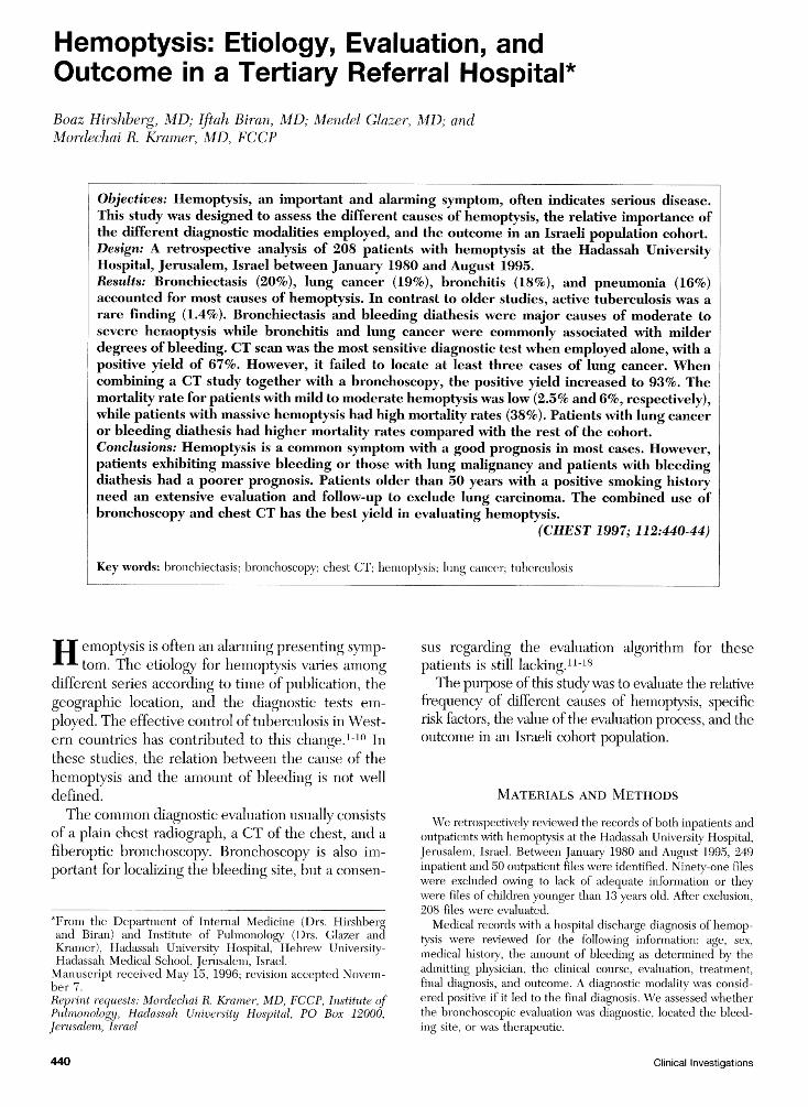
Hemoptysis: Etiology, Evaluation, andOutcome in a Tertiary Referral Hospital*Boaz Hirshberg, MD; Iftah Biran, MD; Mendel Glazer, MD; andMordechai R. Kramer, MD, FCCP
Objectives: Hemoptysis, an important and alarming symptom, often indicates serious disease.This study was designed to assess the different causes of hemoptysis, the relative importance ofthe different diagnostic modalities employed, and the outcome in an Israeli population cohort.Design: A retrospective analysis of 208 patients with hemoptysis at the Hadassah UniversityHospital, Jerusalem, Israel between January 1980 and August 1995.Results: Bronchiectasis (20%), lung cancer (19%), bronchitis (18%), and pneumonia (16%)accounted for most causes of hemoptysis. In contrast to older studies, active tuberculosis was arare finding (1.4%). Bronchiectasis and bleeding diathesis were major causes of moderate tosevere hemoptysis while bronchitis and lung cancer were commonly associated with milderdegrees of bleeding. CT scan was the most sensitive diagnostic test when employed alone, with a
positive yield of 67%. However, it failed to locate at least three cases of lung cancer. Whencombining a CT study together with a bronchoscopy, the positive yield increased to 93%. Themortality rate for patients with mild to moderate hemoptysis was low (2.5% and 6%, respectively),while patients with massive hemoptysis had high mortality rates (38%). Patients with lung canceror bleeding diathesis had higher mortality rates compared with the rest of the cohort.Conclusions: Hemoptysis is a common symptom with a good prognosis in most cases. However,patients exhibiting massive bleeding or those with lung malignancy and patients with bleedingdiathesis had a poorer prognosis. Patients older than 50 years with a positive smoking historyneed an extensive evaluation and follow-up to exclude lung carcinoma. The combined use ofbronchoscopy and chest CT has the best yield in evaluating hemoptysis.
(CHEST 1997; 112:440-44)
Key words: bronchiectasis; bronchoscopy; chest CT; hemoptysis; lung cancer; tuberculosis
TT emoptysis is often an alarming presenting symp--*¦-*¦ torn. The etiology for hemoptysis varies amongdifferent series according to time of publication, thegeographic location, and the diagnostic tests em¬
ployed. The effective control of tuberculosis in West¬ern countries has contributed to this change.110 Inthese studies, the relation between the cause of thehemoptysis and the amount of bleeding is not welldefined.The common diagnostic evaluation usually consists
of a plain chest radiograph, a CT of the chest, and a
fiberoptic bronchoscopy. Bronchoscopy is also im¬portant for localizing the bleeding site, but a consen-
*From the Department of Internal Medicine (Drs. Hirshbergand Biran) and Institute of Pulmonology (Drs. Glazer andKramer), Hadassah University Hospital, Hebrew University-Hadassah Medical School, Jerusalem, Israel.Manuscript received May 15, 1996; revision accepted Novem¬ber 7.Reprint requests: Mordechai R. Kramer, MD, FCCP, Institute ofPulmonology, Hadassah University Hospital, PO Box 12000,Jerusalem, Israel
sus regarding the evaluation algorithm for thesepatients is still lacking.1118The purpose of this study was to evaluate the relative
frequency of different causes of hemoptysis, specificrisk factors, die value of the evaluation process, and theoutcome in an Israeli cohort population.
Materials and MethodsWe retrospectively reviewed the records of both inpatients and
outpatients with hemoptysis at the Hadassah University Hospital,Jerusalem, Israel. Between January 1980 and August 1995, 249inpatient and 50 outpatient files were identified. Ninety-one fileswere excluded owing to lack of adequate information or theywere files of children younger than 13 years old. After exclusion,208 files were evaluated.
Medical records with a hospital discharge diagnosis of hemop¬tysis were reviewed for the following information: age, sex,medical history, the amount of bleeding as determined by theadmitting physician, the clinical course, evaluation, treatment,final diagnosis, and outcome. A diagnostic modality was consid¬ered positive if it led to the final diagnosis. We assessed whetherthe bronchoscopic evaluation was diagnostic, located the bleed¬ing site, or was therapeutic.
440 Clinical Investigations
Hemoptysis was defined as bleeding originating from the lowerrespiratory tract.1 We divided the patients into three groupsbased on the amount of bleeding: trivial (drops of blood, bloodysputum), moderate (<500 mL/24h, 1 to 2 cups), and massive(>500 mL/24 h, more than 2 cups).
ResultsOf the 208 patients evaluated, 127 (61%) were
male and 81 (39%) were female. The average agewas 58±17 years with a range of 20 to 96 years ofage. One hundred ten (53%) patients had a positivesmoking history and most of them were male (90/110, 82%).
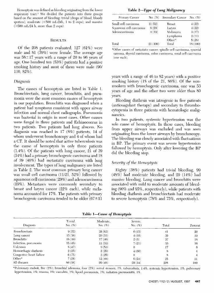
DiagnosisThe causes of hemoptysis are listed in Table 1.
Bronchiectasis, lung cancer, bronchitis, and pneu¬monia were the most common causes of hemoptysisin our population. Bronchitis was diagnosed when a
patient had symptoms consistent with upper airwayinfection and normal chest radiographs. Pneumoniawas bacterial in origin in most cases. Other causeswere fungal in three patients and Echinococcus intwo patients. Two patients had lung abscess. Nodiagnosis was reached in 17 (8%) patients, 14 ofwhom underwent bronchoscopy and 4 of whom hada CT. It should be noted that active tuberculosis wasthe cause of hemoptysis in only three patients(1.4%). Of the patients with lung cancer, 21 of 39(54%) had a primary bronchogenic carcinoma and 18of 39 (46%) had metastatic carcinoma with lunginvolvement. The types of lung malignancy are listedin Table 2. The most common primary lung cancerwas small cell carcinoma (11/21, 52%) followed bysquamous cell carcinoma (29%) and adenocarcinoma(19%). Metastases were commonly secondary tobreast and larynx cancer (22% each), while mela¬noma accounted for 17%. The patients with primarybronchogenic carcinoma tended to be older (67±11
Table 2.Type of Lung MalignancyPrimary7 Cancer No. Secondary Cancer No. (%)
Small cell carcinoma 11 (52)Squamous cell carcinoma 6 (29)Adenocarcinoma 4 (19)
Total 21 (100)
BreastLarynxMelanomaLymphomaOther*Total
4(22)4(22)3(17)2(11)5(28)
18(100)*Other causes of metastatic cancer: spindle cell carcinoma, synovialsarcoma, thyroid carcinoma, colon carcinoma, renal cell carcinoma(one each).
years with a range of 46 to 82 years) with a positivesmoking history (18 of the 21, 86%). Of the non-smokers with bronchogenic carcinoma, one was 53years of age and the other two were older than 80years.
Bleeding diathesis was iatrogenic in five patients(anticoagulant therapy) and secondary to thrombo¬cytopenia in three patients with hematologic malig¬nancies.
In two patients, systemic hypertension was thesole cause of hemoptysis. In these cases, bleedingfrom upper airways was excluded and was seen
originating from the lower airways by bronchoscopy.The bleeding was closely associated with fluctuationsin BP. The primary event was severe hypertensionfollowed by hemoptysis. Only after lowering the BPdid the bleeding stop.Severity of the Hemoptysis
Eighty (38%) patients had trivial bleeding, 99(48%) had moderate bleeding, and 29 (14%?) hadmassive bleeding. Lung cancer and bronchitis wereassociated with mild to moderate amounts of bleed¬ing (90% and 95%, respectively), while patients withbleeding diathesis and bronchiectasis had moderateto severe hemoptysis (78% and 75%, respectively).
Table 1.Cause of Hemoptysis
DiagnosisTrivial,No. (%)
Moderate,No. (%)
Severe,No. (%) Total Percent
BronchiectasisLung cancerBronchitisInfection, pneumoniaUnknownHemorrhagic diathesisCongestive heart failureOther*All diseases
9(22)15 (38)18 (49)15 (45)8(47)2(25)6 (75)7(28)
80 (38)
26 (63)20 (51)17(46)11 (33)9(53)2(25)2(25)12 (48)99 (48)
6(15)4(10)2(5)7(21)04(50)06(24)
29 (14)
413937331788
25208
20191816844
11100
*Pulmonary emboli, five (2%); bronchial adenoma, four (2%); mitral stenosis, 1%; tuberculosis, 1.4%; systemic hypertension, 1%; pulmonaryhypertension, 1%; trauma, 1%; vasculitis, 1%; lipoid pneumonia, 1%; radiation pneumonitis, 1%.
CHEST/112/2/AUGUST, 1997 441
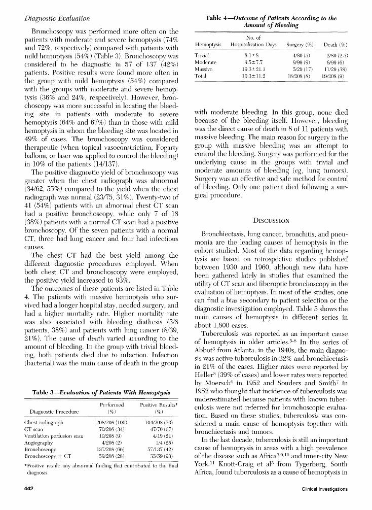
Diagnostic EvaluationBronchoscopy was performed more often on the
patients with moderate and severe hemoptysis (74%and 72%, respectively) compared with patients withmild hemoptysis (54%) (Table 3). Bronchoscopy wasconsidered to be diagnostic in 57 of 137 (42%)patients. Positive results were found more often inthe group with mild hemoptysis (54%) comparedwith the groups with moderate and severe hemop¬tysis (36% and 24%, respectively). However, bron¬choscopy was more successful in locating the bleed¬ing site in patients with moderate to severe
hemoptysis (64% and 67%) than in those with mildhemoptysis in whom the bleeding site was located in49% of cases. The bronchoscopy was consideredtherapeutic (when topical vasoconstriction, Fogartyballoon, or laser was applied to control the bleeding)in 10% of the patients (14/137).The positive diagnostic yield of bronchoscopy was
greater when the chest radiograph was abnormal(34/62, 55%) compared to the yield when the chestradiograph was normal (23/75, 31%). Twenty-two of41 (54%) patients with an abnormal chest CT scanhad a positive bronchoscopy, while only 7 of 18(38%) patients with a normal CT scan had a positivebronchoscopy. Of the seven patients with a normalCT, three had lung cancer and four had infectiouscauses.The chest CT had the best yield among the
different diagnostic procedures employed. Whenboth chest CT and bronchoscopy were employed,the positive yield increased to 93%.The outcomes of these patients are listed in Table
4. The patients with massive hemoptysis who sur¬vived had a longer hospital stay, needed surgery, andhad a higher mortality rate. Higher mortality ratewas also associated with bleeding diathesis (3/8patients, 38%) and patients with lung cancer (8/39,21%). The cause of death varied according to theamount of bleeding. In the group with trivial bleed¬ing, both patients died due to infection. Infection(bacterial) was the main cause of death in the group
Table 3.Evaluation of Patients With Hemoptysis
Table 4.Outcome of Patients According to theAmount ofBleeding
Diagnostic ProcedurePerformed
(%)Positive Results*
(%)Chest radiographCT scanVentilation perfusion scan
AngiographyBronchoscopyBronchoscopy + CT
208/208 (100)70/208 (34)19/208 (9)4/208 (2)
137/208 (66)59/208 (28)
104/208 (50)47/70 (67)4/19 (21)1/4 (25)
57/137 (42)55/59 (93)
HemoptysisNo. of
Hospitalization Days Surgery (%) Death (%)TrivialModerateMassiveTotal
8.1:!9.5d
19.3d10.3-
21.1:11.2
4/80 (5)9/99 (9)5/29(17)
18/208 (8)
2/80 (2.5)6/99 (6)
11/29 (38)19/208 (9)
* Positive result: any abnormal finding that contributed to the finaldiagnosis.
with moderate bleeding. In this group, none diedbecause of the bleeding itself. However, bleedingwas the direct cause of death in 8 of 11 patients withmassive bleeding. The main reason for surgery in thegroup with massive bleeding was an attempt tocontrol the bleeding. Surgery was performed for theunderlying cause in the groups with trivial andmoderate amounts of bleeding (eg, lung tumors).Surgery was an effective and safe method for controlof bleeding. Only one patient died following a sur¬
gical procedure.
Discussion
Bronchiectasis, lung cancer, bronchitis, and pneu¬monia are the leading causes of hemoptysis in thecohort studied. Most of the data regarding hemop¬tysis are based on retrospective studies publishedbetween 1930 and 1960, although new data havebeen gathered lately in studies that examined theutility of CT scan and fiberoptic bronchoscopy in theevaluation of hemoptysis. In most of the studies, onecan find a bias secondaiy to patient selection or thediagnostic investigation employed. Table 5 shows themain causes of hemoptysis in different series inabout 1,800 cases.
Tuberculosis was reported as an important causeof hemoptysis in older articles.58 In the series ofAbbot5 from Atlanta, in the 1940s, the main diagno¬sis was active tuberculosis in 22% and bronchiectasisin 21% of the cases. Higher rates were reported byHeller8 (39% of cases) and lower rates were reportedby Moersch6 in 1952 and Souders and Smith7 in1952 who thought that incidence of tuberculosis wasunderestimated because patients with known tuber¬culosis were not referred for bronchoscopic evalua¬tion. Based on these studies, tuberculosis was con¬sidered a main cause of hemoptysis together withbronchiectasis and tumors.
In the last decade, tuberculosis is still an importantcause of hemoptysis in areas with a high prevalenceof the disease such as Africa3'910 and inner-city NewYork.11 Knott-Craig et al3 from Tygerberg, SouthAfrica, found tuberculosis as a cause of hemoptysis in
442 Clinical Investigations

Table 5.Main Causes of Hemoptysis in Different Series*
Abbott5Souders and
Smith' Moersch6Johnstonand Reisz4
Alaoui etal10
Knott-Craig Santiago et McGuinness Presentet al3al2 et al11Study
Year(s) of studyLocation
No. of cases
Bronchiectasis, %
Carcinoma, %Bronchitis, %Pneumonia, %
Tuberculosis, %Unknown, %Others, %
1940-47 1941-51 1950 1977-85 1985-90 1983-90 1974-81Atlanta Lahey Clinic, Mayo Clinic Kansas City Casablanca, South Africa Los Angeles
497
21
2122
22428
Boston105
28.5
312.41
1.91835
200
26.5
29.59
5.5
21.5
148
19375
73
28
Morocco291
15
343.57
193
18.5
120 (massivebleeding)
51% (all hadTB)
5
73810
264
0.5
292311
6229
1991-92New York
57
25
12512
(aspergilloma)16195
1980-95Jerusalem,
Israel208
20
191816
18*Number in bold indicates highest figure. TB=tuberculosis.
73% of patients and Domoua et al9 from Abidjan,Ivory Coast, reported 49.5% of hemoptysis cases.Alaoui et al10 from Casablanca, Morocco, reportedthat carcinoma, tuberculosis, and bronchiectasiswere the most common findings (34.4%, 18.9%,15.1%, respectively). McGuinness et al11 prospec¬tively studied hemoptysis at Bellevue Hospital, NewYork, and found that bronchiectasis accounted for25% of cases and tuberculosis for 16%. In our
cohort, active tuberculosis was a rare finding, a factreflecting the low incidence of tuberculosis in Israel.The finding of bronchiectasis as a main cause of
hemoptysis in our cohort differs from recently pub¬lished articles.2-4 Johnston and Reisz4 reported thatbronchiectasis caused only 1% of cases in KansasCity between 1977 and 1985. The authors believethat they underdiagnosed this entity because bron-chography was rarely used. They did not statewhether they utilized CT studies in the evaluation oftheir patients. Santiago et al2 in west Los Angelesreported the incidence of bronchiectasis (0.5%).Bronchiectasis was probably underdiagnosed in thisstudy due to the lack of utilization of bronchographyand high-resolution CT scan (unavailable then). Thefinding of bronchiectasis as a main cause for hemop¬tysis in our study is probably secondary to remotenonactive infection by tuberculosis or other infec¬tions. A substantive number of the older Israelipopulation may have been exposed to tuberculosisduring the Holocaust era in Europe or have comefrom countries with high prevalence of tuberculosissuch as Iraq and Morocco. It should be expected thatowing to the decreased incidence of tuberculosis inour area and effective antibiotic therapy, we will seea decline in bronchiectasis as a cause of hemoptysis.The findings of lung cancer and bronchitis as other
main causes reflect the predominantly male cigarettesmoking cohort. These findings are similar to thoseof most of the studies cited in this article.Most patients underwent a thorough investigation
to find the cause of the hemoptysis. We found thatCT had the best yield in the search for a cause but,more importantly, the combined use of both high-resolution CT scan and bronchoscopy increased thepositive yield to 93%. McGuinness et al11 and Set etal14 demonstrated similar findings. They have shownthat bronchoscopy was unreliable in detecting pe¬ripheral lesions while CT was insensitive to earlymucosal abnormalities. In our cohort, CT alonefailed to locate three cases of lung cancer that were
successfully diagnosed with bronchoscopy. Based onthese findings, we conclude that CT scan and fiber¬optic bronchoscopy are complementary modalities.
Bronchoscopy performed in patients with massivehemoptysis had a low yield of positive diagnosticvalue (24%). However, bronchoscopy located thebleeding site in up to 67% of the patients. This is ofvalue early in the course of the illness, especially ifsurgery, such as lobectomy, is indicated due tolife-threatening bleeding.
Patients with primary lung carcinoma were older,mostly male, and had a positive smoking history.Similar results have been described elsewhere.1118Based on these results, we believe that patients olderthan 50 years of age with a positive smoking historyshould undergo a combined diagnostic procedure ofbronchoscopy and CT scan to locate a possiblemalignancy.The amount of bleeding usually correlated well
with the length of hospital stay and the outcome ofthe patients. While patients with minimal hemoptysishad a shorter stay and generally had a good progno-
CHEST/112/2/AUGUST, 1997 443

sis, patients with massive hemoptysis required a
longer hospital stay, more surgical intervention, andhad a graver prognosis.The outcome of hemoptysis is generally good, with
most patients surviving and released home. We identi¬fied two groups with increased risk. One includedpatients with bleeding diathesis who usually had a
greater amount of bleeding with a high mortality rate,probably reflecting the difficulty in controlling thebleeding. The other group included patients with lungmalignancy, mostiy bronchogenic carcinoma. Infectionwas the main cause of death in the groups with trivial tomoderate bleeding. In those groups, none died becauseof the bleeding itself.Although this study gives a good idea of the
etiology of hemoptysis in our cohort, it is limited byits retrospective nature. Furthermore, the evaluationand therapeutic decisions were not based on a presetprotocol but rather on the clinical experience of eachattending physician.
In conclusion, mild or moderate hemoptysis has a
generally good prognosis. Patients exhibiting massivebleeding or patients with lung carcinoma or withbleeding diathesis have a poor outcome, althoughdeath is not always related to the bleeding itself.Patients older than 50 years with a positive smokinghistory are at increased risk for primary lung carci¬noma and therefore need an extensive evaluationand follow-up. The combined use of bronchoscopyand chest CT probably gives the best diagnosticyield. Although active tuberculosis was a rare causeof hemoptysis, bronchiectasis from a remote infec¬tion is still a major cause for hemoptysis in our area.
References1 Fraser RG, Pare P, Pare PD, et al. Hemoptysis. In: FraserRG, Pare P, Pare PD, et al, eds. Diseases of the chest. 3rd ed.Philadelphia: WB Saunders, 1988; 394-96
2 Santiago S, Tobias J, Williams AJ. A reappraisal of the causesof hemoptysis. Arch Intern Med 1991; 151:2449-51
3 Knott-Craig CJ, Oostuizen JD, Rossouw G, et al. Manage¬ment and prognosis of massive hemoptysis. J Thorac Cardio¬vasc Surg 1993; 105:394-97
4 Johnston H, Reisz G. Changing spectrum of hemoptysis:underlying causes in 148 patients undergoing diagnosticfiberoptic bronchoscopy. Arch Intern Med 1989; 149:1661-68
5 Abbott OA. The clinical significance of pulmonary hemor¬rhage: a study of 1,316 patients with chest disease. Dis Chest1948; 14:824-42
6 Moersch HJ. Clinical significance of hemoptysis. JAMA 1952;148:1461-65
7 Souders CR, Smith AT. The clinical significance of hemop¬tysis. N Engl J Med 1952; 247:790-93
8 Heller R. The significance of hemoptysis. Tubercle 1946;26:70-4
9 Domoua K, N'Dhatz M, Coulihaly G, et al. Hemoptysis: mainetiologies observed in a pneumology department in Africa.Rev Pneumol Clin 1994; 50:59-62
10 Alaoui AY, Barial M, Boutahiri A, et al. Clinical characteristicsand etiology in hemoptysis in a pneumology service: 291cases. Rev Mai Respir 1992; 9:295-300
11 McGuinness G, Beacher JR, Harkin TJ, et al. Hemoptysis:prospective high-resolution CT/bronchoscopic correlation.Chest 1994; 105:1155-62
12 O'Neil KM, Lazarus AA. Indications for bronchoscopy. ArchIntern Med 1991; 151:171-74
13 Haponik EF, Chin R. Hemoptysis: clinicians' perspectives.Chest 1990; 97:469-75
14 Set PA, Flower CD, Smith IE, et al. Hemoptysis: comparativestudy of the role of CT and fiberoptic bronchoscopy. Radiol¬ogy 1993; 189:677-80
15 Naidich DP, Funt S, Ettenger NA, et al. Hemoptysis: CT-bronchoscopic correlations in 58 patients. Radiology 1990;177:357-62
16 Haponik EF, Britt EJ, Smith PL, et al. Computed chesttomography in the evaluation of hemoptysis: impact on
diagnosis and treatment. Chest 1987; 91:80-517 Heimer D, Bar-Ziv J, Scharf SM. Fiberoptic bronchoscopy in
patients with hemoptysis and nonlocalizing chest roentgeno-grams. Arch Intern Med 1985; 145:1427-28
18 Poe RH, Israel RH, Marin MG, et al. Utility of fiberopticbronchoscopy in patients with hemoptysis and a nonlocalizingroentgenograms. Chest 1988; 93:70-5
444 Clinical Investigations