Embed Size (px)
Citation preview
Hemolytic anaemia- Classification
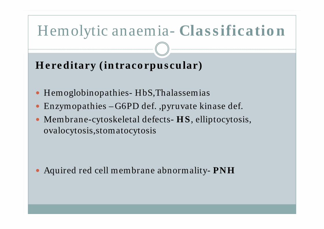
Hereditary (intracorpuscular)
Hemoglobinopathies- HbS,Thalassemias Enzymopathies –G6PD def. ,pyruvate kinase def. Membrane-cytoskeletal defects- HS, elliptocytosis,
ovalocytosis,stomatocytosis
Aquired red cell membrane abnormality- PNH
Classification cont.
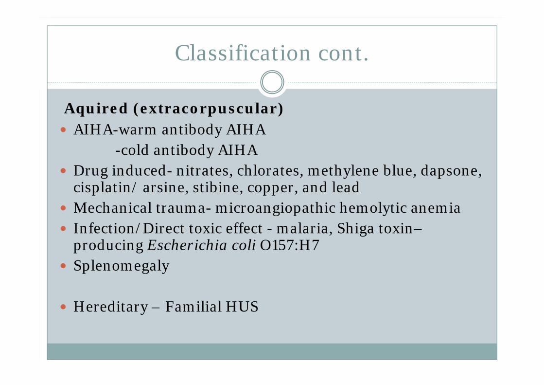
Aquired (extracorpuscular) AIHA-warm antibody AIHA
-cold antibody AIHA Drug induced- nitrates, chlorates, methylene blue, dapsone,
cisplatin/ arsine, stibine, copper, and lead Mechanical trauma- microangiopathic hemolytic anemia Infection/Direct toxic effect - malaria, Shiga toxin–
producing Escherichia coli O157:H7 Splenomegaly
Hereditary – Familial HUS
Common features(HA)
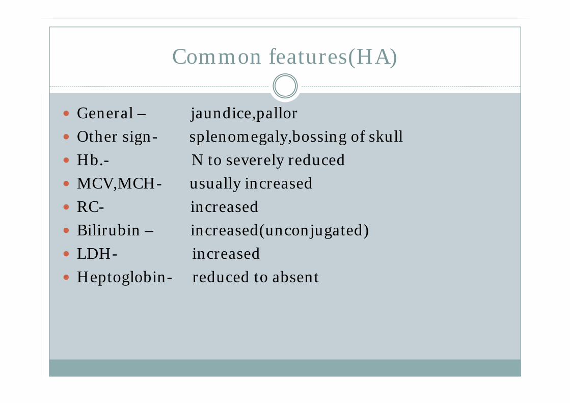
General – jaundice,pallor Other sign- splenomegaly,bossing of skull Hb.- N to severely reduced MCV,MCH- usually increased RC- increased Bilirubin – increased(unconjugated) LDH- increased Heptoglobin- reduced to absent
Investigation
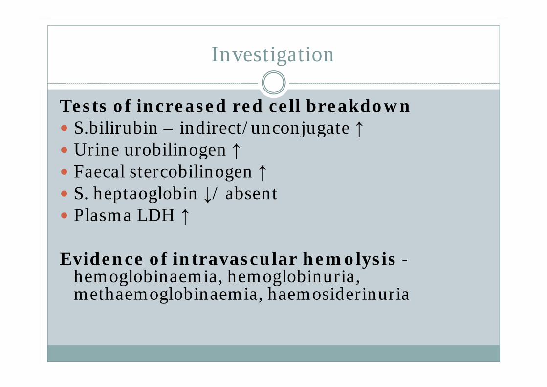
Tests of increased red cell breakdown S.bilirubin – indirect/unconjugate ↑ Urine urobilinogen ↑ Faecal stercobilinogen ↑ S. heptaoglobin ↓/ absent Plasma LDH ↑
Evidence of intravascular hemolysis -hemoglobinaemia, hemoglobinuria, methaemoglobinaemia, haemosiderinuria
Investigation
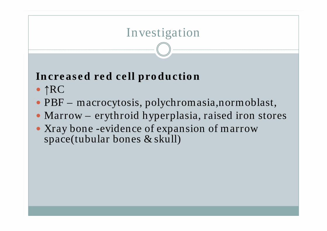
Increased red cell production ↑RC PBF – macrocytosis, polychromasia,normoblast, Marrow – erythroid hyperplasia, raised iron stores Xray bone -evidence of expansion of marrow
space(tubular bones & skull)
Investigation cont.
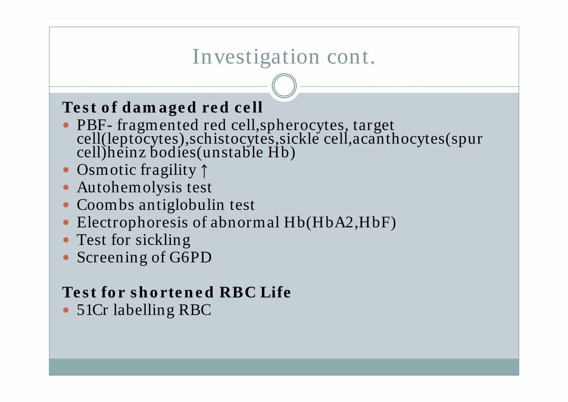
Test of damaged red cell PBF- fragmented red cell,spherocytes, target
cell(leptocytes),schistocytes,sickle cell,acanthocytes(spur cell)heinz bodies(unstable Hb)
Osmotic fragility ↑ Autohemolysis test Coombs antiglobulin test Electrophoresis of abnormal Hb(HbA2,HbF) Test for sickling Screening of G6PD
Test for shortened RBC Life 51Cr labelling RBC
Membrane cytoskeletal defect
Membrane cytoskeletal complex of red cell is integrated. Abnormality of any component-structural failure –
hemolysis Abnormalities almost due to inherited mutations
Hereditary spherocytosis
Common hemolytic anemia ,inherited as an autosomal dominant condition.
Occurs due to defect in protein (spectrin, ankyrin)which anchor lipid bilayer to underlying cytoskeleton.
Clinical features Anemia Splenomegaly Jaundice - ↑unconjugated bilirubin Pigment gall stone -↑bile pigment production Chronic leg ulcers
Investigation
Anaemia(spherocytes) ↑RC Blood film – microcytosis,spherocytes MCV ↓, MCHC- ↑ Osmotic fragility ↑ (pink test) Autohemolysis test - ↑(10-15%) N <4% Direct coomb antiglobin test – negative molecular studies demonstrating a mutation in one of the
genes underlying HS
Treatment
There is currently no treatment aimed at the cause of HS Blood transfusion in case of severe hemolytic crises Folic acid supplementation 5mg/d Splenectomy should be postponed untill 4yrs of age Indication – severe hemolysis, family history death from
similar disease, evidence of cholecystitis & cholelithiasis H . Influenza vaccine must 2 wks before splenectomy
Enzymopathies
Enzymes-role in metabolism of red cell Provide energy in form of ATP Prevent oxidative damage to hemoglobin and other protein. G6PD –critical role (red cell) only source of NADPH that
directly and via glutatione defends these cells against oxidative stress.
G6PD deficiency
Most common congenital shunt defect X linked trait affecting males, females are carrier N G6PD is designated as type B found in 11%
African American males Most common and significant clinical variant is A-
(negative) – confers partial protection against malaria
Hemolytic episodes occur on exposure to oxidant stress (viral, bacterial infection, metabolic acidosis, drugs, fava beans)
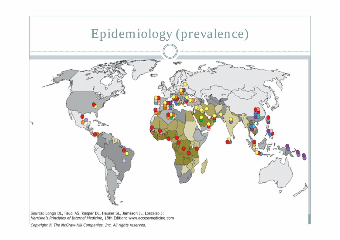
Epidemiology (prevalence)
Clinical Manifestations
Majority-asymptomatic neonatal jaundice (NNJ)-peak incidence of clinical onset is
between day 2 and day 3 Acute hemolytic attack- three types of triggers: (1) fava
beans, (2) infections, and (3) drugs starts with malaise, weakness, and abdominal or lumbar
pain Anemia Jaundice
Investigation
A/c episodes of hemolysis on exposure to oxidant stress –self limited since it affects old RBC only
During a/c hemolysis episodes Rapid fall in hematocrit (25-30%) Intravascular hemolysis test positive Blood film –heinz body (supravital stain crystal violet) –
bite cells (fragmented red cells)
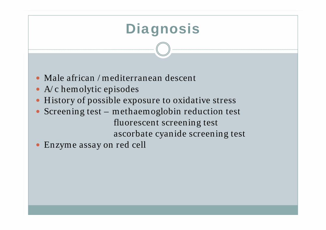
Diagnosis
Male african /mediterranean descent A/c hemolytic episodes History of possible exposure to oxidative stress Screening test – methaemoglobin reduction test
fluorescent screening testascorbate cyanide screening test
Enzyme assay on red cell
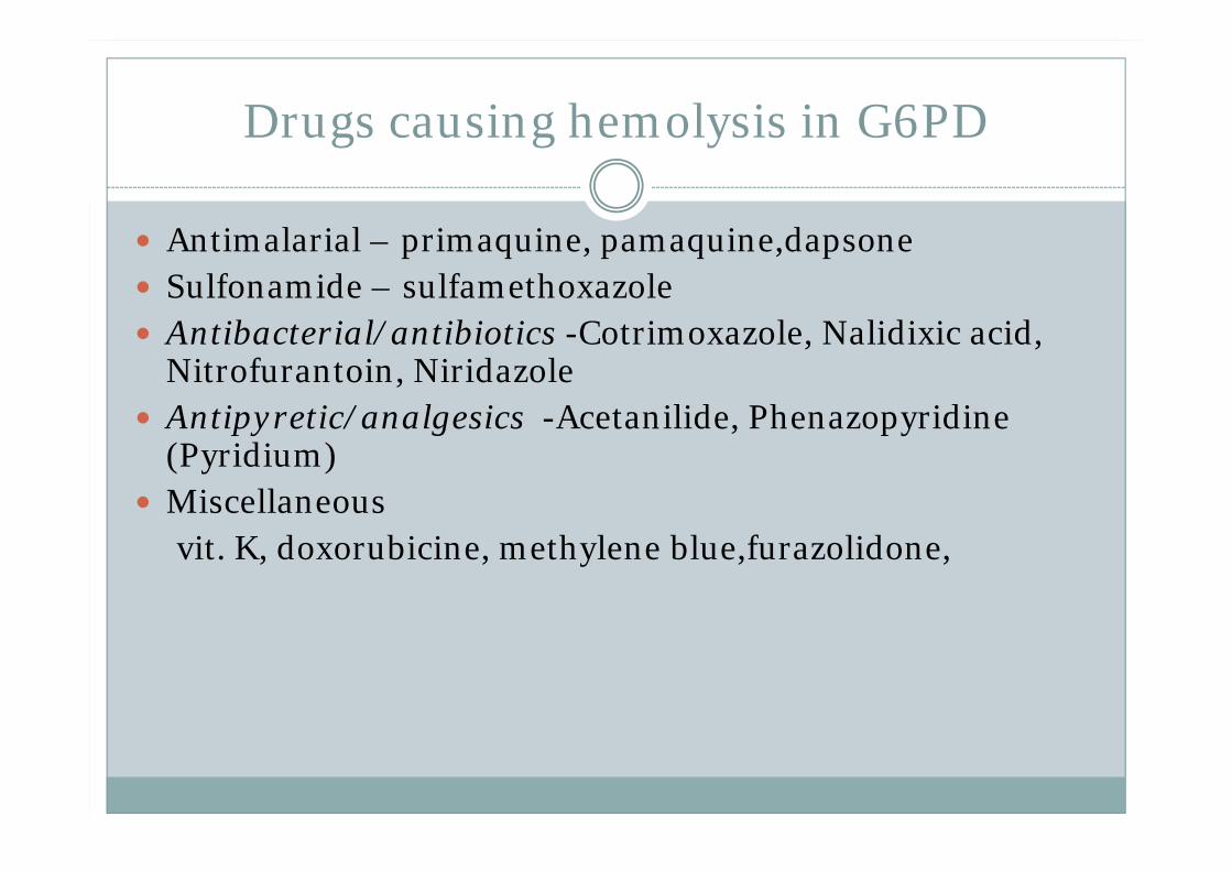
Drugs causing hemolysis in G6PD
Antimalarial – primaquine, pamaquine,dapsone Sulfonamide – sulfamethoxazole Antibacterial/antibiotics -Cotrimoxazole, Nalidixic acid,
Nitrofurantoin, Niridazole Antipyretic/analgesics -Acetanilide, Phenazopyridine
(Pyridium) Miscellaneous
vit. K, doxorubicine, methylene blue,furazolidone,
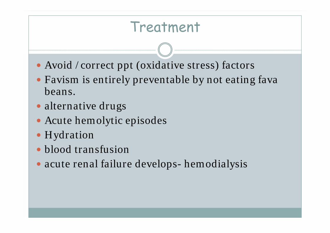
Treatment
Avoid /correct ppt (oxidative stress) factors Favism is entirely preventable by not eating fava
beans. alternative drugs Acute hemolytic episodes Hydration blood transfusion acute renal failure develops- hemodialysis
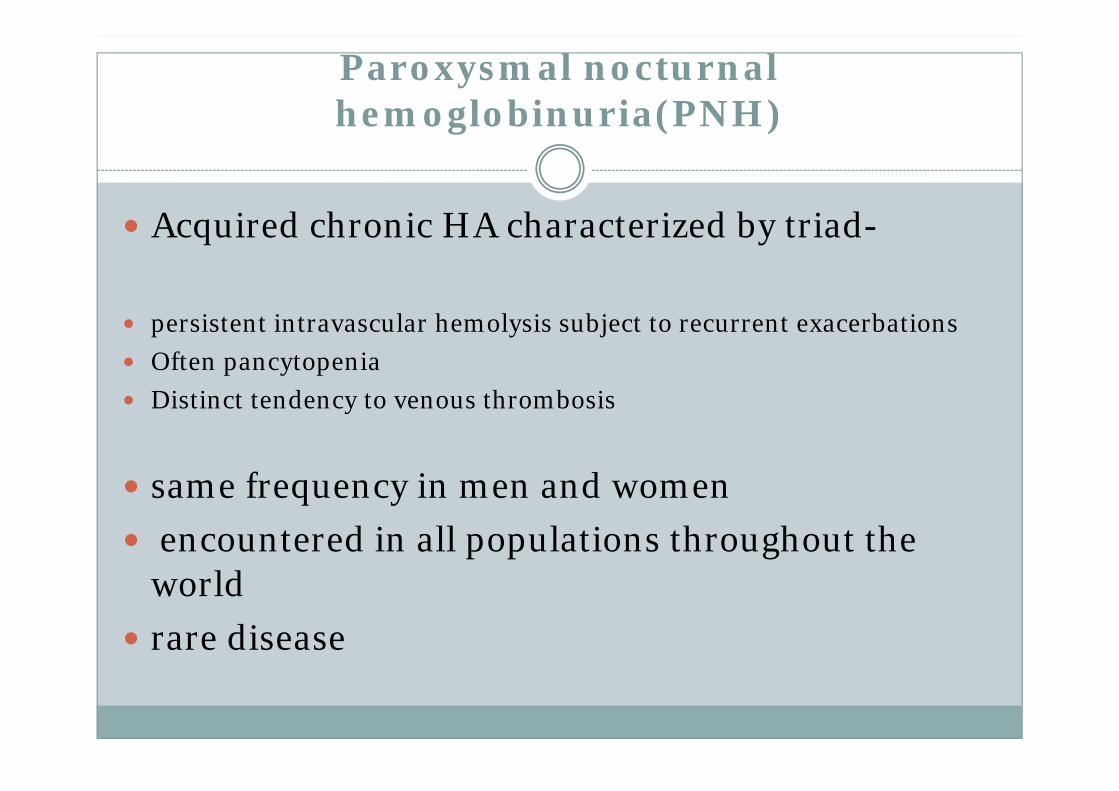
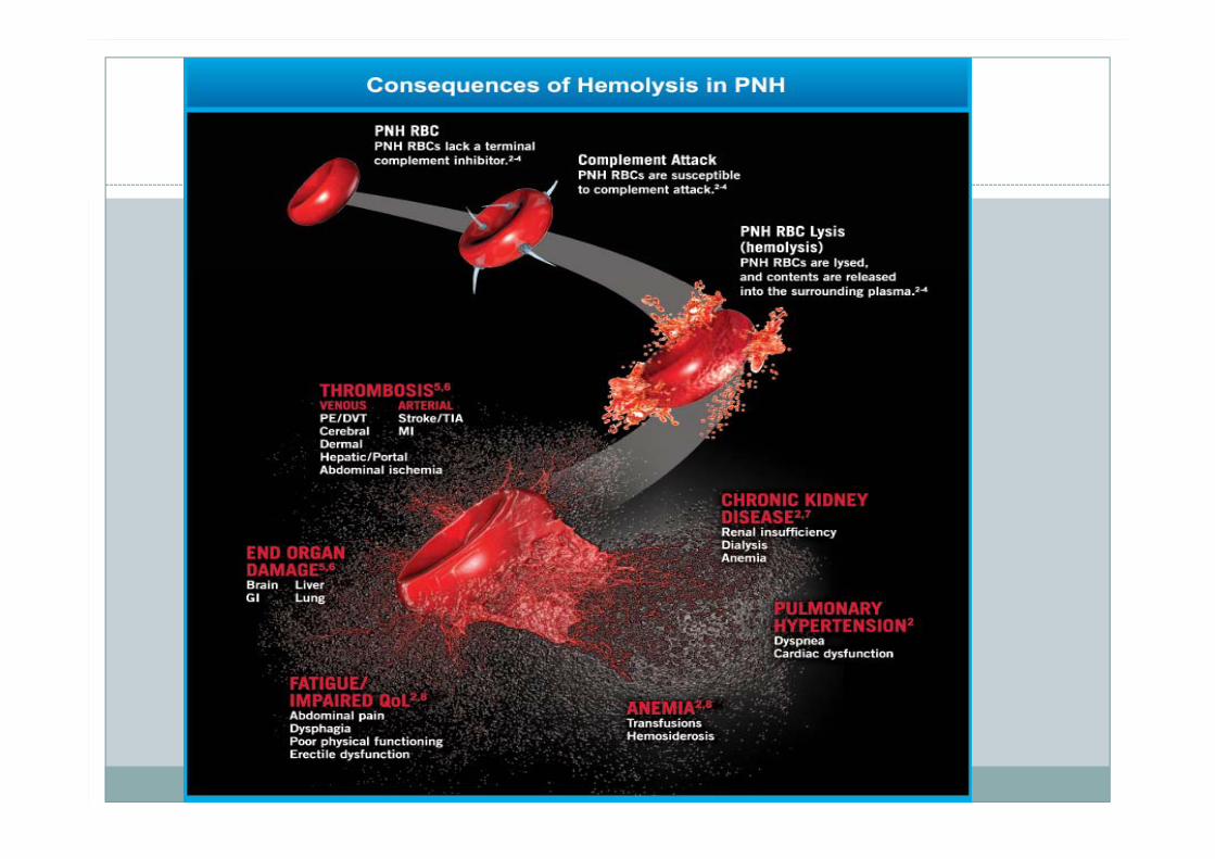
Paroxysmal nocturnal hemoglobinuria(PNH)
Acquired chronic HA characterized by triad-
persistent intravascular hemolysis subject to recurrent exacerbations Often pancytopenia Distinct tendency to venous thrombosis
same frequency in men and women encountered in all populations throughout the
world rare disease

Paroxysmal nocturnal hemoglobinuria
Rarer aquired disorder of red cell membrane defect arising at stem cell level
Defect in stem cell is a mutation affecting myeloid progenitor cells, resulting in partial or complete deficiency of anchor protein which make RBC sensitive to lytic effect of complement.
Decay accelerating factor (DAF) CD55Membrane inhibitor of reactive lysis (MIRL) CD59
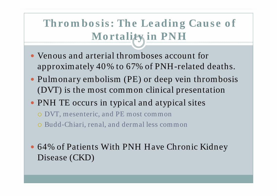
Thrombosis: The Leading Cause of Mortality in PNH
Venous and arterial thromboses account for approximately 40% to 67% of PNH-related deaths.
Pulmonary embolism (PE) or deep vein thrombosis (DVT) is the most common clinical presentation
PNH TE occurs in typical and atypical sites DVT, mesenteric, and PE most common Budd-Chiari, renal, and dermal less common
64% of Patients With PNH Have Chronic Kidney Disease (CKD)
Clinical feature
classical presentation-morning passing "blood instead of urine."
Hemolytic anaemia, thrombocytopenia, neutropenia
Venous thrombosis
Who to test for PNH
PNH Consensus Guidelines and the International PNH Interest Group recommend continued monitoring of patient populations at higher risk for PNH.
Populations include: Coombs-negative hemolytic anemia, hemoglobinuria, aplastic anemia, refractory anemia-myelodysplastic syndrome, unexplained cytopenias, and unexplained thrombosis.
Diagnosis
Hemolytic anaemia –test for intravascular hemolysis Demonstration of lysis of RBC after complement activation
by acid (hams test)or reduction in ionic strength(sucrose lysis test)
Flow cytometry analysis of GPI linked anchor protein CD55,CD59
Relationship between PNH and AA
The natural history of PNH can extend over decades
PNH may evolve into aplastic anemia (AA), and PNH may manifest itself in patients who previously had AA.
Rarely (estimated 1–2% of all cases), PNH may terminate in acute myeloid leukemia.
Treatment
Supportive treatment- transfusion of filtered red cells Folic acid supplements (at least 3 mg/d) are mandatory humanized monoclonal antibody, eculizumab, directed against the
complement protein C5(iv every 14 days) Allogeneic BMT; when an HLA-identical sibling is available, BMT
should be offered to any young patient with severe PNH.
A/c thrombotic event (budd chiari syndrome & cerebral thrombosis)Thrombolytic agents – tpa/heparin/LMWH & warfarin
PNH-AA syndrome,- immunosuppressive treatment with antilymphocyte globulin (ALG or ATG) and cyclosporine
Autoimmune Hemolytic Anemia (AIHA)
most common form of acquired hemolytic anemia next to malaria
autoantibody directed against a red cell antigen
Clinical Features
suspicion of AIHA must be high Anaemia jaundice SplenomegalyDiagnosis Coombs antiglobulin test
Treatment
Transfusion of red cells The first-line – glucocorticoids(1 mg/kg) Rituximab(anti CD 20) remissions upto 80% second-line - long-term immunosuppression with
low-dose prednisone, azathioprine, or cyclosporine. splenectomy
Acquired hemolytic anemia
Mechanical(microangiopathic HA) eg marathon runners,prolonged barefoot ritual dancing,prosthetic heart valves.
Toxins and drugs eg hyperbaric oxygen,nitrates,chlorates,methylene blue,dapsone,cisplatin,arsine,stibine,copper and lead
Infection eg malaria,shiga toxin
splenomegaly