Embed Size (px)
Citation preview
Hemodynamic Monitoring
Charles E. Smith, MD
Professor of Anesthesia
Director, Cardiothoracic Anesthesia
MetroHealth Medical Center
Case Western Reserve University
Cleveland, OhioEmail: [email protected]
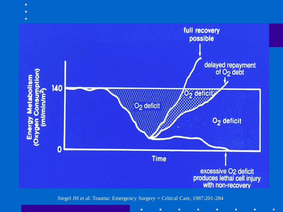
Siegel JH et al: Trauma: Emergency Surgery + Critical Care, 1987:201-284
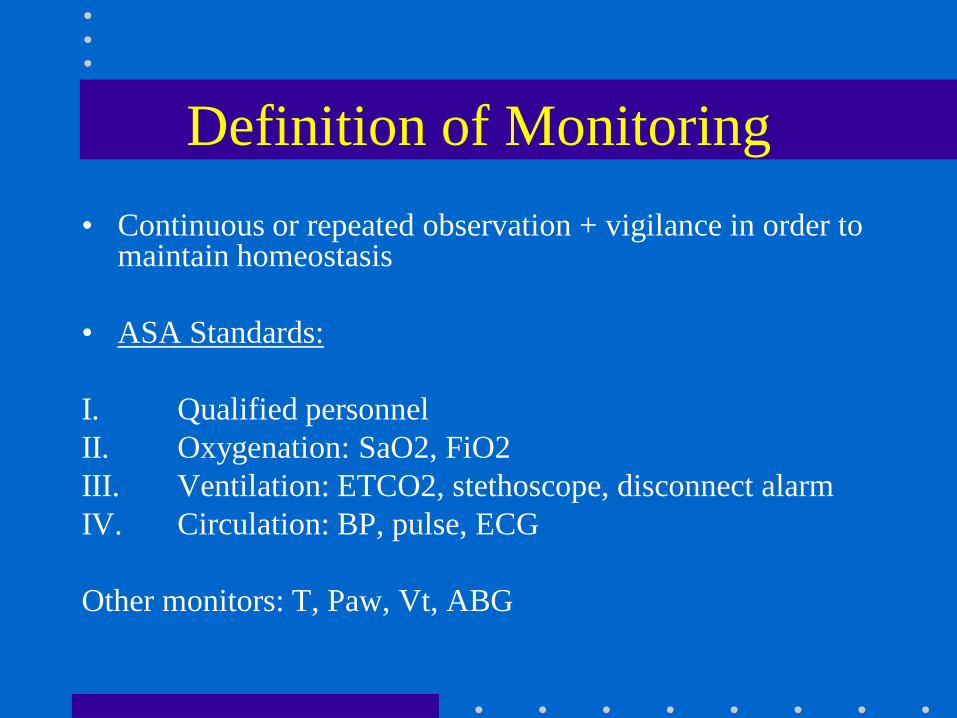
Definition of Monitoring
• Continuous or repeated observation + vigilance in order to maintain homeostasis
• ASA Standards:
I. Qualified personnel
II. Oxygenation: SaO2, FiO2
III. Ventilation: ETCO2, stethoscope, disconnect alarm
IV. Circulation: BP, pulse, ECG
Other monitors: T, Paw, Vt, ABG

Objectives
– Arterial line
– Systolic pressure variation
– Central venous pressure
– Pulmonary artery catheterization
– Cardiac output
– Mixed venous oxygen
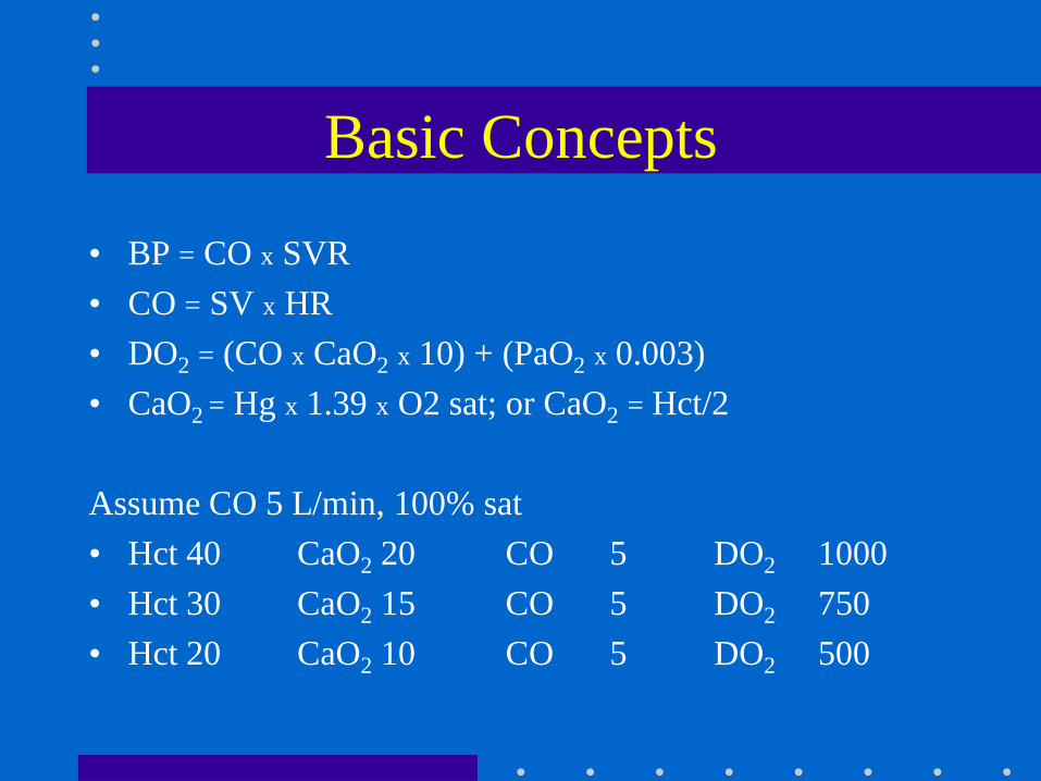
Basic Concepts
• BP = CO x SVR
• CO = SV x HR
• DO2 = (CO x CaO2 x 10) + (PaO2 x 0.003)
• CaO2 = Hg x 1.39 x O2 sat; or CaO2 = Hct/2
Assume CO 5 L/min, 100% sat
• Hct 40 CaO2 20 CO 5 DO2 1000
• Hct 30 CaO2 15 CO 5 DO2 750
• Hct 20 CaO2 10 CO 5 DO2 500
Arterial Line
• Indications:
– Rapid moment to moment BP changes
– Frequent blood sampling
– Circulatory therapies: bypass, IABP, vasoactive
drugs, deliberate hypotension
– Failure of indirect BP: burns, morbid obesity
– Pulse contour analysis: SPV, SV
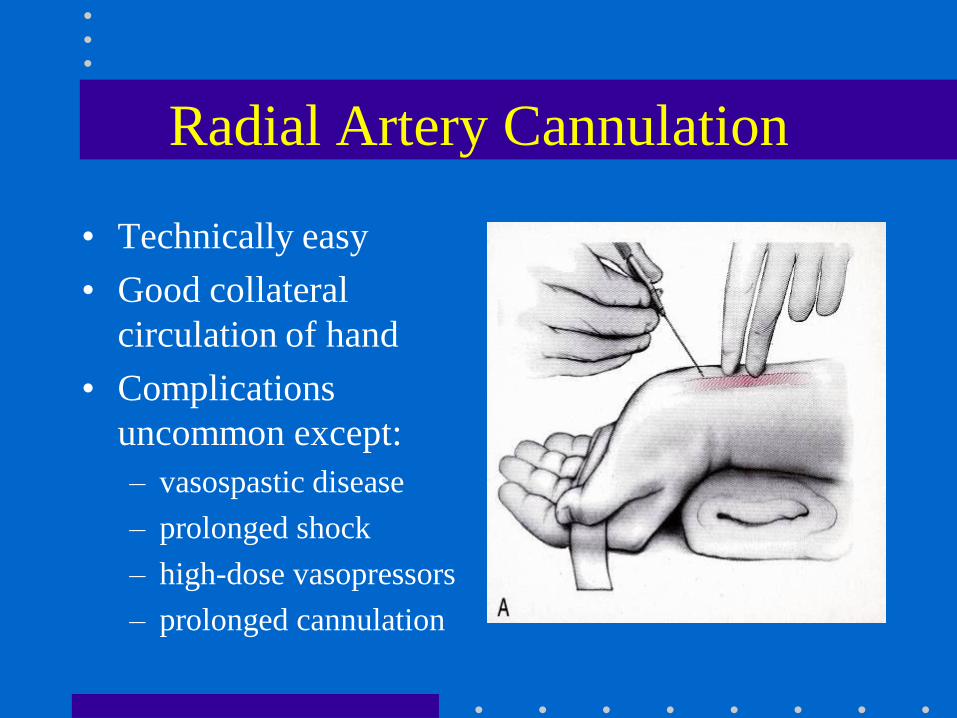
Radial Artery Cannulation
• Technically easy
• Good collateral
circulation of hand
• Complications
uncommon except:
– vasospastic disease
– prolonged shock
– high-dose vasopressors
– prolonged cannulation
Alternative Sites
• Brachial:
– Use longer catheter to traverse elbow joint
– Postop keep arm extended
– Collateral circulation not as good as hand
• Femoral:
– Use guide-wire technique
– Puncture femoral artery below inguinal
ligament (easier to compress, if required)
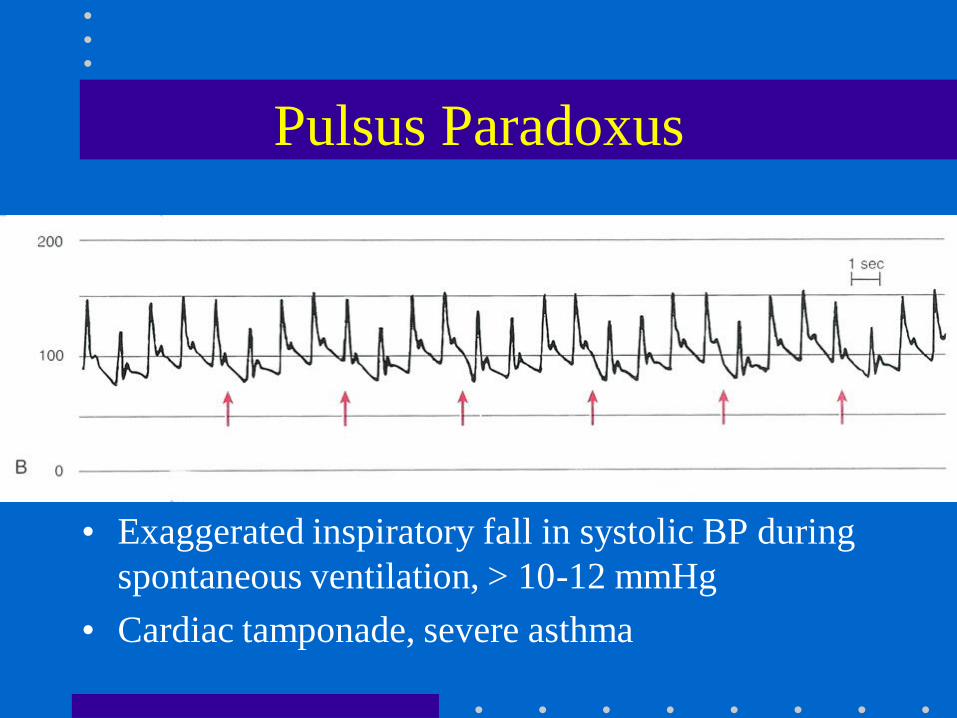
Pulsus Paradoxus
• Exaggerated inspiratory fall in systolic BP during
spontaneous ventilation, > 10-12 mmHg
• Cardiac tamponade, severe asthma
Marik: Anaesth Intensive Care 1993;21:405. Coriat: Anesth Analg 1994;78:46
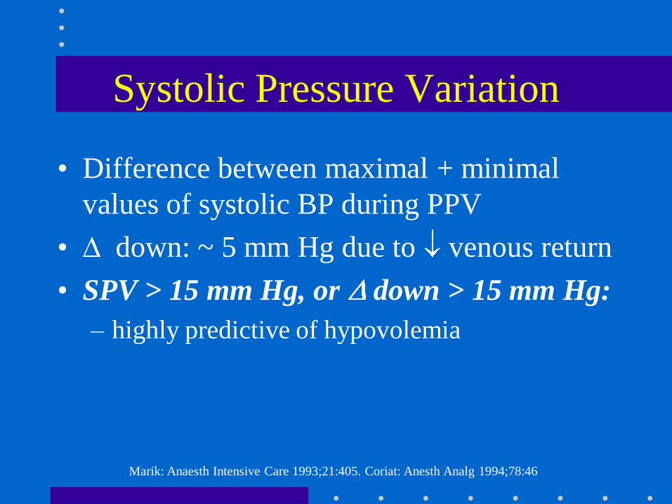
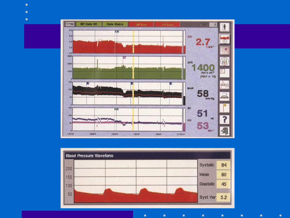
Systolic Pressure Variation
• Difference between maximal + minimal
values of systolic BP during PPV
• down: ~ 5 mm Hg due to venous return
• SPV > 15 mm Hg, or down > 15 mm Hg:
– highly predictive of hypovolemia
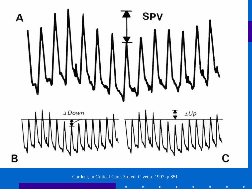
Gardner, in Critical Care, 3rd ed. Civetta. 1997, p 851
Linton R: 1997, 1998, 2000
Pulse Contour Analysis
• 1. Transform BP waveform into volume – time waveform
• 2. Derive uncalibrated SV
– SV x HR = CO
• 3. May calibrate using Li indicator [LidCO] or assume initial SV based on known EF from echo
• Assumptions:
– PPV induces cyclical changes in SV
– Changes in SV results in cyclical fluctuation of BP or SPV
PulseCO SPV + SV
• Predicts SV in response to volume after cardiac
surgery + in ICU [Reuter: BJA 2002; 88:124; Michard: Chest 2002;
121:2000]
• Similar estimates of preload v. echo during
hemorrhage [Preisman: BJA 2002; 88: 716]
• Helpful in dx of hypovolemia after blast injury[Weiss: J Clin Anesth 1999; 11:132]
Pitfalls with SPV + SV
• Inaccurate if
– AI
– IABP
• Problems if
– pronounced peripheral arterial vasoconstriction
– damped art line
– arrhythmias
Central Venous Line
• Indications:
– CVP monitoring
– Advanced CV disease + major operation
– Secure vascular access for drugs: TLC
– Secure access for fluids: introducer sheath
– Aspiration of entrained air: sitting craniotomies
– Inadequate peripheral IV access
– Pacer, Swan Ganz
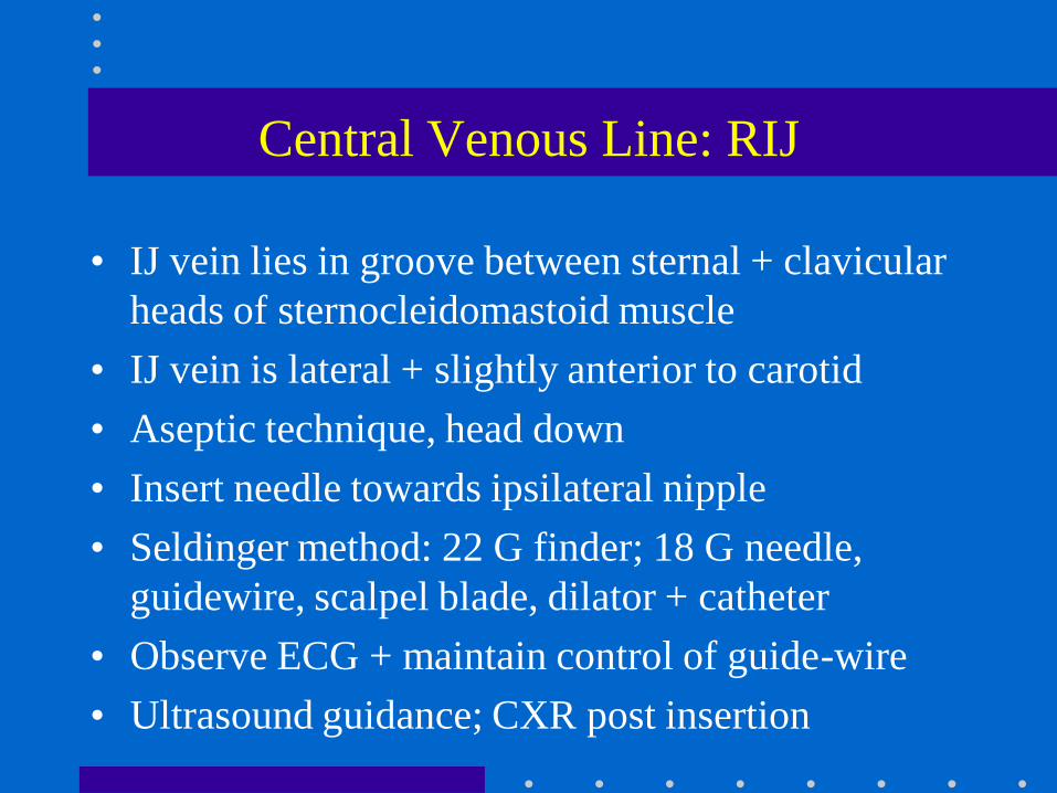
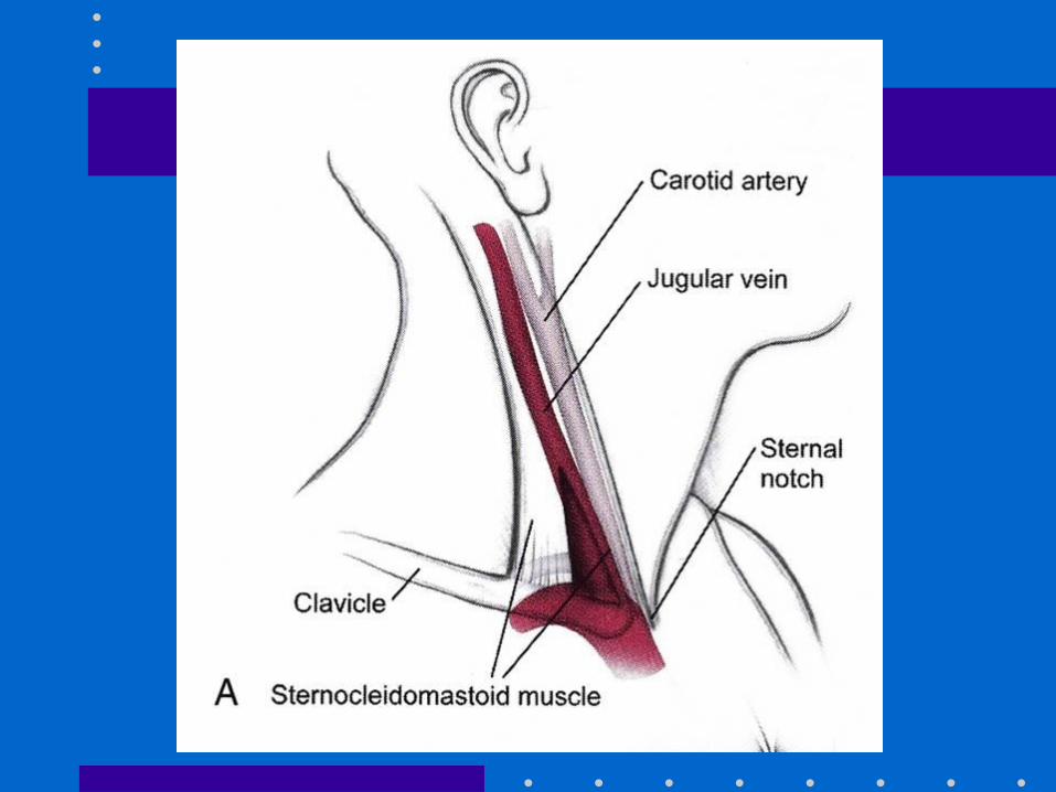
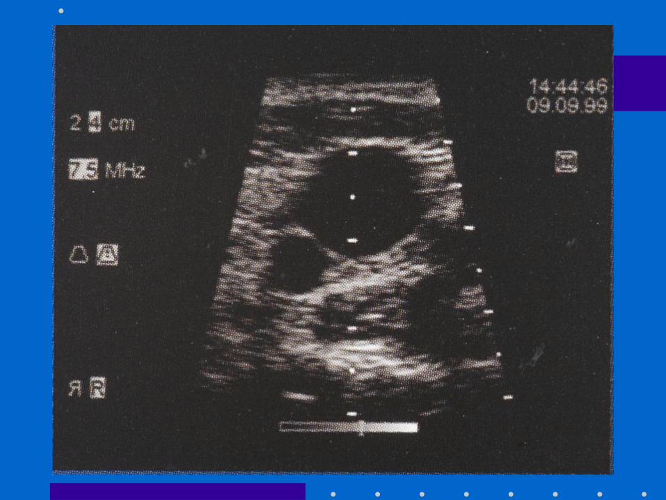
Central Venous Line: RIJ
• IJ vein lies in groove between sternal + clavicular
heads of sternocleidomastoid muscle
• IJ vein is lateral + slightly anterior to carotid
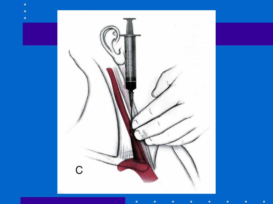
• Aseptic technique, head down
• Insert needle towards ipsilateral nipple
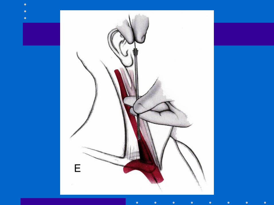
• Seldinger method: 22 G finder; 18 G needle,
guidewire, scalpel blade, dilator + catheter
• Observe ECG + maintain control of guide-wire
• Ultrasound guidance; CXR post insertion
Advantages of RIJ
• Consistent, predictable anatomic location
• Readily identifiable landmarks
• Short straight course to SVC
• Easy intraop access for anesthesiologist at
patient’s head
• High success rate, 90-99%
Types of Central Catheters
• Variety of lengths, gauges, composition + lumens
depending on purpose
• Introducer sheath (8-8.5 Fr):
– Permits rapid fluid/blood infusion or Swan
• Trauma triple-lumen (12 Fr):
– Rapid infusion via 12 g x 2; 16 g for CVP monitoring
• MAC 2: (9 Fr):
– Rapid infusion via distal port; 12 g for CVP
– Also allows for Swan insertion
– More septations + stiffer plastic
Alternative Sites
• Subclavian:
– Easier to insert v. IJ if c-spine precautions
– Better patient comfort v. IJ
– Risk of pneumo- 2%
• External jugular:
– Easy to cannulate if visible, no risk of pneumo
– 20%: cannot access central circulation
• Double cannulation of same vein (RIJ)
– Serious complications: vein avulsion, catheter
entanglement, catheter fracture
CVP Monitoring
• Reflects pressure at junction of vena cava + RA
• CVP is driving force for filling RA + RV
• CVP provides estimate of:
– Intravascular blood volume
– RV preload
• Trends in CVP are very useful
• Measure at end-expiration
• Zero at mid-axillary line
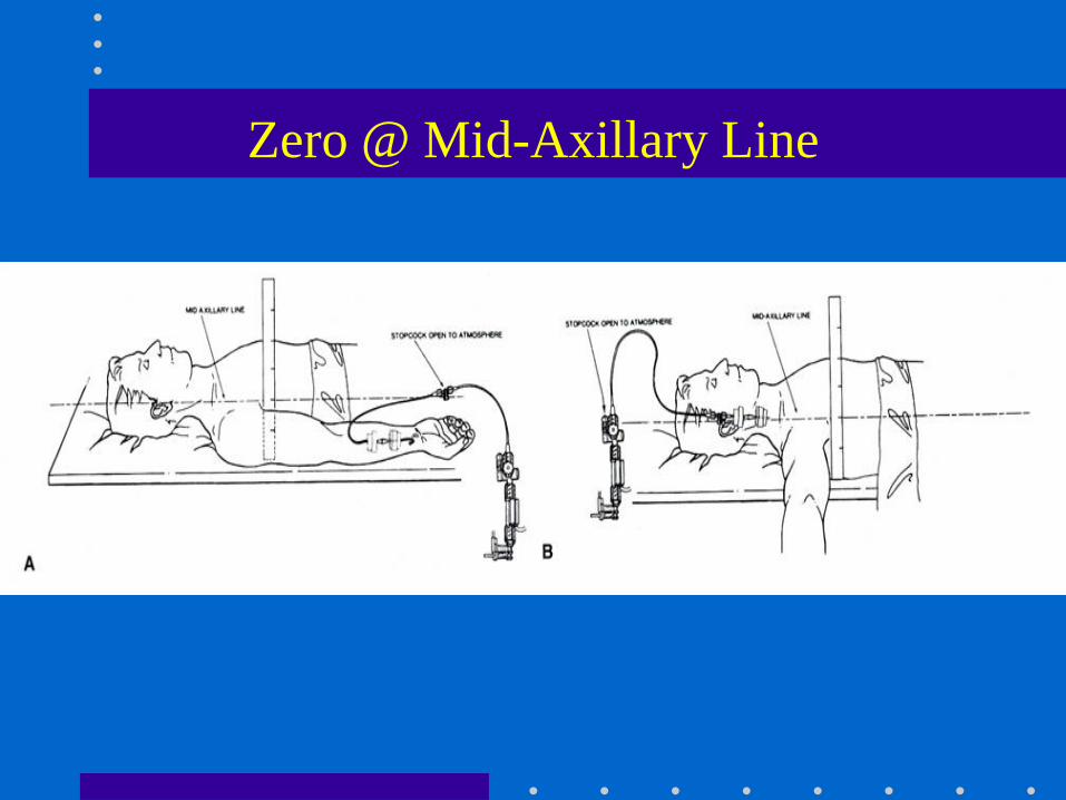
Zero @ Mid-Axillary Line
Mark JB, CV Monitoring, in Miller 5th Edition, 2000, pg 1153
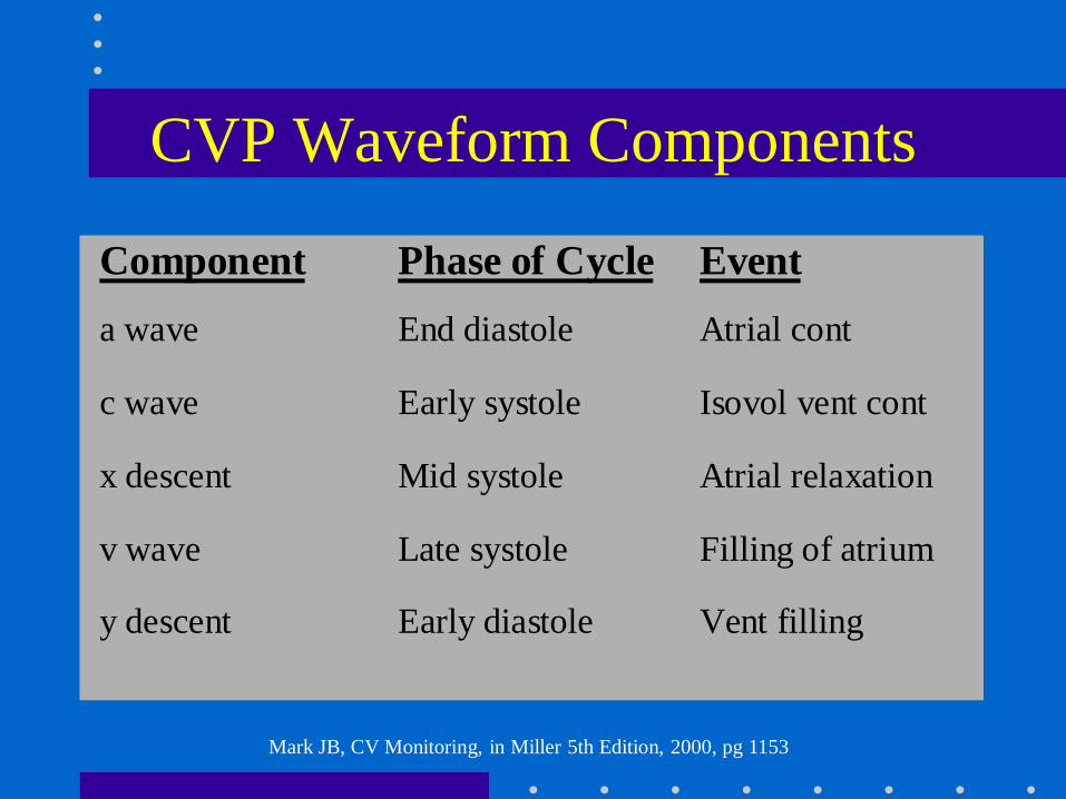
CVP Waveform Components
Component Phase of Cycle Event
a wave End diastole Atrial cont
c wave Early systole Isovol vent cont
x descent Mid systole Atrial relaxation
v wave Late systole Filling of atrium
y descent Early diastole Vent filling
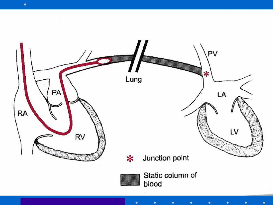
Pulmonary Artery Catheter
• Introduced by Swan + Ganz in 1970
• Allows accurate bedside measurement of
important clinical variables: CO, PAP,
PCWP, CVP to estimate LV filling volume,
+ guide fluid / vasoactive drug therapy
• Discloses pertinent CV data that cannot be
accurately predicted from standard signs +
symptoms
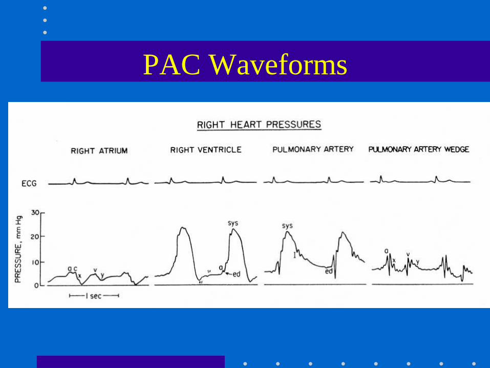
PAC Waveforms
Roizen et al: Anesthesiology 1993;78:380. ASA Newsletter, Aug 2002;66(8):7
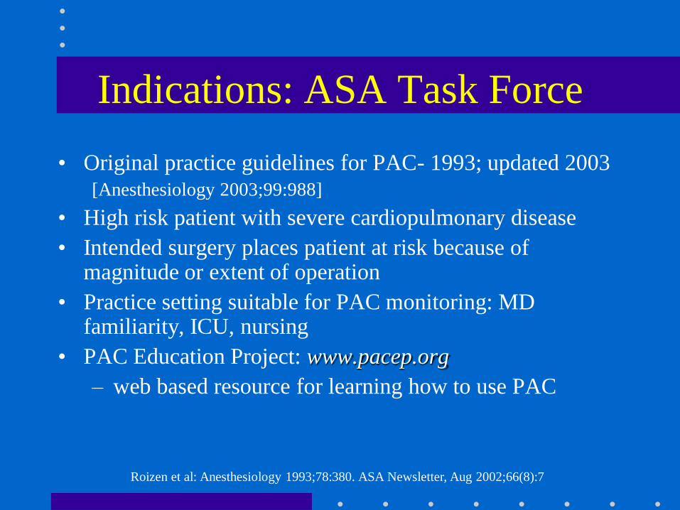
Indications: ASA Task Force
• Original practice guidelines for PAC- 1993; updated 2003[Anesthesiology 2003;99:988]
• High risk patient with severe cardiopulmonary disease
• Intended surgery places patient at risk because of magnitude or extent of operation
• Practice setting suitable for PAC monitoring: MD familiarity, ICU, nursing
• PAC Education Project: www.pacep.org
– web based resource for learning how to use PAC
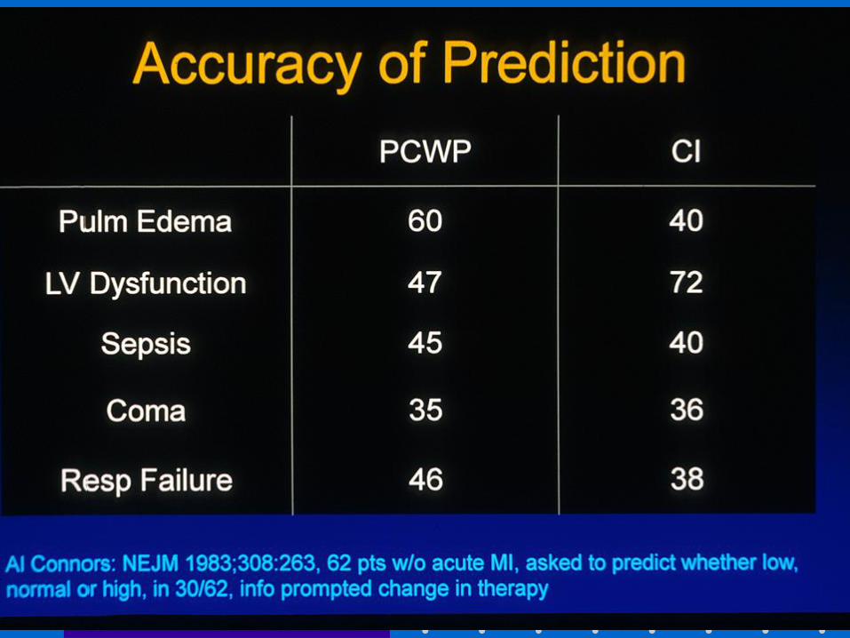
Connors: JAMA 1996;276:916. Mark JB: in Anesthesia 5th Ed. Miller. 2000: pp 1178-80
PAC and Outcome
• Early use of PAC to optimize volume status + tissue perfusion may be beneficial
• PAC is only a monitor. It cannot improve outcome if disease has progressed too far, or if intervention based on PAC is unsuccessful or detrimental
• Many confounding factors: learning bias, skill, knowledge, usage patterns, medical v. surgical illness
Mark JB, in Anesthesia 5th Edition. Miller 2000, pg 1117-1206
PAC: Complications
• Minor in 50%, e.g., arrhythmias
• Transient RBBB- 0.9-5%
– External pacer if pre-existing LBBB
• Misinformation
• Serious: 0.1-0.5%: knotting, pulmonary infarction,
PA rupture (e.g., overwedge), endocarditis,
structural heart damage
• Death: 0.016%
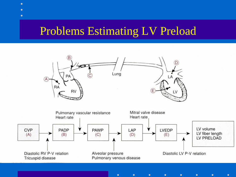
Problems Estimating LV Preload
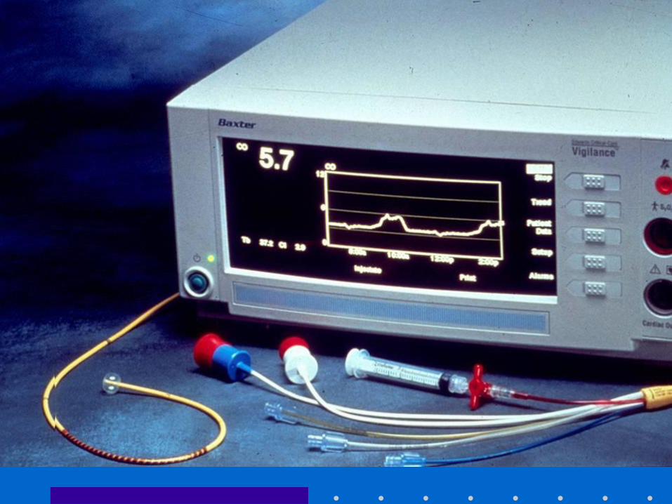
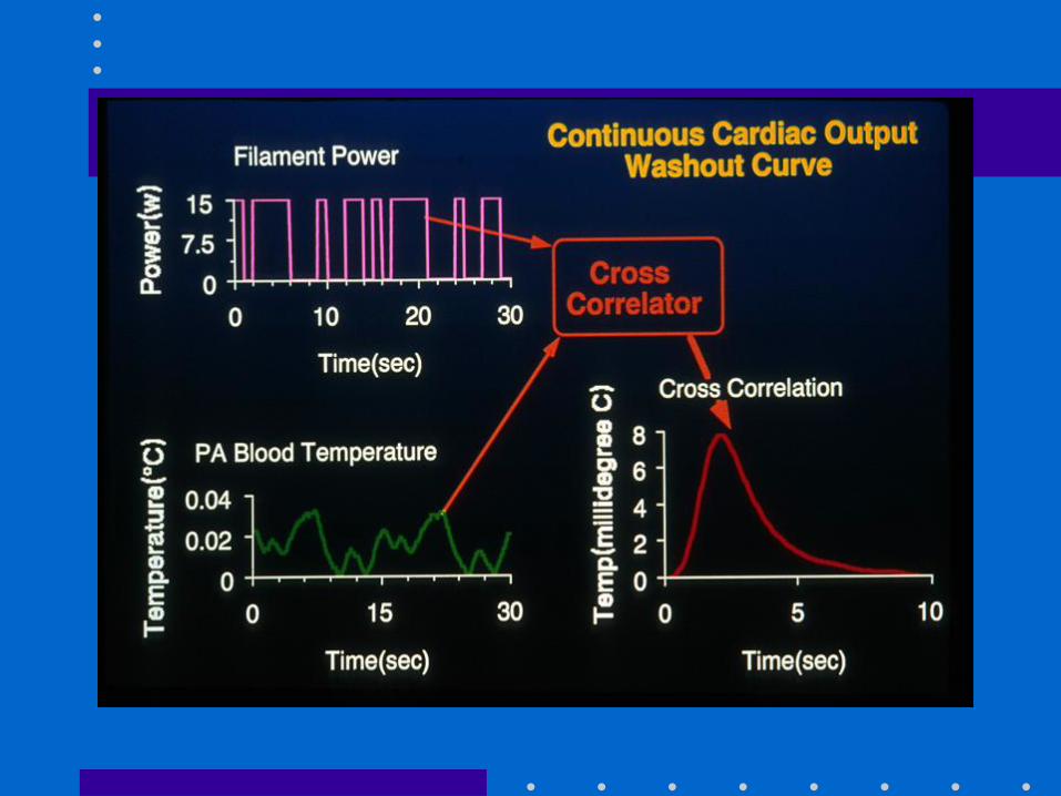
Cardiac Output
• Important feature of PAC
• Allows calculation of DO2
• Thermodilution: inject fixed volume, 10 ml, (of
room temp or iced D5W) into CVP port at end-
expiration + measure resulting change in blood
temp at distal thermistor
• CO inversely proportional to area under curve
Cardiac Output: Technical Problems
• Variations in respiration:
– Use average of 3 measures
• Blood clot over thermistor tip: inaccurate temp
• Shunts: LV + RV outputs unequal, CO invalid
• TR: recirculation of thermal signal, CO invalid
• Computation constants:
– Varies for each PAC, check package insert + manually
enter
Continuous Mixed Venous Oximetry
• Fick Equation
– VO2 = CO [CaO2 - CvO2]
– CvO2 ~ SvO2 b/c most O2 in blood bound to Hg
• If O2 sat, VO2 + Hg remain constant, SvO2 is
indirect indicator of CO
• Can be measured using oximetric Swan or CVP,
or send blood gas from PA / CVP
• Normal SvO2 ~ 65% [60-75]
Mixed Venous Oximetry
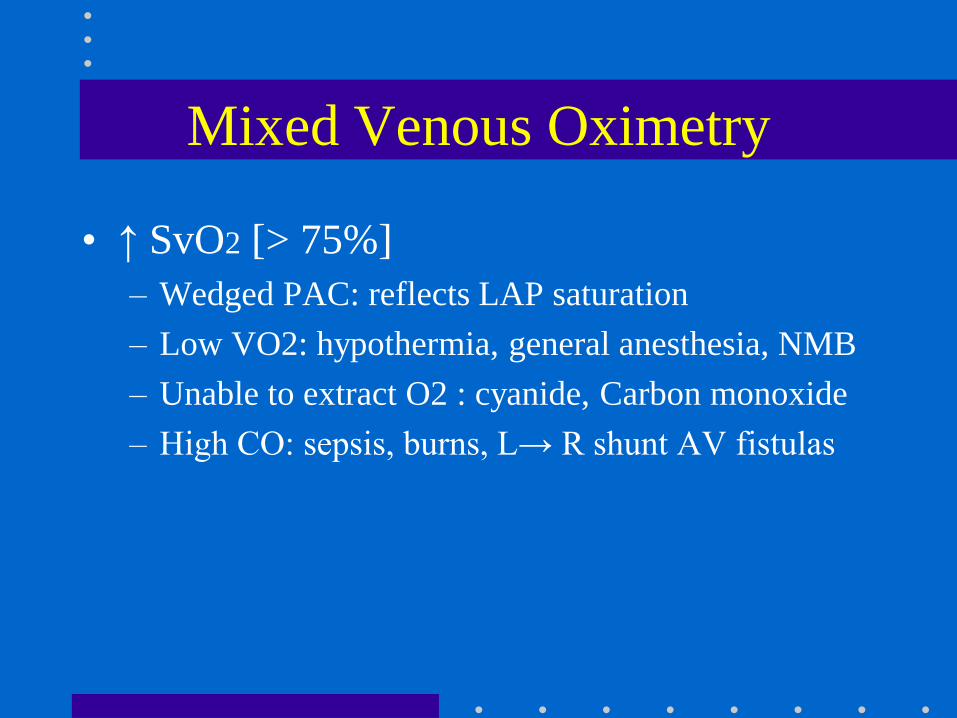
• ↑ SvO2 [> 75%]
– Wedged PAC: reflects LAP saturation
– Low VO2: hypothermia, general anesthesia, NMB
– Unable to extract O2 : cyanide, Carbon monoxide
– High CO: sepsis, burns, L→ R shunt AV fistulas
Mixed Venous Oximetry
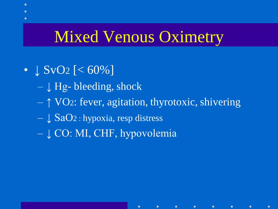
• ↓ SvO2 [< 60%]
– ↓ Hg- bleeding, shock
– ↑ VO2: fever, agitation, thyrotoxic, shivering
– ↓ SaO2 : hypoxia, resp distress
– ↓ CO: MI, CHF, hypovolemia
Summary
• Invasive monitoring routinely performed
– Permits improved understanding of BP, blood
flow, + CV function
– Allows timely detection of hemodynamic
events + initiation of treatment
– Requires correct technique + interpretation
– Complications occur from variety of reasons
– Risk: benefit ratio usually favorable in critically
ill patients