Embed Size (px)
Citation preview
PRENATAL DIAGNOSISPrenat Diagn 2009; 29: 292–293.Published online 16 February 2009 in Wiley InterScience(www.interscience.wiley.com) DOI: 10.1002/pd.2136
RESEARCH LETTER
Hemodynamic effect of fetal supraventricular tachycardiaon the unaffected twin
Sanjeev Aggarwal*, Susan Czaplicki and Kavitha ChintalaDivision of Cardiology, The Carman and Ann Adams Department of Pediatrics, Children’s Hospital of Michigan, Wayne StateUniversity, 3901 Beaubien Blvd, Detroit, USA
KEY WORDS: SVT; twins; hemodynamics
Fetal arrhythmias are diagnosed in 1 to 3% of preg-nancies, of which supraventricular tachycardia (SVT)constitutes 10% (Reed, 1989). Sustained fetal SVT witha heart rate more than 220 beats per minute (bpm)can result in congestive heart failure and nonimmunehydrops, which is associated with a poor outcome (Simp-son and Sharland, 1998). SVT in twin pregnancy isinfrequently reported and poses a therapeutic and eth-ical dilemma in regard to the effect of treatment on theunaffected twin. To date, there is no evidence that fetalSVT in one twin affects hemodynamics of the other twinwho is in normal rhythm. We report for the first time,a case in which fetal SVT in one twin was associatedwith early signs of congestive heart failure, in the othertwin, with subsequent echocardiographic improvementfollowing control of the SVT in the first twin.
A 26-year-old gravida 2, para 0 African-Americanwoman with a twin pregnancy at 27 (3/7) weeks ofgestation was referred to us for evaluation of fetaltachycardia in one of the twins. The course of pregnancywas uneventful until then. Specifically, there was nohistory of hypertension, diabetes or infections. Shewas a nonsmoker and denied use of alcohol or drugs.She was not on any medications. During a regularprenatal visit, auscultation revealed audible arrhythmiain twin A with heart rate in the range of 200 bpm.Ultrasound revealed monochorionic diamniotic twins ofsimilar size (estimated fetal weight of twin A, 992 gand twin B, 959 g). There were no other congenitaldefects. A fetal echocardiogram utilizing M mode andDoppler techniques revealed that Twin A had SVT ata rate of 230 bpm with 1 : 1 conduction to ventricles.Cardiac anatomy was normal. The size and contractilefunction of the ventricles were normal and there was nomitral or tricuspid regurgitation. There was no evidenceof pericardial effusion or congestive heart failure asassessed by the cardiovascular profile (Huhta, 2004).
Fetal echocardiography of Twin B revealed nor-mal cardiac structural anatomy. There was mild car-diomegaly with a cardiothoracic circumference ratio of
*Correspondence to: Sanjeev Aggarwal, Division of Cardiology,The Carman and Ann Adams Department of Pediatrics, Children’sHospital of Michigan, Wayne State University, 3901 BeaubienBlvd, Detroit, USA. E-mail: [email protected]
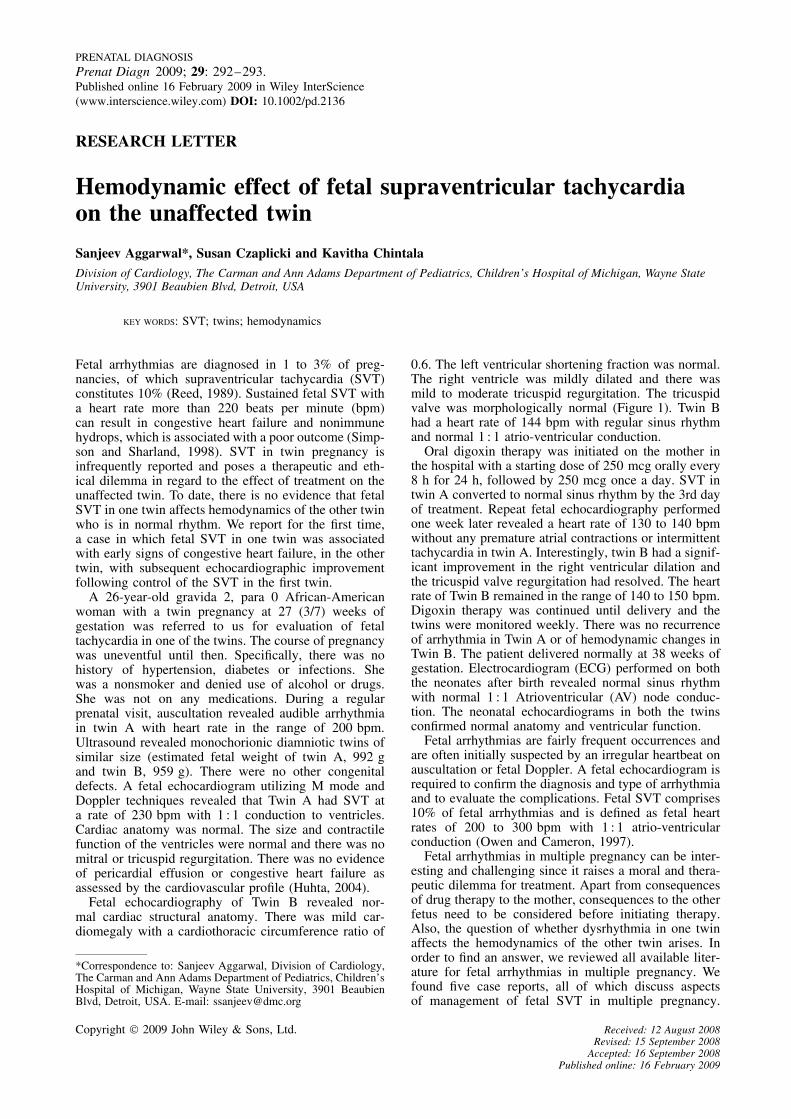
0.6. The left ventricular shortening fraction was normal.The right ventricle was mildly dilated and there wasmild to moderate tricuspid regurgitation. The tricuspidvalve was morphologically normal (Figure 1). Twin Bhad a heart rate of 144 bpm with regular sinus rhythmand normal 1 : 1 atrio-ventricular conduction.
Oral digoxin therapy was initiated on the mother inthe hospital with a starting dose of 250 mcg orally every8 h for 24 h, followed by 250 mcg once a day. SVT intwin A converted to normal sinus rhythm by the 3rd dayof treatment. Repeat fetal echocardiography performedone week later revealed a heart rate of 130 to 140 bpmwithout any premature atrial contractions or intermittenttachycardia in twin A. Interestingly, twin B had a signif-icant improvement in the right ventricular dilation andthe tricuspid valve regurgitation had resolved. The heartrate of Twin B remained in the range of 140 to 150 bpm.Digoxin therapy was continued until delivery and thetwins were monitored weekly. There was no recurrenceof arrhythmia in Twin A or of hemodynamic changes inTwin B. The patient delivered normally at 38 weeks ofgestation. Electrocardiogram (ECG) performed on boththe neonates after birth revealed normal sinus rhythmwith normal 1 : 1 Atrioventricular (AV) node conduc-tion. The neonatal echocardiograms in both the twinsconfirmed normal anatomy and ventricular function.
Fetal arrhythmias are fairly frequent occurrences andare often initially suspected by an irregular heartbeat onauscultation or fetal Doppler. A fetal echocardiogram isrequired to confirm the diagnosis and type of arrhythmiaand to evaluate the complications. Fetal SVT comprises10% of fetal arrhythmias and is defined as fetal heartrates of 200 to 300 bpm with 1 : 1 atrio-ventricularconduction (Owen and Cameron, 1997).
Fetal arrhythmias in multiple pregnancy can be inter-esting and challenging since it raises a moral and thera-peutic dilemma for treatment. Apart from consequencesof drug therapy to the mother, consequences to the otherfetus need to be considered before initiating therapy.Also, the question of whether dysrhythmia in one twinaffects the hemodynamics of the other twin arises. Inorder to find an answer, we reviewed all available liter-ature for fetal arrhythmias in multiple pregnancy. Wefound five case reports, all of which discuss aspectsof management of fetal SVT in multiple pregnancy.
Copyright 2009 John Wiley & Sons, Ltd. Received: 12 August 2008Revised: 15 September 2008
Accepted: 16 September 2008Published online: 16 February 2009
SVT IN TWINS 293
Figure 1—Horizontal four-chamber view of the heart demonstratingdilated right atrium and right ventricle
Tanawattanacharoen et al. reported the successful use ofdigoxin in the management of fetal SVT in one twin(Tanawattanacharoen et al., 2005), while there are tworeports of successful use of transplacental flecanide forSVT and hydrops in one of the twins (Edwards et al.,1999; Gerli et al., 2006). Jones et al. reported successfuluse of transplacental digoxin to control fetal SVT in onefetus among triplets similar to our case report (Jones andGarmel, 2001). There is one report where simultaneousSVT occurred in both twins and oral digoxin was suc-cessful in treating both fetuses (Shima et al., 2004 #3).None of these reports mention any hemodynamic effectson the unaffected fetus.
Fetal SVT is known to cause heart failure andsubsequent hydrops in the affected fetus. The earliestechocardiographic sign of impending cardiac failure isthe dilatation of right atrium and right ventricle (Huhta,2004). Tricuspid valve regurgitation occurs as a resultof increased right ventricular wall stress (Huhta, 2004).Interestingly, in our case, both these findings were notedin twin B who remained in sinus rhythm as confirmed bycontinuous monitoring during the 2-day hospitalizationas well as by fetal echocardiography. In contrast, TwinA, who was in SVT, did not manifest any heart failure.We excluded all other causes of heart failure in twin Bincluding twin to twin transfusion syndrome which isin any case unlikely in twins of similar weights (Barreaet al., 2005). That heart failure in Twin B was inducedby SVT in Twin A was also supported by the fact thatall signs of heart failure resolved when SVT in Twin Awas controlled with drug therapy.
The fetal ventricles at baseline have poor complianceand impaired diastolic relaxation. Occurrence of SVTleads to a marked decrease in the ventricular fillingtimes and this leads to increased systemic venouspressure. The exact reason why this occurred in TwinB is unknown, but we speculate that an imbalance inthe venous pressure in the presence of SVT in twinA led to a net transfer of blood from twin A totwin B. The resultant hemodynamic sequel is similarto that seen in twin to twin transfusion syndrome inthe presence of unbalanced arterio-venous anastomosisin a monochorionic placenta (Bajoria, 1998). In both
conditions, the recipient may develop cardiomegaly anddecreased cardiac function. Besides, hormonal activationduring SVT could have had repercussion on the twin insinus rhythm. To our knowledge, this is the first case inwhich altered hemodynamics in one twin appeared to becaused by SVT in the other.
The optimal management of fetal SVT remains con-troversial. Gestational age, duration of the arrhythmiaand presence or absence of hydrops are important deter-minants of the management strategies for fetal SVT.Since it is difficult to assess whether SVT in a par-ticular fetus is intermittent or sustained and since fetalhydrops can occur in both, most centers now treat allfetal SVTs (Simpson and Sharland, 1998). The mostcommon first line medical management is transplacen-tal administration of digoxin, which, as a monotherapy,is successful in converting 60 to 70% of SVT in non-hydropic fetuses (Simpson and Sharland, 1998). In ourcase, the fetal SVT in one twin converted to normalrhythm after 3 days of starting digoxin. Drug therapyfor the treatment of fetal arrhythmias may be complicatedby adverse effects to both mother and fetus. Presence ofmultiple gestation poses a serious concern and ethicalissues due to adverse effects of medication and/or inter-vention on the unaffected fetuses. However, there wereno ethical problems in the decision for treatment in ourcase because both twins had cardiovascular problemsfor which digoxin was the drug of first choice, resultingin clinical improvement, although via different mecha-nisms. There were no side effects of digoxin either onthe mother or the other unaffected twin.
REFERENCES
Bajoria R. 1998. Vascular anatomy of monochorionic placenta inrelation to discordant growth and amniotic fluid volume. HumReprod 13: 2933–2940.
Barrea C, Alkazaleh F, Ryan G, et al. 2005. Prenatal cardiovascularmanifestations in the twin-to-twin transfusion syndrome recipientsand the impact of therapeutic amnioreduction. Am J Obstet Gynecol192: 892–902.
Edwards A, Peek MJ, Curren J. 1999. Transplacental flecainidetherapy for fetal supraventricular tachycardia in a twin pregnancy.Aust N Z J Obstet Gynaecol 39: 110–112.
Gerli S, Clerici G, Mattei A, Di Renzo GC. 2006. Flecainidetreatment of fetal tachycardia and hydrops fetalis in a twinpregnancy. Ultrasound Obstet Gynecol 28: 117.
Huhta JC. 2004. Guidelines for the evaluation of heart failure in thefetus with or without hydrops. Pediatr Cardiol 25: 274–286.
Jones LM, Garmel SH. 2001. Successful digoxin therapy of fetalsupraventricular tachycardia in a triplet pregnancy. Obstet Gynecol98: 921–923.
Owen P, Cameron A. 1997. Fetal tachyarrhythmias. Br J Hosp Med58: 142–144.
Reed KL. 1989. Fetal arrhythmias: etiology, diagnosis, pathophysiol-ogy, and treatment. Semin Perinatol 13: 294–304.
Shima Y, Baba C, Fujita A, et al. 2004. Simultaneous supraventric-ular tahycardias in both fetuses of a twin gestation. Arch GynecolObstet 270(4): 311–313.
Simpson JM, Sharland GK. 1998. Fetal tachycardias: managementand outcome of 127 consecutive cases. Heart 79: 576–581.
Tanawattanacharoen S, Uerpairojkit B, Prechawat S, Manotaya S,Charoenvidhya D. 2005. Intrauterine therapy for fetal supraventric-ular tachycardia in a twin pregnancy. J Obstet Gynaecol Res 31:94–97.
Copyright 2009 John Wiley & Sons, Ltd. Prenat Diagn 2009; 29: 292–293.DOI: 10.1002/pd