Embed Size (px)
Citation preview
Hemodynamic Disorders, Thromboembolic Disease, and
Shock
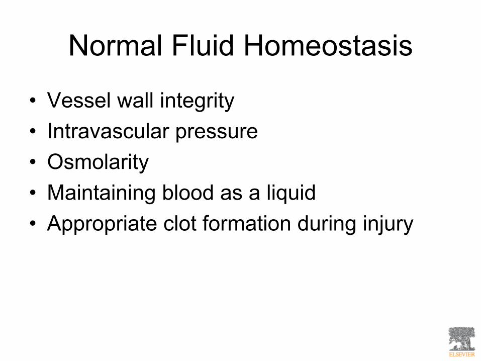
Normal Fluid Homeostasis
• Vessel wall integrity• Intravascular pressure• Osmolarity• Maintaining blood as a liquid• Appropriate clot formation during injury
Downloaded from: Robbins & Cotran Pathologic Basis of Disease (on 9 August 2006 08:47 PM)© 2005 Elsevier
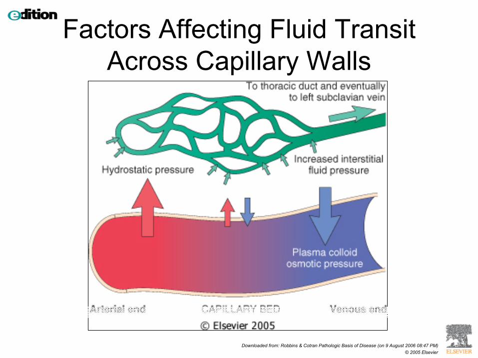
Factors Affecting Fluid Transit Across Capillary Walls
Edema
Increased fluid in interstitial spaces
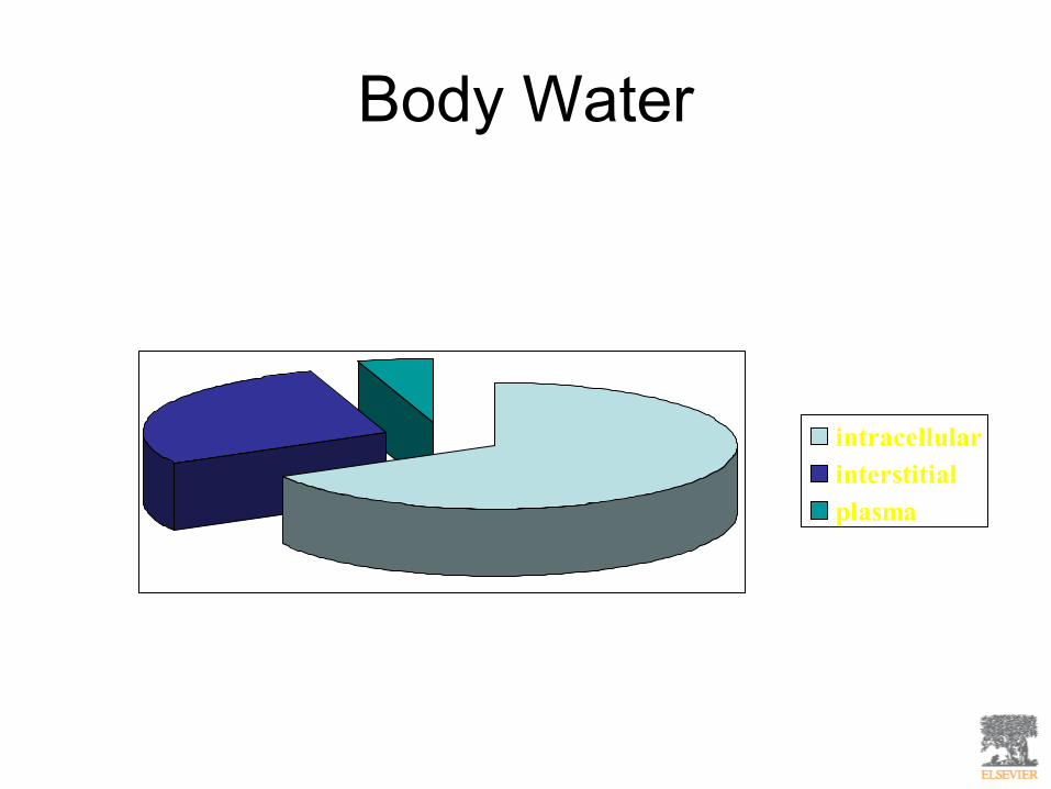
Body Water
intracellularinterstitialplasma
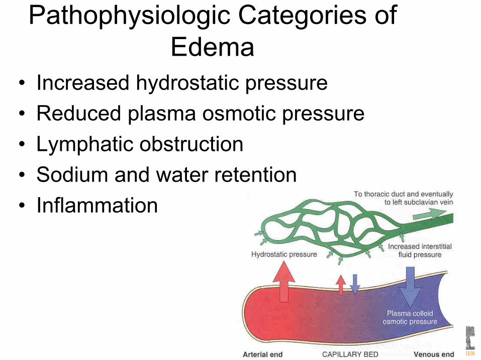
Pathophysiologic Categories of Edema
• Increased hydrostatic pressure• Reduced plasma osmotic pressure• Lymphatic obstruction• Sodium and water retention• Inflammation
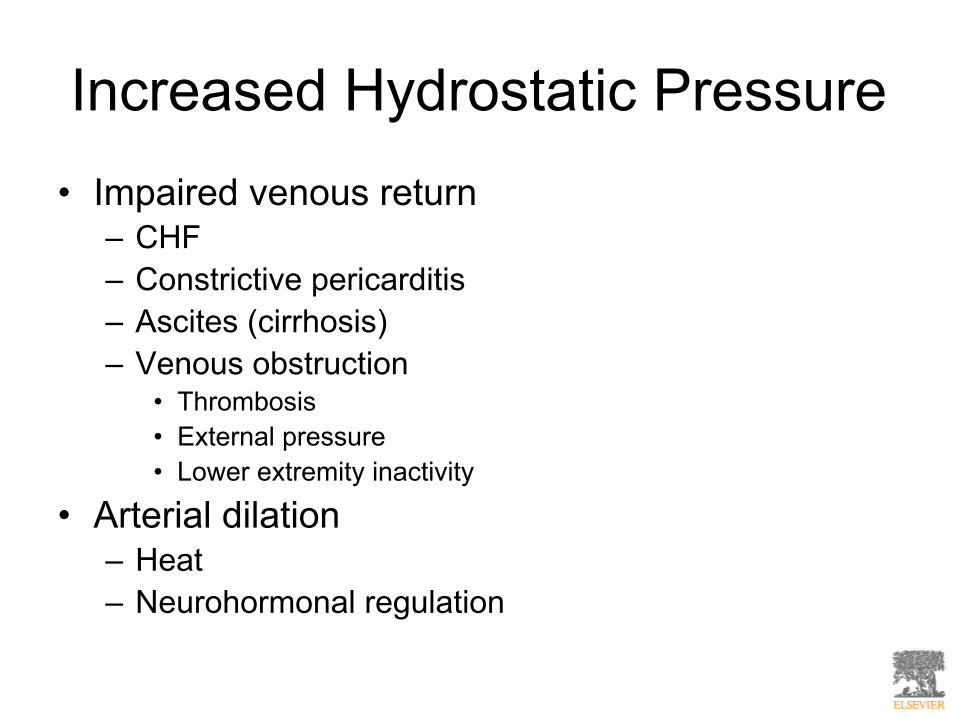
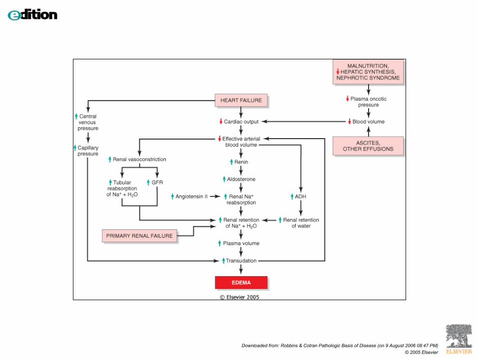
Increased Hydrostatic Pressure• Impaired venous return
– CHF– Constrictive pericarditis– Ascites (cirrhosis)– Venous obstruction
• Thrombosis• External pressure• Lower extremity inactivity
• Arterial dilation– Heat– Neurohormonal regulation
Downloaded from: Robbins & Cotran Pathologic Basis of Disease (on 9 August 2006 08:47 PM)© 2005 Elsevier
Edema: morphology
• Better appreciated grossly than microscopically
• Subcutaneous– Localized– Generalized (anasarca)
• Pulmonary• Brain
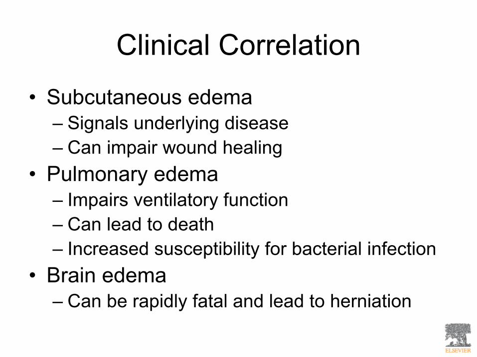
Clinical Correlation
• Subcutaneous edema – Signals underlying disease– Can impair wound healing
• Pulmonary edema– Impairs ventilatory function– Can lead to death– Increased susceptibility for bacterial infection
• Brain edema– Can be rapidly fatal and lead to herniation
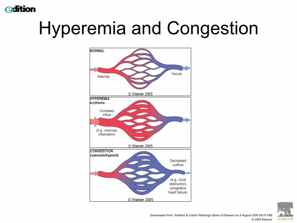
Hyperemia and Congestion
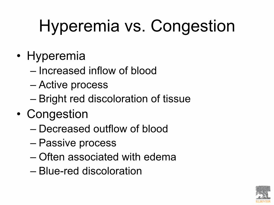
Hyperemia vs. Congestion
• Hyperemia– Increased inflow of blood – Active process– Bright red discoloration of tissue
• Congestion – Decreased outflow of blood – Passive process– Often associated with edema– Blue-red discoloration
Downloaded from: Robbins & Cotran Pathologic Basis of Disease (on 9 August 2006 08:47 PM)© 2005 Elsevier
Hyperemia and Congestion
Congestion
• Commonly occurs with edema• Chronic passive congestion can lead to
tissue hypoxia and cell death• Capillary rupture at the site of congestion
can lead to hemorrhage
Downloaded from: Robbins & Cotran Pathologic Basis of Disease (on 9 August 2006 08:47 PM)© 2005 Elsevier
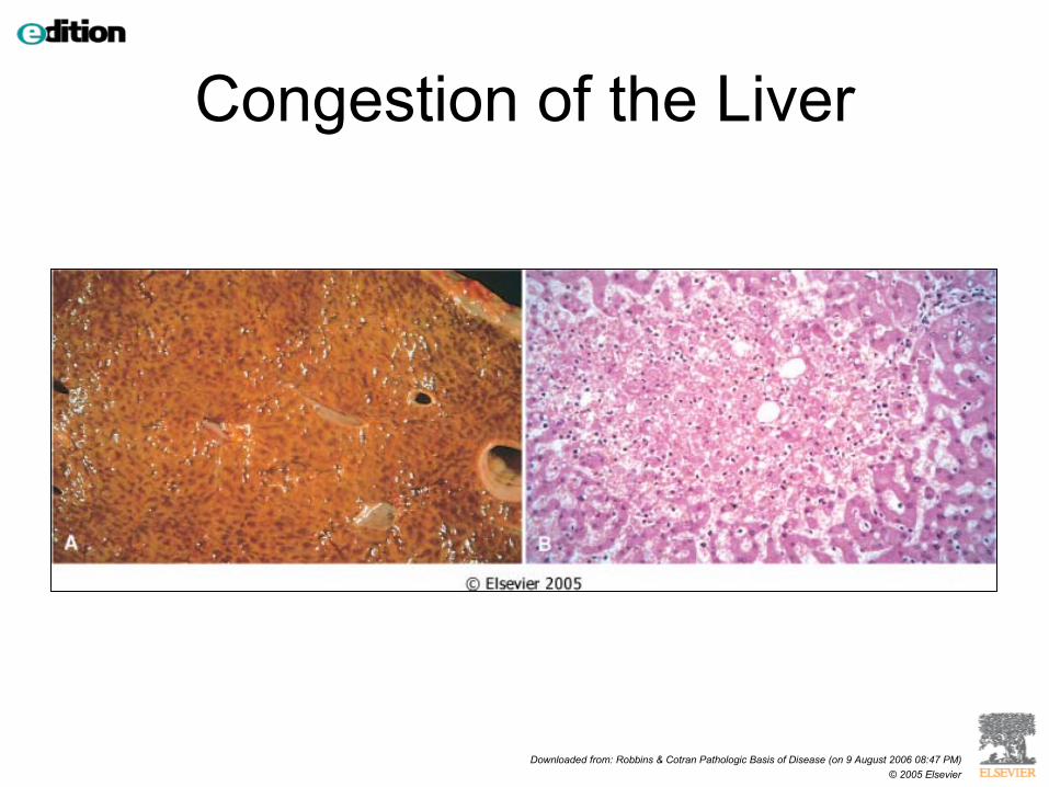
Congestion of the Liver
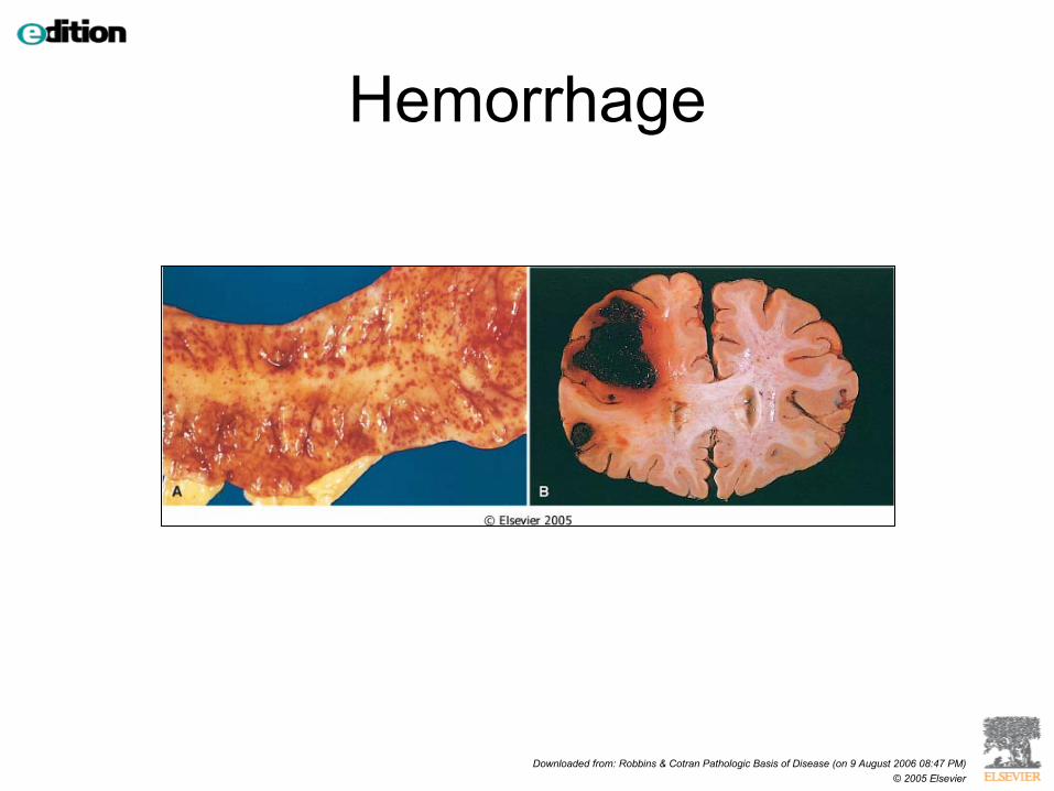
Hemorrhage
Extravasation of blood due to vessel rupture
Patterns of Hemorrhage
• Hematoma: accumulation of blood within a tissue
• Petechiae: 1-2 mm hemorrhages in skin, or mucus membranes, or serosal surfaces
• Purpura: slightly larger >3 mm than petechiae• Ecchymoses: Larger 1-2 cm subcutaneous
hematoma• Hemothorax, hemopericardium,
hemoperitoneum, hemarthrosis
Clinical Significance of Hemorrhage
• Depends on volume and rate of bleeding• >20% may lead to hypovolemic shock• Site of hemorrhage is important• Iron deficiency anemia can develop in
chronic blood loss
Downloaded from: Robbins & Cotran Pathologic Basis of Disease (on 9 August 2006 08:47 PM)© 2005 Elsevier
Hemorrhage
Hemostasis and Thrombosis
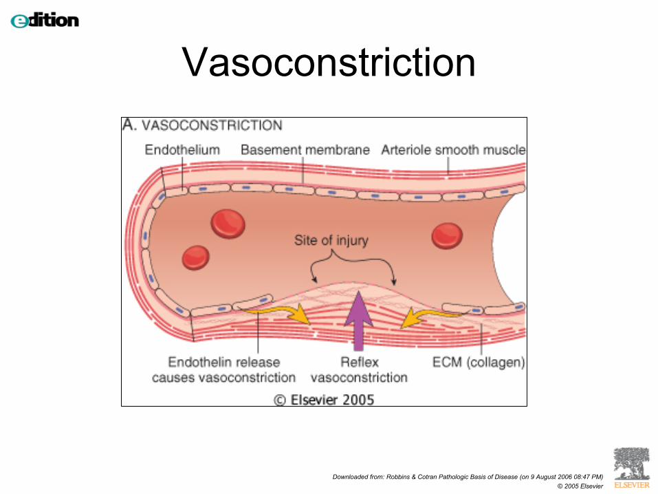
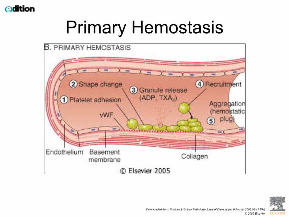
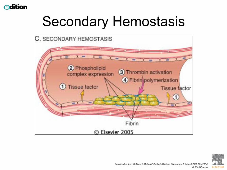
Stages of normal hemostasis• Vasoconstriction• Primary hemostasis
– platelet plug formation• Secondary hemostasis
– Fibrin deposition and polymerization to strengthen plug
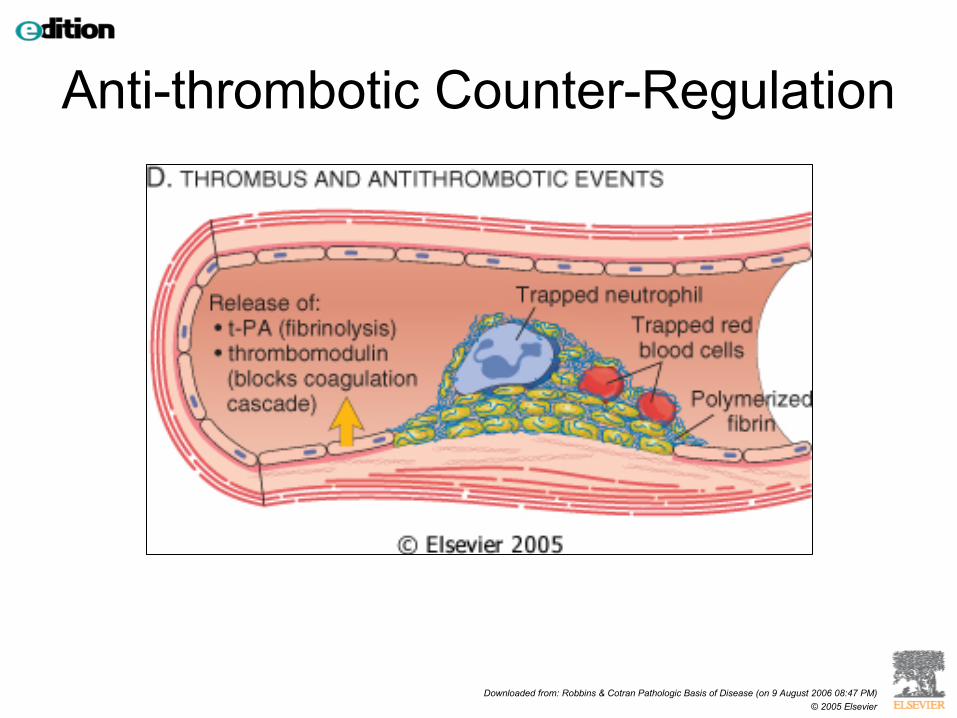
– Coagulation cascade• Antithrombotic mechanisms to limit formation of
thrombotic plug (fibrinolysis)
Downloaded from: Robbins & Cotran Pathologic Basis of Disease (on 9 August 2006 08:47 PM)© 2005 Elsevier
Vasoconstriction
Downloaded from: Robbins & Cotran Pathologic Basis of Disease (on 9 August 2006 08:47 PM)© 2005 Elsevier
Primary Hemostasis
Downloaded from: Robbins & Cotran Pathologic Basis of Disease (on 9 August 2006 08:47 PM)© 2005 Elsevier
Secondary Hemostasis
Downloaded from: Robbins & Cotran Pathologic Basis of Disease (on 9 August 2006 08:47 PM)© 2005 Elsevier
Anti-thrombotic Counter-Regulation
Factors in Normal Hemostasis
• Direct players– Endothelium– Platelets– Coagulation cascade
Downloaded from: Robbins & Cotran Pathologic Basis of Disease (on 9 August 2006 08:47 PM)© 2005 Elsevier
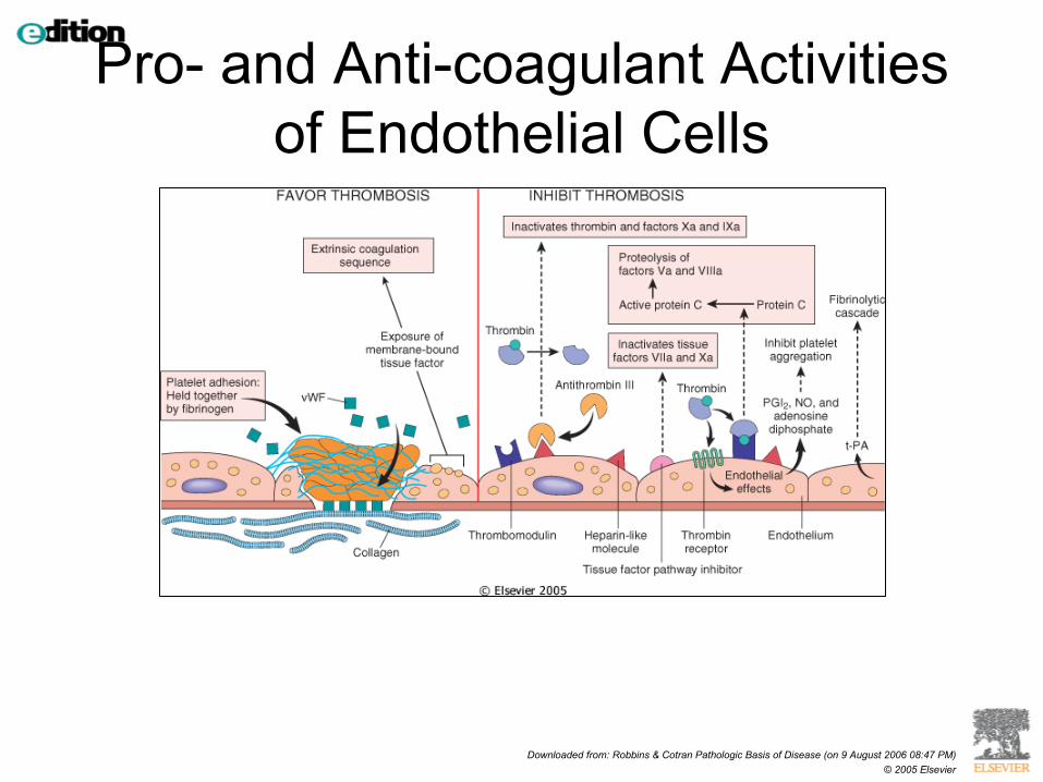
Pro- and Anti-coagulant Activities of Endothelial Cells
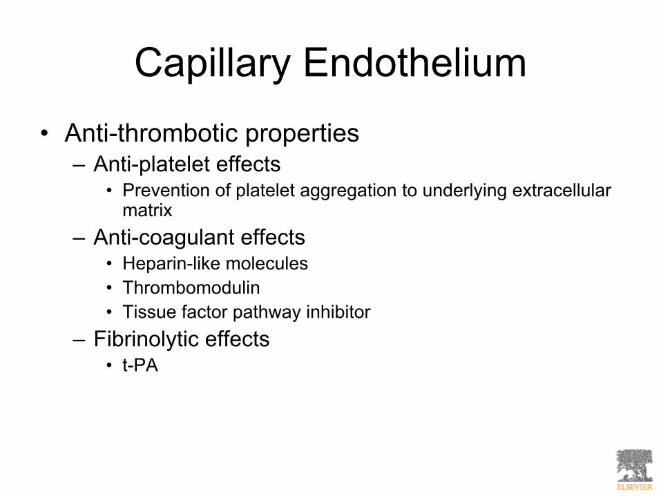
Capillary Endothelium• Anti-thrombotic properties
– Anti-platelet effects• Prevention of platelet aggregation to underlying extracellular
matrix– Anti-coagulant effects
• Heparin-like molecules• Thrombomodulin• Tissue factor pathway inhibitor
– Fibrinolytic effects• t-PA
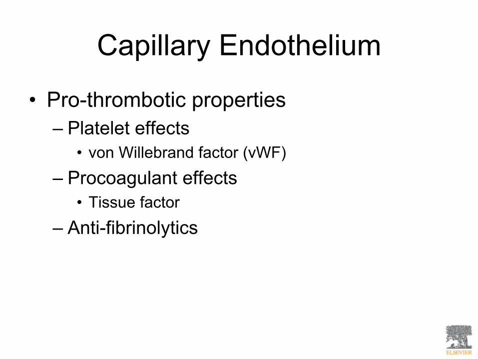
Capillary Endothelium
• Pro-thrombotic properties– Platelet effects
• von Willebrand factor (vWF)– Procoagulant effects
• Tissue factor– Anti-fibrinolytics
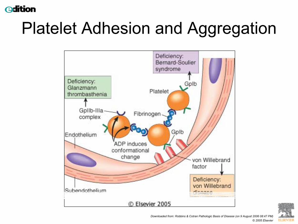
Platelets
• Platelet adhesion– vWF- glycoprotein Ib
• Secretion of granule contents• Aggregation
– Primary hemostatic plug (platelets)– Secondary hemostatic plug (platelets and
fibrin)
Downloaded from: Robbins & Cotran Pathologic Basis of Disease (on 9 August 2006 08:47 PM)© 2005 Elsevier
Platelet Adhesion and Aggregation
Coagulation Cascade
• Series of enzymatic reactions in the plasma
• Conversion of proenzymes into active enzymes
• Culminates in the formation of thrombin, which then converts soluble fibrinogen into fibrin
Coagulation Cascade
• Reactions are assembled on a phospholipid complex (i.e. surface of platelets)
• Requires calcium• Intrinsic and extrinsic pathways
– Factor XII (Hageman factor) activates intrinsic pathway
– Tissue factor activate extrinsic pathway– Interconnections exist
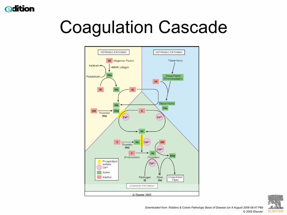
Downloaded from: Robbins & Cotran Pathologic Basis of Disease (on 9 August 2006 08:47 PM)© 2005 Elsevier
Coagulation Cascade
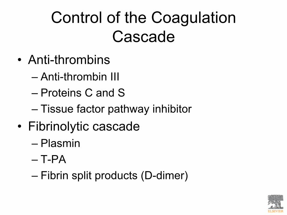
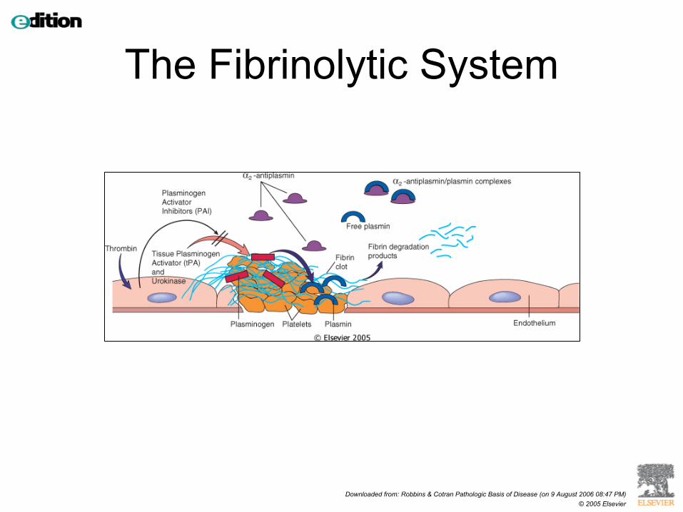
Control of the Coagulation Cascade
• Anti-thrombins– Anti-thrombin III– Proteins C and S– Tissue factor pathway inhibitor
• Fibrinolytic cascade– Plasmin– T-PA– Fibrin split products (D-dimer)
Downloaded from: Robbins & Cotran Pathologic Basis of Disease (on 9 August 2006 08:47 PM)© 2005 Elsevier
The Fibrinolytic System
Thrombosis
Inappropriate activation of a normal hemostatic process
Downloaded from: Robbins & Cotran Pathologic Basis of Disease (on 9 August 2006 08:47 PM)© 2005 Elsevier
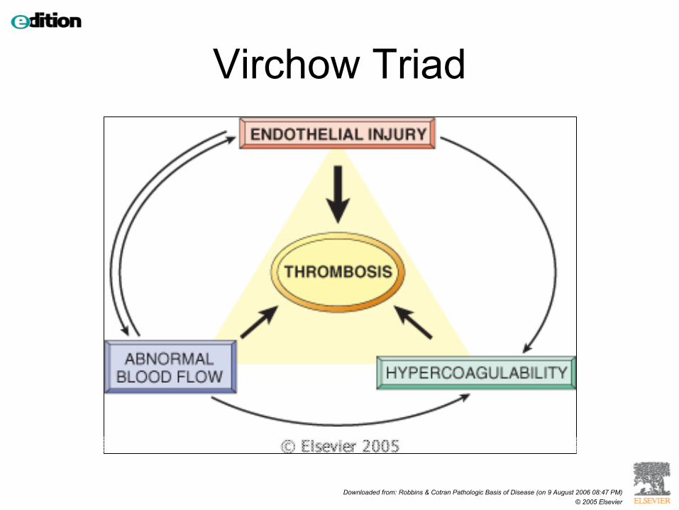
Virchow Triad
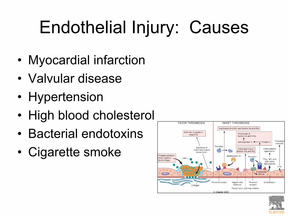
Endothelial Injury: Causes
• Myocardial infarction• Valvular disease• Hypertension• High blood cholesterol• Bacterial endotoxins• Cigarette smoke
Alterations in Normal Blood Flow: Stasis and Turbulence
• Disrupt laminar flow and bring platelets into contact with the endothelium
• Prevent dilution of clotting factors by preventing fresh inflow of blood
• Permit the build up of thrombi by retarding inflow of clotting factor inhibitors
• Promote endothelial cell activation
Stasis and Turbulence: Causes
• Ulcerated atherosclerotic plaques• Aortic aneurysms• Myocardial infarctions• Inactivity
Hypercoagulability
• Any alteration of the coagulation pathways that predisposes to thrombosis
• Inherited• Acquired
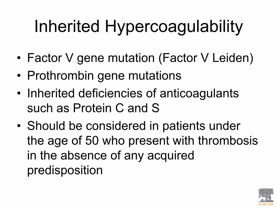
Inherited Hypercoagulability
• Factor V gene mutation (Factor V Leiden)• Prothrombin gene mutations• Inherited deficiencies of anticoagulants
such as Protein C and S• Should be considered in patients under
the age of 50 who present with thrombosis in the absence of any acquired predisposition
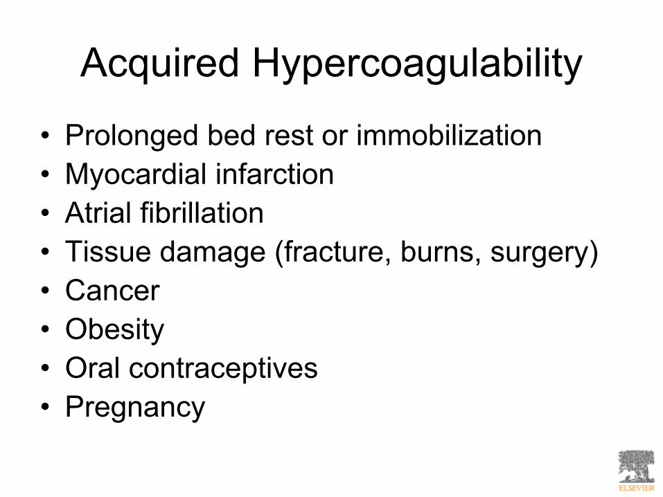
Acquired Hypercoagulability
• Prolonged bed rest or immobilization• Myocardial infarction• Atrial fibrillation• Tissue damage (fracture, burns, surgery)• Cancer• Obesity• Oral contraceptives• Pregnancy
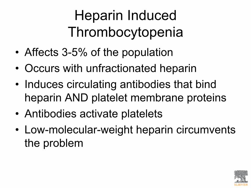
Heparin Induced Thrombocytopenia
• Affects 3-5% of the population• Occurs with unfractionated heparin• Induces circulating antibodies that bind
heparin AND platelet membrane proteins• Antibodies activate platelets• Low-molecular-weight heparin circumvents
the problem
Anti-phospholipid Antibody Syndrome
• High titers of antibodies against phospholipids (cardiolipin)
• Tends to occur in patients with lupus• Recurrent arterial and venous thrombosis,
repeated miscarriages, cardiac valvular vegetations, thrombocytopenia
• Interfere with in-vitro lab tests for coagulability
• Lead to hypercoagulability in-vivo
Morphology of Thrombi
Cardiac and Aortic Thrombi
• Lines of Zahn– Laminations due to alternating platelets/fibrin
and red cells• Mural thrombi
– Applied to the wall of cardiac chambers or the aorta
Downloaded from: Robbins & Cotran Pathologic Basis of Disease (on 9 August 2006 08:47 PM)© 2005 Elsevier
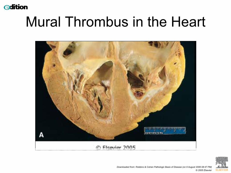
Mural Thrombus in the Heart
Downloaded from: Robbins & Cotran Pathologic Basis of Disease (on 9 August 2006 08:47 PM)© 2005 Elsevier
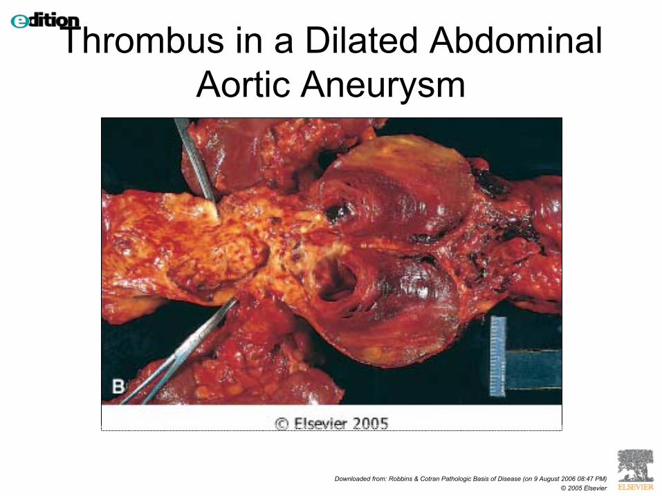
Thrombus in a Dilated Abdominal Aortic Aneurysm
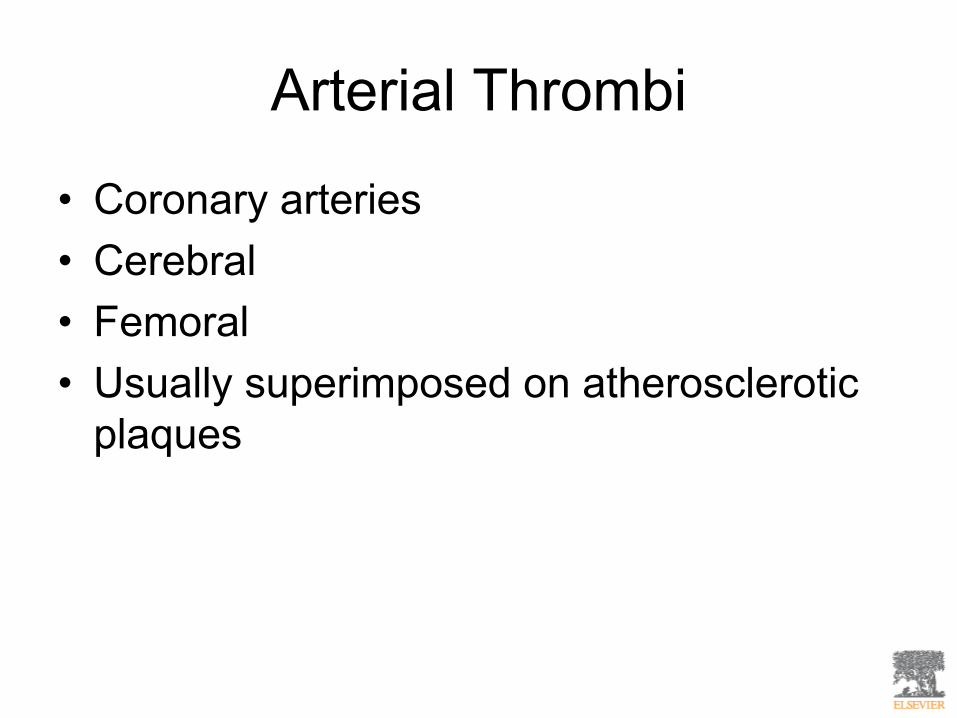
Arterial Thrombi
• Coronary arteries• Cerebral• Femoral• Usually superimposed on atherosclerotic
plaques
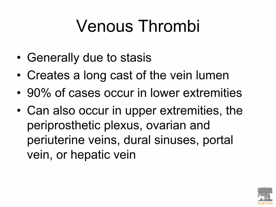
Venous Thrombi
• Generally due to stasis• Creates a long cast of the vein lumen• 90% of cases occur in lower extremities• Can also occur in upper extremities, the
periprosthetic plexus, ovarian and periuterine veins, dural sinuses, portal vein, or hepatic vein
Downloaded from: Robbins & Cotran Pathologic Basis of Disease (on 9 August 2006 08:47 PM)© 2005 Elsevier
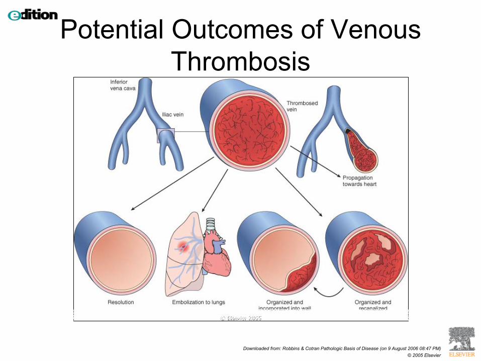
Potential Outcomes of Venous Thrombosis
Clinical Correlation of Thrombi
• Cause obstruction of arteries and veins• Possible sources of emboli
Venous Thrombosis (Phlebothrombosis)
• Superficial and deep veins of the leg• Local congestion with swelling, pain,
tenderness or may be asymptomatic• Deep thrombi more likely to embolize• Major danger is pulmonary embolism
Clinical Settings of DVT
• Cardiac failure• Trauma, surgery, burns• Pregnancy and post-partum states• Disseminated cancer• Advanced age, immobilization, bed rest
Arterial and Cardiac Thrombosis
• Atherosclerosis is major initiator• Mural thrombi• Valve disease• Arterial thrombi can also embolize
– brain, kidneys, spleen
Disseminated Intravascular Coagulation
• Sudden or insidious onset of widespread thrombi in the microcirculation
• Diffuse circulatory insufficiency• Rapid consumption of platelets and fibrin• Activation of fibrinolytic mechanisms can
of all into a bleeding disorder• Potential complication of any condition
associated with widespread activation of thrombin
Embolism
Detached intravascular solid, liquid, or gaseous mass that is carried by the
blood to a site distant from its point of origin.
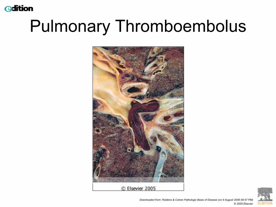
Pulmonary Thromboembolism
• 200,000 deaths per year in United States• Most originate from deep leg vein thrombi• Carried through the circulation, pass
through the right side of the heart, and into the pulmonary vasculature
Pulmonary Thromboembolism
• Can occlude:– Main pulmonary artery– Bifurcation of the pulmonary artery (saddle
embolus)– Pass into smaller arterioles
• Can be clinically silent, or result in sudden death, pulmonary hemorrhage, or cause pulmonary hypertension
Downloaded from: Robbins & Cotran Pathologic Basis of Disease (on 9 August 2006 08:47 PM)© 2005 Elsevier
Pulmonary Thromboembolus
Systemic Thromboembolism
• Refers to emboli traveling within the arterial circulation
• Most arise from intracardiac mural thrombi• Remainder originates from thrombi
associated with ulcerated atherosclerotic plaques
• Lower extremities (75%), brain (10%), intestines, kidneys, and spleen
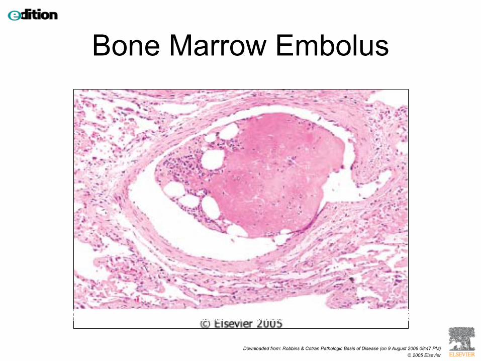
Other types of Emboli
• Bone marrow/fat emboli• Air embolism• Amniotic fluid embolism
Downloaded from: Robbins & Cotran Pathologic Basis of Disease (on 9 August 2006 08:47 PM)© 2005 Elsevier
Bone Marrow Embolus
Infarction
An area of ischemic necrosis caused by occlusion of either the arterial
supply or the venous drainage of a particular issue
Infarction
• Nearly all infarcts result from thrombotic or embolic events
• Almost all result from arterial occlusion• Other mechanisms
– Local vasospasm– Hemorrhage within an atherosclerotic plaque– Extrinsic compression of a vessel
Downloaded from: Robbins & Cotran Pathologic Basis of Disease (on 9 August 2006 08:47 PM)© 2005 Elsevier
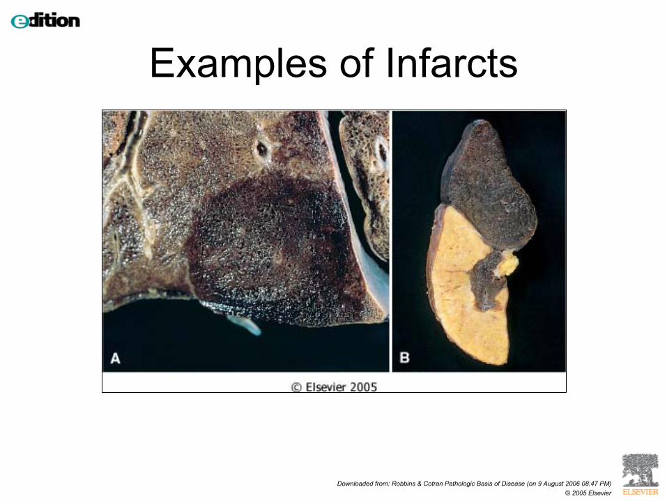
Examples of Infarcts
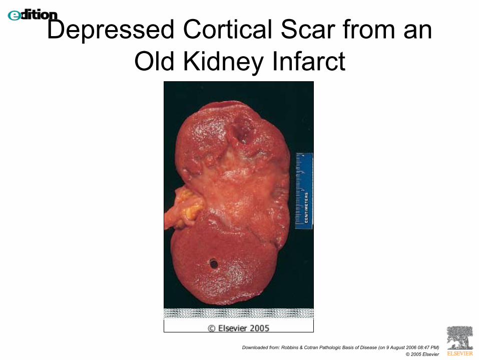
Morphologic Characteristics of Infarcts
• Can be red in color due to hemorrhage, or pale due to lack of blood supply
• Wedge shaped• Coagulative necrosis
– Most replaced by scar tissue• Inflammation incited by necrotic tissue
Downloaded from: Robbins & Cotran Pathologic Basis of Disease (on 9 August 2006 08:47 PM)© 2005 Elsevier
Depressed Cortical Scar from an Old Kidney Infarct
Factors that Influence Development of an Infarct
• Nature of the vascular supply• Rate of development of the occlusion• Vulnerability to hypoxia• Oxygen content of blood

Shock
Systemic hypoperfusion due to a reduction either in cardiac output or in the effective circulating blood volume
Shock
• Cardiogenic– Myocardial pump failure
• Hypovolemic– Loss of blood, fluid loss
• Septic– systemic microbial infection
Septic Shock
• 25 to 50% mortality rate• #1 cause of death in intensive care units• Spread and expansion of an initially
localized infection into the bloodstream
Septic Shock
• Most caused by gram-negative bacilli that produce endotoxin
• Bacterial wall component (LPS) activates immune cells, complement, and results in a cytokine cascade
Effects of Septic Shock
• Systemic vasodilatation (hypotension)• Diminished myocardial contractility• Widespread endothelial injury and
activation• Activation of the coagulation system,
leading to DIC• Multi-organ system failure
Stages of Shock
• Nonprogressive stage– Reflex compensatory mechanisms are
activated to maintain perfusion of vital organs– Tachycardia, peripheral vasoconstriction,
renal conservation of fluid
Stages of Shock
• Progressive stage– Tissue hypoperfusion and onset of worsening
circulatory and metabolic imbalances– Lactic acidosis– Blunting a vasomotor response– Confusion, decrease in urine output
Stages of Shock
• Irreversible stage– Severe tissue injury– Survival not possible– Myocardial contractile function worsens– Renal shutdown– Ischemic bowel
Morphology Of Shock
• Brain– Ischemic
encephalopathy• Heart
– Widespread coagulation necrosis, subendocardial hemorrhage
• Kidneys– Acute tubular necrosis
• Lungs– Diffuse alveolar
damage• Gastrointestinal tract
– Mucosal hemorrhage and necrosis
• Liver– Central hemorrhagic
necrosis


![Sepsis and Hemodynamic Support in 2017 [Read-Only]...Sepsis and Hemodynamic Support in 2017 ... Carleen Risaliti 2 Review fluid resuscitation guidelines in septic shock Discuss volume](https://img.pdfslide.us/doc/110x75/5ea9a1f51936e552541087a7/sepsis-and-hemodynamic-support-in-2017-read-only-sepsis-and-hemodynamic-support.jpg)