Embed Size (px)
Citation preview
Neuropsychologia 42 (2004) 1852–1857
Note
Hemispheric integration and differences in perception ofa line-motion illusion in the divided brain
Michael C. Corballisa,∗, Kylie J. Barnetta, Mara Fabrib, Aldo Paggib, Paul M. Corballisca Department of Psychology, Research Center for Cognitive Neuroscience, University of Auckland, Private Bag 92019, Auckland, New Zealand
b Institute of Human Physiology, University of Ancona, 60020 Ancona, Italyc School of Psychology, Georgia Institute of Technology, Atlanta, GA, USA
Received 26 August 2003; received in revised form 6 November 2003; accepted 7 April 2004
Abstract
Five people lacking the corpus callosum (two callosotomized, three with agenesis of the corpus callosum) and neurologically normalsubjects were shown vertical lines that appeared instantaneously between pairs of rectangles in one or other visual field. When one ofthe rectangles flashed prior to the presentation of the line, and the line was in the same visual field, all subjects perceived the line asspreading from the flashed rectangle to the other. Normal subjects and one of the callosotomized subjects showed a slight but significantright visual-field advantage, perhaps reflecting a left-hemispheric superiority in processing rapid temporal events. The illusion was alsoinduced when the line and the flash were in opposite visual fields in one of the callosotomized, one of the acallosal subjects, and about halfof the normal subjects, implying interhemispheric integration even in the absence of the corpus callosum.© 2004 Elsevier Ltd. All rights reserved.
Keywords:Split brain; Callosal agenesis; Motion perception; Hemispheric differences; Hemispheric integration
1. Introduction
In a recent review of research on the split brain,Gazzaniga (2000)concluded that “split brain patients can-not cross-integrate visual information between their twovisual hemifields (p. 1299).” It is well known that patientswho have undergone callosotomy or complete forebraincommissurotomy typically cannot name words or objectsflashed in the left visual field, indicating that little or no in-formation about these stimuli, whether visual or semantic,crosses from the right cerebral hemisphere to the left, andthese patients seldom score better than chance in judgingwhether letters, digits, shapes, or colors flashed in oppo-site visual fields are the same or different (e.g.,Corballis& Corballis, 2001; Johnson, 1984). Nevertheless there aresome exceptions. For example, one split-brained patient,L.B., had relatively little difficulty deciding whether slopinglines flashed in opposite visual fields were aligned or not(Corballis & Trudel, 1993) even though he could not judgewhether colors or shapes in opposite visual fields were thesame or different.
∗ Corresponding author. Tel.:+64 9 373 7599x8561;fax: +64 9 373 7450.
E-mail address:[email protected] (M.C. Corballis).
There is also evidence that split-brained patients can per-ceive apparent motion across the midline. When a stimulusappears in one visual field and is replaced by a stimulus inthe other, split-brained people can decide whether the pro-gression is left-to-right or right-to-left (Naikar & Corballis,1996; Ramachandran, Cronin-Golomb, & Myers, 1986), al-thoughGazzaniga (1987)has suggested that this is based oninference rather than genuine perception of apparent motion.To demonstrate this, he showed that one split-brained man,J.W., could not distinguish the succession of two stimulifrom the appearance of a single stimulus in one visual field.J.W. was presumably inferring that if a stimulus appearedin one field, it must have “moved” from the other visualfield. Another split-brained man, L.B., was able to makethis discrimination (Naikar & Corballis, 1996), althoughagain it is possible that he could judge when stimuli hadappeared in both fields without perceiving apparent motion,and judged motion from the appearance of a stimulus in thecurrently attended field.
In the present study, we examine both interhemisphericdifferences and interhemispheric integration of a line-motionillusion in the split brain. If a line appears instantaneouslybetween two markers, and if one of the markers is brieflyilluminated prior to the appearance of the line, the linemay appear to propagate from the illuminated marker to-
0028-3932/$ – see front matter © 2004 Elsevier Ltd. All rights reserved.doi:10.1016/j.neuropsychologia.2004.04.014
M.C. Corballis et al. / Neuropsychologia 42 (2004) 1852–1857 1853
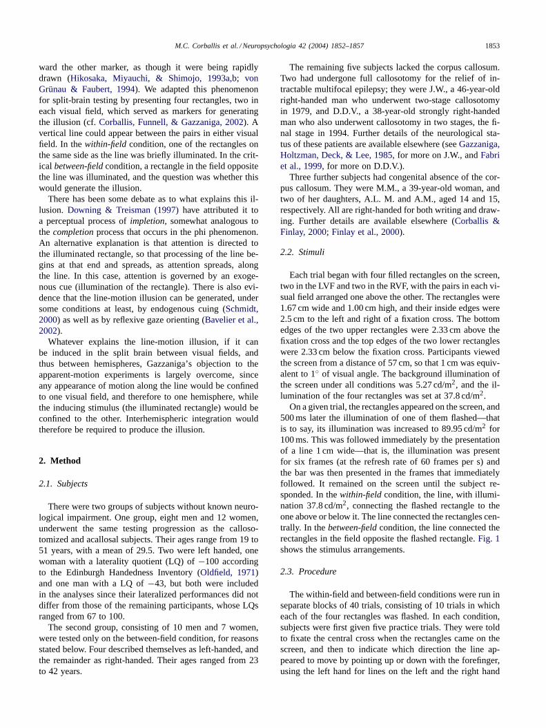
ward the other marker, as though it were being rapidlydrawn (Hikosaka, Miyauchi, & Shimojo, 1993a,b; vonGrünau & Faubert, 1994). We adapted this phenomenonfor split-brain testing by presenting four rectangles, two ineach visual field, which served as markers for generatingthe illusion (cf.Corballis, Funnell, & Gazzaniga, 2002). Avertical line could appear between the pairs in either visualfield. In thewithin-field condition, one of the rectangles onthe same side as the line was briefly illuminated. In the crit-ical between-fieldcondition, a rectangle in the field oppositethe line was illuminated, and the question was whether thiswould generate the illusion.
There has been some debate as to what explains this il-lusion. Downing & Treisman (1997)have attributed it toa perceptual process ofimpletion, somewhat analogous tothe completionprocess that occurs in the phi phenomenon.An alternative explanation is that attention is directed tothe illuminated rectangle, so that processing of the line be-gins at that end and spreads, as attention spreads, alongthe line. In this case, attention is governed by an exoge-nous cue (illumination of the rectangle). There is also evi-dence that the line-motion illusion can be generated, undersome conditions at least, by endogenous cuing (Schmidt,2000) as well as by reflexive gaze orienting (Bavelier et al.,2002).
Whatever explains the line-motion illusion, if it canbe induced in the split brain between visual fields, andthus between hemispheres, Gazzaniga’s objection to theapparent-motion experiments is largely overcome, sinceany appearance of motion along the line would be confinedto one visual field, and therefore to one hemisphere, whilethe inducing stimulus (the illuminated rectangle) would beconfined to the other. Interhemispheric integration wouldtherefore be required to produce the illusion.
2. Method
2.1. Subjects
There were two groups of subjects without known neuro-logical impairment. One group, eight men and 12 women,underwent the same testing progression as the calloso-tomized and acallosal subjects. Their ages range from 19 to51 years, with a mean of 29.5. Two were left handed, onewoman with a laterality quotient (LQ) of−100 accordingto the Edinburgh Handedness Inventory (Oldfield, 1971)and one man with a LQ of−43, but both were includedin the analyses since their lateralized performances did notdiffer from those of the remaining participants, whose LQsranged from 67 to 100.
The second group, consisting of 10 men and 7 women,were tested only on the between-field condition, for reasonsstated below. Four described themselves as left-handed, andthe remainder as right-handed. Their ages ranged from 23to 42 years.
The remaining five subjects lacked the corpus callosum.Two had undergone full callosotomy for the relief of in-tractable multifocal epilepsy; they were J.W., a 46-year-oldright-handed man who underwent two-stage callosotomyin 1979, and D.D.V., a 38-year-old strongly right-handedman who also underwent callosotomy in two stages, the fi-nal stage in 1994. Further details of the neurological sta-tus of these patients are available elsewhere (seeGazzaniga,Holtzman, Deck, & Lee, 1985, for more on J.W., andFabriet al., 1999, for more on D.D.V.).
Three further subjects had congenital absence of the cor-pus callosum. They were M.M., a 39-year-old woman, andtwo of her daughters, A.L. M. and A.M., aged 14 and 15,respectively. All are right-handed for both writing and draw-ing. Further details are available elsewhere (Corballis &Finlay, 2000; Finlay et al., 2000).
2.2. Stimuli
Each trial began with four filled rectangles on the screen,two in the LVF and two in the RVF, with the pairs in each vi-sual field arranged one above the other. The rectangles were1.67 cm wide and 1.00 cm high, and their inside edges were2.5 cm to the left and right of a fixation cross. The bottomedges of the two upper rectangles were 2.33 cm above thefixation cross and the top edges of the two lower rectangleswere 2.33 cm below the fixation cross. Participants viewedthe screen from a distance of 57 cm, so that 1 cm was equiv-alent to 1◦ of visual angle. The background illumination ofthe screen under all conditions was 5.27 cd/m2, and the il-lumination of the four rectangles was set at 37.8 cd/m2.
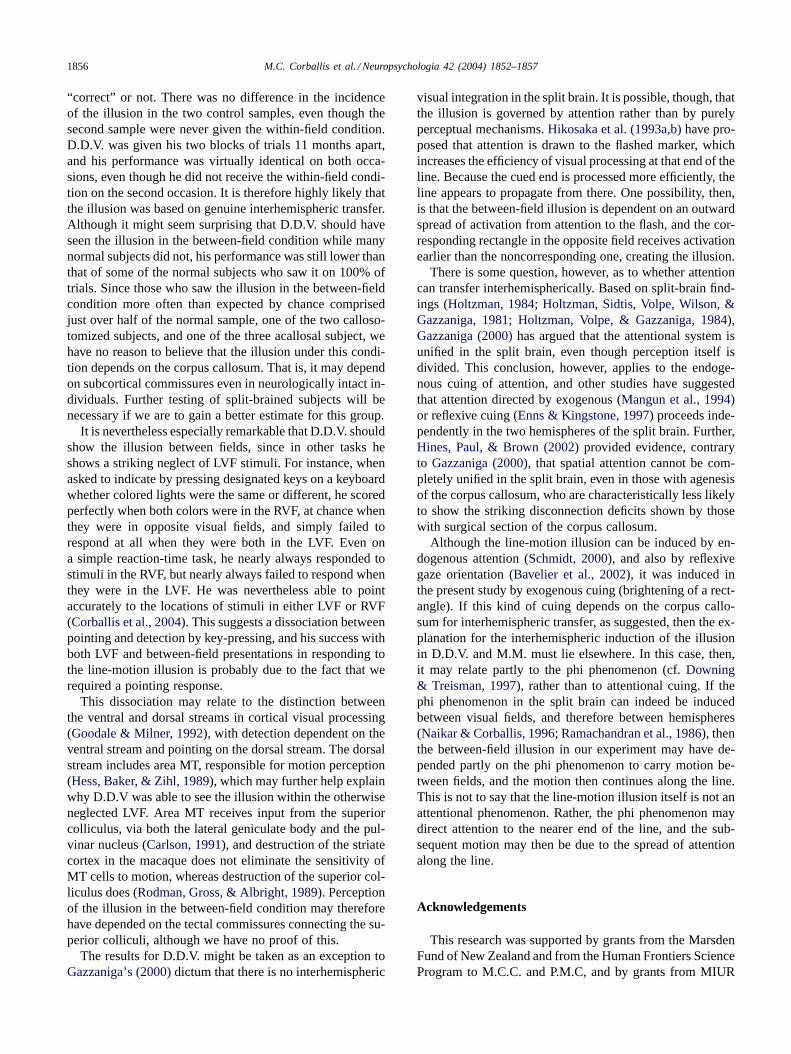
On a given trial, the rectangles appeared on the screen, and500 ms later the illumination of one of them flashed—thatis to say, its illumination was increased to 89.95 cd/m2 for100 ms. This was followed immediately by the presentationof a line 1 cm wide—that is, the illumination was presentfor six frames (at the refresh rate of 60 frames per s) andthe bar was then presented in the frames that immediatelyfollowed. It remained on the screen until the subject re-sponded. In thewithin-fieldcondition, the line, with illumi-nation 37.8 cd/m2, connecting the flashed rectangle to theone above or below it. The line connected the rectangles cen-trally. In thebetween-fieldcondition, the line connected therectangles in the field opposite the flashed rectangle.Fig. 1shows the stimulus arrangements.
2.3. Procedure
The within-field and between-field conditions were run inseparate blocks of 40 trials, consisting of 10 trials in whicheach of the four rectangles was flashed. In each condition,subjects were first given five practice trials. They were toldto fixate the central cross when the rectangles came on thescreen, and then to indicate which direction the line ap-peared to move by pointing up or down with the forefinger,using the left hand for lines on the left and the right hand
1854 M.C. Corballis et al. / Neuropsychologia 42 (2004) 1852–1857
Fig. 1. Illustration of stimulus sequence (not to scale). Frame 1 shows four rectangles, two on each side of the fixation cross. In Frame 2, one of therectangles is illuminated (flashed) for 100 ms. In Frame 3, a vertical line instantaneously joins two of the rectangles. In this example, the flash and theline are in opposite hemifields.
for lines on the right. Fixation was checked by visual mon-itoring. Responses were forced choice; if subjects were notsure, they were to guess. One reason for using a pointingresponse rather than a button press is that an earlier studyhad shown D.D.V. to show marked neglect of stimuli in theleft visual field when required to respond by pressing a key,but was readily able to point to stimuli in the left visual field(Corballis, Corballis, & Fabri, 2004).
For the first group of neurologically normal subjects,and for the five subjects lacking the corpus callosum, thewithin-field condition was run before the between-field con-dition, to ensure that the subjects first saw the illusion underoptimal conditions. Since prior perception of the illusion un-der the within-field conditions might conceivably have influ-enced their reports in the between-field condition, the secondgroup of neurologically normal subjects were tested underthe between-field condition only. Normal subjects in the firstgroup and the three acallosal subjects were given one blockof 40 experimental trials under each condition, and those inthe second group were give just one block of 40 trials in
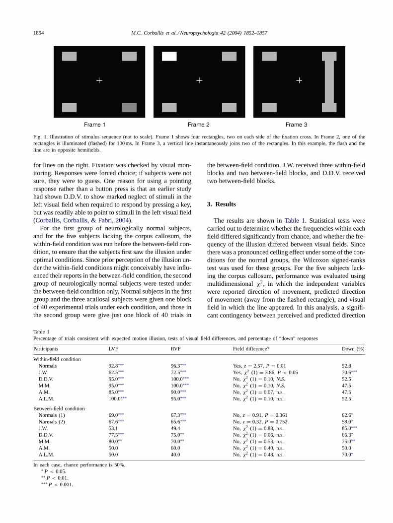
Table 1Percentage of trials consistent with expected motion illusion, tests of visual field differences, and percentage of “down” responses
Participants LVF RVF Field difference? Down (%)
Within-field conditionNormals 92.8∗∗∗ 96.3∗∗∗ Yes, z = 2.57, P = 0.01 52.8J.W. 62.5∗∗∗ 72.5∗∗∗ Yes, χ2 (1) = 3.86, P < 0.05 70.6∗∗∗D.D.V. 95.0∗∗∗ 100.0∗∗∗ No, χ2 (1) = 0.10, N.S. 52.5M.M. 95.0∗∗∗ 100.0∗∗∗ No, χ2 (1) = 0.10, N.S. 47.5A.M. 85.0∗∗∗ 90.0∗∗∗ No, χ2 (1) = 0.07, n.s. 47.5A.L.M. 100.0∗∗∗ 95.0∗∗∗ No, χ2 (1) = 0.10, n.s. 52.5
Between-field conditionNormals (1) 69.0∗∗∗ 67.3∗∗∗ No, z = 0.91, P = 0.361 62.6∗Normals (2) 67.6∗∗∗ 65.6∗∗∗ No, z = 0.32, P = 0.752 58.0∗J.W. 53.1 49.4 No,χ2 (1) = 0.88, n.s. 85.0∗∗∗D.D.V. 77.5∗∗∗ 75.0∗∗ No, χ2 (1) = 0.06, n.s. 66.3∗M.M. 80.0∗∗ 70.0∗∗ No, χ2 (1) = 0.53, n.s. 75.0∗∗A.M. 50.0 60.0 No,χ2 (1) = 0.40, n.s. 50.0A.L.M. 50.0 40.0 No,χ2 (1) = 0.48, n.s. 70.0∗
In each case, chance performance is 50%.∗ P < 0.05.∗∗ P < 0.01.∗∗∗ P < 0.001.
the between-field condition. J.W. received three within-fieldblocks and two between-field blocks, and D.D.V. receivedtwo between-field blocks.
3. Results
The results are shown inTable 1. Statistical tests werecarried out to determine whether the frequencies within eachfield differed significantly from chance, and whether the fre-quency of the illusion differed between visual fields. Sincethere was a pronounced ceiling effect under some of the con-ditions for the normal groups, the Wilcoxon signed-rankstest was used for these groups. For the five subjects lack-ing the corpus callosum, performance was evaluated usingmultidimensionalχ2, in which the independent variableswere reported direction of movement, predicted directionof movement (away from the flashed rectangle), and visualfield in which the line appeared. In this analysis, a signifi-cant contingency between perceived and predicted direction
M.C. Corballis et al. / Neuropsychologia 42 (2004) 1852–1857 1855
of movement indicates that the illusion was perceived moreoften than expected by chance, while a significant triple con-tingency between these two factors and visual field indicatesa significant difference between visual fields.
3.1. Within-field condition
As Table 1shows, there was a clear illusion of movementin the predicted direction for all subjects in the within-fieldcondition. It was weakest in J.W., but even he saw theillusion in the predicted direction significantly more thanexpected by chance. Moreover, both J.W. and the normalsample showed a significant RVF advantage, perhaps re-flecting a left-hemispheric advantage. For D.D.V. and thethree acallosals there may have been too few trials to elicita reliable visual-field effect.
3.2. Between-field condition
Among the normal participants, the percentage of reportsin the predicted direction was considerably lower than inthe within-field condition. When analyzed using individualχ2-tests, 12 of the 20 subjects in the first group, when testedindividually, showed the illusion significantly (P < 0.05)more often than expected by chance. For the second groupwho were tested only under this condition, 9 of the 17 sub-jects showed the illusion significantly (P < 0.05) more oftenthan expected by chance.
The two control samples did not differ significantly withrespect to the percentage of times they saw the illusion in theexpected direction (F(1, 35)= 0.84,P = 0.366), nor did thesample interact significantly with field (F(1, 25)= 0.006,P= 0.941). Since the results were very similar for the two nor-mal samples, there is no suggestion that subjects in the firstsample were influenced by having seen the illusion underthe more optimal within-field condition. No subject showeda reverse illusion; individual percentages of seeing the il-lusion in the expected direction ranged from 45 to 100%.Typically subjects either scored at chance (50%), sometimesreporting the same direction on all trials, or else they sawthe illusion in the expected direction on more than 50% oftrials. Some of those who failed to see the illusion on ex-perimental trials were able to do so later when encouragedto view the stimulus array more holistically. The differencesbetween visual fields were not significant, and were if any-thing in the reverse direction to that under the within-fieldcondition, perhaps because the inducing flash was in thehemisphere opposite the line.
Although J.W. was not above chance, the other calloso-tomized subject, D.D.V. was clearly above chance, seeingthe illusion on at least 75% of trials in both visual fields.The acallosal subject M.M. also saw the illusion more oftenthan expected by chance in both visual fields, although hertwo acallosal daughters did not.
Table 1shows that, overall, there was a bias toward re-sponding “down”, perhaps as a result of experience with
gravity. This may conceivably have reduced sensitivity tothe illusion, although it is perhaps more likely that subjectsresorted to this bias when they did not see the illusion. Notshown inTable 1are cases when the bias varied between vi-sual fields, as was the case in three of the five cases lackingthe corpus callosum: J.W. responded “down” on 95% of RVFtrials but only 45% of LVF trials; A.M. responded “down” on70% of RVF trials but 30% of LVF trials; A.L.M. responded“down” on 50% of RVF trials but 90% of LVF trials. In eachcase the differences were significant atP = 0.011 or better.This suggests different strategies in the disconnected hemi-spheres, even though the illusion was not present in eithervisual field.
4. Discussion
It is clear first that the illusion was strongly induced in thewithin-field condition. The normal subjects showed an over-all RVF advantage under the within-field condition, whichmay reflect a left-hemispheric advantage for the processingof rapid temporal sequences (Nicholls, 1996). The illusionwas relatively weak in J.W., although still above chance, andhe too showed a RVF advantage. The other subjects lack-ing the corpus callosum showed stronger illusory motion,but there were no significant field effects, perhaps becauseperformance was close to ceiling.
The illusion was also present in the between-field condi-tion in the majority (21 out of 37) of normal subjects, and inthe callosotomized patient D.D.V. and the acallosal subjectM.M. It is perhaps not surprising that J.W., did not see theillusion in the between-field condition, given that it was rel-atively weakly induced in the within-field condition. SinceM.M., like other people with callosal agenesis (Lassonde& Jeeves, 1994), does not show the dramatic disconnectionshown by those with surgical section of the corpus callosum(Corballis & Finlay, 2000), her ability to detect the motionin the between-field condition is not so surprising as is thatof D.D.V.
It is unlikely that the illusion in the between-field condi-tion was based on inference, asGazzaniga (1987)argued inthe case of the interhemispheric phi phenomenon. The il-lusion of motion along the line is entirely contained withinone visual field, while the inducing stimulus is in the other,and the sequence of events is too rapid for an eye move-ment to locate both events in the same visual field. The onlypossible source of inference might be that the hemispherereceiving the flash might signal to the other hemispherewhich direction to point in. There was no evidence that thisoccurred through external cross-cutting; for example, bothhands were visible to the experimenters, who recorded thepointing movements, and the response was always made bythe hand contralateral to the visual field in which the flashoccurred. Further, there was no reason for subjects to expectthe movement to be away from the flash, since they werenever given feedback as to whether their responses were
1856 M.C. Corballis et al. / Neuropsychologia 42 (2004) 1852–1857
“correct” or not. There was no difference in the incidenceof the illusion in the two control samples, even though thesecond sample were never given the within-field condition.D.D.V. was given his two blocks of trials 11 months apart,and his performance was virtually identical on both occa-sions, even though he did not receive the within-field condi-tion on the second occasion. It is therefore highly likely thatthe illusion was based on genuine interhemispheric transfer.Although it might seem surprising that D.D.V. should haveseen the illusion in the between-field condition while manynormal subjects did not, his performance was still lower thanthat of some of the normal subjects who saw it on 100% oftrials. Since those who saw the illusion in the between-fieldcondition more often than expected by chance comprisedjust over half of the normal sample, one of the two calloso-tomized subjects, and one of the three acallosal subject, wehave no reason to believe that the illusion under this condi-tion depends on the corpus callosum. That is, it may dependon subcortical commissures even in neurologically intact in-dividuals. Further testing of split-brained subjects will benecessary if we are to gain a better estimate for this group.
It is nevertheless especially remarkable that D.D.V. shouldshow the illusion between fields, since in other tasks heshows a striking neglect of LVF stimuli. For instance, whenasked to indicate by pressing designated keys on a keyboardwhether colored lights were the same or different, he scoredperfectly when both colors were in the RVF, at chance whenthey were in opposite visual fields, and simply failed torespond at all when they were both in the LVF. Even ona simple reaction-time task, he nearly always responded tostimuli in the RVF, but nearly always failed to respond whenthey were in the LVF. He was nevertheless able to pointaccurately to the locations of stimuli in either LVF or RVF(Corballis et al., 2004). This suggests a dissociation betweenpointing and detection by key-pressing, and his success withboth LVF and between-field presentations in responding tothe line-motion illusion is probably due to the fact that werequired a pointing response.
This dissociation may relate to the distinction betweenthe ventral and dorsal streams in cortical visual processing(Goodale & Milner, 1992), with detection dependent on theventral stream and pointing on the dorsal stream. The dorsalstream includes area MT, responsible for motion perception(Hess, Baker, & Zihl, 1989), which may further help explainwhy D.D.V was able to see the illusion within the otherwiseneglected LVF. Area MT receives input from the superiorcolliculus, via both the lateral geniculate body and the pul-vinar nucleus (Carlson, 1991), and destruction of the striatecortex in the macaque does not eliminate the sensitivity ofMT cells to motion, whereas destruction of the superior col-liculus does (Rodman, Gross, & Albright, 1989). Perceptionof the illusion in the between-field condition may thereforehave depended on the tectal commissures connecting the su-perior colliculi, although we have no proof of this.
The results for D.D.V. might be taken as an exception toGazzaniga’s (2000)dictum that there is no interhemispheric
visual integration in the split brain. It is possible, though, thatthe illusion is governed by attention rather than by purelyperceptual mechanisms.Hikosaka et al. (1993a,b)have pro-posed that attention is drawn to the flashed marker, whichincreases the efficiency of visual processing at that end of theline. Because the cued end is processed more efficiently, theline appears to propagate from there. One possibility, then,is that the between-field illusion is dependent on an outwardspread of activation from attention to the flash, and the cor-responding rectangle in the opposite field receives activationearlier than the noncorresponding one, creating the illusion.
There is some question, however, as to whether attentioncan transfer interhemispherically. Based on split-brain find-ings (Holtzman, 1984; Holtzman, Sidtis, Volpe, Wilson, &Gazzaniga, 1981; Holtzman, Volpe, & Gazzaniga, 1984),Gazzaniga (2000)has argued that the attentional system isunified in the split brain, even though perception itself isdivided. This conclusion, however, applies to the endoge-nous cuing of attention, and other studies have suggestedthat attention directed by exogenous (Mangun et al., 1994)or reflexive cuing (Enns & Kingstone, 1997) proceeds inde-pendently in the two hemispheres of the split brain. Further,Hines, Paul, & Brown (2002)provided evidence, contraryto Gazzaniga (2000), that spatial attention cannot be com-pletely unified in the split brain, even in those with agenesisof the corpus callosum, who are characteristically less likelyto show the striking disconnection deficits shown by thosewith surgical section of the corpus callosum.
Although the line-motion illusion can be induced by en-dogenous attention (Schmidt, 2000), and also by reflexivegaze orientation (Bavelier et al., 2002), it was induced inthe present study by exogenous cuing (brightening of a rect-angle). If this kind of cuing depends on the corpus callo-sum for interhemispheric transfer, as suggested, then the ex-planation for the interhemispheric induction of the illusionin D.D.V. and M.M. must lie elsewhere. In this case, then,it may relate partly to the phi phenomenon (cf.Downing& Treisman, 1997), rather than to attentional cuing. If thephi phenomenon in the split brain can indeed be inducedbetween visual fields, and therefore between hemispheres(Naikar & Corballis, 1996; Ramachandran et al., 1986), thenthe between-field illusion in our experiment may have de-pended partly on the phi phenomenon to carry motion be-tween fields, and the motion then continues along the line.This is not to say that the line-motion illusion itself is not anattentional phenomenon. Rather, the phi phenomenon maydirect attention to the nearer end of the line, and the sub-sequent motion may then be due to the spread of attentionalong the line.
Acknowledgements
This research was supported by grants from the MarsdenFund of New Zealand and from the Human Frontiers ScienceProgram to M.C.C. and P.M.C, and by grants from MIUR
M.C. Corballis et al. / Neuropsychologia 42 (2004) 1852–1857 1857
(Cofin 2001) and CNR (99.02520.CT04) to M.F. We thankDr. Tullio Manzoni (Institute of Human Physiology, Univer-sity of Ancona) for his cooperation, and all of the subjects fortheir support and cooperation. This research was approvedby the University of Auckland Human Participants EthicsCommittee, and carried out in accordance with the ethicalstandards laid down in the 1964 Declaration of Helsinki.
References
Bavelier, D., Schneider, K. A., & Monacelli, A. (2002). Reflexive gazeorienting induces the line-motion illusion.Vision Research, 42, 2817–2827.
Carlson, N. R. (1991).Physiology of behavior(4th ed.) Boston: Allynand Bacon.
Corballis, M. C., & Corballis, P. M. (2001). Interhemispheric visualmatching in the split brain.Neuropsychologia, 39, 1395–1400.
Corballis, M. C., Corballis, P. M., & Fabri, M. (2004). Redundancy gainin simple reaction time following partial and complete callosotomy.Neuropsychologia, 42, 71–81.
Corballis, M. C., & Finlay, D. C. (2000). Interhemisphericvisual integration in three cases of familial callosal agenesis.Neuropsychology, 14, 60–70.
Corballis, M. C., & Trudel, C. I. (1993). The role of the forebraincommissures in interhemispheric integration.Neuropsychology, 7, 306–324.
Corballis, P. M., Funnell, M. G., & Gazzaniga, M. S. (2002). Aninvestigation of the line motion effect in a callosotomy patient.Brainand Cognition, 48, 327–332.
Downing, P. E., & Treisman, A. M. (1997). The line-motion illusion:attention or impletion?Journal of Experimental Psychology: HumanPerception: Journal of Experimental Psychology: Human Perception& Performance, 23, 768–779.
Enns, J. T., Kingstone, A. (1997). Hemispheric cooperation in visualsearch: Evidence from normal and split-brain observers. In S.Christman (Ed.),Cerebral asymmetries in sensory and perceptualprocesses(pp. 197–231). Amsterdam: North-Holland.
Fabri, M., Polonara, G., Quattrini, A., Salvolini, U., Del Pesce, M., &Manzoni, T. (1999). Role of the corpus callosum in the somatosensoryactivation of the ipsilateral cerebral cortex: An fMRI study ofcallosotomized subjects.European Journal of Neuroscience, 11, 3983–3994.
Finlay, D. C., Peto, T., Payling, J., Hunter, M., Fulham, W. R., &Wilkinson, I. (2000). A study of three cases of familial relatedagenesis of the corpus callosum.Journal of Clinical and ExperimentalNeuropsychology, 22, 731–742.
Gazzaniga, M. S. (1987). Perceptual and attentional processes followingcallosal section in humans.Neuropsychologia, 25, 119–133.
Gazzaniga, M. S. (2000). Cerebral specialization and interhemisphericcommunication: Does the corpus callosum enable the human condition?Brain, 123, 1293–1326.
Gazzaniga, M. S., Holtzman, J. D., Deck, M. D. E., & Lee, B. C. P. (1985).MRI assessment of human callosal surgery with neuropsychologicalcorrelates.Neurology, 35, 682–685.
Goodale, M. A., & Milner, A. D. (1992). Separate visual pathways forperception and action.Trends in Neurosciences, 15, 20–25.
Hess, R. H., Baker, C. L., & Zihl, J. (1989). The “motion blind” patient:Low-level spatial and temporal filters.Journal of Neuroscience, 9,1628–1640.
Hikosaka, O., Miyauchi, S., & Shimojo, S. (1993a). Focal visualattention produces illusory temporal order and motion sensation.VisionResearch, 33, 1219–1240.
Hikosaka, O., Miyauchi, S., & Shimojo, S. (1993b). Voluntary andstimulus-induced attention detected as motion sensation.Perception,22, 517–526.
Hines, R. J., Paul, L. K., & Brown, W. S. (2002). Spatial attention inagenesis of the corpus callosum: Shifting attention between visualfields. Neuropsychologia, 40, 1804–1814.
Holtzman, J. D. (1984). Interactions between cortical and subcorticalvisual areas: Evidence from human commissurotomy patients.VisionResearch, 24, 801–813.
Holtzman, J. D., Sidtis, J. J., Volpe, B. T., Wilson, D. H., & Gazzaniga, M.S. (1981). Dissociation of spatial information for stimulus localizationand the control of attention.Brain, 104, 861–872.
Holtzman, J. D., Volpe, B. T., Gazzaniga, M. S. (1984). Spatial orientationfollowing commissural section. In R. Parasuraman, D. R. Davies(Eds.), Varieties of attention(pp. 375–394). Orlando, FL: AcademicPress.
Johnson, L. E. (1984). Bilateral cross integration by human forebraincommissurotomy subjects.Neuropsychologia, 22, 167–175.
M. Lassonde, M. A. Jeeves (Eds.). (1994).Callosal agenesis: A naturalsplit brain? New York: Plenum Press.
Mangun, G. R., Hillyard, S. A., Luck, S. J., Hany, T., Plager, R., &Clark, V. P. et al., (1994). Monitoring the visual world: Hemisphericasymmetries and subcortical processes in attention.Journal ofCognitive Neuroscience, 6, 267–275.
Naikar, N., & Corballis, M. C. (1996). The perception of apparent motionacross the retinal midline after commissurotomy.Neuropsychologia,34, 297–309.
Nicholls, M. E. R. (1996). Temporal processing asymmetries between thecerebral hemispheres: Evidence and implications.Laterality, 2, 97–137.
Oldfield, R. C. (1971). The assessment and analysis of handedness: TheEdinburgh Handedness Inventory.Neuropsychologia, 9, 97–113.
Ramachandran, V. S., Cronin-Golomb, A., & Myers, J. J. (1986).Perception of apparent motion by commissurotomy patients.Nature,320, 358–359.
Rodman, H. R., Gross, C. G., & Albright, T. D. (1989). Afferent basisof visual response properties in area MT of the macaque. Effects ofstriate cortex removal.Journal of Neuroscience, 9, 2033–2050.
Schmidt, W. C. (2000). Endogenous attention and the illusory line motionillusion. Journal of Experimental Psychology: Human Perception andPerformance, 26, 980–996.
von Grünau, M., & Faubert, J. (1994). Inter- and intra-attribute effects inmotion induction.Perception, 23, 913–928.