Embed Size (px)
Citation preview

Hemiacetal can undergo ring opening reaction: this is designated the reducing end of the disaccharide.
Non-reducing end

Which of these are reducing sugars?
Which are non-reducing?
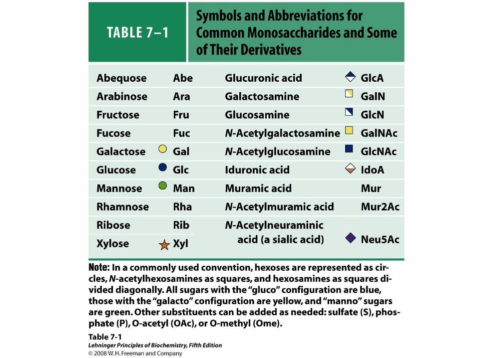


Figure 11-13 Electron micrograph of the cellulose fibers in the cell wall of the alga
Chaetomorpha melagonium.

Figure 11-14 The primary structure of cellulose.
Pag
e 36
5
What is this linkage?

Figure 11-15 Proposed structural model of cellulose.
Pag
e 36
5

Plant cell wall architecture
extensin
Pectins
Cellulose

Common sugars found in plant polysaccharides

A spotted June beetle (Pelidnota punctata), showing its surface armor (exoskeleton) of chitin.

Figure 11-16 Structure of chitin.
Pag
e 36
6
O O
Linkage?

Figure 11-17a -Amylose. The D-glucose residues of-amylose are linked by (1 4) bonds (red).
Pag
e 36
6

Figure 11-17b -Amylose. This regularly repeating polymer forms a left-
handed helix.
Pag
e 36
6

Figure 11-18a Amylopectin. Its primary structure near one of its (1 6)
branch points (red).
Pag
e 36
7

Figure 11-18b Amylopectin. (b) Its bushlike
structure with glucose residues at branch points indicated in red.
Pag
e 36
7

Figure 11-20 The disaccharide repeating units of the common glycosaminoglycans.
Pag
e 36
8
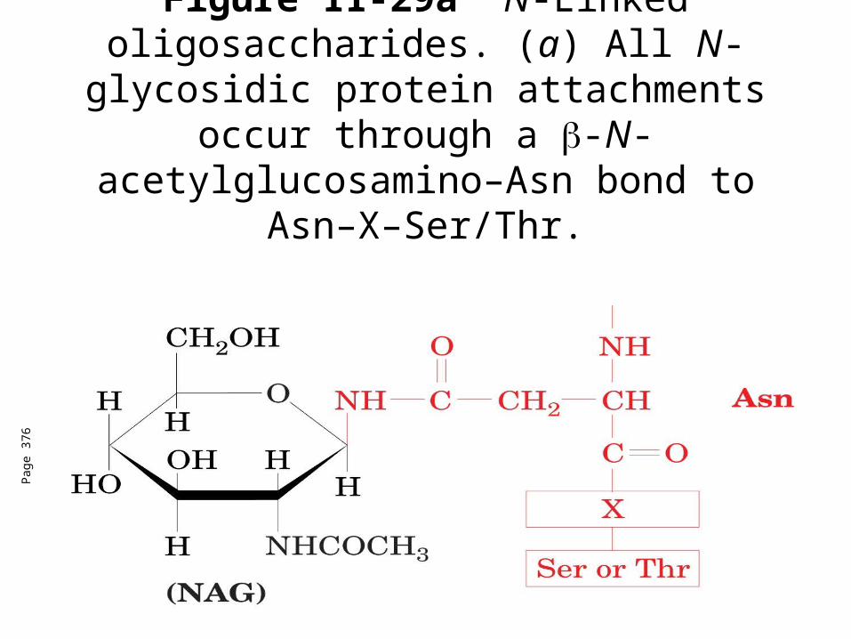
Figure 11-29a N-Linked oligosaccharides. (a) All N-glycosidic protein attachments
occur through a -N-acetylglucosamino–Asn bond to Asn–X–Ser/Thr.
Pag
e 37
6

Figure 11-29c N-Linked oligosaccharides. (c) Some examples of N-
linked oligosaccharides.
Pag
e 37
6

Figure 11-30 Some common O-glycosidic attachments of oligosaccharides to
glycoproteins (red).
Pag
e 37
6

• Hemagglutinin binding to sialic acid-containing receptor.
• Watch a layperson’s video of flu infection


FIGURE 7-35 Roles of oligosaccharides in recognition and adhesion at the cell surface.

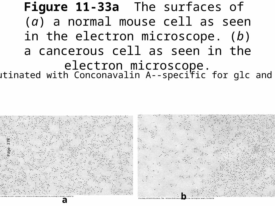
Figure 11-33a The surfaces of (a) a normal mouse cell as seen in the electron microscope. (b) a cancerous cell as seen in the
electron microscope.
Pag
e 37
8
a b
Agglutinated with Conconavalin A--specific for glc and man


Sugar determination by acetylation followed byGC/MS
5 10 15 20 25minutes
0.0
2.5
5.0
7.5
10.0
12.5
15.0
17.5
MCounts gal.40012.SMS 40:650 40:650
Gas chromatogram of D-galactitol hexacetate
Spectrum of Galactitol-1,2,3,4,5,6-hexacetate

FIGURE 7-37 Separation and quantification of the oligosaccharides in a group of glycoproteins. A mixture of proteins extracted from kidney tissue was treated to release oligosaccharides from glycoproteins, and the oligosaccharides were analyzed by matrix-assisted laser desorption/ionization mass spectrometry (MALDI MS). Each distinct oligosaccharide produces a peak at its molecular mass, and the area under the curve reflects the quantity of that oligosaccharide.

• LEHNINGER • PRINCIPLES OF BIOCHEMISTRY
• Fifth Edition
David L. Nelson and Michael M. Cox
© 2008 W. H. Freeman and Company
CHAPTER 13Bioenergetics and Biochemical
Reaction Types

Living cells are not at equilibrium!
Concentrations of reactants and products are typically far from the equilibrium values (Q Keq).
We must consider “steady state” concentrations of these species for the determination of G.
G = Go' + RTlnQ


Homeostaticconditions

Fig 16.2

See Figure 16.3
Catabolicpathways
Anabolicpathways

Figure 16.20


The energy charge of most cells ranges from 0.8 to 0.95


Pag
e 56
7
Figure 16-21b Some overall coupled reactions involving ATP. (b) The phosphorylation of ADP by phosphoenolpyruvate to form ATP and pyruvate.


Oxidized cofactors Reduced cofactors
Reduced substrates Oxidized substrates+ +


Fig 17.3


The Rossman fold. Structural motif is found in the NAD-binding site of many dehydrogenases. (a) Consists of a pair of structurally similar motifs, with three parallel β sheets and two α helices (β-α-β-α-β). (b) Nucleotide-binding domain of lactate dehydrogenase with NAD (ball-and-stick structure) bound in an extended conformation through hydrogen bonds and salt bridges to the paired β-α-β-α-β motifs of the Rossman fold (shades of green).


“Alfonse, Biochemistry makes my head hurt!!”\


![MS- K2.5 METABOLISME FRUKTOSA.ppt [Read-Only]ocw.usu.ac.id/course/download/1110000095-metabolism-system/mbs127... · FRUCTOSE METABOLISM The disaccharide sucrose from diet cleaved,](https://img.pdfslide.us/doc/110x75/5c85a0a809d3f279718d2379/ms-k25-metabolisme-read-onlyocwusuacidcoursedownload1110000095-metabolism-systemmbs127.jpg)