Embed Size (px)
Citation preview

Hematology - Part 2Andrea Lu, MDAssociate Program DirectorMay 2020
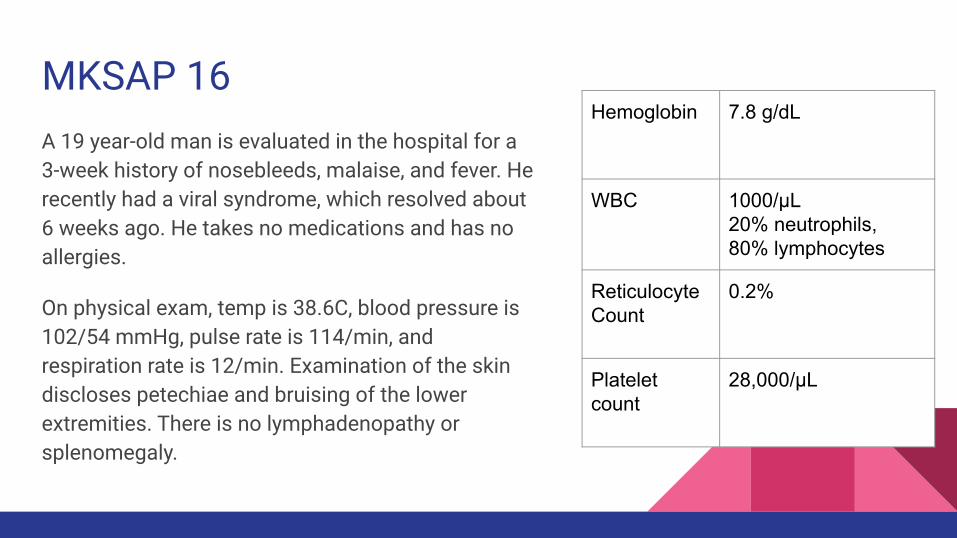
MKSAP 16A 19 year-old man is evaluated in the hospital for a 3-week history of nosebleeds, malaise, and fever. He recently had a viral syndrome, which resolved about 6 weeks ago. He takes no medications and has no allergies.
On physical exam, temp is 38.6C, blood pressure is 102/54 mmHg, pulse rate is 114/min, and respiration rate is 12/min. Examination of the skin discloses petechiae and bruising of the lower extremities. There is no lymphadenopathy or splenomegaly.
Hemoglobin 7.8 g/dL
WBC 1000/μL20% neutrophils, 80% lymphocytes
Reticulocyte Count
0.2%
Platelet count
28,000/μL
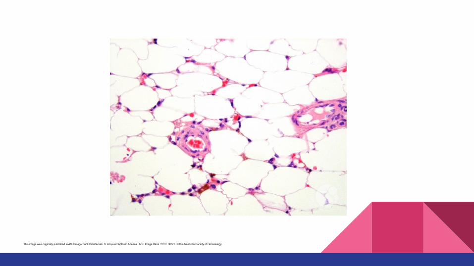
This image was originally published in ASH Image Bank.Schafernak, K. Acquired Aplastic Anemia.. ASH Image Bank. 2016; 60876. © the American Society of Hematology.
Which of the following is the most likely diagnosis?
A. Acute myeloid leukemiaB. Aplastic anemiaC. Chronic lymphocytic anemiaD. Myelodysplasia
Aplastic Anemia - Bone marrow failure characterized by pancytopenia - Caused by:
- Medications: methimazole, PTU, sulfonamids, anticonvulsants - Infections: HIV, EBV- Immune disorders: SLE, graft-vs-host dz- idiopathic
- Bone marrow: hypocellular, decreased precursors- Treatment
- Allogeneic HSCT - >50 yo: Immunosuppression (Cyclosporine + Antithymocyte Globulin/Prednisone)
Pure Red Cell Aplasia- normocytic/macrocytic anemia, low retic count
- WBC, platelets usually normal
- Bone Marrow: absent/decreased RBC precursors- Causes
- Parvovirus b19 infection - Thymoma- Hematologic malignancies
- Large granular lymphocyte leukemia (CD57+)- Medications: phenytoin, isoniazid - idiopathic- anti-EPO antibodies in patients receiving EPO
- Treatment: immunosuppression (prednisone, cyclosporine, cyclophosphamide), IVIG
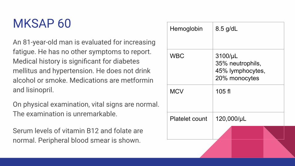
MKSAP 60An 81-year-old man is evaluated for increasing fatigue. He has no other symptoms to report. Medical history is significant for diabetes mellitus and hypertension. He does not drink alcohol or smoke. Medications are metformin and lisinopril.
On physical examination, vital signs are normal. The examination is unremarkable.
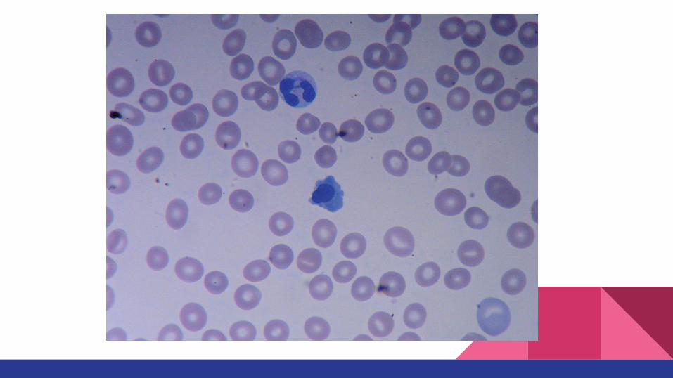
Serum levels of vitamin B12 and folate are normal. Peripheral blood smear is shown.
Hemoglobin 8.5 g/dL
WBC 3100/μL35% neutrophils, 45% lymphocytes, 20% monocytes
MCV 105 fl
Platelet count 120,000/μL
Which of the following is the most appropriate diagnostic test to perform next to evaluate this patient’s anemia?
A. BCR-ABL testingB. Bone marrow biopsyC. ImmunoelectrophoresisD. JAK2 mutation testing
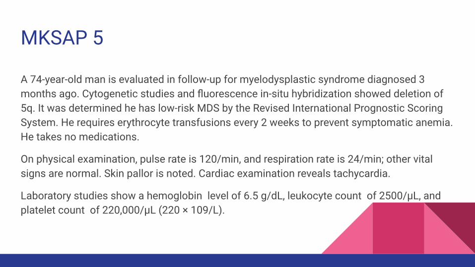
MKSAP 5
A 74-year-old man is evaluated in follow-up for myelodysplastic syndrome diagnosed 3 months ago. Cytogenetic studies and fluorescence in-situ hybridization showed deletion of 5q. It was determined he has low-risk MDS by the Revised International Prognostic Scoring System. He requires erythrocyte transfusions every 2 weeks to prevent symptomatic anemia. He takes no medications.
On physical examination, pulse rate is 120/min, and respiration rate is 24/min; other vital signs are normal. Skin pallor is noted. Cardiac examination reveals tachycardia.
Laboratory studies show a hemoglobin level of 6.5 g/dL, leukocyte count of 2500/µL, and platelet count of 220,000/µL (220 × 109/L).
Which of the following is the most appropriate treatment?
A. Allogeneic hematopoietic stem cell transplantationB. Antithymocyte globulin and cyclosporineC. ImatinibD. Lenalidomide
Myelodysplastic Syndromes (MDS)- Ineffective hematopoiesis causing dysplastic, hypercellular bone marrow and
peripheral cytopenias - Risk is transformation to AML - Can be 2/2: chemotherapy, XRT, chemical exposure (benzene)- Smear: normocytic/macrocytic RBCs, ovalocytes, dysplastic PMNs- Bone Marrow: hypercellular, blasts (<20%)- Prognosis depends on bone marrow blasts, degree of cytopenias, and
cytogenics- Goals of treatment: management of cytopenias, reducing risk of AML
- Transfusions, EPO - Azacytidine, decitabine- -5q cytogenic abnormality: lenalidomide- Allogeneic HSCT
Chronic Myeloid Leukemia (CML)
- Myeloproliferative neoplasms with uncontrolled production of mature/maturing granulocytes
- Median age: 50-60- Usually asymptomatic, but can present with constitutional symptoms,
abdominal discomfort/splenomegaly, bleeding, gout- Lymphadenopathy is uncommon- Labs: high PMN count, thrombocytosis, eosinophilia, basophilia - Diagnosis: BCR-ABL/Philadelphia chromosome t(9:22)
Chronic Myeloid Leukemia (CML)
- Natural History of Disease- Chronic Phase: Indolent - Accelerated Phase: more dysplastic, higher PMN counts- Blast Crisis: AML
- Treatment- TKIs: Imatinib, dasatinib, nilotinib- Be aware of effects: fluid retention, QT prolongation - Contraindicated in pregnancy - Refractory: Bosutinib, ponatinib- Allogeneic HSCT for accelerated/blast phase
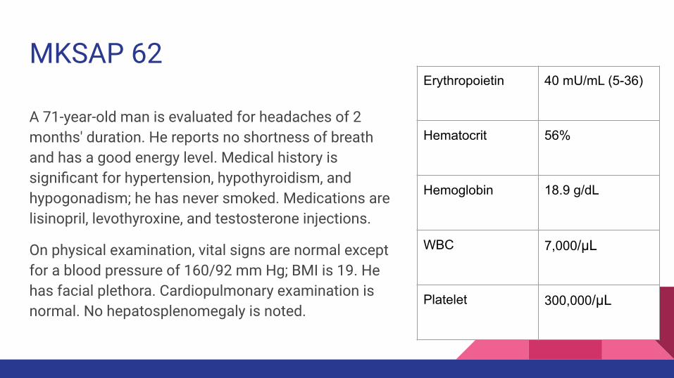
MKSAP 62
A 71-year-old man is evaluated for headaches of 2 months' duration. He reports no shortness of breath and has a good energy level. Medical history is significant for hypertension, hypothyroidism, and hypogonadism; he has never smoked. Medications are lisinopril, levothyroxine, and testosterone injections.
On physical examination, vital signs are normal except for a blood pressure of 160/92 mm Hg; BMI is 19. He has facial plethora. Cardiopulmonary examination is normal. No hepatosplenomegaly is noted.
Erythropoietin 40 mU/mL (5-36)
Hematocrit 56%
Hemoglobin 18.9 g/dL
WBC 7,000/μL
Platelet 300,000/μL

Which of the following is the most likely cause of this patient’s findings?
A. Levothyroxine B. LisinoprilC. Polycythemia veraD. Testosterone
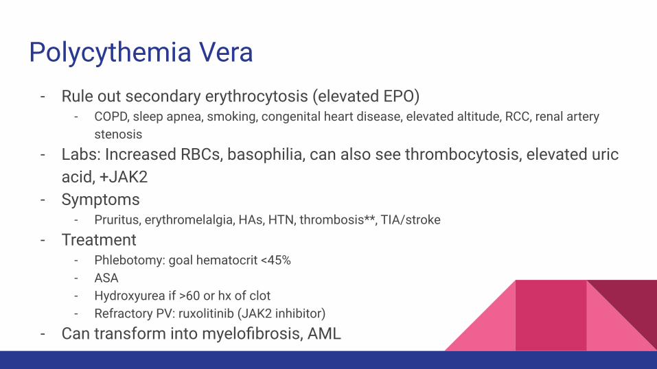
Polycythemia Vera- Rule out secondary erythrocytosis (elevated EPO)
- COPD, sleep apnea, smoking, congenital heart disease, elevated altitude, RCC, renal artery stenosis
- Labs: Increased RBCs, basophilia, can also see thrombocytosis, elevated uric acid, +JAK2
- Symptoms- Pruritus, erythromelalgia, HAs, HTN, thrombosis**, TIA/stroke
- Treatment- Phlebotomy: goal hematocrit <45%- ASA- Hydroxyurea if >60 or hx of clot - Refractory PV: ruxolitinib (JAK2 inhibitor)
- Can transform into myelofibrosis, AML
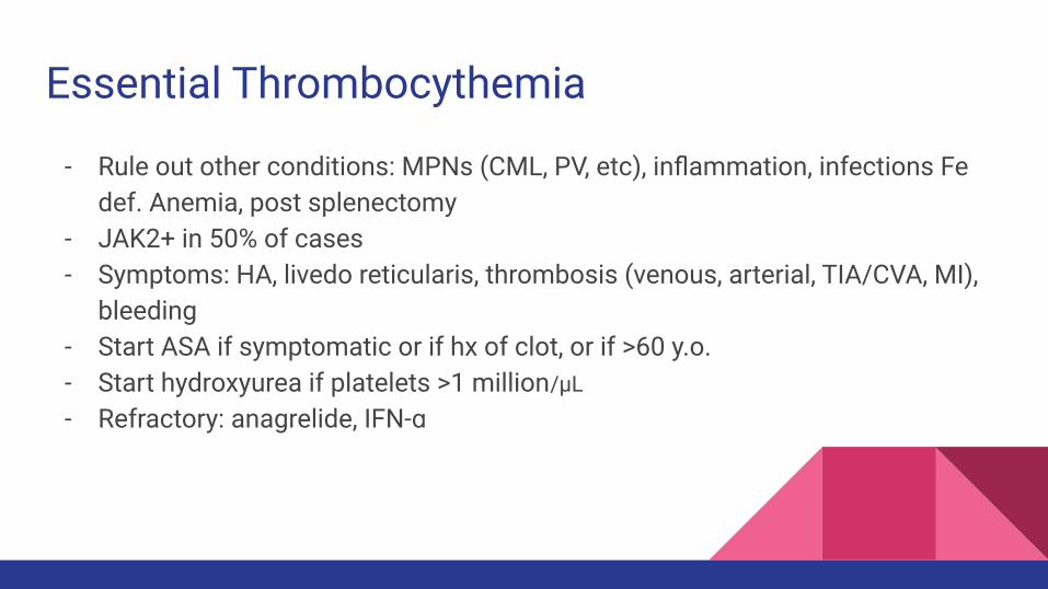
Essential Thrombocythemia
- Rule out other conditions: MPNs (CML, PV, etc), inflammation, infections Fe def. Anemia, post splenectomy
- JAK2+ in 50% of cases - Symptoms: HA, livedo reticularis, thrombosis (venous, arterial, TIA/CVA, MI),
bleeding- Start ASA if symptomatic or if hx of clot, or if >60 y.o.- Start hydroxyurea if platelets >1 million/µL- Refractory: anagrelide, IFN-ɑ
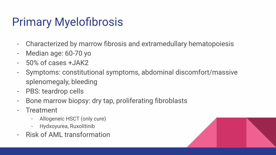
Primary Myelofibrosis
- Characterized by marrow fibrosis and extramedullary hematopoiesis- Median age: 60-70 yo- 50% of cases +JAK2- Symptoms: constitutional symptoms, abdominal discomfort/massive
splenomegaly, bleeding- PBS: teardrop cells - Bone marrow biopsy: dry tap, proliferating fibroblasts- Treatment
- Allogeneic HSCT (only cure) - Hydxoyurea, Ruxolitinib
- Risk of AML transformation
MKSAP 6A 45-year-old man is evaluated for shortness of breath with exertion and lower extremity edema of 6 months' duration. He reports no chest pain, fever, or cough. He takes no medications.
On physical examination, vital signs are normal. Jugular venous distention is noted. Cardiopulmonary examination reveals crackles at the base of the lungs. He also has 1+ lower extremity edema.
Laboratory studies are remarkable for eosinophilia, with an absolute eosinophil count of 2500/µL (2.5 × 10⁹/L). A review of the medical record shows an eosinophil count of 2900/µL (2.9 × 10⁹/L) 1 year ago.
An electrocardiogram shows sinus rhythm with low-voltage QRS and nonspecific ST changes. An echocardiogram shows findings compatible with a restrictive cardiomyopathy.
Which of the following is the most appropriate initial management?
A. Evaluation for helminth infectionB. Fat pad biopsyC. PrednisoneD. Repeat laboratory evaluation in 3 months
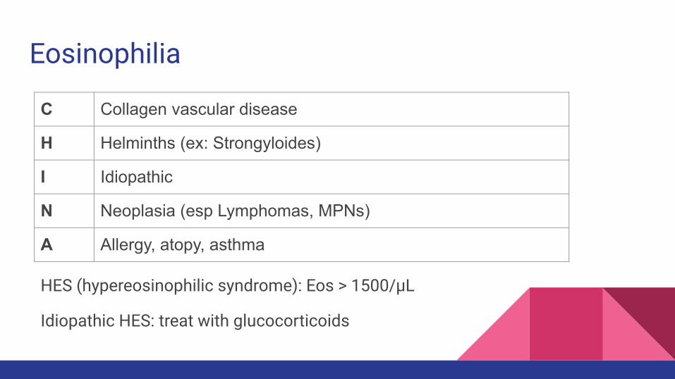
Eosinophilia
HES (hypereosinophilic syndrome): Eos > 1500/µL
C Collagen vascular disease
H Helminths (ex: Strongyloides)
I Idiopathic
N Neoplasia (esp Lymphomas, MPNs)
A Allergy, atopy, asthma
Idiopathic HES: treat with glucocorticoids
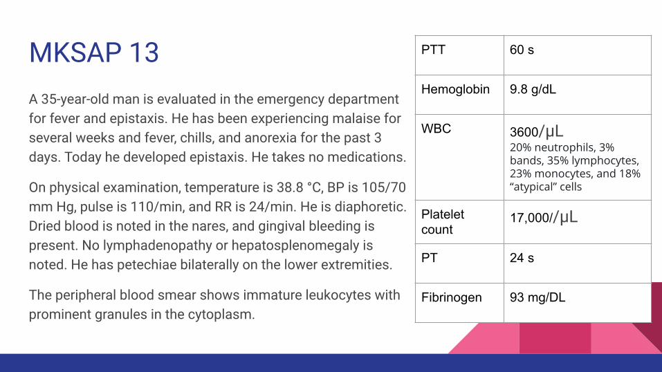
MKSAP 13A 35-year-old man is evaluated in the emergency department for fever and epistaxis. He has been experiencing malaise for several weeks and fever, chills, and anorexia for the past 3 days. Today he developed epistaxis. He takes no medications.
On physical examination, temperature is 38.8 °C, BP is 105/70 mm Hg, pulse is 110/min, and RR is 24/min. He is diaphoretic. Dried blood is noted in the nares, and gingival bleeding is present. No lymphadenopathy or hepatosplenomegaly is noted. He has petechiae bilaterally on the lower extremities.
The peripheral blood smear shows immature leukocytes with prominent granules in the cytoplasm.
PTT 60 s
Hemoglobin 9.8 g/dL
WBC 3600/µL20% neutrophils, 3% bands, 35% lymphocytes, 23% monocytes, and 18% “atypical” cells
Platelet count
17,000//µL
PT 24 s
Fibrinogen 93 mg/DL
Which of the following is the most likely diagnosis?
A. Acute promyelocytic leukemiaB. Aplastic anemiaC. Chronic granulocytic leukemiaD. Immune thrombocytopenic purpura
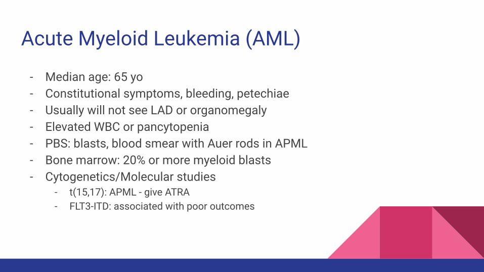
Acute Myeloid Leukemia (AML)
- Median age: 65 yo- Constitutional symptoms, bleeding, petechiae - Usually will not see LAD or organomegaly - Elevated WBC or pancytopenia - PBS: blasts, blood smear with Auer rods in APML- Bone marrow: 20% or more myeloid blasts - Cytogenetics/Molecular studies
- t(15,17): APML - give ATRA- FLT3-ITD: associated with poor outcomes
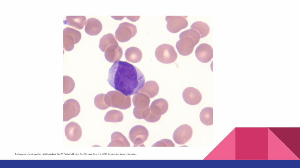
This image was originally published in ASH Image Bank. Carll TC, Parilla M. AML - Auer Rod. ASH Image Bank. 2018; 61328. © the American Society of Hematology.
AML
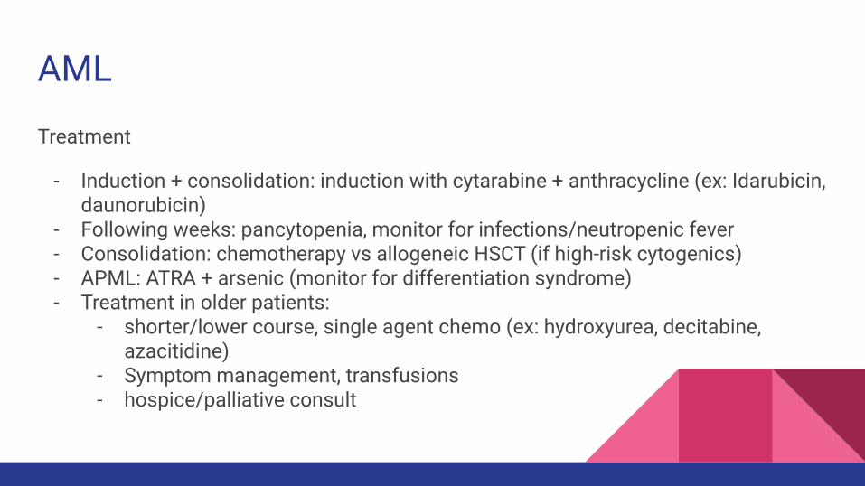
Treatment
- Induction + consolidation: induction with cytarabine + anthracycline (ex: Idarubicin, daunorubicin)
- Following weeks: pancytopenia, monitor for infections/neutropenic fever- Consolidation: chemotherapy vs allogeneic HSCT (if high-risk cytogenics)- APML: ATRA + arsenic (monitor for differentiation syndrome)- Treatment in older patients:
- shorter/lower course, single agent chemo (ex: hydroxyurea, decitabine, azacitidine)
- Symptom management, transfusions - hospice/palliative consult
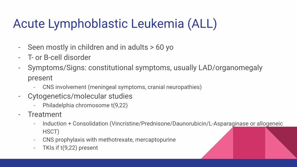
Acute Lymphoblastic Leukemia (ALL)
- Seen mostly in children and in adults > 60 yo- T- or B-cell disorder- Symptoms/Signs: constitutional symptoms, usually LAD/organomegaly
present - CNS involvement (meningeal symptoms, cranial neuropathies)
- Cytogenetics/molecular studies- Philadelphia chromosome t(9,22)
- Treatment - Induction + Consolidation (Vincristine/Prednisone/Daunorubicin/L-Asparaginase or allogeneic
HSCT)
- CNS prophylaxis with methotrexate, mercaptopurine- TKIs if t(9,22) present
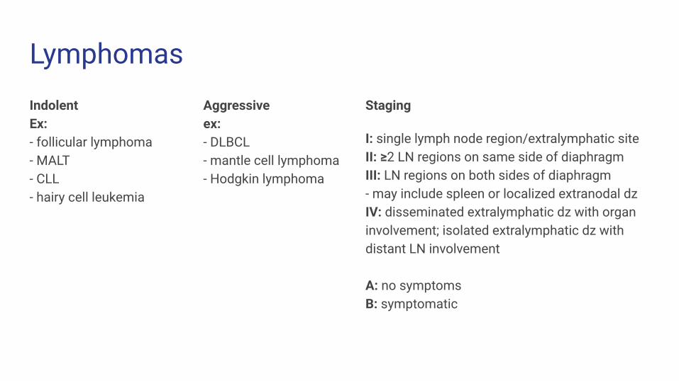
LymphomasIndolentEx: - follicular lymphoma- MALT- CLL- hairy cell leukemia
Aggressiveex:- DLBCL- mantle cell lymphoma- Hodgkin lymphoma
Staging
I: single lymph node region/extralymphatic siteII: ≥2 LN regions on same side of diaphragmIII: LN regions on both sides of diaphragm- may include spleen or localized extranodal dzIV: disseminated extralymphatic dz with organ involvement; isolated extralymphatic dz with distant LN involvement
A: no symptomsB: symptomatic
MKSAP 120
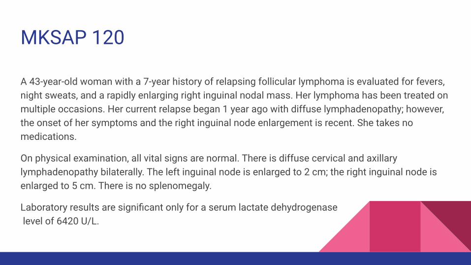
A 43-year-old woman with a 7-year history of relapsing follicular lymphoma is evaluated for fevers, night sweats, and a rapidly enlarging right inguinal nodal mass. Her lymphoma has been treated on multiple occasions. Her current relapse began 1 year ago with diffuse lymphadenopathy; however, the onset of her symptoms and the right inguinal node enlargement is recent. She takes no medications.
On physical examination, all vital signs are normal. There is diffuse cervical and axillary lymphadenopathy bilaterally. The left inguinal node is enlarged to 2 cm; the right inguinal node is enlarged to 5 cm. There is no splenomegaly.
Laboratory results are significant only for a serum lactate dehydrogenase level of 6420 U/L.

Which of the following is the most appropriate management?
A. Biopsy of the right inguinal lymph nodeB. Oral idelalisibC. Radiation therapy to he right inguinal nodeD. Repeat a course of chemotherapy
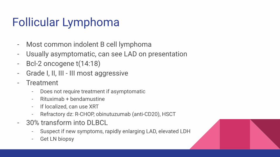
Follicular Lymphoma
- Most common indolent B cell lymphoma- Usually asymptomatic, can see LAD on presentation - Bcl-2 oncogene t(14:18)- Grade I, II, III - III most aggressive - Treatment
- Does not require treatment if asymptomatic - Rituximab + bendamustine - If localized, can use XRT- Refractory dz: R-CHOP, obinutuzumab (anti-CD20), HSCT
- 30% transform into DLBCL - Suspect if new symptoms, rapidly enlarging LAD, elevated LDH- Get LN biopsy
Mucosa-Associated Lymphoid Tissue Lymphoma
- MALTomas commonly associated with H. Pylori, but can be seen with other chronic infections, autoimmune disorders
- Sjogren’s, Hashimotos
- Other sites besides stomach: salivary glands, thyroid, colon, lung, eyes - Treatment
- Gastric MALToma - PPI+antibiotic therapy against H Pylori - Other localized MALTomas: XRT- Systemic dz: rituximab ± chemotherapy
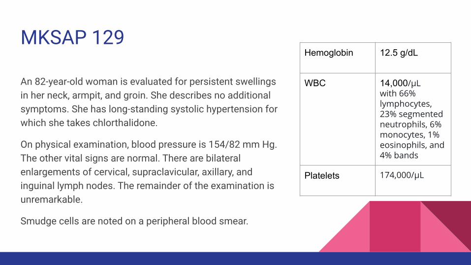
MKSAP 129
An 82-year-old woman is evaluated for persistent swellings in her neck, armpit, and groin. She describes no additional symptoms. She has long-standing systolic hypertension for which she takes chlorthalidone.
On physical examination, blood pressure is 154/82 mm Hg. The other vital signs are normal. There are bilateral enlargements of cervical, supraclavicular, axillary, and inguinal lymph nodes. The remainder of the examination is unremarkable.
Smudge cells are noted on a peripheral blood smear.
Hemoglobin 12.5 g/dL
WBC 14,000/µL with 66% lymphocytes, 23% segmented neutrophils, 6% monocytes, 1% eosinophils, and 4% bands
Platelets 174,000/µL
Which of the following is the most appropriate diagnostic test to perform next?
A. Bone marrow aspiration and biopsyB. CT scan of the chest, abdomen, and pelvisC. Cytogenetic studies on peripheral bloodD. Flow cytometry on peripheral blood
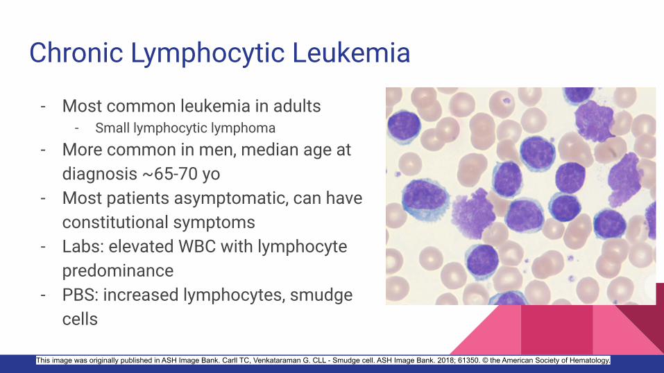
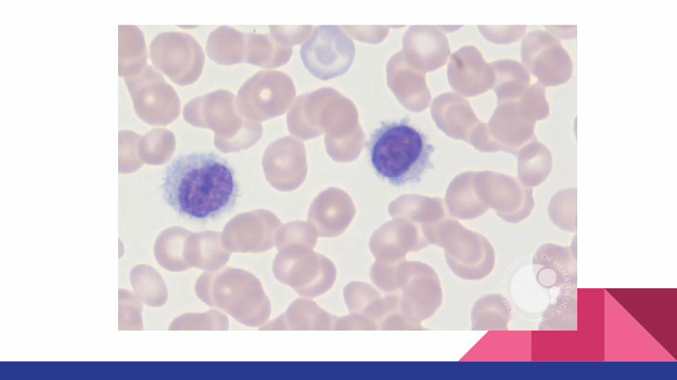
Chronic Lymphocytic Leukemia
- Most common leukemia in adults- Small lymphocytic lymphoma
- More common in men, median age at diagnosis ~65-70 yo
- Most patients asymptomatic, can have constitutional symptoms
- Labs: elevated WBC with lymphocyte predominance
- PBS: increased lymphocytes, smudge cells
This image was originally published in ASH Image Bank. Carll TC, Venkataraman G. CLL - Smudge cell. ASH Image Bank. 2018; 61350. © the American Society of Hematology.
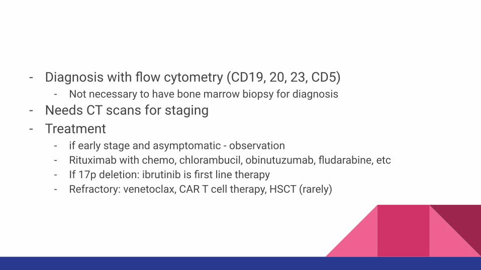
- Diagnosis with flow cytometry (CD19, 20, 23, CD5)- Not necessary to have bone marrow biopsy for diagnosis
- Needs CT scans for staging - Treatment
- if early stage and asymptomatic - observation - Rituximab with chemo, chlorambucil, obinutuzumab, fludarabine, etc - If 17p deletion: ibrutinib is first line therapy - Refractory: venetoclax, CAR T cell therapy, HSCT (rarely)
CLL
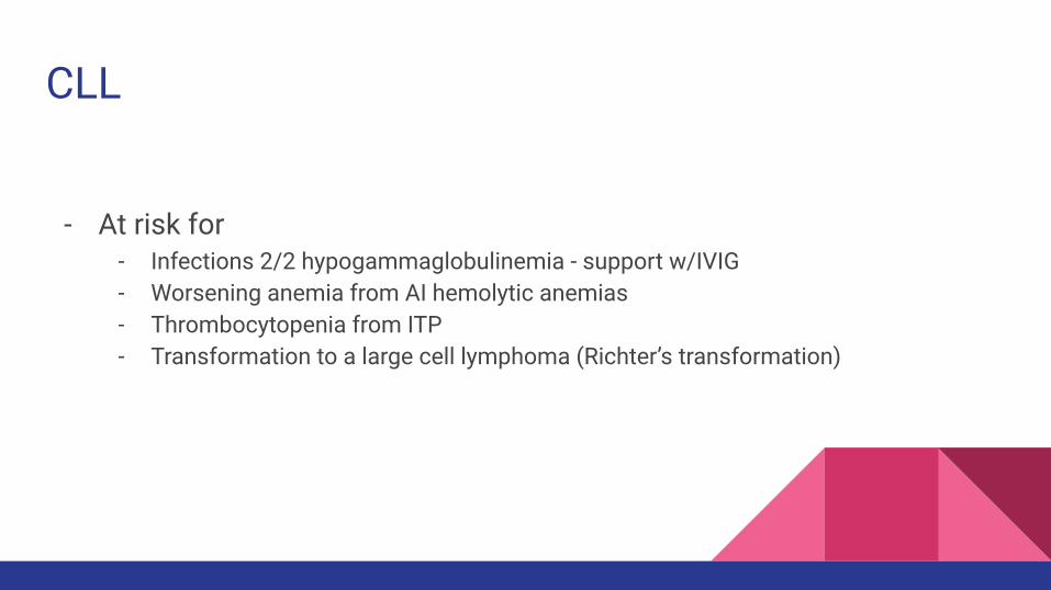
- At risk for - Infections 2/2 hypogammaglobulinemia - support w/IVIG- Worsening anemia from AI hemolytic anemias- Thrombocytopenia from ITP - Transformation to a large cell lymphoma (Richter’s transformation)
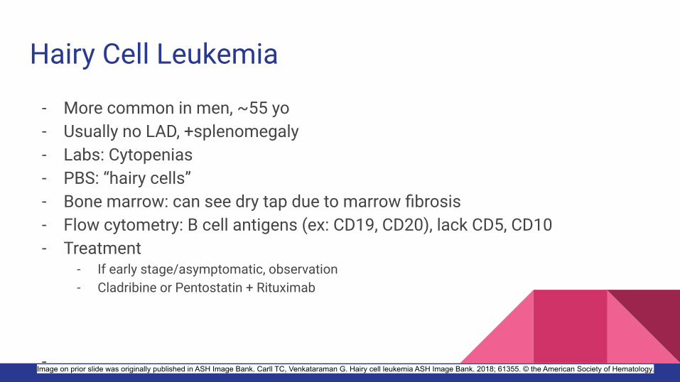
Hairy Cell Leukemia
- More common in men, ~55 yo - Usually no LAD, +splenomegaly - Labs: Cytopenias- PBS: “hairy cells” - Bone marrow: can see dry tap due to marrow fibrosis - Flow cytometry: B cell antigens (ex: CD19, CD20), lack CD5, CD10- Treatment
- If early stage/asymptomatic, observation- Cladribine or Pentostatin + Rituximab
-Image on prior slide was originally published in ASH Image Bank. Carll TC, Venkataraman G. Hairy cell leukemia ASH Image Bank. 2018; 61355. © the American Society of Hematology.
Diffuse Large B Cell Lymphoma
- Median age: 50-60 yo - Considered AIDS-defining malignancy- Most present at stage III/IV. Usually symptomatic at presentation from
enlarging LAD, splenomegaly, B symptoms present - Treatment:
- R-CHOP, R-hyper-CVAD+XRT- Refractory: Autologous HSCT or CAR-T
Burkitt Lymphoma
- Endemic (associated with EBV), Sporadic, Immunodeficiency-associated (Ex: HIV)
- Very rapid growth, enlarging neck mass - Elevated LDH - Monitor for TLS, start prophylaxis before treatment
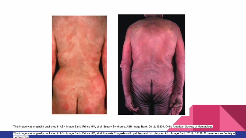
This image was originally published in ASH Image Bank. Prince HM, et al. Mycosis Fungoides with patches and thin plaques. ASH Image Bank. 2012; 15199. © the American Society of Hematology.
This image was originally published in ASH Image Bank. Prince HM, et al. Sezary Syndrome. ASH Image Bank. 2012; 15204. © the American Society of Hematology.
Cutaneous T Cell Lymphomas
- Mycosis fungoides (skin) and Sezary syndrome (skin+blood) - Can eventually infiltrate organs- Skin manifestations: raised plaques, diffuse skin erythema,
skin ulcers- Pruritus and Infections are common - Treatment
- Early Stage: Topical steroids, UV light therapy, retinoids- Advanced stage: electron-beam radiation therapy,
photopheresis- Allogeneic HSCT
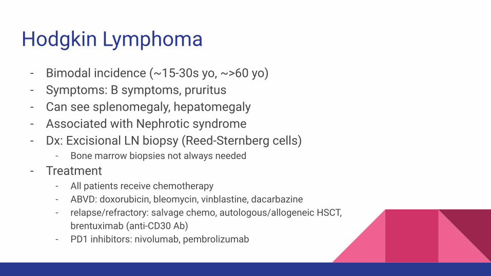
Hodgkin Lymphoma- Bimodal incidence (~15-30s yo, ~>60 yo)- Symptoms: B symptoms, pruritus - Can see splenomegaly, hepatomegaly- Associated with Nephrotic syndrome - Dx: Excisional LN biopsy (Reed-Sternberg cells)
- Bone marrow biopsies not always needed
- Treatment- All patients receive chemotherapy - ABVD: doxorubicin, bleomycin, vinblastine, dacarbazine - relapse/refractory: salvage chemo, autologous/allogeneic HSCT,
brentuximab (anti-CD30 Ab)- PD1 inhibitors: nivolumab, pembrolizumab
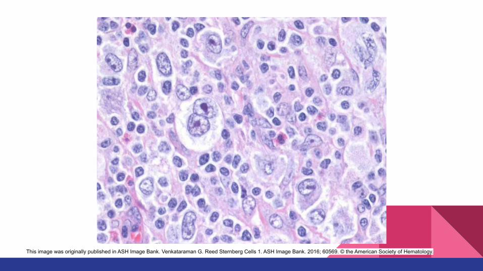
This image was originally published in ASH Image Bank. Venkataraman G. Reed Sternberg Cells 1. ASH Image Bank. 2016; 60569. © the American Society of Hematology.
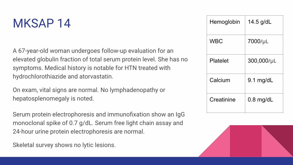
MKSAP 14
A 67-year-old woman undergoes follow-up evaluation for an elevated globulin fraction of total serum protein level. She has no symptoms. Medical history is notable for HTN treated with hydrochlorothiazide and atorvastatin.
On exam, vital signs are normal. No lymphadenopathy or hepatosplenomegaly is noted.
Serum protein electrophoresis and immunofixation show an IgG monoclonal spike of 0.7 g/dL. Serum free light chain assay and 24-hour urine protein electrophoresis are normal.
Skeletal survey shows no lytic lesions.
Hemoglobin 14.5 g/dL
WBC 7000/µL
Platelet 300,000/µL
Calcium 9.1 mg/dL
Creatinine 0.8 mg/dL
Which of the following is the most appropriate management?
A. Kidney biopsyB. MRI of the cervical, thoracic, and lumbar spineC. Repeat laboratory studies in 6 monthsD. Serum β2microglobulin I measurement
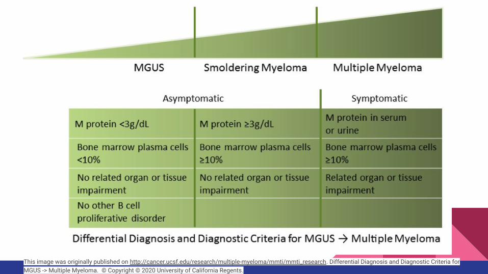
Monoclonal gammopathy of undetermined significance (MGUS)
- MGUS: M protein < 3 g/dL (or <500 mg/24 h of urinary monoclonal FLCs), clonal plasma cells <10% of bone marrow, asymptomatic/no end-organ damage
- Initial evaluation: Check M protein level, FLC level - Usually noted incidentally- At higher risk for osteoporosis/fractures
- Will need screening
- Risk of transformation into MM/Plasma cell dyscrasias- IgM gammopathy - M protein 1.5 g/dL or more- Abnormal serum FLC ratio
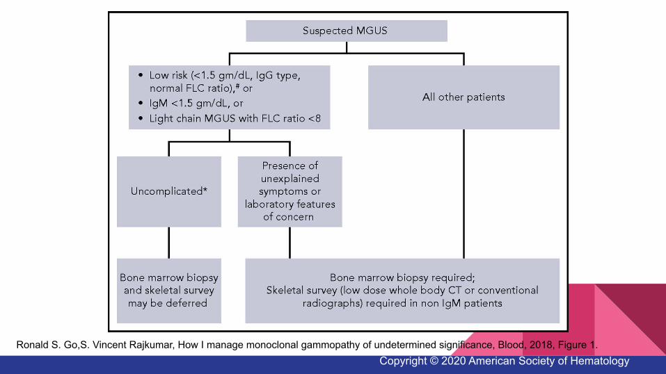
Ronald S. Go,S. Vincent Rajkumar, How I manage monoclonal gammopathy of undetermined significance, Blood, 2018, Figure 1.Copyright © 2020 American Society of Hematology
Multiple Myeloma- Median age: 65-70 yo- Symptoms/signs: fatigue, dyspnea, hypercalcemia, renal failure, bone
pain/fractures, recurrent infections, neuropathy - SPEP, UPEP, serum free light chains, bone surveys (MRI best), bone marrow
biopsyβ2-microglobulin and albumin
- Diagnosis: - M protein (serum or urine)- Bone marrow: >10% plasma cells, or plasmacytoma- Sign of Organ dysfunction
- Calcium >11 - Cr >2 or CrCl <40 - Hgb <10 - Lytic lesions or osteoporosis
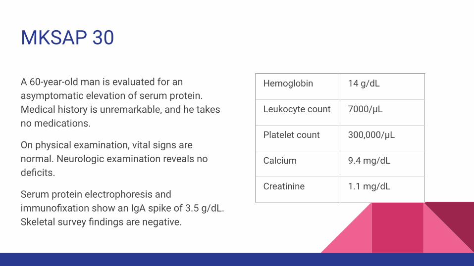
MKSAP 30
A 60-year-old man is evaluated for an asymptomatic elevation of serum protein. Medical history is unremarkable, and he takes no medications.
On physical examination, vital signs are normal. Neurologic examination reveals no deficits.
Serum protein electrophoresis and immunofixation show an IgA spike of 3.5 g/dL. Skeletal survey findings are negative.
Hemoglobin 14 g/dL
Leukocyte count 7000/µL
Platelet count 300,000/µL
Calcium 9.4 mg/dL
Creatinine 1.1 mg/dL
Which of the following is the most appropriate next test?
A. Bone scanB. CT of the chest, abdomen and pelvisC. Whole body MRID. No further testing
This image was originally published on http://cancer.ucsf.edu/research/multiple-myeloma/mmti/mmti_research. Differential Diagnosis and Diagnostic Criteria for MGUS -> Multiple Myeloma. © Copyright © 2020 University of California Regents.
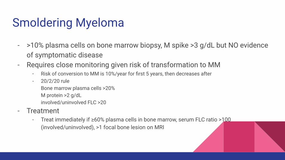
Smoldering Myeloma
- >10% plasma cells on bone marrow biopsy, M spike >3 g/dL but NO evidence of symptomatic disease
- Requires close monitoring given risk of transformation to MM- Risk of conversion to MM is 10%/year for first 5 years, then decreases after- 20/2/20 rule
Bone marrow plasma cells >20%M protein >2 g/dLinvolved/uninvolved FLC >20
- Treatment- Treat immediately if ≥60% plasma cells in bone marrow, serum FLC ratio >100
(involved/uninvolved), >1 focal bone lesion on MRI
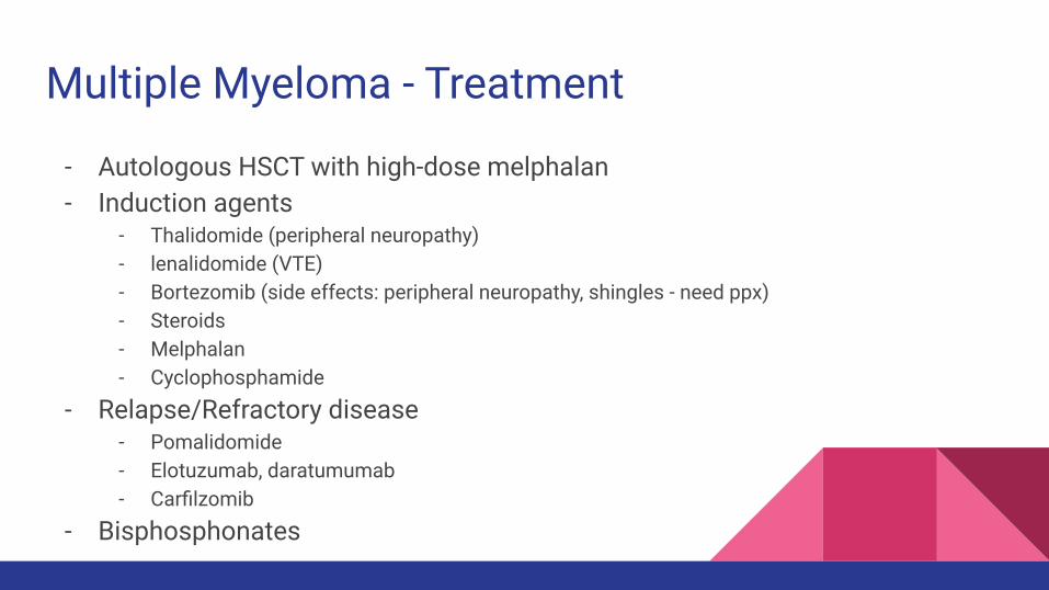
Multiple Myeloma - Treatment
- Autologous HSCT with high-dose melphalan- Induction agents
- Thalidomide (peripheral neuropathy)- lenalidomide (VTE) - Bortezomib (side effects: peripheral neuropathy, shingles - need ppx)- Steroids- Melphalan- Cyclophosphamide
- Relapse/Refractory disease- Pomalidomide- Elotuzumab, daratumumab- Carfilzomib
- Bisphosphonates
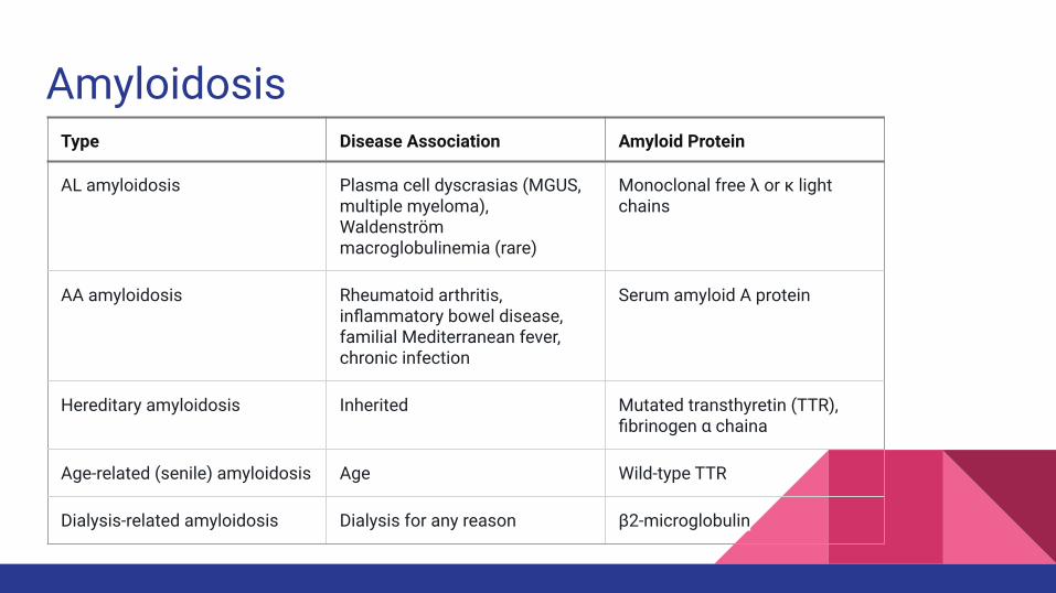
AmyloidosisType Disease Association Amyloid Protein
AL amyloidosis Plasma cell dyscrasias (MGUS, multiple myeloma), Waldenström macroglobulinemia (rare)
Monoclonal free λ or κ light chains
AA amyloidosis Rheumatoid arthritis, inflammatory bowel disease, familial Mediterranean fever, chronic infection
Serum amyloid A protein
Hereditary amyloidosis Inherited Mutated transthyretin (TTR), fibrinogen α chaina
Age-related (senile) amyloidosis Age Wild-type TTR
Dialysis-related amyloidosis Dialysis for any reason β2-microglobulin
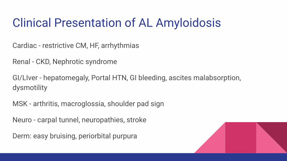
Clinical Presentation of AL Amyloidosis
Cardiac - restrictive CM, HF, arrhythmias
Renal - CKD, Nephrotic syndrome
GI/LIver - hepatomegaly, Portal HTN, GI bleeding, ascites malabsorption, dysmotility
MSK - arthritis, macroglossia, shoulder pad sign
Neuro - carpal tunnel, neuropathies, stroke
Derm: easy bruising, periorbital purpura
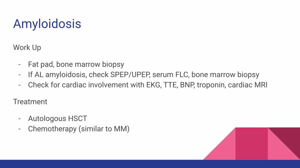
Amyloidosis
Work Up
- Fat pad, bone marrow biopsy- If AL amyloidosis, check SPEP/UPEP, serum FLC, bone marrow biopsy- Check for cardiac involvement with EKG, TTE, BNP, troponin, cardiac MRI
Treatment
- Autologous HSCT- Chemotherapy (similar to MM)
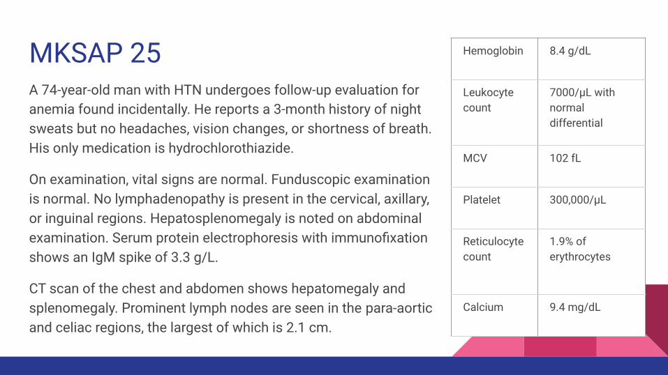
MKSAP 25A 74-year-old man with HTN undergoes follow-up evaluation for anemia found incidentally. He reports a 3-month history of night sweats but no headaches, vision changes, or shortness of breath. His only medication is hydrochlorothiazide.
On examination, vital signs are normal. Funduscopic examination is normal. No lymphadenopathy is present in the cervical, axillary, or inguinal regions. Hepatosplenomegaly is noted on abdominal examination. Serum protein electrophoresis with immunofixation shows an IgM spike of 3.3 g/L.
CT scan of the chest and abdomen shows hepatomegaly and splenomegaly. Prominent lymph nodes are seen in the para-aortic and celiac regions, the largest of which is 2.1 cm.
Hemoglobin 8.4 g/dL
Leukocyte count
7000/µL with normal differential
MCV 102 fL
Platelet 300,000/µL
Reticulocyte count
1.9% of erythrocytes
Calcium 9.4 mg/dL
Which of the following is the most appropriate diagnostic test to perform next?
A. Bone marrow biopsyB. CT imaging-guided needle biopsy of a para-aortic lymph node C. Excisional biopsy of a para-aortic lymph nodeD. Serum viscosity testing
Waldenström Macroglobulinemia
- Indolent B cell non-Hodgkin lymphoma producing clonal IgM - Diagnosis
- IgM monoclonal gammopathy- ≥10 percent of the bone marrow are clonal lymphoplasmacytic cells
- Can have constitutional symptoms, bleeding, abdominal discomfort from organomegaly, kidney disease
- Increased viscosity - HA, AMS, change in vision, nystagmus, ataxia - Treatment
- If asymptomatic - observation - Rituximab ± chemotherapy, + steroids- Ibrutinib - If hyperviscosity present - plasmapheresis

The End

Good Luck!