Embed Size (px)
DESCRIPTION
Hematology Oncology Study Questions for Medical School
Citation preview
QuestionsNormal RangesNormal % of MyeloblastsNormal range for ANCNormal range for WBCNormal range for Platelets
Anemia
LeukemiaAML is a cancer of which type?
What is leukostasis?Least common leukemia in children:
Lymphoma is a cancer of a ___________.What is a lymphocytic leukemia?
Lymphoma
Sickle cell disease patients up to age 5 need are given what prophylaxis?
Most common leukemia in children:
2nd most common?
All lymphomas can have a leukemic phase.
Which leukemia has these 3 stages:- Chronic- Accelerating- Blast crisis
What blood malignancy is associated with autoimmune hemolytic anemia (AIHA)?
Treatment for (Diffuse Large B-Cell Lymphoma) DLBCL?
Clinical/Labs
What does the Coomb's test check for?
Treatments
What is the treatment for leukostasis?
If patient has impressive cerival adenopathy, get CXR because they might have a mediastinal mass.
Burkitt's Lymphoma often has acute abdominal symptoms and can be mistaken for appendicitis.
What might you check for in a lab for:
Hemolytic anemia
Patient has:
Elevated LDHElevated bilirubinLow HbLow haptoglobin
Probably cause?
Chemo has a maximum effect 2 weeks after starting treatment.
It takes 2 weeks for blood cell count to bounce back.
Total of 4 weeks.
Class of drugs that end in "ib"i.e. Imatinib (Gleevac)
Answers Notes
5%1.5 to 8 (1000's)4,500 to 10,000150,000 to 400,000
Penicillin for susceptibility to infection
Myeloblasts
*Seen in advanced AML (and others)CLL (<1%)
Lymphocytes *Not a cancer of lymph node!
CML
CLL
Myeloblasts aggregating in the peripheral blood -> Sludging
Most common: ALL (80%)2nd most common: AML
Cancer of circulating lymphocyte
R-CHOP
Autoimmune Hemolytic Anemia
Hemolytic anemia
Elevated LDHElevated bilirubinLow HbLow haptoglobin
Luekopheresis Hydroxyurea -> myelosuppression
Questions
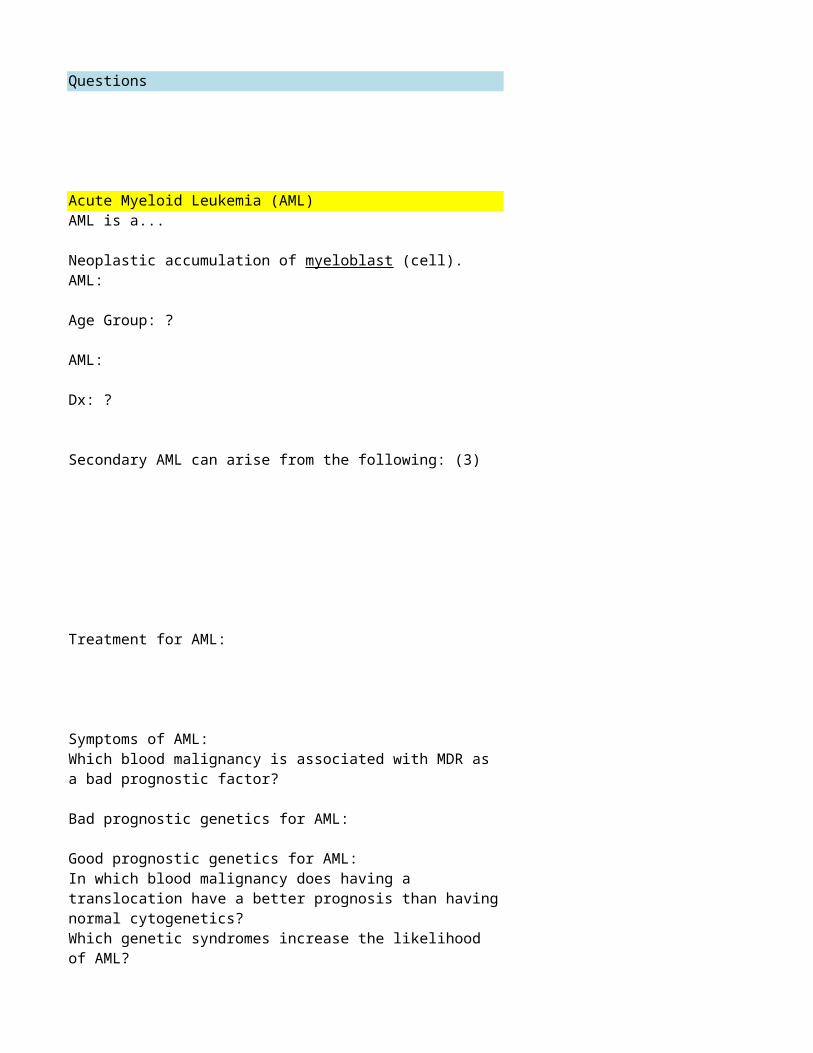
Acute Myeloid Leukemia (AML)
Secondary AML can arise from the following: (3)
Treatment for AML:
Symptoms of AML:
Bad prognostic genetics for AML:
Good prognostic genetics for AML:
Which genetic syndromes increase the likelihood of AML?
AML is a...
Neoplastic accumulation of myeloblast (cell).
AML:
Age Group: ?
AML:
Dx: ?
Which blood malignancy is associated with MDR as a bad prognostic factor?
In which blood malignancy does having a translocation have a better prognosis than having normal cytogenetics?
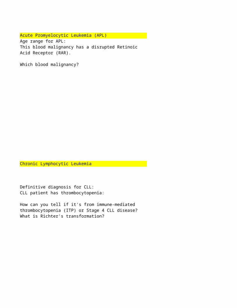
Acute Promyelocytic Leukemia (APL)Age range for APL:
Chronic Lymphocytic Leukemia
Definitive diagnosis for CLL:
What is Richter's transformation?
This blood malignancy has a disrupted Retinoic Acid Receptor (RAR).
Which blood malignancy?
CLL patient has thrombocytopenia:
How can you tell if it's from immune-mediated thrombocytopenia (ITP) or Stage 4 CLL disease?
Answers Notes
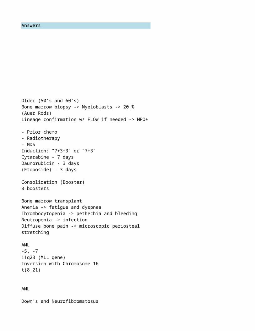
Older (50's and 60's)
*85% relapse after remission
AML
AML
Down's and Neurofibromatosus
Bone marrow biopsy -> Myeloblasts -> 20 % (Auer Rods)Lineage confirmation w/ FLOW if needed -> MPO+
- Prior chemo- Radiotherapy- MDS
Induction: "7+3+3" or "7+3"Cytarabine - 7 daysDaunorubicin - 3 days(Etoposide) - 3 days
Consolidation (Booster)3 boosters
Bone marrow transplant
Anemia -> fatigue and dyspneaThrombocytopenia -> pethechia and bleedingNeutropenia -> infectionDiffuse bone pain -> microscopic periosteal stretching
-5, -711q23 (MLL gene)
Inversion with Chromosome 16t(8,21)
*Better to have translocation than have normal cytogenetics
23's and 30's
APL
Bone marrow biopsy -> Look for megakaryocytesCLL progression to Diffuse Large B-Cell Lymphoma
Peripheral blood FLOW cytometry:
CD5 (abberrant T-cell marker) plus CD5,19,20 (normal B-cell marker)
Questions140811.1 Intro and Normal HematopoiesisInfants have hematopoiesis in which bones?
When does hematopoeisis start in utero?Where does hematopoeisis take place at 3 weeks?Where does hematopoeisis take place at 12 weeks?Where does hematopoeisis take place at 27 weeks?Where does hematopoeisis take place at 34 weeks?
Where does hematopoeisis take place 3 weeks after birth?
6 stages of RBC development:Mean diameter of RBCCentral pallor of RBC should be _____ of cell diameterWhat are the 3 granulocytes?Which granulocytes have pink granules?Which granulocytes have blue granules?
What are the 6 stages of neutrophil development?How many lobes in a normal neutrophil?How many lobes in a normal eosinophil?
As we mature, hematopoiesis gets restricted to these bones: (3)
Where does hematopoeisis take place 12 weeks post-partum?
High eosinophil count might indication what type of infection?
Allergens can bind to IgE on a basophil, which leads to a release of histamines.Monocytes circulate in the blood for 24 hours.
Monocytes are white blood cells that are like "vacuum cleaners" because they eat up fungi, pyogenic bacteria, mycobacteria, and old or defective RBCs.

2 morphological stages for lympocytes:
NK cell surface markers:
B-cell surface markers:
T-cell surface markers:Plasma (memory) cell surface marker:
What is the largest collection of lymphoid tissue in a person?25% of our lymphocytes are where in the body?
140811.3 Laboratory Evaluation of hematologic parameters
When in life is RBC (red blood count) the highest?At what age does HbF decrease to a normal level?Normal % for HbF:
The nucleus of a lymphocyte is about the same size as a RBC.
Megakaryocytes rarely get out into peripheral circulation, but if they do, they get caught up.
Where?
T cells form PALS (Periarterial lymphatic sheaths) around the central arteries of the spleen.
The red pulp of the spleen is composed of these 2 structural components and this very important cell:
If a patient's WBC count is too high or too low, what should you look at next?

MCV tells you:
What is MCH and MCHC?Normal range for MCV:Normal range for RDW:
Reticulocyte morphology:
# of RBCs decreases with age.
T/F?
Basophilic stippling in RBCs is actually the precipitation of undegraded RNA and seen in (condition) lead toxicity.
You see basophilic stippling in a patient's peripheral blood smear:
Immediately think: --> (Dx)
You see Target cells in a patient's peripheral blood smear:
Possible Dx's (4):
You see Schistocytes (helmet cells) in a patient's peripheral blood smear:
You should think these Dx's: (2)
You see Acanthocytes (thorn cells) in a patient's peripheral blood smear:
Immediately think: --> (Dx)
In Rouleaux, RBCs abnormally adhere to each other due to increased plasma protein production.
You see Rouleaux in a patient's peripheral blood smear:
Immediately think: --> (Dx)
What part of the body do you take a bone marrow biopsy?
What do you do with a bone marrow aspirate? (Tests)
What do you use from a patient to run a metabolic panel?What is SPEP?
What is a sickle cell screen?
140811.4 Synthesis & catabolism of Normal HemoglobinHeme molecules have a ring structure called:What is the charge of iron in a heme molecule?
Howell Jolly Bodies are remnants of nuclear chormatin normally removed by macrophages in the spleen.
You see Howell Jolly Bodies in a patient's peripheral blood smear:
Immediately think: --> (Condition)
You see Spherocytes in a patient's peripheral blood smear:
Immediately think: --> (Dx)
What is the normal range for myeloblast percentage?
Globin chains:
Alterations in the amino acid sequences result in different globin chains.
Globin genes:
The alpha genes are on chromosome 16.
Globin genes:
The beta, delta, and gamma genes are on chromosome 11:
Humans have 2 copies of these hemoglobin chains per chromosome: (2)
What is hemoglobin A1C?
What is the Embden-Meyerhof pathway?
What are 2 important functions of 2,3-BPG for RBCs?
What is methemoglobin?
Hemoglobin A:
Which globin chains?
Hemoglobin A2:
Which globin chains?
Hemoglobin F:
Which globin chains?
Normal % range for:- HbA2 - HbF
Bohr Effect:
Low pH (acidic environment) shifts oxygen curve L/R?
Higher relative levels of 2,3-BPG (bisphosphoglycerate) shifts oxygen curve L/R?
Fever and acidosis:
Oxygen curve shifts L or R?
Hb values for newborns:
Higher or Lower? Why?
Without the enzyme, G6PD, no NADPH is formed, which is needed for to metabolize glutathione.
Which cells break down RBCs in the spleen (extravascularly)?
This enzyme deficiency, which can cause hemolysis, is the most common enzye deficiency in the world.
G6PD deficiency puts the patient at risk for hemolysis when his cells are exposed to oxidative stress.
What 3 important things should you know about the Embden-Meyerhof Pathway?
Causes of Hemolysis:Intracorpuscular Hemolysis VS Extracorpuscular Hemolysis
(Just Click and Read)
Free hemoglobin binds to haptoglobin and the complex is removed by the liver.
Hemosiderin in the urine can indicate what type of hemolysis?
Within the splenic macrophage cytoplasm, RBC hemoglobin is broken down into 3 parts:
Globin: --> ExplainIron: --> ExplainProtoporphyrin --> Explain
Nutritional requirements for Chronic Hemolysis:
What is the purpose of a Direct (or Indirect) Coomb's Test?
Evaluating for Hemolysis:
Increase of Decrease?LDH (Lactate Dehydogenase): Haptoglobin:Reticulocyte Count:Total Bilirubin:Indirect Bilirubin:Coomb's Test: (+/-)?
Answers Notes
All of them
3 weeks *When the yolk sac developsYolk sac *First 6 weeksLiver *6-18 weeksLiver and Spleen *18-30 weeksLiver, Spleen, and Bone Marrow *30 weeks - 8 weeks after birth
Liver, Spleen, and Bone Marrow *30 weeks - 8 weeks after birth
Bone marrow only *After 10 weeks after birth
8 um1/3Neutrophils, eosinophils, and basophilsEosinophilsBasophils
3 to 6 lobes2 lobes (bi-lobed)
Parasite or helminth infection
- breast bones- pelvic bones- proximal part of long bones
- Pronormoblast- Basophilic normoblast- Polychromatophilic normoblast- Orthochromatic normoblast- Reticulocyte- RBC
- Myeloblast- Promyelocyte- Myelocyte- Metamyelocyte- Band- Neutrophil
*Could also be season allergies or allergic reaction
Lymphoblast and lymphocyte
CD 38
Lung microvasculature *Smallest capillaries
SpleenWhite pulp of the spleen
At birth3 months
2%
*Cell surface markers need to be identified to further differentiate** Can't do it morphologically
CD 16CD 56
CD 10CD 19CD 20CD 79asIg k/d
CD 3CD 4CD 5CD 7CD 8
- Sinusoids- Splenic Cords ("Billroths" cords)
- Monocytes/macrophages
*Dark alley analogy where new cells can squeeze through small windows to escape monocytes but old cells get stuck and are destroyed**Extensive RBC damage leads to blood getting trapped in spleen
WBC differential
Check "NLMEB" and make sure percentages are okay.
0 *Anemia is not a normal finding in the elderly
60-80 fLLess than 12%
*It's a sign of ineffective hematopoiesis
Lead toxicity
Multiple myeloma
Mean corpuscular volume
Tells you the size of the RBC
MCH - Mean corpuscular hemoglobin- Avg. Weight of Hb / Cell
MCHC - Mean corpuscular hemoglobin concentration- Avg. Concentration of Hb / Cell
- Larger than an RBC- Less central pallor (Giemsa stain)- Pricipitated RNA (methylene blue stain)
*Reticulocyte count not part of CBC - Need to order separately
*Basophilic stippling in RBCs is actually the precipitation of undegraded RNA and seen in (condition) lead toxicity.
Anything that results in extra membrane or decreased cell volume
- Hemoglobinopathies/Thalassemias - Iron deficiency anemia- Drug-induced hemolytic anemia- Liver disease
Conditions w/ increased fibrin:
- DIC / MAHA- TTP
Liver disease
(Or something else that would affect the membrane morphology)
*Liver dysfunction -> decreased synthesis of cholesterol, which is needed for normal cell membrane
Patient is functionally/surgically asplenic.
Flatter part of Posterior Superior Iliac CrestLess than 5%Smear, FLOW, Cytogenetics (karyotype), FISH
SerumSerum Protein Electrophoresis
Add picture
Protoporhyrin IX2+ (ferrous state)
*Because they are more important
Heriditary spherocytosis(Can also be acquired: physical damage of RBCs)
RBCs are mixed with a reducing agen in a tube
Turbidity = Hemolysis = Sickle Cell
Alpha and gamma
(Hemoglobin A and Hemoglobin F)
Shifts right (Oxygen more easily released into tissues)
Shifts right (Oxygen more easily released into tissues)
Shifts right (Oxygen more easily released into tissues)
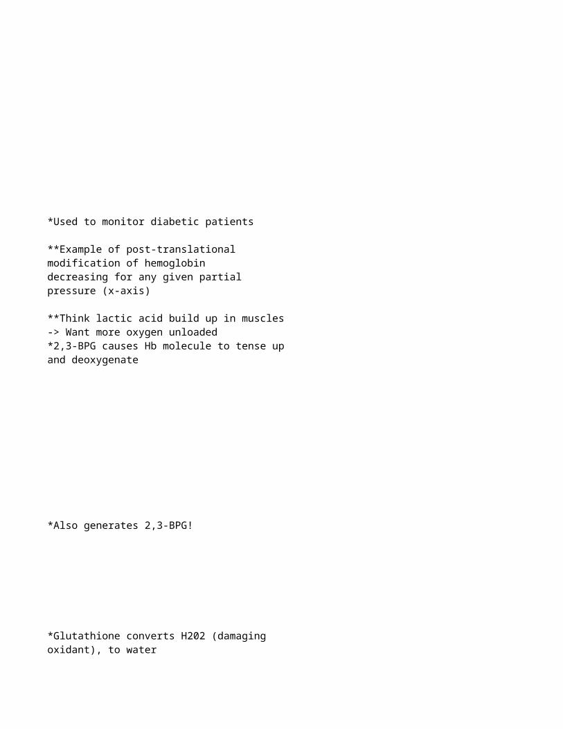
*Also generates 2,3-BPG!
2 alpha2 beta
2 alpha2 delta
2 alpha2 gamma
HbA2: 3.5% HbF: 1-2%
Glycated (Glycosylated) hemoglobin as a result of glucose being added to the beta chain
*Used to monitor diabetic patients
**Example of post-translational modification of hemoglobin
*Oxygen saturation (y-axis) is decreasing for any given partial pressure (x-axis)
**Think lactic acid build up in muscles -> Want more oxygen unloaded
*2,3-BPG causes Hb molecule to tense up and deoxygenate
Higher
Because they have fetal hemoglobin, which binds oxygen more tightly. Therefore need higher concentrations to unload oxygen to tissues.
Anaerobic glycolytic pathways for RBCs
- Glucose gets metabolized to lactic acid- Input 2 ATP to get 4 ATP = Net gain of 2 ATP
- Shifts oxygen curve right -> better oxygen delivery to tissues
- Reduces methemoglobin (Fe3+) to hemoglobin (Fe2+)
Hemoglobin with iron in a Fe3+ state*CAN'T Bind Oxygen!
*Glutathione converts H202 (damaging oxidant), to water
G6PD deficiency
*Example: Antibiotics
Macrophages
*G6PD is essential to prevent oxidative stress, cell death, and lysis.
- Energy (Input 2 ATP to get 4 ATP = Net 2 ATP- 2,3-BPG reduces methemoglobin into hemoglobin- G6PD from hexose monophosphate shunt prevents oxidative damage (hemolysis)
Intra:RBC membrane disorders- heriditary spherocytosis or elliptocytosisRBC enzyme disorders- G6PD deficiency- Pyruvate kinase deficiencyHemoglobin Disorders- Methemoglobinemia- Thalassemia- Sickle Cell Disease
Extra:- Antibodies- Sludging, trapping, destruction in spleen- Trauma (prosthetic valces, DIC, etc)- Chemicals (w/ oxidant properties)- Infectious destruction (malaria, babesiosis, etc)
Intravascular Hemolysis
- Hb breaks down into alpha-beta dimers, which are small enough to be filtered by the glomerulus. - Some dimers are taken up into the renal tubular cells and the iron is stored as hemosiderin. - Renal tubular cells slough off into urine -> can be detected.
Globin: --> Amino Acids (recycled)Iron: --> Binds to Transferrin -> Goes to Liver, Spleen, Bone Marrow (recycled)Protoporphyrin --> Unconjugated Bilirubin -> Binds to Albumin and sent to Liver -> Eventually excreted
LDH (Lactate Dehydogenase): IncreaseHaptoglobin: DecreaseReticulocyte Count: IncreaseTotal Bilirubin: IncreaseIndirect Bilirubin: IncreaseCoomb's Test: (+/-)? Positive
Folic Acid: Supplementation requiredVitamin B12: Usually not required (Body has 10 yr store)Iron: Not required (iron gets recycled and stored)
Test for Autoimmune Hemolytic Anemia- Agglutination = Positive Test
Direct test uses patient's RBCs (antigen already stuck on them)Indirect uses store bought RBCs and patient's serum (which should have antibodies in it)
Questions140818.4 Myeloid Disorders - Beaty
Answers Notes
What protein is iron bound to when stored intracellularly? Ferritin
What protein is iron bound to when traveling in the blood? Transferrin
Suppresses EPO production
Enterocytes transport iron into the bloodstream via ferroportin.
What is the role of the acute phase reactant, hepcidin, when it comes to anemia of chronic disease
Sequesters iron into storage sites, preventing use by erythroid precursors
The congenital defect that causes sideroblastic anemia most commonly involves the enzyme Aminolevulinic synthase (ALAS)
What are the lab findings for sideroblastic anemia?FerratinTIBCSerum Iron% Saturation
Ferratin - IncreaseTIBC - DecreaseSerum Iron - Increase% Saturation - Increase

QuestionsChapter 6.1 Leukopenia and LeukocytosisCD marker for Hematopoietic Stem Cells:
Chapter 6.2 Acute Leukemia
Acute leukemia is a neoplastic proliferation of blasts (cell type).
Acute leukemia is defined as a greater than 20% blasts in the bone marrow.
Acute Leukemia
Why is the WBC (in the blood) high?
What is the phenotypic marker for lymphoblasts?What is the phenotypic marker for myeloblasts?
You see Auer Rods in a blood smear.
What kind of cell are you looking at?
Presence of Auer rods indicates that your probably dealing with which malignancy?
Acute Lymphoblastic Leukemia (ALL) most commonly arises in:
Children of AdultsB-ALL is the most common type of ALL.

AML is more commonly seen in which decades of life?
AML risk factors:
Chapter 6.3 Chronic Leukemia
Primary cause of death in CLL patients?CLL is a neoplasm of which cell?CLL can progress to which condition?CLL is associated with which autoimmune condition?
Would you see lymphadenopathy in Hair Cell Leukemia?Would you see splenomegaly in Hairy Cell Leukemia?
Cytogenetics (genotype) you should know for B-ALL: (2 Translocations)
Which has the better prognosis?
Patient, who is a teenager, presents with a mediastinal (thymic mass):
Likely diagnosis?
Acute promyelocytic leukemia is characterized by: (translocation)
Acute promyelocytic leukemia (APL):
Pathogenesis
Acute promyelocytic leukemia (APL):
Treatment
Chronic Leukemia - neoplastic proliferation of mature circulating lymphocytes
Cells co-express CD5 and CD 20:
Likely diagnosis:
Hairy Cell Leukemia - neoplastic proliferation of mature B-cellsHairy Cells are positive for TRAP
ATLL is associated with which virus?
Hairy Cell Leukemia:
Treatment
Adult T-Cell Lymphoma Leukemia (ATLL):
Neoplastic proliferation of mature CD4+ T-Cells
Mycosis fungoides:
Neoplastic proliferation of mature CD4+ T-Cells
Patient has aggregates of neoplastic T-cells in the epidermis (Pautrier microabscesses):
Likely Dx?
When cancer cells in Myscosis Fungoides spreads to the blood it's called Sezary Syndrome. The cells have a cerebriform (characteristic) nuclei in the blood smear.

Answers Notes
Overproduction of blasts -> Leaks out into blood
TdTMyeloperoxidase (MPO)
Myeloblast -> Probably looking at AML
Acute Myeloid Leukemia (AML)
Children *Associated with Down Syndrome (after age 5)
*TdT is a DNA polymerase**TdT is absent in myeloid blasts and mature lymphcytes
*Auer Rods are crystallized MPO (seen only in Myeloblasts)
T-ALL
50-60 yo
t(15;17)
RAR receptor disrupted -> promyelocytes accumulate *Promyelocytes also contain auer rods
CLLInfection due to hypogammaglobulinemiaB-CellDiffuse Large B-Cell LymphomaAutoimmune hemolytic anemia
No - cells are trapped in bone marrowYes - expansion of red pulp
t(12;21) - Good prognosist(9;22) - Poor Prognosis
*t(9;22) also known as the Philadelphia Chromosome*t(9;22) more classically associated with CML
ATRA (all-trans retinoic acid)- derivative of vitamin A- causes blasts to mature
Preexisting dysplasia (Myelodysplastic Syndrome)Prior exposure to alkylating agents (chemo)Radiotherapy

Add picture
2-CDA*Adenosine deaminase inhibitor**Adenosine accumulates to toxic levels in neoplastic B cells
HTLV-1*Japan and Carribean
Mycosis fungoides
*Cells infiltrate skin producing rashes, plaques, or nodules

Questions140819.1 Intro to Lymphomas 2014
What is the difference between Leukemia and Lymphoma?Primary lymphoid tissues (2):
Secondary lymphoid tissues (4):
Thymic epithelial cells interact with lymphocytes to help w
What are the high grade lymphomas? (4)
What are the intermediate grade lymphomas? (3)
Most common low grade non-Hodgkin lymphoma
Where are the primary and secondary follicles in a lymph node?
Where in the lymph node does an dentritic cell present an antigen to a T-cell?
Plasma cells and sinuses containing macrophages and histiocytes can be found in which part of the lymph node?
What kind of cells are in the Primary follicles of a lymph node?Secondary follicle?
In a secondary follicle, what kinds of cells are in the dark zone?Light zone?
In the secondary follicle, ________________, destroy B-cells with wrong antibodies.
What are some lymphomas that can have a leukemic phase? (4)
Follicular Lymphoma:
HighIntermediateor Low Grade?

Grading for Follicular Lymphoma is based on:
What is the treatment for Diffuse Large B-Cell Lymphoma?What are the 3 types of Burkitt's Lymphoma?
Which lymphoma involves bones of the jaw and face?
Cytogenetic abnormality necessary for Burkitt's Lymphoma:
Follicular Lymphoma:
Median age?
Histology shows "small-cleaved cells", centrocytes, and centromeres.
Most likely dx?
85% of patients with Follicular Lymphoma will have t(14;18) (chromosomal abnormality).
If a patient has a t(14;18), what lymphoma might you suspect? (2)
What is the most common of the intermediate grade lymphomas?
Burkitt's Lymphoma (especially African) is associated with an infection by which virus?
Histology shows "a starry sky pattern".
Most likely dx?
Explain the presence of "starry sky" in histology of Burkitt's Lymphoma
Patient has a C-myc abnormality on chromosome 8.
What kind of cancer?

140819.2 Intro to Lymphoproliferative Disorders 2014
Which lymphoma is more common?
Non-Hodgkin or Hodgkin?
Normal age range for Hodgkin lymphoma?
Non-Hodgkin?
What type of cell is a Reed-Sternberg cell? Which kind of malignancy is it associated with?
Patient has nodal malignancy with predictable, contiguous spread.
What kind of lumphoma?
What kind of translocation would you see in a patient w/ Follicular lymphoma?
Answers Notes
Bone Marrow and Thymus
Cortex
Paracortex
Medulla
Tingible Body Macrophages
LowFollicular Lymphoma
Leukemias are cancers of the myeloid system and Lymphomas are cancers of the lymphoid system (lymphocytes)
Think cell type and location.
*Every lymphoma is a cancer of lymphocytes*Lymphoma can have a leukemic phase (lympocytes have the potential to circulate in the blood)
Lymph Nodes, Spleen, Tonsils, Clusters in GI tract and pulmonary tract
*Macrophages and histiocytes (in the medullary sinuses) capture antigen and process it
Primary: Naïve B CellSecondary: Proliferating B Cells (after encountering antigen)
Dark zone: CentroblastsLight zone: Centrocytes
Burkitt's Lymphoma/LeukemiaPre-B Cell ALL/LymphomaPre-T Cell ALL/LymphomaAdult T-Cell Lymphoma (HTLV-1)
Diffuse Large B-Cell LymphomaAnaplastic Large Cell LymphomaMantle Cell Lymphoma
Burkitt's Lymphoma/LeukemiaPre-B Cell ALL/LymphomaPre-T Cell ALL/LymphomaCLL/SLL
60-70 yo
Follicular LymphomaCentroblasts per HPF (high power field)
Diffuse Large B-Cell Lymphoma
African, American, Immunodeficiency-associated
EBVAfrican Burkitt's
Burkitt's Lymphoma
Burkitt's Lymphoma
*Translocation (14;18) not diagnostic because it could also be Diffuse Large B-cell Lymphoma
- Follicular Lymphoma- Diffuse Large B-cell Lymphoma
R-CHOP----Rituximab----Cyclophosphamide----Hydroxy-doxorubicin----Oncovin----Prednisone
*Refers to either the "white cells" (macrophages and histiocytes) or the vacuoles
Starry sky appearance refers to either the "white cells" (macrophages and histiocytes) or the vacuoles.
Burkitt's cells multiply so fast that they spontaneously die. The "white cells" are macrophages and histiocytes cleaning up the debris.
Chromosome 8: C-myc*can happen in a variety of different ways (translocations)
Malignant B-Cell associated with Hodgkin Lymphoma
Hodgkin Lymphoma
t(14;18)
Non-Hodgkin (70% of malignant lymphomas)
*Hodgkin (30%)
Non-Hodgkin (Young Adult: 20's and 30's)
*Hodgkin (Adults)
*Add Picture
Add picture
Questions140820.3 Chemotherapy
MESNA is indicated for:
MESNA mechamism of action:Which class of chemo drugs cause myelosuppression?
Dangerous side effect of Cisplatin
Side effect for Carboplatin:Interesting side effect of Oxaliplatin:
Common side effects for alkylating agents: (5)
Hemorrhagic Cystitis is caused by an accumulation of Acrolein.
How do you prevent hemorrhagic cystitis?
What is typically the cause of the dose limiting toxicity for alkylating agents?
If a patient has impaired renal function, would you give him Cisplatin or Carboplatin?
Platinum analogues:
Indications?
Which class of chemo drugs causes IgE-mediated hypersensitivity?
Answers Notes
*MESNA must be given before chemo treatment
*Must be given before chemo treatment
Binds acrolein (uroprotectant)- Alkylating agents
Myelosuppression
Carboplatin *Cisplatin is very nephrotoxic
Myelosuppresion/decreased platelet countCold-sensitizing peripheral neuropathy
Platinum analogues (alkylating agent)
- Vigorous hydration- MESNA (mercaptoethane sodium)
- Cyclophosphamide > 1g/m2/dose- Ifosfamide (any dose)
*Preventative; Too late if hemorrhagic cystitis is present
- Nephrotoxic, but renally cleared---Positive feedback cycle- Patient must have good renal function
*Also nausea/ vomiting"SPLAT" in Cisplatin
Solid Tumors- breast, lung, testicular, cervical/ovarian, colorectal, bladder, lymphoma
- myelosuppression- secondary leukemias- infertility- alopecia- nausea/vomiting