Embed Size (px)
Citation preview
Color index: Red: Important Gray: Extra, notes
Editing file
Hematology 438 teamwork
| Anemia
| objectives
● To understand the normal control of erythropoiesis
● To understand the pathophysiology of anemia
● To recognize the general features of anemia
● To understand the basis of anemia classification
● To understand iron metabolism
● To understand how iron deficiency
● To understand anemia of chronic disease may arise and how to manage it.
| Hemoglobin
αβ
βα
⁺⁺Fe
⁺⁺Fe
⁺⁺Fe
⁺⁺Fe
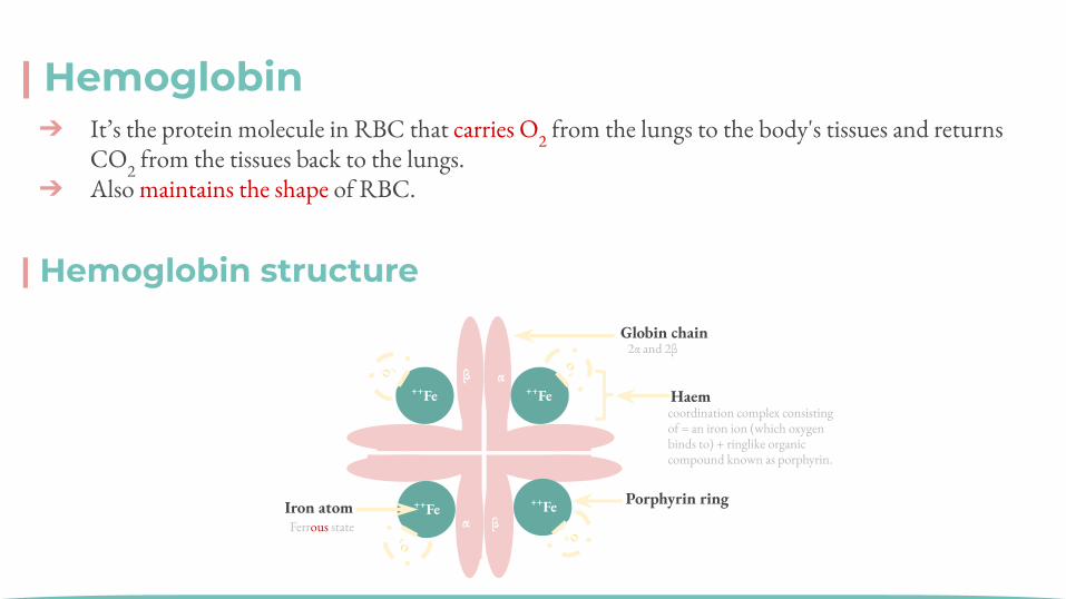
Globin chain
Haem
Porphyrin ring Iron atom
O2
O2
O 2
O2
| Hemoglobin structure
2α and 2βββ
Ferrous state
coordination complex consisting of = an iron ion (which oxygen binds to) + ringlike organic compound known as porphyrin.
➔ It’s the protein molecule in RBC that carries O2 from the lungs to the body's tissues and returns CO2 from the tissues back to the lungs.
➔ Also maintains the shape of RBC.
| Hematopoiesis
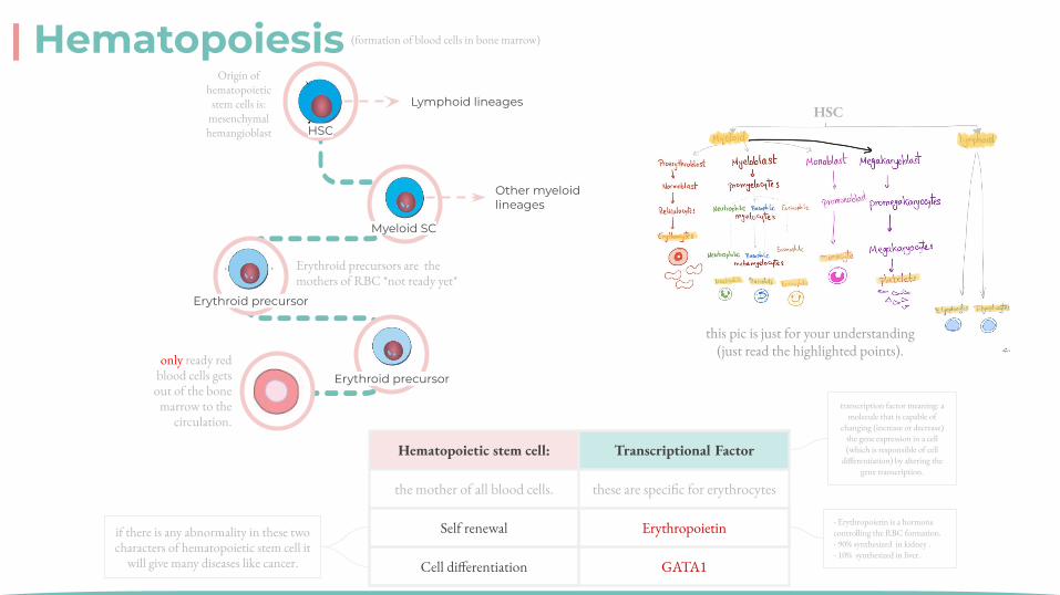
Hematopoietic stem cell: Transcriptional Factor
the mother of all blood cells. these are specific for erythrocytes
Self renewal Erythropoietin
Cell differentiation GATA1
(formation of blood cells in bone marrow)ββ
- Erythropoietin is a hormone controlling the RBC formation.- 90% synthesized in kidney .- 10% synthesized in liver.
if there is any abnormality in these two characters of hematopoietic stem cell it
will give many diseases like cancer.
HSC
this pic is just for your understanding (just read the highlighted points).
transcription factor meaning: a molecule that is capable of
changing (increase or decrease) the gene expression in a cell (which is responsible of cell
differentiation) by altering the gene transcription.
Lymphoid lineages
Myeloid SC
Other myeloid lineages
Erythroid precursor
Erythroid precursoronly ready red
blood cells gets out of the bone
marrow to the circulation.
Erythroid precursors are the mothers of RBC *not ready yet*
HSC
Origin of hematopoietic
stem cells is: mesenchymal
hemangioblast
وش حذفتوا؟
Hematocrit
Cell
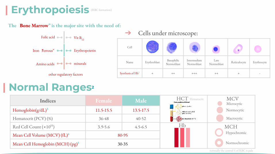
Name Erythroblast BasophilicNormoblast
IntermediateNormoblast
LateNormoblast Reticulocyte Erythrocyte
Synthesis of Hb1 + ++ +++ ++ + -
Cell
Name Erythroblast BasophilicNormoblast
IntermediateNormoblast
LateNormob
lastReticulocyte Erythrocyte
| Erythropoiesis
➔ Cells under microscope:
| Normal Ranges2
Indices Female MaleHemoglobin(g/dL)3 11.5-15.5 13.5-17.5
Hematocrit (PCV) (%) 36-48 40-52
Red Cell Count (×10¹²) 3.9-5.6 4.5-6.5
Mean Cell Volume (MCV) (fL)4 80-95
Mean Cell Hemoglobin (MCH) (pg)5 30-35
HbMacrocytic
Normocytic
MicrocyticMCV
Normochromic
HypochromicMCH
The “Bone Marrow” is the major site with the need of:
Folic acid
Iron “Ferrous”
Amino acids minerals
Erythropoietin
Vit B12
other regulatory factors
HCT
(RBC formation)
normally the central ⅓ of RBC is pale
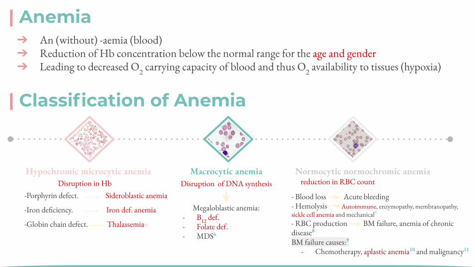
-Porphyrin defect. Sideroblastic anemia
-Iron deficiency. Iron def. anemia
-Globin chain defect. Thalassemia
| Anemia ➔ An (without) -aemia (blood)➔ Reduction of Hb concentration below the normal range for the age and gender➔ Leading to decreased O2 carrying capacity of blood and thus O2 availability to tissues (hypoxia)
| Classification of Anemia
Disruption of DNA synthesis
Megaloblastic anemia:- B12 def.- Folate def. - MDS6
- Blood loss Acute bleeding- Hemolysis Autoimmune, enzymopathy, membranopathy, sickle cell anemia and mechanical7
- RBC production BM failure, anemia of chronic disease8
BM failure causes:9
- Chemotherapy, aplastic anemia10 and malignancy11
Macrocytic anemiaHypochromic microcytic anemia Normocytic normochromic anemiareduction in RBC countDisruption in Hb
protein chain abnormality
poor diet or absorption problems.
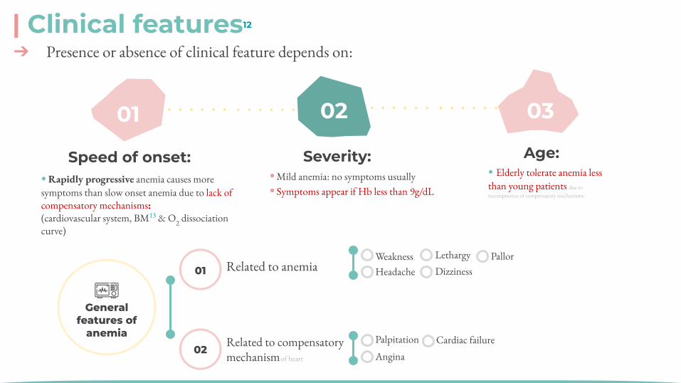
| Clinical features12
Specific Features
General features of
anemia
Related to anemia 01
02Related to compensatory mechanism
Weakness Headache
Pallor Lethargy Dizziness
Palpitation
Angina Cardiac failure
01
02
03
04
Specific signs are associated with particular types of anemia
Spoon nail with iron deficiency
Leg ulcers with sickle cell anemia
Jaundice with hemolytic anemia
Bone deformities in thalassemia major
• Rapidly progressive anemia causes more symptoms than slow onset anemia due to lack of compensatory mechanisms:(cardiovascular system, BM13 & O2 dissociation curve)
➔ Presence or absence of clinical feature depends on:
Speed of onset:
01 02 03
• Mild anemia: no symptoms usually• Symptoms appear if Hb less than 9g/dL
Severity:• Elderly tolerate anemia less than young patients due to incompetence of compensatory mechanisms.
Age:
of heart
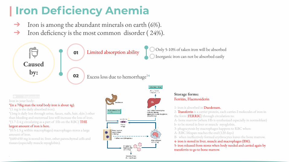
1- iron is absorbed in Duodenum.2- Transferrin is a carrier protein, each carries 2 molecules of iron in the form (FERRIC) through circulation to:A- bone marrow (where Hb is synthesized especially in normoblast)b- to be stored in liver or muscle myoglobin.3- phagocytosis by macrophages happens to RBC when:A- RBC lifespan reaches the end (120 days) B- when ineffectively formed erythrocytes leaves the bone marrow.4- iron is stored in liver, muscle and macrophages (BM).5- iron released from stores when body needed and carried again by transferrin to go to bone marrow.
| Iron Deficiency Anemia ➔ Iron is among the abundant minerals on earth (6%).➔ Iron deficiency is the most common disorder ( 24%).
Storage forms: Ferritin, Haemosiderin
Limited absorption ability
02
01
Excess loss due to hemorrhage14
Only 5-10% of taken iron will be absorbed Inorganic iron can not be absorbed easily
Caused by:
➔ ExplanationIron in your body:*(in a 70kg man the total body iron is about 4g).*(1 mg is the daily absorbed iron).*(1mg is daily lost through urine, faeces, nails, hair, skin ) other than bleeding and menstrual loss will increase the loss of iron.*(1.7-2.4 g circulating as a part of Hb on the RBC) THE largest amount of iron is here.*(0.5-1.5 g within macrophages) macrophages stores a large amount of iron.*(600-650 mg is stored in: liver, other parenchymal cells and tissues (especially muscle myoglobin).
.
➔ PathomaIron is consumed in heme (meat-derived) and non-heme (vegetable-derived) forms.
1. Absorption occurs in the duodenum. Enterocytes have heme & non-heme (DMT1) transporters; the heme form is more readily absorbed
2. Enterocytes transport iron across the cell membrane into blood via ferroportin3. Transferrin transports iron in the blood & delivers it to liver & bone marrow
macrophages for storage 4. Stored intracellular iron is bound to ferritin, which prevents iron from forming
free radicals via the Fenton reaction
1
2
2a
2b
3
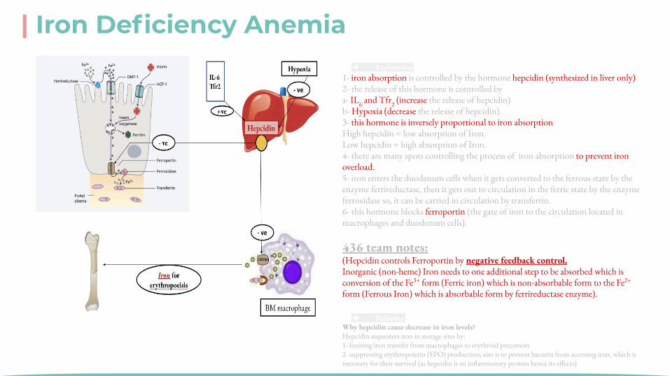
➔ Explanation1- iron absorption is controlled by the hormone hepcidin (synthesized in liver only) 2- the release of this hormone is controlled by a- IL6 and Tfr2 (increase the release of hepcidin)b- Hypoxia (decrease the release of hepcidin).3- this hormone is inversely proportional to iron absorption: High hepcidin = low absorption of Iron.Low hepcidin = high absorption of Iron.4- there are many spots controlling the process of iron absorption to prevent iron overload.5- iron enters the duodenum cells when it gets converted to the ferrous state by the enzyme ferrireductase, then it gets out to circulation in the ferric state by the enzyme ferroxidase so, it can be carried in circulation by transferrin.6- this hormone blocks ferroportin (the gate of iron to the circulation located in macrophages and duodenum cells). 436 team notes: (Hepcidin controls Ferroportin by negative feedback control.Inorganic (non-heme) Iron needs to one additional step to be absorbed which is conversion of the Fe3+ form (Ferric iron) which is non-absorbable form to the Fe2+ form (Ferrous Iron) which is absorbable form by ferrireductase enzyme).
| Iron Deficiency Anemia
➔ Pathoma: Why hepcidin cause decrease in iron levels?Hepcidin sequesters iron in storage sites by:1- limiting iron transfer from macrophages to erythroid precursors 2- suppressing erythropoietin (EPO) production; aim is to prevent bacteria from accessing iron, which is necessary for their survival (as hepcidin is an inflammatory protein hence its effects)
| Iron Absorption
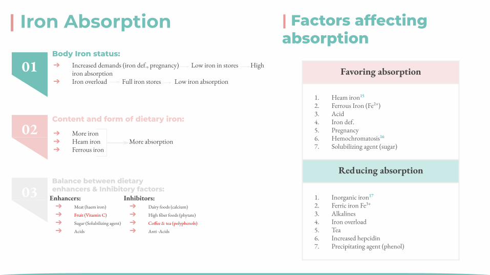
Content and form of dietary iron:Body Iron status:
Balance between dietary enhancers &
Inhibitory factors:
Factors affecting absorption:
➔ Increased demands (iron def., pregnancy) Low iron in stores High iron absorption
➔ Iron overload Full iron stores Low iron absorption
➔ More iron ➔ Heam iron More absorption➔ Ferrous iron
Enhancers Inhibitors
Meat (haem iron) Fruit (Vitamin C)Sugar (Solubilizing agent) Acids
Dairy foods (calcium) High fiber foods (phytate)Coffee & tea (polyphenoles)Anti -Acids
Favoring absorption Reducing absorption
1. Heam iron2. Ferrous Iron (Fe++)3. Acid4. Iron def.5. Pregnancy6. Hemochromatosis7. Solubilizing agent (sugar)
1. Inorganic iron 2. Ferric iron Fe+++3. Alkalines4. Iron overload 5. Tea6. Increased hepcidin7. Precipitating agent (phenol)
02
01
03
Body Iron status:
Content and form of dietary iron:
Balance between dietary enhancers & Inhibitory factors:
Enhancers: ➔ Meat (haem iron)
➔ Fruit (Vitamin C)
➔ Sugar (Solubilizing agent)
➔ Acids
Inhibitors:➔ Dairy foods (calcium)
➔ High fiber foods (phytate)
➔ Coffee & tea (polyphenols)
➔ Anti -Acids
| Factors affecting absorption
Favoring absorption
1. Heam iron15
2. Ferrous Iron (Fe2+)3. Acid4. Iron def.5. Pregnancy6. Hemochromatosis16
7. Solubilizing agent (sugar)
Reducing absorption
1. Inorganic iron17 2. Ferric iron Fe3+
3. Alkalines4. Iron overload 5. Tea6. Increased hepcidin7. Precipitating agent (phenol)
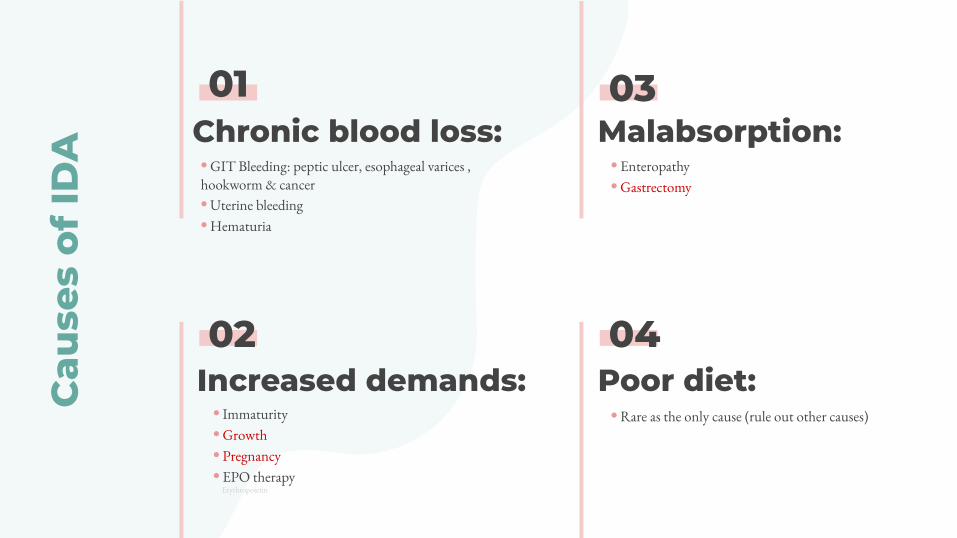
01Chronic blood loss:
02Increased demands:
03Malabsorption:
04Poor diet:C
ause
s of
IDA • GIT Bleeding: peptic ulcer, esophageal varices ,
hookworm & cancer• Uterine bleeding• Hematuria
• Immaturity• Growth• Pregnancy• EPO therapy
• Enteropathy• Gastrectomy
• Rare as the only cause (rule out other causes)
Erythropoietin
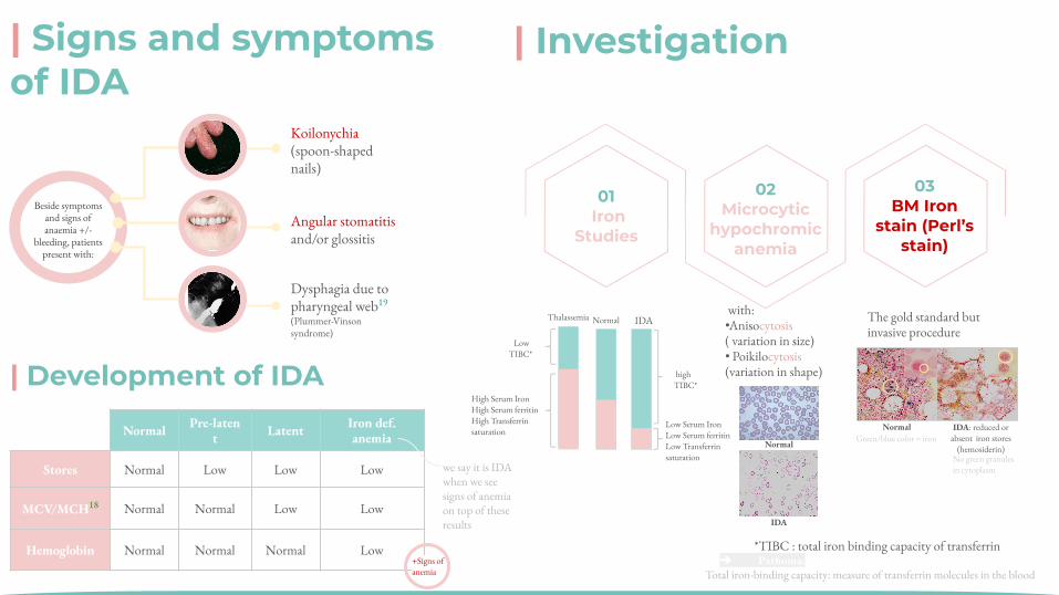
| Signs and symptoms of IDA
01 Iron
Studies
03BM Iron
stain (Perl’s stain)
The gold standard but invasive procedure
02Microcytic
hypochromic anemia
with: •Anisocytosis( variation in size) • Poikilocytosis(variation in shape)
Low TIBC*
High Serum IronHigh Serum ferritinHigh Transferrin saturation
high TIBC*
Thalassemia IDA Normal
IDA: reduced or absent iron stores
(hemosiderin)
Normal
| Investigation
*TIBC : total iron binding capacity of transferrin➔ Pathoma:
Total iron-binding capacity: measure of transferrin molecules in the blood
Beside symptoms and signs of anaemia +/-
bleeding, patients present with:
Koilonychia (spoon-shaped nails)
Angular stomatitis and/or glossitis
Dysphagia due to pharyngeal web19 (Plummer-Vinson syndrome)
| Development of IDA
Normal Pre-latent Latent Iron def.
anemia
Stores Normal Low Low Low
MCV/MCH18 Normal Normal Low Low
Hemoglobin Normal Normal Normal Low
Low Serum IronLow Serum ferritinLow Transferrin saturation
ز:) :(
we say it is IDA when we see signs of anemia on top of these results
+Signs of anemia
Green/blue color = iron
No green granules in cytoplasm
Normal
IDA
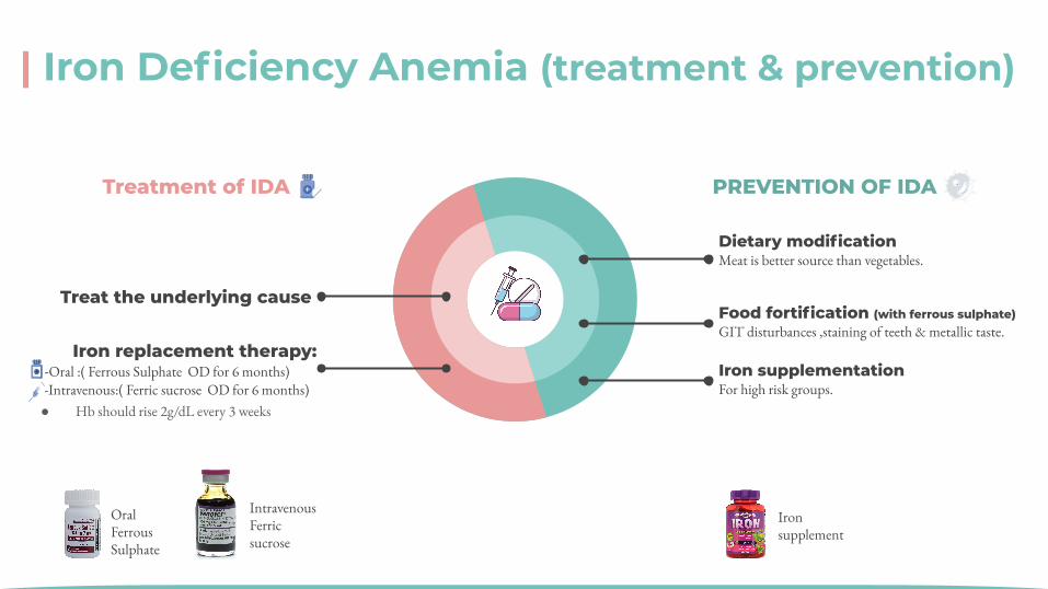
| Iron Deficiency Anemia (treatment & prevention)
Dietary modificationMeat is better source than vegetables.
Iron supplementationFor high risk groups.
Treat the underlying cause
Iron replacement therapy:-Oral :( Ferrous Sulphate OD for 6 months) -Intravenous:( Ferric sucrose OD for 6 months)
Treatment of IDA PREVENTION OF IDA
Food fortification (with ferrous sulphate)GIT disturbances ,staining of teeth & metallic taste.
● Hb should rise 2g/dL every 3 weeks
Oral Ferrous Sulphate
Intravenous Ferric sucrose
Iron supplement
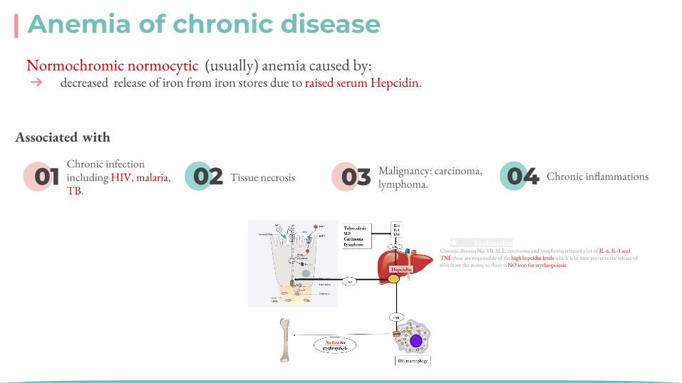
| Anemia of chronic disease Normochromic normocytic (usually) anemia caused by:➔ decreased release of iron from iron stores due to raised serum Hepcidin.
Fe+3 Fe+2
Ferri reductase
BM
-ve -ve
No Iron for erythropoiesis
TbSLE
CarcinomaLymphoma
+ve
IL-6IL-1TNF
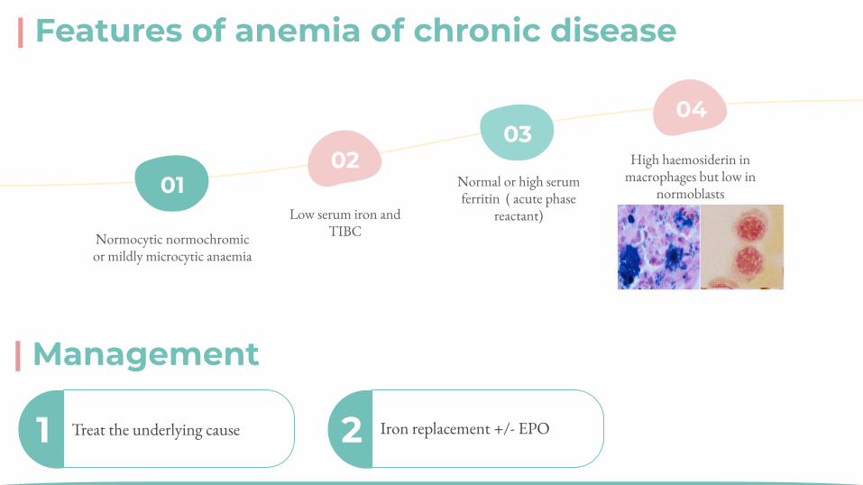
Features of anemia of chronic disease
Normocytic normochromic or mildly microcytic anaemia
Low serum iron and TIBC
Normal or high serum ferritin ( acute phase reactant)
High haemosiderin in macrophages but low in normoblasts
Management:➔ Treat the underlying cause➔ Iron replacement +/- EPO
اه ي ا ر وز
ل ا ا
1
2
3
4
Normocytic normochromic or mildly microcytic anaemia
Normal or high serum ferritin ( acute phase reactant)
Low serum iron and TIBCHigh haemosiderin in macrophages but low in normoblasts
Features of anemia of chronic disease:
01 02 03 04
Associated with
Chronic infection including HIV, malaria, TB.
Associated with
Chronic infection including HIV, malaria
Tissue necrosis
Malignancy
Chronic inflammations
Tissue necrosis Malignancy: carcinoma, lymphoma.
Chronic inflammations
➔ ExplanationChronic diseases like TB, SLE, carcinoma and lymphoma released a lot of IL-6, IL-1 and TNF these are responsible of the high hepcidin levels which is in turn prevents the release of iron from the stores, so there is NO iron for erythropoiesis.
| Features of anemia of chronic disease
Normocytic normochromic or mildly microcytic anaemia
Normal or high serum ferritin ( acute phase
reactant)
High haemosiderin in macrophages but low in
normoblastsLow serum iron and
TIBC
0102
0304
Management:➔ Treat the underlying cause➔ Iron replacement +/- EPO
1
2
3
4
Normocytic normochromic or mildly microcytic anaemia
Normal or high serum ferritin ( acute phase reactant)
Low serum iron and TIBCHigh haemosiderin in macrophages but low in normoblasts
Features of anemia of chronic disease:
1 Treat the underlying cause 2 Iron replacement +/- EPO
| Management
Dr. Notes
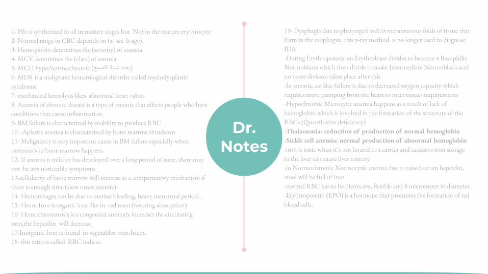
19- Dysphagia due to pharyngeal web is membranous folds of tissue that form in the esophagus. this x-ray method is no longer used to diagnose IDA-During Erythropoiesis, an Erythroblast divides to become 4 Basophilic Normoblasts which then divide to make Intermediate Normoblasts and no more division takes place after this -In anemia, cardiac failure is due to decreased oxygen capacity which requires more pumping from the heart to meet tissues requirements -Hypochromic Microcytic anemia happens as a result of lack of hemoglobin which is involved in the formation of the structure of the RBCs (Quantitative deficiency)-Thalassemia: reduction of production of normal hemoglobin-Sickle cell anemia: normal production of abnormal hemoglobin-iron is toxic when it’s not bound to a carrier and excessive iron storage in the liver can cause liver toxicity -in Normochromic Normocytic anemia due to raised serum hepcidin, stool will be full of iron -normal RBC has to be biconcave, flexible and 8 micrometer in diameter. -Erythropoietin (EPO) is a hormone that promotes the formation of red blood cells.
1- its imp to know that Hb is synthesized in all immature stages but Not in the mature erythrocyte2- in CBC. it depends on 1- sex 2-age.3- determines the (severity )of anemia.4- determine the (class) of anemia 5- hypo/ normochromic (یحدد نسبة التصبغ)6- its a malignant hematological disorder called myelodysplastic. syndrome7- mechanical like abnormal heart valves when the break the RBC8- is a type of anemia that affects people who have conditions that cause inflammation.9- can NOT produce RBC10 - bone marrow shutdown11- especially when metastasis to bone marrow happens12- If your anemia is mild or has developed over a long period of time, you may not notice any symptoms.13- cellularity of bone marrow will increase14- first cause of anemia15- uterine bleeding, heavy menstrual period…16- organic iron like in:red meat17- congenital anomaly increases the circulating iron,the hepcidin will decrease.18- in vegetables: non-haem.19- usually we drink tea after eating red meat which decreases the iron absorption.
-Origin of hematopoietic stem cells is: mesenchymal hemangioblast-There are two types of transcriptional factors, 1-for self renewal 2-for differentiation: which are (Erythropoietin and GATA1)-During Erythropoiesis, an Erythroblast divides to become a 4 Basophilic Normoblasts which then divide to make Intermediate Normoblasts and no more division takes place after this -60% of bone marrow is cellular while the rest is backup space (it becomes more cellular during anemia)-In anemia, cardiac failure is due to decreased oxygen capacity which requires more pumping from the heart to meet tissues requirements -Hypochromic microcytic anemia happens as a result of lack of hemoglobin which is involved in the formation of the structure of the RBCs (Quantitative deficiency)-Thalassemia: reduction of production of normal hemoglobin-Sickle cell anemia: normal production of abnormal hemoglobin-iron is toxic when it’s not bound to a carrier and excessive iron storage in the liver can cause liver toxicity -Ferrous form (Fe2+) is the favorable form for absorption from small intestine-children with undiscovered anemia have less IQ compared to other children -in Normochromic Normocytic anemia due to raised serum hepcidin, stool will be full of iron
1- Hb is synthesized in all immature stages but Not in the mature erythrocyte2- Normal range in CBC depends on (a- sex b-age).3- Hemoglobin determines the (severity) of anemia.4- MCV determines the (class) of anemia 5- MCH hypo/normochromic (یحدد نسبة التصبغ)6- MDS is a malignant hematological disorder called myelodysplastic syndrome7- mechanical hemolysis likes abnormal heart valves8- Anemia of chronic disease is a type of anemia that affects people who have conditions that cause inflammation.9- BM failure is characterized by inability to produce RBC10 - Aplastic anemia is characterized by bone marrow shutdown11- Malignancy is very important cause in BM failure especially when metastasis to bone marrow happens12- If anemia is mild or has developed over a long period of time, there may not be any noticeable symptoms13-cellularity of bone marrow will increase as a compensatory mechanism if there is enough time (slow onset anemia)14- Hemorrhages can be due to uterine bleeding, heavy menstrual period…15- Heam Iron is organic iron like in: red meat (favoring absorption)16- Hemochromatosis is a congenital anomaly increases the circulating iron,the hepcidin will decrease.17-Inorganic Iron is found in vegetables: non-haem.18- this ratio is called RBC indices.
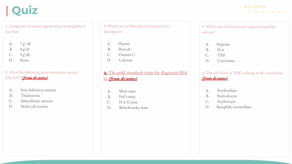
| Quiz 1- Symptoms of anemia appear when hemoglobin is less than:
A. 7 g\ dLB. 8 g\dlC. 9 g\dL D. None
2- All of the following cause microcytic anemia EXCEPT: (from dr.notes)
A. Iron deficiency anemia B. ThalassemiaC. Sideroblastic anemia D. Sickle cell anemia
3- Which one of these factors increase iron absorption?
A. Phytate B. Phenols C. Vitamin C D. Calcium
4- The gold standard stain for diagnosis IDA is: (from dr.notes)
A. Silver stainB. Perl’s stainC. H & E stainD. Bielschowsky stain
Key answers: 1-C 2-D 3-C 4-B 5-A 6-C
5- Which one of these factors suppress hepcidin activity?
A. Hypoxia B. IL-6C. TNFD. Carcinoma
6- The only form of RBCs that go to the circulation: (from dr.notes)
A. ErythroblastB. Reticulocyte C. Erythrocyte D. Basophilic normoblast

● Amirah Alzahrani ● Deema Almaziad ● Jude Alotaibi● Njoud Almutairi ● Nouf Albraikan● Noura Almazroa● Razan Alzohaifi● Rema Almutawa● Renad Alhaqbani● Renad Almutawa● Taif Alotaibi ● Wejdan Alnufaie
THANKS | TEAM LEADERS | Abdulaziz Alghamdi
| Elaf Almusahel
| TEAM MEMBERS
● Abdullah Alghamdi● Hashem Bassam● Mashal Abaalkhail ● Moath Aljehani● Mohammed Alasmari● Mohammed H.Alshehri● Mohammed Alkhamees● Mohammed Alshalan● Naif Alsolais● Saud Bin Queid
= Done by
= Note taker
= Textbooks note taker