Embed Size (px)
Citation preview
International Journal of Surgical PathologyXX(X) 1 –5© The Author(s) 2013Reprints and permission: sagepub.com/journalsPermissions.navDOI: 10.1177/1066896912475082ijsp.sagepub.com
Introduction
Hemangioblastomas are rare benign vascular tumors of the central nervous system (CNS) and are composed of endothelial cells, pericytes, mast cells, and stromal cells. They account for 2.5% of all intracranial tumors and 8% to 12% of all posterior fossa tumors.1-3 In about 80% of patients the tumor is a sporadic, single lesion of the brain-stem, upper cervical spinal cord, or cerebellum.4 The high-est incidence is in young adults, with a slight male predominance. Approximately 20% of patients with intra-cranial hemangioblastomas have Von Hippel–Lindau dis-ease.5 Hemangioblastomas have also been reported outside the CNS, for example, in peripheral nerves,6,7 liver,8,9 lung,10 pancreas,1 retroperitoneum,10 kidney,3,11-13 pan-creas,11 urinary bladder,11 soft tissues of the ankle and popliteal fossa,13,14 and nasal skin.15 We present the first documented case of hemangioblastoma located in the left colon.
Case ReportIn May 2009, a 75-year-old woman undergoing adjuvant chemotherapy for breast cancer (pT2, pN2a M0) at our
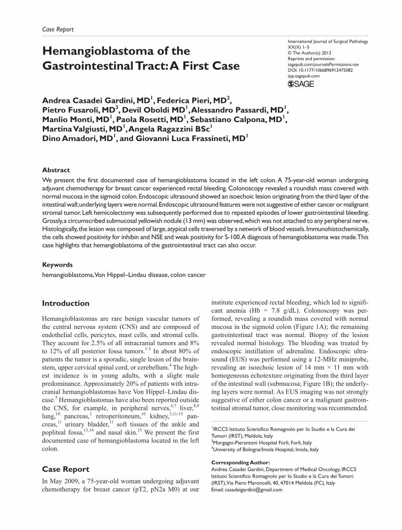
institute experienced rectal bleeding, which led to signifi-cant anemia (Hb = 7.8 g/dL). Colonoscopy was per-formed, revealing a roundish mass covered with normal mucosa in the sigmoid colon (Figure 1A); the remaining gastrointestinal tract was normal. Biopsy of the lesion revealed normal histology. The bleeding was treated by endoscopic instillation of adrenaline. Endoscopic ultra-sound (EUS) was performed using a 12-MHz miniprobe, revealing an isoechoic lesion of 14 mm × 11 mm with homogeneous echotexture originating from the third layer of the intestinal wall (submucosa; Figure 1B); the underly-ing layers were normal. As EUS imaging was not strongly suggestive of either colon cancer or a malignant gastroin-testinal stromal tumor, close monitoring was recommended.
475082 IJSXXX10.1177/1066896912475082International Journal of Surgical PathologyGardini et al
1IRCCS Istituto Scientifico Romagnolo per lo Studio e la Cura dei Tumori (IRST), Meldola, Italy2Morgagni-Pierantoni Hospital Forlì, Forlì, Italy3University of Bologna/Imola Hospital, Imola, Italy
Corresponding Author:Andrea Casadei Gardini, Department of Medical Oncology, IRCCS Istituto Scientifico Romagnolo per lo Studio e la Cura dei Tumori (IRST), Via Piero Maroncelli, 40, 47014 Meldola (FC), Italy Email: [email protected]
Hemangioblastoma of the Gastrointestinal Tract: A First Case
Andrea Casadei Gardini, MD1, Federica Pieri, MD2, Pietro Fusaroli, MD3, Devil Oboldi MD1, Alessandro Passardi, MD1, Manlio Monti, MD1, Paola Rosetti, MD1, Sebastiano Calpona, MD1, Martina Valgiusti, MD1, Angela Ragazzini BSc1 Dino Amadori, MD1, and Giovanni Luca Frassineti, MD1
Abstract
We present the first documented case of hemangioblastoma located in the left colon. A 75-year-old woman undergoing adjuvant chemotherapy for breast cancer experienced rectal bleeding. Colonoscopy revealed a roundish mass covered with normal mucosa in the sigmoid colon. Endoscopic ultrasound showed an isoechoic lesion originating from the third layer of the intestinal wall; underlying layers were normal. Endoscopic ultrasound features were not suggestive of either cancer or malignant stromal tumor. Left hemicolectomy was subsequently performed due to repeated episodes of lower gastrointestinal bleeding. Grossly, a circumscribed submucosal yellowish nodule (13 mm) was observed, which was not attached to any peripheral nerve. Histologically, the lesion was composed of large, atypical cells traversed by a network of blood vessels. Immunohistochemically, the cells showed positivity for inhibin and NSE and weak positivity for S-100. A diagnosis of hemangioblastoma was made. This case highlights that hemangioblastoma of the gastrointestinal tract can also occur.
Keywords
hemangioblastoma, Von Hippel–Lindau disease, colon cancer
Case Report
2 International Journal of Surgical Pathology XX(X)
A B
C D
A
E F
Figure 1. A) Colonoscopy showing a roundish mass covered with normal mucosa in the sigmoid colon. B) Endoscopic ultrasound showing an isoechoic lesion. C) CT scan without contrast showing hypodense lesion. D) After contrast infusion, the lesion appeared hyperdense. E) Vascularization of the inferior mesenteric artery. F) Outflow from the superior mesenteric artery.

Casadei Gardini 3
At the 6-month follow-up, both endoscopy and EUS imaging was unchanged. A computed tomography (CT) scan performed without contrast revealed a hypodense lesion (Figure1C); after contrast infusion the lesion appeared hyperdense (Figure 1D). Vascularization of the inferior mesenteric artery was visible (Figure 1E), as was the outflow from the superior mesenteric artery (Figure 1F). Following repeated episodes of lower gastrointestinal bleeding, the patient underwent left hemicolectomy in October 2010.
Macroscopic FeaturesThe tumor (13 mm × 11 mm) was localized in the submu-cosal layer of the large bowel and was well demarcated from the surrounding fibroadipose tissue. The cut section of the tumor was solid and yellow, with no evidence of necrosis or hemorrhage. The lesion was not associated with any visible nerves.
Microscopic Features
The tumor consisted of sheets of variously sized polygonal, epithelioid cells with eosinophilic cytoplasm and irregular, pleomorphic nuclei (Figure 2A and B). There was a sub-population of tumor cells with bizarre nuclei and micro-vacuolated cytoplasm, indicating the presence of lipids. A very prominent branching thin-walled vascular network was present. A number of vessels were ectasic, showing a hemangiopericytomatous pattern (Figure 2C). Mitotic fig-ures were rare. There was no evidence of necrosis.
Immunohistochemical FindingsSlides for immunohistochemical staining were prepared from formalin-fixed, paraffin-embedded tissue. Staining was carried out using Bond-Max automated immunos-tainer with heat-induced antigen retrieval and a polymer-based immunohistochemical detection system. The
I
A
IHG
C
B
FED
Figure 2. A-C) Microscopic features of the lesion. D) Neoplastic cells strongly positive for inhibin. E) Neoplastic cells strongly positive for neuron-specific enolase. F) Neoplastic cells negative for pan-cytokeratin. G) Neoplastic cells negative for c-kit. H) Neoplastic cells negative for muscle-specific actin. I) Immunostaining for CD34 highlighted an abundance of non neoplastic vessels.

4 International Journal of Surgical Pathology XX(X)
neoplastic cells were strongly and diffusely positive for α-inhibin (Figure 2D) and neuron-specific enolase (Figure 2E). Positivity for S100 protein was weak and multifocal. The tumor was negative for pan-cytokeratin (Figure 2F), c-kit (Figure 2G), DOG1, HMB45, muscle-specific actin (Figure 2H), desmin, chromogranin, and synaptophysin. Immunostaining for CD34 highlighted an abundance of nonneoplastic vessels (Figure 2I). A diagnosis of intestinal hemangioblastoma was made. On the basis of the histo-logical findings, it was decided not administer adjuvant chemotherapy or radiotherapy. Almost 3 years have passed since surgery and the patient is disease-free and in good clinical condition.
DiscussionAs far as we know, ours is the first reported case of hemangioblastoma of the gastrointestinal tract. CT and EUS imaging did not facilitate diagnosis, which ulti-mately was based on the presence of typical morphology and immunophenotype. The recognition of hemangio-blastoma depends, to a large extent, on awareness of the existence of such tumors. However, its occurrence in extraneural sites is so rare that such a hypothesis may not even be included in the differential diagnosis, which takes into account lipogenic tumors, melanoma, meta-static clear cell carcinoma, gastrointestinal stromal tumor, capillary hemangioma, and paraganglioma. Large, multivacuolated cells of hemangioblastoma are similar to lipogenic cells.
Well-differentiated liposarcoma is normally paucivas-cular, in contrast to the hemangioblastoma of our patient, which was not surrounded by areas of adipose tissue. Multivacuolated cells with centrally located vesicular nuclei and a rich capillary network are typical of hiber-noma, but hibernoma cells do not show the pronounced nuclear atypia of our case. However, the strong positivity for α-inhibin16 helped in formulating the differential diag-nosis. We also took the possibility of melanoma into con-sideration as an in situ melanoma had been removed from the patient’s leg in October 2007, but this was rejected as tumor dimensions remained unchanged for almost 2 years and mitoses were rare in the biopsy specimen. Furthermore, HMB45 was completely negative. From a morphological point of view, differential diagnosis of hemangioblastoma includes metastatic carcinoma, in particular renal clear cell carcinoma and adrenocortical carcinoma. Taking into account the patient’s breast cancer history, it was also nec-essary to rule out metastatic lobular carcinoma of the breast. The absence of pancytokeratin, cytokeratin 7, and HMB45 and the expression of S-100 and α-inhibin excluded the hypothesis of carcinoma. In view of the ana-tomical location of the lesion, we also included gastroin-testinal stromal tumor in the differential diagnosis, but
immunohistochemistry for DOG1 and c-kit was com-pletely negative. Like CNS hemangioblastomas, our patient’s tumor was characterized by prominent vascular proliferation, but the presence of peculiar stromal cells led to the exclusion of hemangioma or hemangiopericytoma. The lesion also had a very prominent network of thin-walled branching blood vessels similar to that of paragan-glioma but lacked its typical nested architecture.
Given the rarity of this tumor and the patient’s history of breast cancer and melanoma, genetic counseling was also performed because hemangioblastoma sporadically occurs in association with Von Hippel–Lindau disease. The patient’s professional activity had not exposed her to specific occupational carcinogens. The genealogical tree did not reveal a family history or any other clinical mani-festation of the syndrome. Molecular genetic analysis, a more specific instrument to confirm or exclude Von Hippel–Lindau disease, was offered to the patient, but was refused.
Cerebral hemangioblastomas frequently exhibit overex-pression of erythropoietin, which manifests as an increase in hemoglobin and of hematocrit values. This was not seen in our patient because of the numerous episodes of rectal bleeding. Furthermore, hemangioblastomas are benign but our case showed a marked nuclear atypia, which is unusual for this tumor. It was thus decided to monitor the patient closely with an annual CT scan and biannual clinical fol-low-up with full blood counts. Colonoscopy was performed 1 year after surgery and will be repeated at 3 and 5 years. Given the small dimensions of the tumor and the negative resection margins, it was decided not to administer chemo-therapy or adjuvant radiotherapy.
The identification of hemangioblastoma depends, to a large extent, on both the clinical manifestation of the dis-ease and radiological images. Surgery is performed and diagnosis is confirmed by histological examination. Because of its rarity, hemangioblastoma was not included in the dif-ferential diagnosis of our patient by either the clinicians (oncologist and gastroenterologist) or radiologist involved. Immunohistochemical results (coexpression of α-inhibin, NSE, and S100), together with salient histological features (vacuolated epithelioid cells associated with a prominent vascular component), are other elements required for a cor-rect diagnosis.
In conclusion, our experience shows that hemangioblas-toma can also occur in the gastrointestinal tract, indicating that such a hypothesis should be taken into consideration in the differential diagnosis of similar clinical cases.
Acknowledgment
The authors wish to thank Gráinne Tierney and Ursula Elbling for editing the article. They also thank Prof. Chistopher Fletcher (Professor of Pathology at Harvard Medical School , Boston) for reviewing the histological slides.

Casadei Gardini 5
Declaration of Conflicting InterestsThe author(s) declared no potential conflicts of interest with respect to the research, authorship, and/or publication of this article.
FundingThe author(s) received no financial support for the research, authorship, and/or publication of this article.
References
1. Bird AV, Mendelow H. Lindau’s disease in a South Afri-can family: a report on three further cases. Br J Surg. 1959;47:173-176.
2. Frank TS, Trojanowski JQ, Roberts SA, Brooks JJ. A detailed immunohistochemical analysis of cerebel-lar hemangioblastoma: an undifferentiated mesenchymal tumor. Mod Pathol. 1989;2:638-651.
3. Hardwig P, Robertson DM. von Hippel-Lindau disease: a familial, often lethal, multi-system phakomatosis. Ophthal-mology. 1984;91:263-270.
4. Ho KL. Ultrastructure of cerebellar capillary hemangioblas-toma. I. Weibel-Palade bodies and stromal cell histogenesis. J Neuropathol Exp Neurol. 1984;43:592-608.
5. Gnarra JR, Tory K, Weng Y, et al. Mutations of the VHL tumour suppressor gene in renal carcinoma. Nat Genet. 1994;7:85-90.
6. Brodkey JA, Buchignani JA, O’Brien TF. Hemangio-blastoma of the radial nerve: case report. Neurosurgery. 1995;36:198-200.
7. Giannini C, Scheithauer BW, Hellbusch LC, et al. Periph-eral nerve hemangioblastoma. Mod Pathol. 1998;11: 999-1004.
8. McGrath FP, Gibney RG, Morris DC, Owen DA, Erb SR. Case report: multiple hepatic and pulmonary haemangio-blastomas—a new manifestation of von Hippel-Lindau dis-ease. Clin Radiol. 1002;45:37-39.
9. Rojiani AM, Owen DA, Berry K, et al. Hepatic hemangio-blastoma. An unusual presentation in a patient with von Hippel-Lindau disease. Am J Surg Pathol. 1991;15:81-86.
10. Fanburg-Smith JC, Gyure KA, Michal M, Katz D, Thomp-son LD. Retroperitoneal peripheral hemangioblastoma: a case report and review of the literature. Ann Diagn Pathol. 2000;4:81-87.
11. Christoferson LA, Gustafson MB, Petersen AG. Von Hippel-Lindau’s disease. JAMA. 1961;178:280-282.
12. Moller PM. Another family with von Hippel Lindau dis-ease. Acta Ophthalmol Scand. 1952;30:154-165.
13. Michal M, Vanecek T, Sima R, et al. Primary capillary hemangioblastoma of peripheral soft tissues. Am J Surg Pathol. 2004;28:962-966.
14. Patton KT, Satcher RL Jr, Laskin WB. Capillary hemangio-blastoma of soft tissue: report of a case and review of the literature. Hum Pathol. 2005;36:1135-1139.
15. Boyd AS, Zhang J. Hemangioblastoma arising in the skin. Am J Dermatopathol. 2001;23:482-484.
16. Hoang MP, Amirkhan RH. Inhibin alpha distinguishes hemangioblastoma from clear cell renal cell carcinoma. Am J Surg Pathol. 2003;27:1152-1156.