Embed Size (px)
Citation preview
Helminths: An Introduction to Pathogenic Worms in Man Introduction Table 1 below summarizes Helminths and provides an overview of 18 different worms that create havoc with humans. This table is not by any means inclusive, but is representative of the major parasitic critters that are pathogenic to man.
Helminths
Platyhelminthes Nemathelminthes
Cestoda Trematoda Nematoda
Tapeworms Flukes Round worms
D. latum, Taenia spp., Echinococcus, H. nana
S. japonicum, S. haematobium, S.
mansoni, P. westermani, C. sinensis, F. hepatica
A. lumbricoides, E. vermicularis, N. americanus, A.
duodenale, S. stercoralis, T. spiralis, T. trichiura
Table 1. A summary of Helminths. The classical method of identifying ova and parasites (O&P) is by examining fecal samples microscopically. Those samples may be examined by wet mounts of the fresh sample, wet mounts of the preserved specimen, wet mounts of concentrated specimen or by staining a fecal smear. A wet mount is made by mixing a small amount of the fresh or preserved feces in normal saline. A drop of this suspension is applied to a microscope slide and a cover slip is positioned over the drop. Since this sample is to be examined under oil immersion, the cover slip must be sealed to the microscope slide. A mixture of paraffin and vaseline melted together and "painted" onto the edges of the cover slip serves this purpose. Specimens may be concentrated, i.e., remove the garbage and collect the O&P, by one of two techniques. These techniques are summarized in Table 2.
1
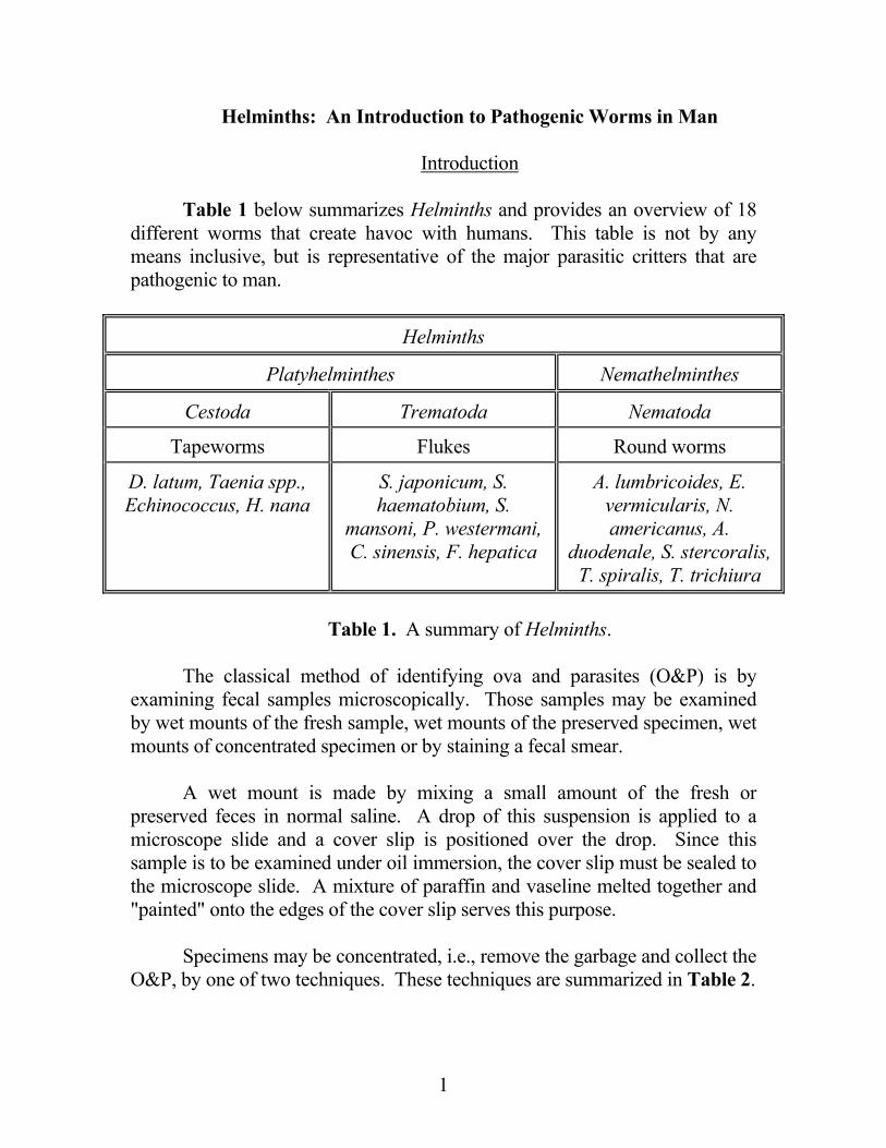
Technique Pros Cons
Formalin-Ether Efficient in recovering most helminth eggs;
moderately effective in recovering schistosome
eggs.
Lose H. nana eggs; decreases the
concentration of G. lamblia cysts; EXPLOSIVE!
O&P settle to bottom after centrifugation
Formalin-ZnSO4 Flotation
Clears up specimen; decreases distortion of
parasites
Unsatisfactory for schistosome ova
O&P float to top of supernatant after centrifugation Table 2. Summary of the two methods of obtaining O&P for microscopic exam. Samples obtained by concentration methods are examined quickly under the microscope. Table 3 summarizes one permanent staining technique for the microscopic visualization of O&P.
Technique Comment
Wheatley Trichrome Stain Destaining increases visualization of some O&P
Trichrome Stain REACTIONS Cytosol of cysts and trophozoites are blue-green with a purplish tinge; the nucleus, RBC and bacteria are red to purple red; yeasts are green and the background of the slides is green
Table 3. Summary of the Wheatley Trichrome O&P Stain method. In any event, the one variable which remains constant is that to visualize O&P, one must know their appearance. Following the NCID’s format of life
2
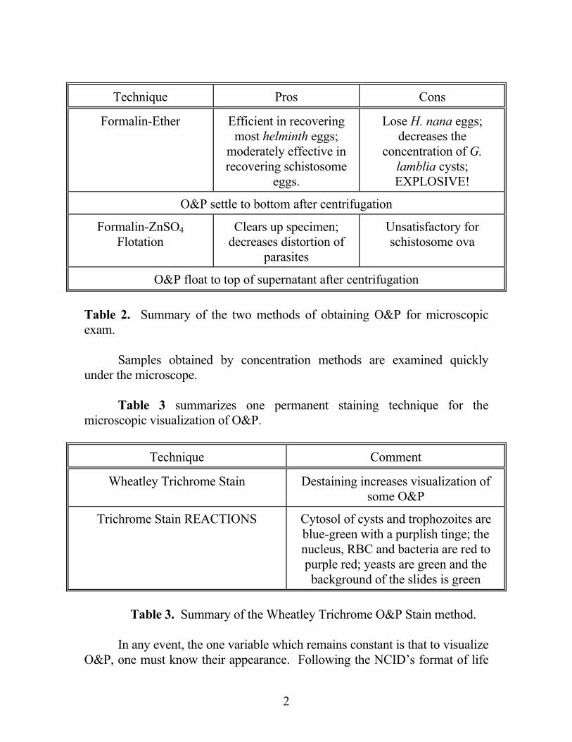
cycle with micrographs, once again, below are two representative cestodes (tape worms):
Diphyllobothrium. latum
http://www.dpd.cdc.gov/dpdx/HTML/ImageLibrary/Diphyllobothriasis_il.htm
3
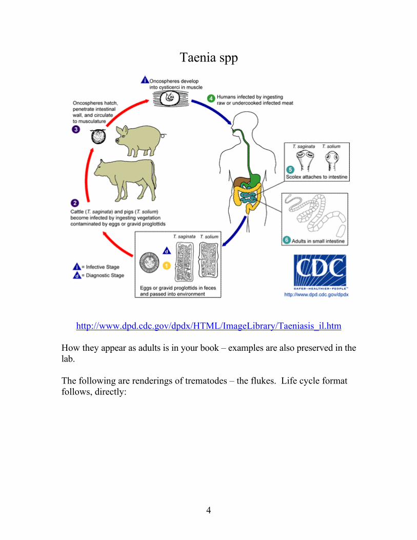
Taenia spp
http://www.dpd.cdc.gov/dpdx/HTML/ImageLibrary/Taeniasis_il.htm
How they appear as adults is in your book – examples are also preserved in the lab. The following are renderings of trematodes – the flukes. Life cycle format follows, directly:
4
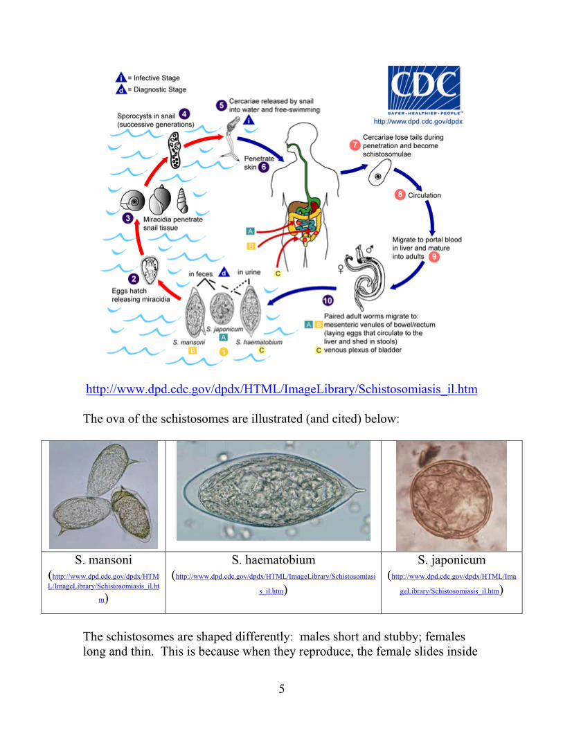
http://www.dpd.cdc.gov/dpdx/HTML/ImageLibrary/Schistosomiasis_il.htm
The ova of the schistosomes are illustrated (and cited) below:
S. mansoni
(http://www.dpd.cdc.gov/dpdx/HTML/ImageLibrary/Schistosomiasis_il.ht
m)
S. haematobium (http://www.dpd.cdc.gov/dpdx/HTML/ImageLibrary/Schistosomiasi
s_il.htm)
S. japonicum (http://www.dpd.cdc.gov/dpdx/HTML/Ima
geLibrary/Schistosomiasis_il.htm)
The schistosomes are shaped differently: males short and stubby; females long and thin. This is because when they reproduce, the female slides inside
5
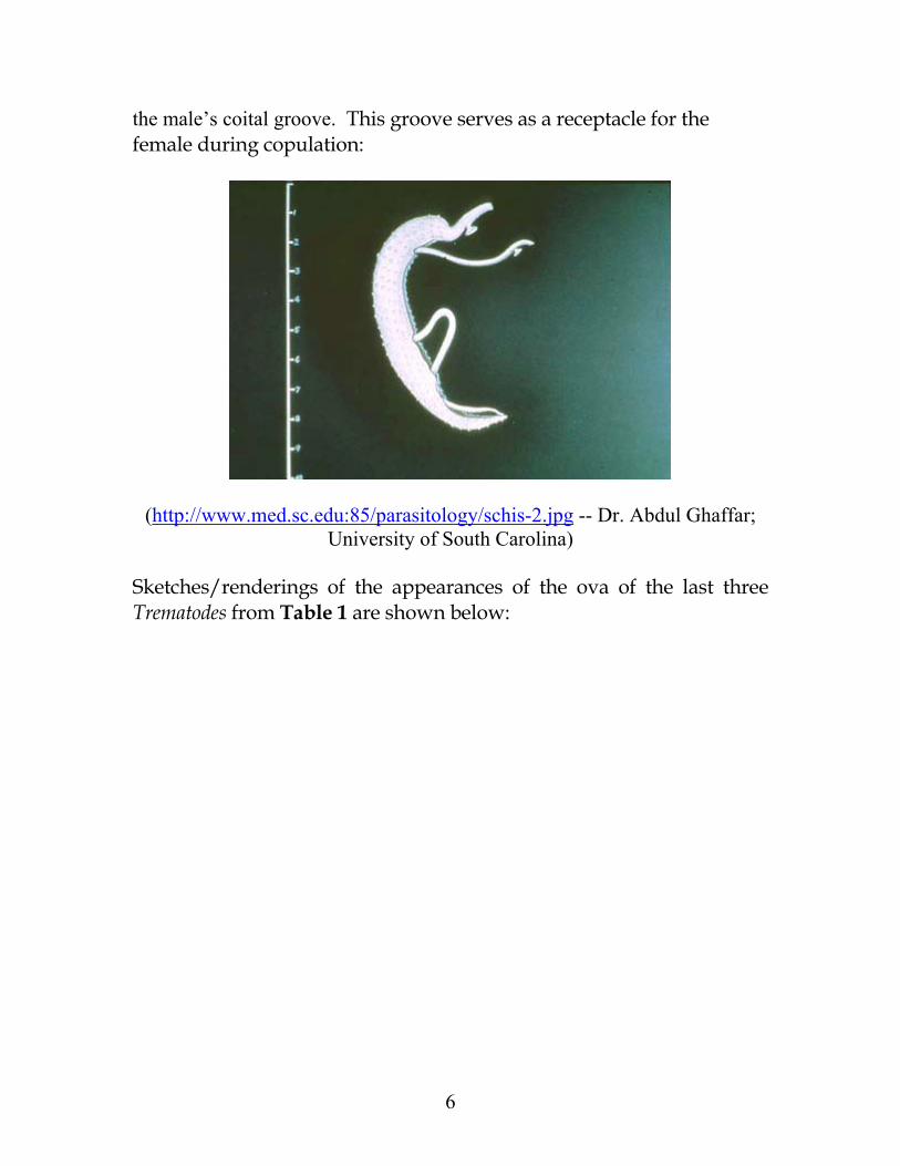
the male’s coital groove. This groove serves as a receptacle for the female during copulation:
(http://www.med.sc.edu:85/parasitology/schis-2.jpg -- Dr. Abdul Ghaffar; University of South Carolina)
Sketches/renderings of the appearances of the ova of the last three Trematodes from Table 1 are shown below:
6
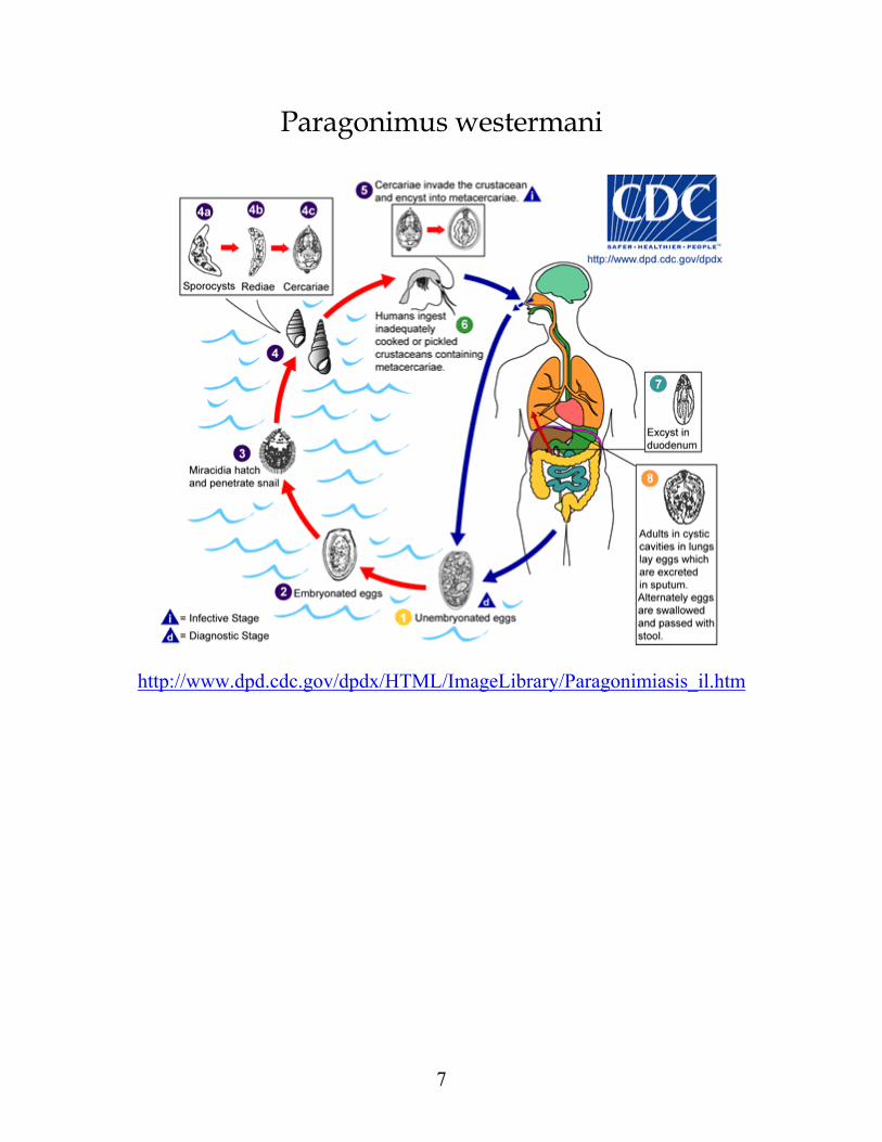
Paragonimus westermani
http://www.dpd.cdc.gov/dpdx/HTML/ImageLibrary/Paragonimiasis_il.htm
7
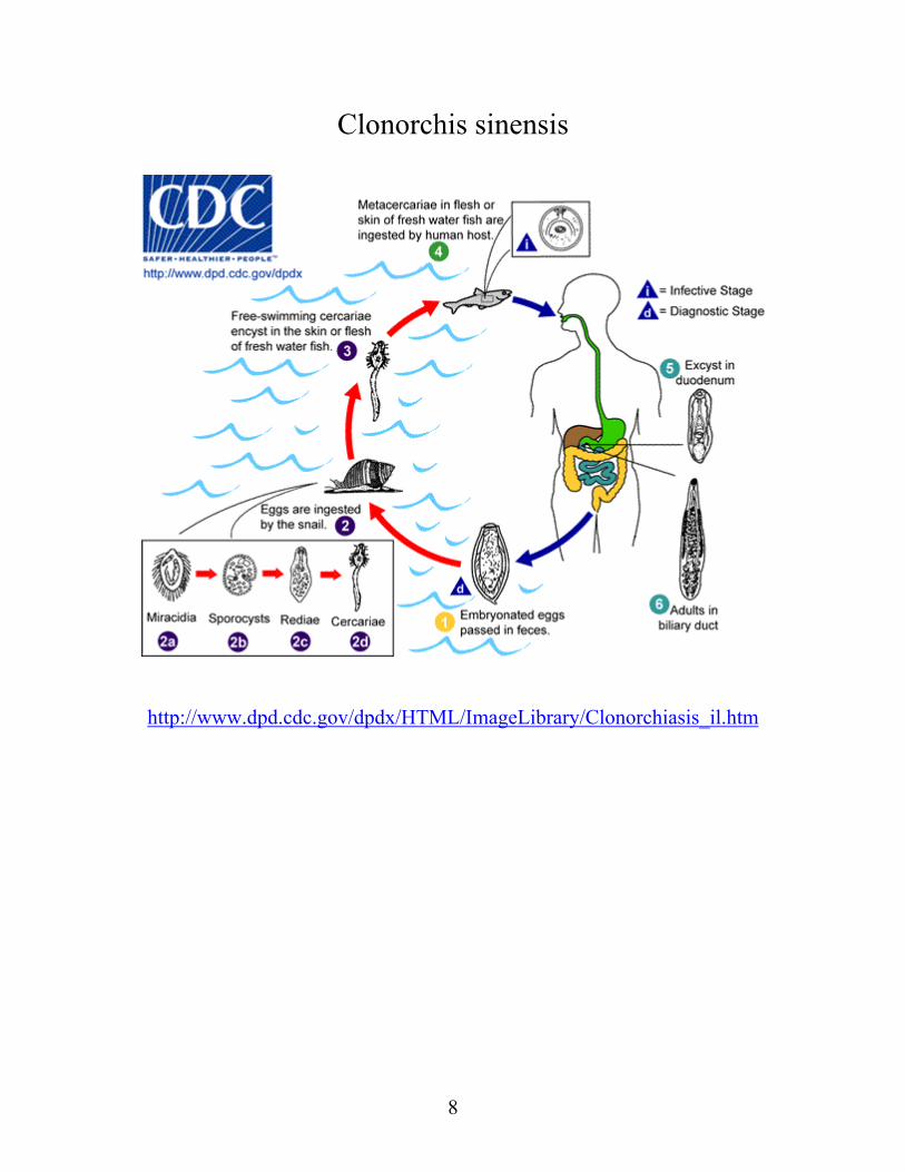
Clonorchis sinensis
http://www.dpd.cdc.gov/dpdx/HTML/ImageLibrary/Clonorchiasis_il.htm
8
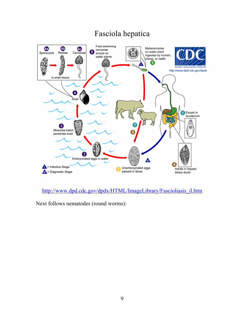
Fasciola hepatica
http://www.dpd.cdc.gov/dpdx/HTML/ImageLibrary/Fascioliasis_il.htm
Next follows nematodes (round worms):
9
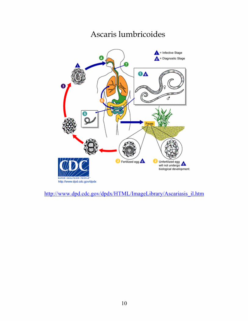
Ascaris lumbricoides
http://www.dpd.cdc.gov/dpdx/HTML/ImageLibrary/Ascariasis_il.htm
10
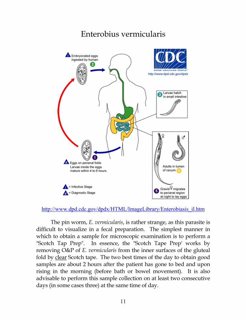
Enterobius vermicularis
http://www.dpd.cdc.gov/dpdx/HTML/ImageLibrary/Enterobiasis_il.htm
The pin worm, E. vermicularis, is rather strange, as this parasite is difficult to visualize in a fecal preparation. The simplest manner in which to obtain a sample for microscopic examination is to perform a "Scotch Tap Prep". In essence, the "Scotch Tape Prep' works by removing O&P of E. vermicularis from the inner surfaces of the gluteal fold by clear Scotch tape. The two best times of the day to obtain good samples are about 2 hours after the patient has gone to bed and upon rising in the morning (before bath or bowel movement). It is also advisable to perform this sample collection on at least two consecutive days (in some cases three) at the same time of day.
11
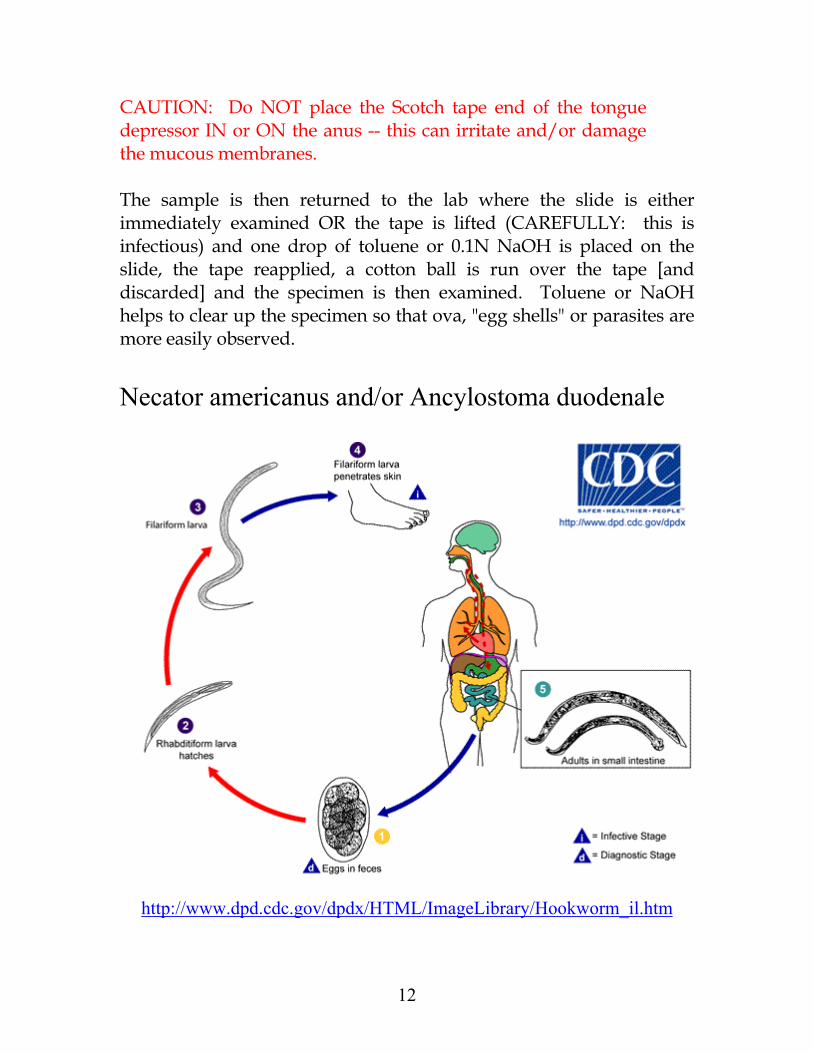
CAUTION: Do NOT place the Scotch tape end of the tongue depressor IN or ON the anus -- this can irritate and/or damage the mucous membranes. The sample is then returned to the lab where the slide is either immediately examined OR the tape is lifted (CAREFULLY: this is infectious) and one drop of toluene or 0.1N NaOH is placed on the slide, the tape reapplied, a cotton ball is run over the tape [and discarded] and the specimen is then examined. Toluene or NaOH helps to clear up the specimen so that ova, "egg shells" or parasites are more easily observed. Necator americanus and/or Ancylostoma duodenale
http://www.dpd.cdc.gov/dpdx/HTML/ImageLibrary/Hookworm_il.htm
12
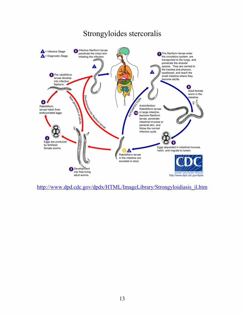
Strongyloides stercoralis
http://www.dpd.cdc.gov/dpdx/HTML/ImageLibrary/Strongyloidiasis_il.htm
13
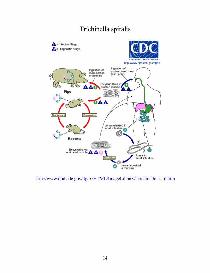
Trichinella spiralis
http://www.dpd.cdc.gov/dpdx/HTML/ImageLibrary/Trichinellosis_il.htm
14
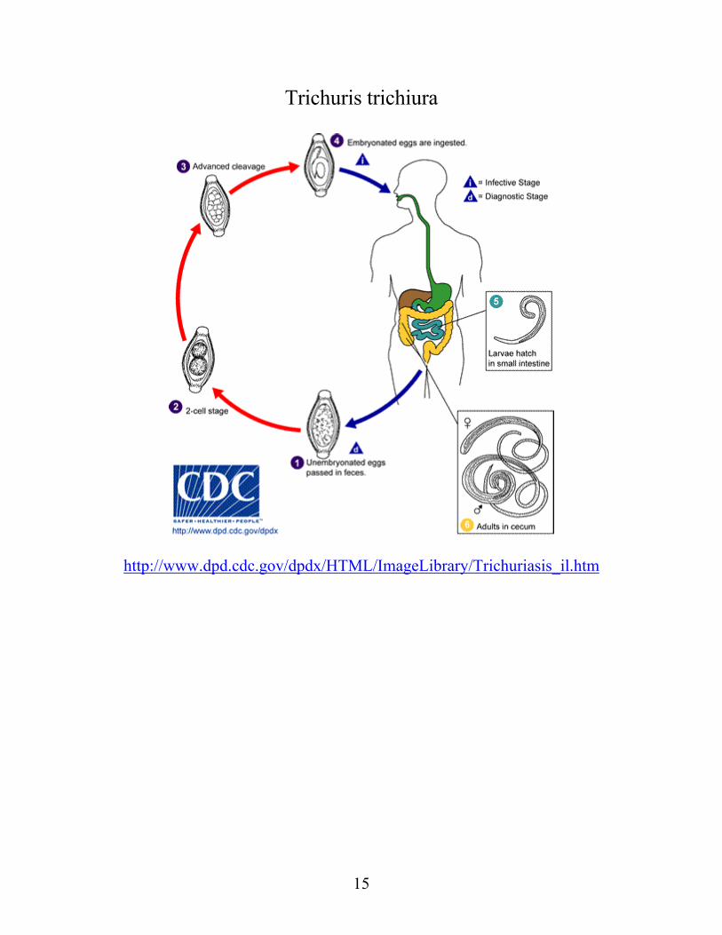
Trichuris trichiura
http://www.dpd.cdc.gov/dpdx/HTML/ImageLibrary/Trichuriasis_il.htm
15
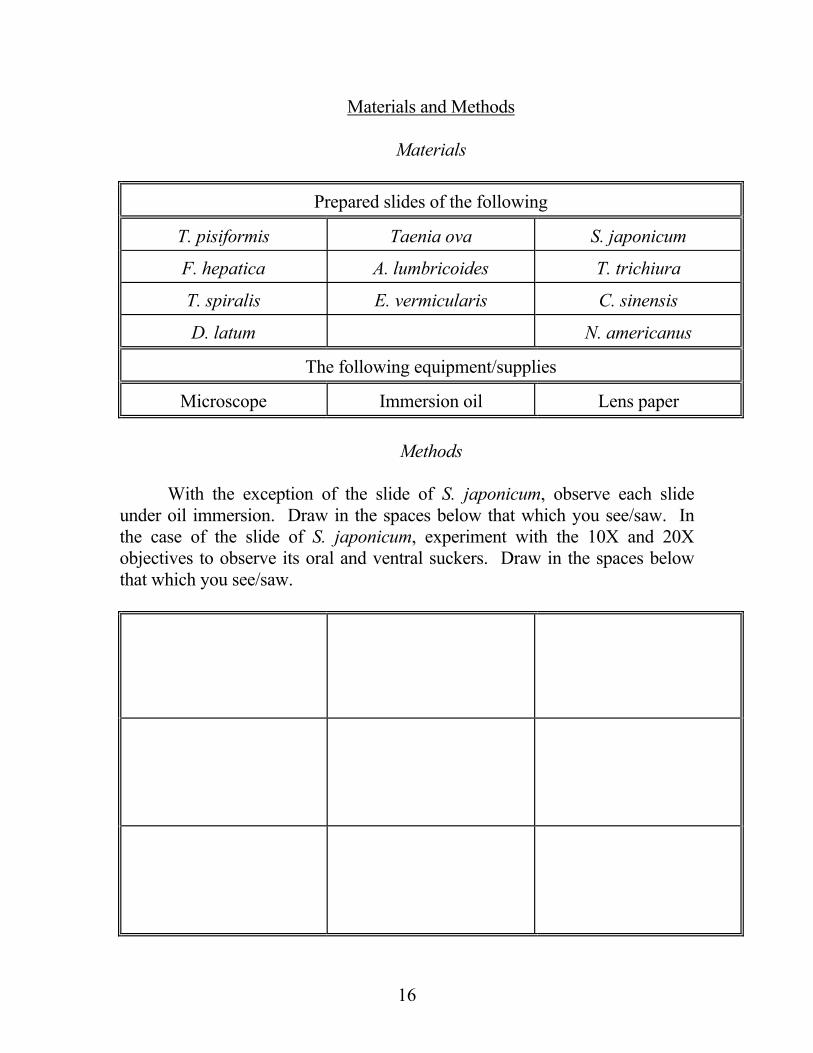
Materials and Methods Materials
Prepared slides of the following
T. pisiformis Taenia ova S. japonicum
F. hepatica A. lumbricoides T. trichiura
T. spiralis E. vermicularis C. sinensis
D. latum N. americanus
The following equipment/supplies
Microscope Immersion oil Lens paper Methods With the exception of the slide of S. japonicum, observe each slide under oil immersion. Draw in the spaces below that which you see/saw. In the case of the slide of S. japonicum, experiment with the 10X and 20X objectives to observe its oral and ventral suckers. Draw in the spaces below that which you see/saw.
16
17
REFERENCES 1.Davidsohn, I. and Wells, B.B.: Todd-Sanford Clinical Diagnosis by
Laboratory Methods, Thirteenth Edition. (W.B. Saunders Co.: Philadelphia) ©1965.
2.Henry, J.B., Ed.: Todd, Sanford and Davidsohn's Clinical
Diagnosis and Management by Laboratory Methods, Sixteenth Edition. (W.B. Saunders Co.: Philadelphia) ©1979.
3.Jawetz, E., Ed.: Medical Microbiology, Eighteenth Edition.
(Appleton and Lange: San Mateo) ©1989. 4.Noble, E.R. and Noble, G.A.: Parasitology: The Biology of Animal
Parasites, Third Edition. (Lea and Febiger: Philadelphia) ©1973.