Embed Size (px)
Citation preview

Cong. Anom., 38: 9-23,1998
Review
Heat Shock Proteins in Normal and Stressed Mammalian Embryonic
Deve I op m en t
David WALSH', Xiao-Ou ZHU', Jane GRANTHAM', Rosanne TAYLOR', Minoru INOUYE3 and Marshall J. EDWARDS' 'School of Anatomy, Department of Medicine, The University of New South Wales,
Sydney 2052, NSW, Australia,
'Animal Sciences University of Sydney 2006, NSW, Australia, and
'Research Institute for Environmental Medicine, University of Nagoya, Nagoya, Aichi,
Japan.
ABSTRACT The heat shock response is associated with a characteristic inhibition of
normal transcription and translation with enhanced synthesis of a multigene family of Heat Shock Proteins (HSP), a common cellular mechanism to all living organisms including the most primitive species. Elevated body temperature or hyperthermia can result from many agents including fever, hot weather and heavy exercise. In more modern surroundings
induced hyperthermia can be achieved by hot baths, saunas, specific drugs, electromagnetic radiation and ultrasound. Hyperthermia during pregnancy has been shown to cause a wide
spectrum of effects in all species studied, including humans. The outcome depends on the
duration and severity of the heat shock experienced by both the mother and the embryo and the stage of embryonic or fetal development. In severe exposures in utero embryonic death
and resorption or abortion are probably the most common outcomes. In less severe exposures major or minor developmental defects can result, with the CNS a major target for its effects. Heat shock can interrupt cell differentiation at critical stages of organogenesis which may lead to apoptosis. All living organisms appear to have developed protective
mechanisms collectively known as the heat shock response. Heat shock triggers the abrupt
suspension in the normal protein synthesis and the concurrent induction of heat shock genes (hsp) and the synthesis of a set of multi-gene protein families known as the heat shock
proteins (HSP). Each hsp has at least two copies, one which appears to function in normal embryonic development (cognate) and another that is stress-induced (inducible) and can
result in acquired thermotolerance, offering some protection against further damage. The
inducible HSP are usually activated at critical inductive stages of organ development, suggesting they play a major role in cell differentiation. The HSPs appear to protect cells through their chaperone functions by binding to adhesive sites on newly synthesised or heat damaged and partially unfolded structural and functional proteins. This prevents the
Received February 23, 1998 All correspondence should be addressed to Dr. D. Walsh.

10 D. Walsh et al.
formation of functionless aggregates. Heat shock transcription factors (HSF) bind to a heat
shock element (HSE) and initiate hsp activation. Although this research has defined some
pathways indicating how and why heat can cause some defects, a means of preventing them
has not yet emerged. Key words: heat shock proteins, hyperthermia, cell cycle, apoptosis, CNS development
Heat Shock Proteins and the Cellular Response Heat shock response
Ritossa (1962) described a “puffing” pattern in the polytene chromosomes of Drosophila pupae which had been exposed to abnormally high incubation temperatures. Subsequent research showed that the gene activity
associated with this reaction initiated the “heat shock response” which offered some protection against thermal
damage to tissues. It is now known that the response can be induced in a very wide range of animals, whether
unicellular or multicellular, embryonic or mature, and vertebrate or invertebrate by exposure to elevated
temperatures. The response has also been identified and studied in plants. It is characterised by the abrupt
suspension of normal protein synthesis soon after exposure to a sufficient dose of heat. This is followed by a rapid induction of synthesis by the heat shock genes (hsp) of a number of families of proteins termed the heat shock proteins (HSP) (Lindquist, 1986; Hightower, 1991). As the response was subsequently found to be elicited by some chemical and toxic agents it is also, perhaps more correctly termed the “stress response”. Its conservation throughout the milennia of evolution from primitive to more complex organisms indicates the fundamental importance of the response to recovery from injury and survival.
Heat shock and the cell cycle Walsh and Morris (1989) studied the expression of stress proteins at various stages of the cell cycle of the
neural plate of cultured E9.5 rat embryos. Flow cytometry was used to determine the levels of hsp27, 71 (inducible), 73 (constitutive) and 90 mRNA. The heat inducible HSP70 was found in unstressed embryos at very low levels only during late S phase and G2+M phases and as the cell passed into the GO phase, it fell to undetectable levels. There was steady expression of hsc73 throughout the cell cycle while hsp90 was expressed strongly at GO-G1 and hsp71 at G2-M phases. After exposure to 42°C for 10 mins, levels of hsp27 and 71
mRNA were elevated at all stages of the cycle, and all hsp in GO-G1 and late S, while hsc73 and hsp 90 were elevated particularly at late S and G2-M. Exposure to the 43°C produced very strong expression of hsp27 throughout the cycle, and both hsc73 and hsp71 were elevated especially during S and G2-M.
It is possible that the different levels of response at these stages reflect different levels of heat damage to the exposed cells. This proposal is reinforced by the lower levels of hsp mRNA found after the combined treatments
of 42°C followed by 43”C, which does not result in defects. Even though the total dose of heat was larger in this group, the levels of all hsp mRNA were less than for either of the single treatments.
Flow cytometry studies (Walsh and Morris, 1989) found that heat caused a partial synchronisation of the generation cycle in neurectodermal cells by causing delays to progression for 1-2 hr at the G1-S boundaries
(Figure 2). There was also a barrier to cell progression at the S-G2 boundary in cells given non-teratogenic exposures (42°C for 10 mins or 42°C folowed by 43°C for 7.5 mins). Squash preparations from the brains of
E21 guinea pig embryos also showed blocks to progression through mitosis before prophase and during
metaphase (Edwards et al., 1974).
Heat shock protein in development 11
Heat shock and apoptosis
Apoptosis is an active form of cell death characterised by condensation of chromatin, which marginates
against the nuclear membrane and later fragments, and shrinkage and condensation of the cellular contents within the intact cell membranes (Alison and Sarraf, 1992). In this form of cell death, the cellular contents remain isolated from surrounding tissues within the membranes which avoids inflammatory reactions. Apoptosis (programmed cell death) is a normal, gene-controlled event in embryogenesis which allows orderly disposal of cells during organogenesis when organ and body parts are being moulded into templates of their mature shapes (Glucksmann, 195 1). It is also the normal means of disposal of supernumerary cells, for example,
cells in the developing brain which fail to make appropriate connections to their intended targets. Another form of apoptosis, which has not been programmed genetically in embryos, is caused by a number of toxic agents, including hyperthermia.
In histological studies of embryos cultured at 40.5"C and 4 1 T , Cockroft and New (1978) showed severe cell death particularly in the CNS. Neuroepithelial cell death was also the most notable feature in the developing CNS of heated E21 guinea pig embryos (Edwards et al., 1974; Wannere et at., 1975; Upfold et al., 1989), as well as in E8 mouse (Shiota, 1988) and El4 rat embryos in vivo (Skreb and Frank, 1963; Harding and Edwards, 1993) and E9.5-10.5 rat embryos in culture (Mirkes, 1985; Walsh et al., 1987, 1991, 1993). Cell death is the most prominent feature underlying the neural defects caused by heat and appears most severe in ectodermal structures. It is usually less severe in mesodermal cells and is infrequently found in endoderm. Mitotic cells of the neuroepithelium of E21 guinea pig embryos are the most sensitive to heat, clumped chromosomes being found immediately after a spike elevation of approximately 2°C. These cells die quickly and are phagocytosed
by adjacent cells. After 4-8 hr, with an elevation of at least 3-3.5"C, S-phase cells and some mesodermal cells die and this form of cell death has been identified as apoptosis. During the period between exposure and the appearance of apoptosis, normal mitotic activity in the neuroepithelium ceases. The onset of apoptosis is accompanied by a synchronised burst of mitotic activity (Edwards et al., 1974; Wanner et al., 1975; Upfold et al., 1989). The damaged M and S phase cells are removed by phagocytosis from macrophages and adjacent normal neuroepithelial cells over the next 24 hr. In E9.5 cultured rat embryos (Walsh et al., 1991, 1993) a
temperature of 42°C (+3.5"C) for 10 mins causes minor rates of cell death in the G2 or mitotic phases of the cell cycle at approximately 5 hr and also 12-1.5 hr after exposure. No developmental abnormalities result from this dose of heat. However, a more severe exposure, known to be teratogenic (43°C for 7.5 mins), causes extensive
apoptosis in the neuroectoderm, particularly in the ridges of the neural folds at 3-5 hr following exposure. The death of S-phase cells in heated embryos is distinct from programmed apoptosis which is a normal
embryological process (Glucksmann, 1951). In normal development, at the time of neurulation and the onset of rapid proliferation of neurones, apoptotic cells are rarely found. It was suggested that once dividing cells are committed to enter a cycle, apoptosis is initiated and can be prevented only when acceptable progression is made through the various phases of the cell cycle (Kung et al., 1990). It is apparent that thermal damage causes severe disruption of the cycle, possibly by denaturation of proteins and this might initiate the process of apoptosis. Following teratogenic doses of heat, large numbers of apoptotic cells are found in areas which are primordia of the malformed organs. Sufficient heat damage to the functional proteins of dividing embryonic cells might well prevent the successful completion of the immediate or the subsequent cycle and allow the apoptotic program to he implemented.

12 D. Walsh et al.
Hyperthermia and Birth Defects Background
Hyperthermia is a condition of elevation of body temperature over the normal levels and heat shock is the response of cells to hyperthermia. Under normal conditions hyperthermia can have many causes including febrile infections, heavy exercise, a hot and humid environment, exposures to sauna baths, hot tubs, ultrasound and various electromagnetic and other radiations, as well as by a number of drugs and organic compounds
(Lomax, 1987). Combinations of these agents and conditions, especially if involving a hot, humid environment,
can accentuate the liability to, and the degree of hyperthermia.
Although the teratogenic effects of hyperthermia on birds have been known since the last century (Dareste,
1877; Alsop, 1919), its possible effects in mammals were not examined systematically before 1960. Before this time, many studies had shown that maternal hyperthermia in many different species of farm and laboratory animals was a potent cause of prenatal death of embryos (Bell, 1987).
It is now accepted that maternal hyperthermia causes disturbances of prenatal development in animals, including humans (Edwards, 1986; Edwards et al., 1995). Significant experimental demonstration of its teratogenic effects has been published for mice (Finnell et al., 1986, 198Sa: Webster and Edwards, 1984), rats in vivo (Skreb and Frank,1963; Edwards, 1967; Lary, 1982; Webster et al., 198.5; Germain et al., 198.5) and in v i m
(Cockroft and New, 1975; Walsh et al., 198.5; Mirkes, 198.5; Kimmel et al., 1993), guinea pigs (Edwards,
1969a; Hutchinson and Bowler, 1984; Upfold et al., 1989), hamsters (Kilham and Ferm, 1976), sheep (Hartley
et al., 1974), pigs (Done et al., 1982) and monkeys (Poswillo et al., 1974; Hendrickx et al., 1979).
Methods of induction and conditions causing hyperthermia Numerous methods have been used experimentally to cause elevations of embryonic and fetal temperatures.
Most methods have involved elevation of the body temperatures of the mother, or of the maternal abdomen, at
various stages of pregnancy and include the following: exposures in hot air chambers or incubators (Edwards 1967, 1969a; Done et a1.,1982; Hartley et al., 1974: Hendrickx et al., 1979), immersion of the lower abdomen in
warm water (Chernoff et a1.,198.5; Shiota 1988; Webster et al., 198.5), immersion of the exterionsed pregnant
uterus of anaesthetised rats in warm water (Skreb and Frank, 1963), electromagnetic exposure (Lary et al., 1982:
Brown-Woodman et al., 1988) and microwave irradiation of the abdomen and pregnant uterus (Fukui et al.,
1992), maternal fever induced by the injection of boiled milk (Brinsmade and Rubsaamen, 1957) or endotoxins (Hellmann, 1977; Hilbelink, 1986) and administration of LSD to the mother (Freedman et al., 1981).
While many different methods have been used to elevate embryonic temperatures, the malformations resulting from these different agents all conform with a generally consistent pattern of defects. Therefore it can reasonably be assumed that the defective development was a result of temperature elevation and not due to
some other effect of the agent used to cause the elevation. Also, as malformations have been produced by elevated temperatures in embryos in culture (Cockroft and New, 1975), and in incubating chickens (Alsop,
1919), it is apparent that the defects are due to the direct effects of heat on the embryo and not caused indirectly through metabolic or toxic changes induced in the mother.
Developmental defects Hyperthermia can cause a wide range of defects, the type depending mainly on the stage of embryonic
development when the exposure occurs. The incidence and severity of defects depend largely on the severity of exposure (dose of heat). A consistent feature of experimental studies with many different species of animals has

Heat shock protein in development 13
been that the central nervous system (CNS) is the most susceptible organ to damage. Defects of the CNS can be severe and bizarre, such as anencephaly, exencephaly, encephalocele, microphthalmia or subtle such as various degrees of micrencephaly (Edwards, 1969b) associated with learning deficits (Lyle et al., 1977). The most sensitive period for neural tube defects (NTD) and microphthalmia is at the neural plate stage of development, just prior to and during the early stages of tube closure, E8 in mice (Shiota, 1988), E9.5 in rats (Webster et al., 1985; Walsh et al., 1987) and E13-14 in guinea pigs (Harding and Edwards, 1993). The most sensitive stage for
micrencephaly is just prior to and during formation of the cortical plate (E13-14 in rats and E21 in guinea pigs) and precedes the intense phase of neuroepithelial proliferation which produces the neurones of the cerebral cortex. Another stage of sensitivity for micrencephaly in guinea pigs is during glial cell proliferation (days E39-46). The deficits in brain growth caused during neurogenesis and glial cell proliferation are permanent and produce severe learning problems (Edwards et al., 1976; Lyle et al., 1977). Hyperthermia during the phase of intense myelin production (days E53-60) is less severe, catching up during postnatal growth but does not result in learning deficits. Other CNS defects in guinea pigs include neurogenic clubfoot (after exposure about days E20-23), arthrogryposis (after exposures between days E3042) and cranial nerve defects. Most of the defects of the CNS in experimental animals are at times accompanied by other abnormalities such as coloboma, facial clefting, maxillary hypoplasia, hypoplasias of teeth and toes, vertebral and tail abnormalities, exomphalos and
renal agenesis (Edwards et al., 1995). Most instances of defects in humans mirror the patterns in experimental animals in the range of abnormalities
and relative timing of the exposure to hyperthermia. It is notable that abortion is a common sequel to a febrile episode (Kline et al., 1985) and it is probable that this is the outcome in most instances of maternal heat exposure (Graham and Edwards, 1988). Hyperthermia is associated with an increase in the plasma levels of prostaglandins in the ewe and fetal lamb (Andrianakis et al., 1989) and is accompanied by increased uterine activity and fetal distress (Morishima et al., 1975). There are many reports of retrospective and prospective studies on the occurrence of NTD after maternal exposures to a range of conditions leading to elevations of temperature (Layde et al., 1980; Shiota, 1982; Hunter, 1984; Milunsky et al., 1992) and it has been estimated that maternal hyperthermia at the time of neural tube closure is the cause of about 10% of cases of NTD
(Graham and Edwards, 1988). Other defects include microphthalmia (Spraggett and Fraser, 1982), neuronal heterotopias, microcephaly, micrognathia, mid-face hypoplasia, clefts of the lip and palate, hypotonias, neurogenic contractures, seizures, mental deficiency (Graham and Edwards, 1988) Moebius syndrome (Graham et al., 1988) and Hirschsprung disease (Lipson, 1988). Recently data concerning the association between elevated maternal body temperature and abnormal fetal development has been recognised from a prospective study involving the California Teratogen Information Service (CTIS). Women with high fevers during the first 4 weeks of post-conception of above 102°F demonstrated an increase in major and minor malformations (Chambers et al., 1997). From the minor group the results suggested the effects on facial and brain morphogenesis leading to possible gross motor delay and learning defects (Mattson et al., 1997).
Thresholds of hyperthermia and malformations Experimental data from a number of species has shown that in simple, uncomplicated cases of hyperthermia,
defects can be caused by elevations over the normal body temperature of 2°C and above. However, the teratogenic threshold dose of heat has been shown to be a product of the temperature elevation and the duration of elevation. Recorded instances of defects being caused at elevations lower than 2°C even for extended periods of time are uncommon, although embryonic resorption and abortions might be associated with these lower
14 D. Walsh et a1
elevations. In E21 guinea pig embryos, with a normal maternal body temperature of 39.5"C, a slow elevation of
2°C to 41.5"C over a period of 60 mins causes an increase in the incidence of mirencephaly. Similarly in E9.5 rat embryos, an elevation of 2.5"C for 60 mins (over the normal temperature of 38.5"C to 41°C) or a brief
"spike" pattern of elevation of 4.5"C results in arrange of defects of the head (Germain et al., 1985). With E9.5 rat embryos in culture at 40°C or 41°C for 43 hr (an elevation of 2°C and 3°C over the control incubation
temperature of 38°C) a similar pattern of neural tube and facial defects was found (Cockroft and New, 1975). Also in El0 rat embryos, a slow elevation over a period of about 3 0 4 0 mins to 39.240.4"C (an elevation over
temperatures of control animals of approximately 2-2.5"C), caused a spectrum of skeletal malformations
(Kimmel et al., 1993). A systematic study of the interactions between temperature elevation and duration of elevation, showed that as the temperature elevation becomes higher, the time required to cause malformation is
reduced logarithmically (Germain et al., 1985). One conclusion which might be drawn from these studies is that
i t appears more appropriate to relate critical thresholds to an elevation above normal level for a species or
genotype, rather than to a single, actual temperature which applies to all species.
Although it is relatively simple to record the duration of temperature elevations in experimental animals, it is
not a simple matter in the human clinical situation in which most elevations would be in the lower ranges. In
fact, recording of temperatures in humans appears to be unusual (Edwards et al., 1995). All experimental
evidence to date indicates that maternal elevations less than 2°C even for extended periods appear not to be
associated with embryonic damage leading to birth defects, and for this reason, to quote a simple threshold
maternal temperature of 2°C without a duration appears to be justified.
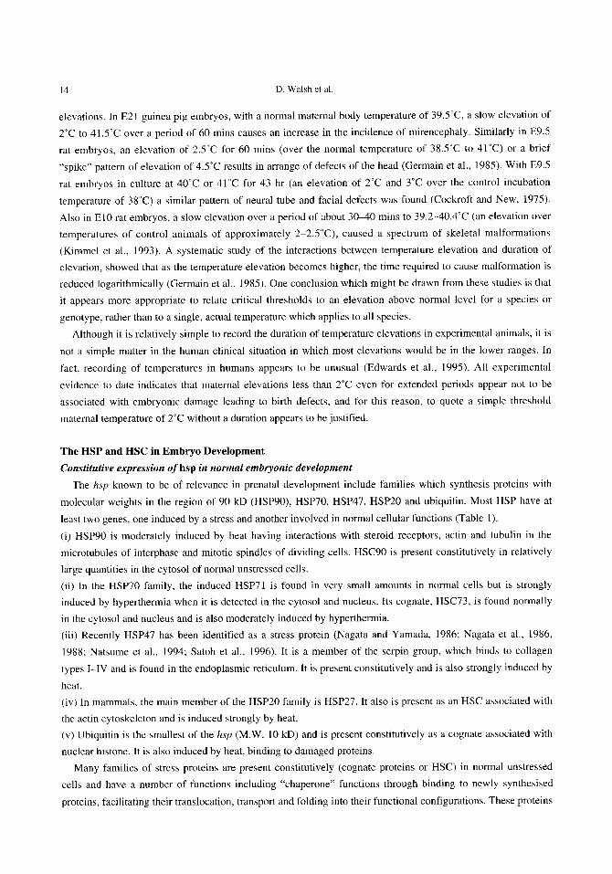
The HSP and HSC in Embryo Development Constitutive expression of hsp in normal embryonic development
The hsp known to be of relevance in prenatal development include families which synthesis proteins with
molecular weights in the region of 90 kD (HSP90), HSP70, HSP47, HSP20 and ubiquitin. Most HSP have at
least two genes, one induced by a stress and another involved in normal cellular functions (Table 1).
(i) HSP90 is moderately induced by heat having interactions with steroid receptors, actin and tubulin in the
microtubules of interphase and mitotic spindles of dividing cells. HSC90 is present constitutively in relatively
large quantities in the cytosol of normal unstressed cells.
(ii) In the HSP70 family, the induced HSP71 is found in very small amounts in normal cells but is strongly induced by hyperthermia when it is detected in the cytosol and nucleus. Its cognate, HSC73, is found normally in the cytosol and nucleus and is also moderately induced by hyperthermia.
(iii) Recently HSP47 has been identified as a stress protein (Nagata and Yamada, 1986; Nagata et al., 1986, 1988; Natsume et al., 1994; Satoh et al., 1996). It is a member of the serpin group, which binds to collagen
types I-IV and is found in the endoplasmic reticulum. It is present constitutively and is also strongly induced by
heat.
(iv) In mammals, the main member of the HSP20 family is HSP27. It also is present as an HSC associated with
the actin cytoskeleton and is induced strongly by heat. (v) Ubiquitin is the smallest of the hsp (M.W. 10 kD) and is present constitutively as a cognate associated with nuclear histone. It is also induced by heat, binding to damaged proteins.
Many families of stress proteins are present constitutively (cognate proteins or HSC) in normal unstressed
cells and have a number of functions including "chaperone" functions through binding to newly synthesised
proteins, facilitating their translocation, transport and folding into their functional configurations. These proteins
Heat shock protein in development 15
Table 1 Heat Shock Proteins in early mammalian embryos HSP Location Function
37°C 42°C hspl l0 nucleosomes nucleosomes hsp 100 lysosome lysosome hsp90 cytoplasm cytoplasdnucleus bind to actin
filaments bind to steroid receptors
g v 7 8 ER lumen ER lumen
g v 7 5 mitochondria mitochondria matrix
hsp73 cytoplasm cytoplasm & nucleus
hsp7 1 cytoplasm nucleus
hsp60 mitochondria mitochondria
hsp47 endoplasmic lysosomes reticulum
hsp27 cytoplasm cytoplasm
attach to nascent proteins glucose deprivat- ion response
uncoating clatharin cages
molecular chaperones protein folding & transport
ATP binding bind nasent proteins bind to HSF
intermembrane protein transport
collagen binding & transformation, mRNA splicing
actin cytoskeleton regulators phosphory lation
Ubiquitin cytoplasm cytoplasm protein degradation
were named chaperones because they protect newly synthesised proteins against inappropriate binding which forms functionless aggregates. Newly heat-induced HSP and some HSC bind to uncovered, adhesive sites on thermally sensitive proteins in the cytoplasm. Others translocate to the nucleus binding to, and protecting chromatin protein complexes (Lindquist, 1986; Hightower, 199 1 ; Schlesinger, 1990). The families with
chaperone functions include HSP90, HSP70, HSP47 and the small HSP. The heat inducible HSP and HSC appear to assist in reconstitution of heat damaged proteins after binding to them by an orderly disengagement to
allow the resumption of the correct functional structure of the rescued protein.
The role of HSP in normal embryonic development As a number of the HSP families are found in normally developing embryos, it can reasonably be assumed
that they perform chaperone roles in normal embryogenesis. The HSC90 family has been detected in the E9.5
16 D. Walsh et al.
rat neuroectoderm and is highly expressed at the GO phase (stationary, resting phase) of the cell cycle, with a
possible function of maintaining cells at this phase until a signal initiates their entry into the active phase of the
generation cycle (Walsh et al., 1993). HSC70 is expressed constitutively in pre-implantation mouse embryos, (Mezger et al., 1991) where it has
been identified as early as the 2 cell stage. It is also found at high levels at the 8 cell stage and during subsequent development where this level is maintained. It is also normally present in rat embryos, being at high levels in the cytosol and nucleus during differentiation of the neural plate, neural tube closure and organogenesis (Walsh et al., 1989).
The functions of HSC47 have been studied recently using E7.5-11 .5 rat embryos in culture (Walsh et al., 1997). In E7.5 embryos it was found only in the endoderm cells. At E8.5, it was present in ectoderm, mesoderm and endoderm of the embryo plus the allantois and yolk sac. During neural tube formation at E9.5 it was present
in cells of the neuroectoderm and mesoderm and its synthesis and location was coordinated closely with the production and location of collagen IV, including the basement membrane and extracellular matrix. However, collagen IV was not identified in the neuroepithelium of E9.5 embryos and did not appear until after the commencement of neural tube closure. At later stages, it was confined to more regionally specific areas of the brain, heart, pharyngeal arches and somites and again its location coincided with the patterns of distribution of collagens I and IV.
In vitro experimental studies Walsh et a]. (1985; 1987; 1989; 1993) studied the expression of HSC and HSP using E9.5 rat embryos in
culture. The embryo culture was carried out using a modified method of New et al. (1973) and involved cuture from day E8.5 to E11.5 in a cabinet at 38.5"C, the normal temperature of rats (Germain et al., 1985). After culture for 2.5 hr at 38.5"C, E9.5 embryos were allotted randomly to one of four groups: (i) a control group which were maintained at 38.5"C until day El 1.5; (ii) a group heated to 42°C for 10 mins and then returned to 38.5"C until day El 1.5; (iii) a group exposed to 43°C for 7.5 mins then cultured returned to 38.5"C until day El 1.5 and (iv) a group exposed to 42°C for 10 mins, returned to 38.5"C for 1 hr and exposed to 43°C for 7.5 mins and then returned to 38.5"C until day El 1.5.
In these conditions HSP90, HSC73 and HSP23-27 were demonstrated in normally developing embryos but HSP7 I was not detected. At 1 .5 hr after the 43°C exposure a reduction in total protein synthesis was observed and at 2-3 hr there was a large increase in the synthesis of HSP71, 73 and 90, which returned to normal by 6-8 hr (Walsh et a]., 1989; 1991; 1993; 1994).
The heat shock response induces changes in the levels of hsp27,71 and 90 mRNA. Northern analysis did not detect 71 kD mRNA in control embryos but it was present in small quantities at 5-10 mins after the 42°C exposure and within 2 mins after 43"C, reaching a peak level at 90, mins with a decline to normal levels in 5 hr. The levels in embryos exposed to 42/43"C were much less than 43°C group. HSC73 was present in all samples. Small amounts were found in embryos after exposrue to 43°C and 42/43"C. HSP27 was induced rapidly and after 42°C it rose to a moderate level between 0.5-3 hr and then declined to normal levels by 6 hr. Much higher expression followed exposre to 43°C and the 42/43"C exposrue resulted in levels lower than both the 42°C and 43°C responses. The response by hsp90 mRNA following 42f43"C was higher than that for 43°C which was higher than those for 42°C. This indicates that the hsp90 mRNA response is proportional for the total dose of heat.
In situ hybridisation studies using hsp cDNA probes showed constitutive expression of hsp71 only in the

Heat shock protein in development 17
allantois and ectoplacental cone of control E9.5 embryos. After an exposure to 43"C, maximum hsp71
expression at 90-120 mins was found in the neuroectoderm of the neural plate, its underlying mesoderm and at
low levels in the endoderm. Expression was most marked around the anterior neuropore. In 10.5 and 11.5 day embryos given 43°C the overall hsp71 expression was reduced being greatest around the mid- and hindbrain areas.
Mirkes (9185, 1987) used four treatment groups of day 10 (6-10 somite) rat embryos. Controls were cultured throughout at 37°C. A second group was at 37°C throughout except for 30 mins at 42T , given 1 hr after the commencement of the culture. A third group was exposed to 37°C throughout, with 43°C for 30 mins given at
2.5 hr, while a fourth group received exposures of 42°C and 43°C as above, separated by 1 hr at 37°C. At examination on day 11, the 42°C group did not differ from controls, the 43°C embryos had a high mortality and level of defects, while the 42/43"C embryos had partial protection against mortality and malformation. Within 1
hr, the 43°C treatment induced the synthesis of eight proteins of 28, 31.5, 33.5, 34, 39, 69, 78 and 82 kD. Two proteins (33.5 and 34 kD) were also detected in control embryos. Synthesis of all induced proteins ceased 3 to 9
hr after exposure except 28 kD which ceased at 1-3 hr. Usually, normal protein synthesis is curtailed during the heat shock response but in these experiments, the 43°C embryos did not show this response. After the 42°C exposure, synthesis of 31.5, 39 and 69 kD proteins was induced and the combined treatment induced all proteins except for the 28 kD at about half the level of the 43°C treatment.
Fisher et al. (1995) also induced heat shock in day 10 rat embryos in vivo and in vitro to examine the role of the heat shock response in the genesis of defects of somite segmentation which has been shown to result in
vertebral and rib anomalies of the mid thoracic region (Cuff et a1.,1993). After exposures in vivo to 42-42.5"C
for 5 mins or in vitro to 4242.5"C for 15 mins, they found enhanced synthesis of a 70 kD protein for 1 to 4 hr and of a 90 kd protein for 1 to 8 hr. The 70 kD protein was identified by Western blot as an inducible HSP72 which accumulated and remained in the neuroectoderm for 2-27 hr after exposure. It was not detected in the somite mesoderm. There was a lag period of 18 hr between accumulation of HSP72 and the appearance of abnormal segmentation. The severity of damage to somites was related to the dose of heat. The absence of heat shock response in the somite mesoderm was believed to explain its sensitivity to heat.
Mirkes and Dogget (1991) compared the response in days 9 (pre-somite), 10 (6-10 somite), 1 1 (21-25 somite) and 12 (31-35 somite) rat embryos which after equilibration for 1 hr at 37°C were given 43°C for 15-60 mins and then returned to 37°C for 1 hr before processing. Northern blot analysis of hsp70 mRNA showed a response which varied with the stage of development and the dose of heat. Day 9 embryos showed the greatest response which decreased during days 10 to 12, especially with the 60 mins dose. Agents other than heat can elicit a stress response.
HSP47 expression in E7.5 rat embryos, studied by immunohistochemical techniques was found to be confined to parietal endoderm cells (Walsh et el., 1997). At 8.5 days it was found in ectoderm, mesoderm and endoderm but on day 9.5 it was widespread throughout the ectoderm, mesoderm and endoderm plus the allantois and yolk sac and later it was found in more specific areas including the brain, pharyngeal arches, heart and somites. Except for ectodermal tissues, its distribution coincided with the expression of collagens I and IV except in ectodermal tissues in which they were not identified before neural tube closure.
HSP and Signal Transduction Pathways Extracellular events causing changes in the immediate cellular environment initiate responses which act
through a signal induction pathway involving mitogen activated proteins (MAP) and stress activated proteins
18 D. Walsh et al.
(SAP). As the signals are being transmitted to the nucleus, they are amplified by kinase cascades in multiple
biochemical pathways through which specific programs of gene activity are induced. The activated MAP and SAP kinases regulate physiological processes by phosphorylation of proteins (Finnin et al., 1997). Normal embryonic development depends on rigid control of cellular proliferation and differentiation. MAP and SAP kinases are regulated tightly by phosphorylation (Clarke. 1994) and they regulate other proteins such as HSP, transcription factors, other protein kinases and protein phosphorylases, growth factor receptors and cytoskeletal
proteins, by phosphorylation. In organisms as diverse as yeast and mammals, MAP and SAP kinases are the targets of a series of three-
tiered kinase cascades. The first group of kinases to be activated are the MAP/SAP kinase kinase kinases
including the serinelthreonine kinases, MEKKl and Raf-1 (a product of the c-ruf proto-oncogene). They then phosphorylate MAPlSAP kinase kinases (MEK, including MEKl, SEKl and RKK) which in turn phosphorylate MAP and SAP kinases. MEK activates both MAP and SAP kinases by phosphorylating the threonine and tyrosine residues in specific motifs (Davis, 1994). The best understood example of activation of MAP/SAP kinase involves the phosphorylation of the p44 and p42 MAP kinases (extracellular-signal regulated kinases, ERK-I and ERK-2). ERK-I and ERK-2 were first identified as the kinases which phosphorylate ribosomal
subunit kinase as a response growth factor stimulation. They were then shown to respond to a range of stimuli which engage receptor tyrosine kinases, non-tyrosine kinase receptors and G-protein coupled receptors (Davis.
1994; Marshall, 1994; Waskiewicz and Cooper, 1995). When a growth factor binds to its receptor, it induces
receptor dimerisation and phosphorylation, leading to recruitment of intracellular adaptor proteins and
GDP/GTP exchange factors such as Grb-2, SOS and Ras to the receptor complex. The SAP kinases are a distinct sub-group of the MAP-like kinases. Two jun (JNK) isoforms, p46 JNKl and
p54 JNK2 were originally identified as kinases that phosphorylate the activation domain of the transcription factor c-jun (Davis, 1994). JNKs are activated by environmental stress, UV irradiation and stimulation with pro- inflammatory cytokines by phosphorylation of threonine and tyrosine in a T-P-Y motif. This activation cascade
is similar to, but distinct from the cascade that activated ERKs. Dual phosphorylation of another SAPKs-p38 MAPK or reactivating kinase-p38 MAPK or reactivating kinase (RK) occurs in response to osmotic shock or
when cells are stimulated under the conditions that lead to JNK activation.
Like ERK-I and ERK-2, SAP kinases phosphorylate a number of proteins including nuclear transcription factors (Han, 1994). In contrast, nerve growth factor (NGF) withdrawal from cells can result in apoptosis leading to sustained activation of JNK and p38 kinases. Using JNK, p38 and ERK signaling pathways it was shown that activating JNK and p38 and concurrent inhibition of ERK are critical for inducing apoptosis. Therefore a dynamic balance between growth factor activated ERK and stress activated JNK and p39 pathways seems to be important in determining whether a cell survives or undergoes apoptosis (Zhengui Xia et al., 1995; Verheij et al., 1996). Aberrant or constitutive activation of MAP and SAP kinases resulting from disrupted down-regulation of the active MAP/SAP kinases appears to lead to oncogenic changes.
HSP and Salicylate Acid The effects of salicylate on the HSP synthesis and the heat shock response were investigated using whole in
vitrz, embryo cultures. Increased synthesis of HSP60 were observed in neural plates following exposure to both salicylate and heat shock. It is known that sodium salicylate activates HSF-HSE binding but does not appear to induce heat shock gene transcription. At concentrations of 20 mM salicylate activation of the HSF-HSE binding was comparable to that obtained with a 42°C heat shock. However no increase in transcription rates of either
Heat shock protein in development 19
hsp70 or 90 were observed. HSF-DNA binding activity in salicylate-treated cells was equal to or greater than that in heat-shocked cells. HeLa cells were also treated with salicylate then heat shocked at 42°C. Salicylate treatment did not inhibit thermal induction of heat shock gene transcription (Jurivich et al., 1992). In a later experiment, it was found that salicylate activated the same heat shock transcription factor as heat, i.e. HSFl . However, the salicylate-induced form of HSFl did not undergo extensive phosphorylation as did the heat inducible form, inducing a threonine phosphorylation of HSFl, whereas heat predominantly induced HSFl serine phosphorylation (Jurivich et al., 1995).
The molecular mechanisms of salicylates includes inhibitory effects on prostaglandin synthesis. This is achieved by inhibiting a transcription factor called nuclear factor (NFKB). Salicylates prevent the phosporylation and degradation the inhibitor IkB. In contrast, hyerthermia caused an elevation of prostaglandin (PG) plasma levels (Andrianakis et al., 1989). Inhibition of PG synthesis caused fetal distress and at times, fetal death. Rossi et al. (1997), showed that prostaglandin A1 (PGAI) acted as an intracellular signal, inhibiting NFKB phosphorylation. PGAI also caused the activation of the HSF which induced hsp70 expression in cultured human cells (Ella et al., 1996). It is likely that a number of other factors also modulate the heat shock response, including endogenous glucocorticoids (Coelho et al., 1995).
Concluding Remarks The stress response has been highly conserved through evolution from primitive to complex organisms which
indicates a critical function in many life processes. The roles and mechanisms of HSP in facilitating folding and
unfolding, transport and reconstitution of proteins within the cells of embryonic and mature organisms need further investigation. The cellular processes which induce and regulate the functions of HSP during normal and stressed conditions are still incompletely understood. With the use of recently developed transgenic animal models for gene knockout or overexpression and antisense strategies a more complete understanding of HSPs in prenatal development should be obtained.
ACKNOWLEDGMENTS
This work was supported in part by a Grant-in-Aid for International Scientific Research (Joint Research) No. 08044268 from the Ministry of Education, Science, Sports and Culture, Japan.
REFERENCES
Alison, M.R. and Sarraf, C.E. (1992) Apoptosis: A gene- directed programme of cell death. J. Roy. Coll. Phys., 26: 25-35.
Alsop, F.M. (1919) The effect of abnormal temperatures upon the developing nervous system in the chick embryos. Anat. Rec., 15: 307-33 1 .
Andrianakis, P., Walker, D.D., Ralph, M.M. and Thorburn, G.D. (1989) Effect of inhibi t ing prostaglandin synthesis in pregnant sheep 4-amino antipyrine under normothermic and hyperthermic conditions. Am. .I. Obstet. Gynecol., 161: 241-247.
Bell, A.W. (1987) Consequences of severe heat stress for fetal development. In: Heat Stress: Physical Exertion and Environment, (Hales, J.R.S.and Richards, D.A.B. ed.), Excerpta Medica, Elsevier Science. Amsterdam, 313-333.
Brinsrnade, A.B. and Rubsaamen, H. (1957) Zur teratogenetischen Wirkung von unspezifischem Fieher auf den sich entwickelnden Kaninchenembryo. Bietr. Path. Anat., 117: 154-164.
Brown-Woodman, P.D.C.. Hadley, J.A., Waterhouse. J . and Webster, W.S. (1988) Teratogenic effect, of
20 D. Walsh et al.
exposure to radiofrequency radiation (27.12 Mhz) from a shortwave diathermy unit. Ind. Health, 26: 1-10,
Chambers, C.D., Hohnson, K.A., Felix, R.J., Dick, L.M., Jones, K.L. (1997) Hyperthermia in pregnancy: A prospective cohort study. Teratology, 55: 45. (Abst.)
Chernoff, G.F., Golden, J.A. and Seymour, M.A. (1985) Neural tube closure in mouse embryos following a hyperthermic exposure.
Clarke, P.R. (1994) Signal transduction. Switching of
Cockroft, D.L. and New, D.A.T. (1975) Effects of hyperthermia on rat embryos in culture. Nature, 258: 604-606.
MAP kinase. Current Biology, 4: 647.
Cockroft, D.L. and New, D.A.T. (1978) Abnormalities induced in cultured rat embryos by hyperthermia. Teratology, 17: 277-284.
Coelho, M.M., Luheshi, G., Hopkins, S.J., Pela, I.R. and Rothwell, N.J. (1995) Multiple mechanisms mediate antipyretic action o f glucocorticoids. Am. J. Physiol., 269: R527-535.
Cuff, J.M., Kimmel, G.L., Kimmel, C.A., Heredia, D.J., Tudor, N. and Chen, J. (1993) The relationship between abnormal somite development and thoracic skeletal defects in rats following heat exposure. Teratology, 48: 259-266.
Dareste, C. (1877) Recherches sur la production artificielle des monstruosites, ou essais de teratogenie experimentale. Reinwald, Paris.
Davis, R.J. (1994) MAPKs: New JNK expands the group. Trends Biochem. Sci., 19 (11): 470473.
Done, J.T.. Wrathall. A.E. and Richardson, C. (1982) Fetopathogenicity of maternal hypethermia at mid- gestation. Proc. Int. Pig. Vet. Soc., Mexico, p. 252.
Edwards, M.J. (1967) Congenital malformations in the rat following induced hyperthermia during gestation. Teratology, 1: 173-1 77.
Edwards, M.J. (1969a) Congenital defects in guinea pigs: Fetal resorptions, abortions and malformations following induced hyperthermia during early gestation. Teratology, 2: 3 13-328.
Edwards, M.J. (1969b) Congenital defects in guinea pigs: Retardation of brain growth of guinea pigs following hyperthermia during gestation. Teratology, 2: 329-336.
Edwards, M.J. (1986) Hyperthermia as a teratogen: A review of experimental studies and their clinical significance. Teratogenesis Carcinog. Mutagen., 6: 563-583.
Edwards, M.J., Mulley, R.C., Ring, S. and Wanner, R.A. ( I 974) Mitotic cell death and delay of mitotic activity
in guinea-pig embryos following brief maternal hyperthermia. J. Embryol. Exp. Morph., 32: 593402.
Edwards, M.J., Shiota, K., Smith, M.S.R. and Walsh, D.A. (1995) Hyperthermia and birth defects. Reprod. Toxicol., 9: 41 1 4 2 5 .
Edwards, M.J., Wanner, R.A. and Mulley, R.C. (1976) Growth and development of the brain in normal and heat-retarded guinea pigs. Neuropathol. Appl. Neurobiol., 2: 439450.
Ella, G., Amici, C., Rossi, A. and Santoro, M.G. (1996) Modulation of prostaglandin Al- induced thermotolerance by quercetin in human leukemic cells: Role of heat shock protein. Cancer Res., 56: 210-217.
Finnell, R.H., Moon, S.P., Abbott, L.C., Golden, J.A. and Chernoff, G.F. (1986) Strain differenced in heat- induced neural tube defects in mice. Teratology, 33: 247-252.
Finnin, P.J., Richardson, I.B., Grumont, R.J. and Gerondakis, S . (1997) Regulating MAP and SAP kinases. Life Sci., 9: 4, 4 0 4 4 .
Fisher, B.R., Heredia, D.J. and Brown K.M. (1995) Induction of hsp72 in heat treated rat embryos: A tissue specific response. Teratology, 52: 90-1 00.
Freedman, M.S., Clark, B.D., Cruz, T.F., Gurd, J.W. and Brown, I.R. (1981) Selective effects of LSD and hyperthermia on the synthesis of synaptic proteins and glycoproteins. Brain Res., 207: 129-145.
Fukui, Y., Hoshino, K., Inouye, M. and Kameyama, Y. (1 992) Effects of hyperthermia induced by microwave irradiation on brain development in mice. J. Radiat. Res., 33: 1-10,
Germain, M-A., Webster, W.S. and Edwards, M.J. (1985) Hyperthermia as a teratogen: Parameters determining hyperthermia-induced head defects in the rat .
Teratology, 31: 265-272. Gluckaman, A. (1951) Cell deaths in normal vertebrate
ontogeny. Biol. Rev., 26: 59-86. Graham, J.M. and Edwards, M.J. (1988) Teratogenic
effects of maternal hyperthermia. Ann. Res. Inst. Environ. Med. Nagoya, 40: 365-374.
Graham, J.M., Edwards, M.J., Lipson, A.H., Webster, W.S. and Edwards, M. (1988) Gestational hyperthermia as a cause for Moebius syndrome. Teratology, 37: 461462.
Han, J., Lee, J.D., Bibbs, L. and Ulevitch, R.J. (1994) A
MAP kinase targeted by endotoxin and hyperosmolarity in mammalian cells. Science, 265: 808-8 1 I .
Harding, A.J. and Edwards, M.J. (1993) Micrencephaly in rats caused by maternal hyperthermia on days 13 and

Heat shock protein in development 21
14 of pregnancy. Cong. Anom., 33: 203-209. Hartley, W.J., Alexander, G. and Edwards, M.J. (1974)
Brain cavitation and micrencephaly in lambs exposed to prenatal hyperthermia. Teratology, 9: 299-303.
Heikkila, J.J., Miller, J.G.O., Schultz, G.A., Kloc, M., and Browder, L.W. (1985) Heat shock gene expression during early animal development. In: Changes in Eukaryote Gene Expression in Response to Environmental Stress, (Atkinson, B.G. and Walden, D.B. ed.), Academic Press, Orlando, 135-138.
Hellmann, W. (1977) Endotoxin fever and anomalies of development i n rabbits. Arzneimittel Forsch., 29: 1062-1 064.
Hendrickx, A.G., Stone, G.W., Hendrickson, R.V. and Matayoshi, K. (1979) Teratogenic effects of hyperthermia in the bonnet monkey (Macaaca radialu). Teratology, 19: 177-1 82.
Hightower, L.E. (1991) Heat shock, stress proteins, chaperons and proteotoxicity. Cell, 66: 1-20.
Hilbelink, D.R., Chen, L.T. and Bryant, M. (1986) Endotoxin-induced hyperthermia in pregnant golden hamsters. Teratogenesis Carcinog. Mutagen., 6: 209-2 17.
Hutchinson, R. and Bowler, K (1984) The effect of hyperthermia on the development of the brain in the guinea pig. Dev. Brain Res., 14: 219-227.
Hunter, A.G.W. (1984) Neural tube defects in eastern Ontario and western Quebec: Demography and family data. Am. J. Med. Genet., 19: 45-63.
Johnson, H.A. and Pavelec, M. (1972) Thermal injury due to normal body temperature. Am. J. Pathol., 66: 557-564.
Kilham, L. and Ferm, V.H. (1976) Exencephaly in fetal hamsters following exposure to hyperthermia. Teratology, 14: 323-326.
Kimmel, C.A., Cuff, J.M., Kimmel, G.L., Heredia, D.J., Tudor, N. , Silverman, P.M. and Chen, J . (1993) Skeletal development following heat exposure in the rat. Teratology, 47: 229-242.
Kline, J., Stein, Z., Susser, M. and Warburton, D. (1985) Fever during pregnancy and spontaneous abortion. Am. J. Epidemiol., 121: 832-842.
Kung, A.L., Sherwood, S.W. and Schimke, R.T. (1990) Cell line specific differences in the control of cell cycle progression in the abscence of mitosis. Proc. Nat. Acad. Sci. U.S.A., 87: 9553-9557.
Lary, J.M., Conover, D.L., Foley, E.D. and Hanser, P.L. (1982) Teratogenic effects of 27.12 MHz radiofrequency in rats. Teratology, 26: 299-309.
Layde, P.M., Edmonds, L.D. and Erickson, J.D. (1980) Maternal fever and neural tube defects. Teratology, 21: 195-108.
Lindquist, S. (1986) The heat shock response. Ann. Rev. Biochem., 55: 1151-1 191.
Lipson, A. (1988) Hirschsprung disease in the offspring of mothers exposed to hyperthermia during pregnancy. Am. J. Med. Genet., 29: 117-124.
Lomax, P. (1987) Implications of drugs for heat and exercise tolerance. In: Heat Stress: Physical Exertion and Environment, (Hales, J.R.S. and Richards, D.A.B. ed.), Exerpta Medica, Elsevier Science, Amsterdam, 3 9 9 4 18.
Lyle, J.G., Edwards, M.J. and Jonson, K. (1977) Critical periods and the effects of prenatal heat stress on the learning and brain growth of mature guinea pigs. Biobehav. Rev., 1: 1-17.
Marshall, C.J. (1994) MAP kinase kinase kinase, MAP kinase kinase and MAP kinase. Curr. Opin. Gene Devel., 4 (1): 84-89.
Mattson, S.N., Gramling, L.J., Goodman, A.M., Chambers, C.D., Johnson, K.A., Harris, J.A., Riley, E.P. and Hones, K.L. (1997) Neurobehavioural follow-up of children born to women infected with varicellar during pregnancy. Teratology, 55: 46. (Abst.)
Mezger, V., Legagneux, V., Babinet, C. Morange, M. and Bensaude, 0. (1991) Heat shock synthesis in preimplantation mouse embryos and embryonal carcinoma cells. In: Heat Shock and Development, (Hightower, L. and Nover, L. ed.), Springer Verlag, Berlin, 153-166.
Milunsky, A., Ulcickas, M., Rothman, K.J., Willett, W., Jick, S.S. and Jick, H. (1992) Maternal heat exposure and neural tube defects. JAMA, 268: 882-885.
Mirkes. P.E. (1985) Effects of acute exposure to elevated temperatures on rat embryo growth and development in vitro. Teratology, 32: 259-266.
Mirkes, P.E. (1987) Hyperthermia-induced heat shock response and thermotolerance in postimplantation rat embryos. Dev. Biol., 119: 115-122.
Mirkes, P.E. and Doggett, B. (1991) Accumulation of heat shock protein 72 (hsp72) in postimplantation rat embryos after exposure to various periods of hyperthermia ( 4 0 T 4 3 " C ) in virro: Evidence that heat shock protein 72 is a biomarker of heat-induced embryotoxicity. Teratology, 46: 30 1-309.
Morange, M., Diu, A., Bensaude, 0. and Babinet, C. (1984) Altered expression of heat shock proteins in embryonal carcinoma cells and mouse early embryonic

22 D. Walsh et al.
cells. Mol. Cell. Biol., 4: 730-735. Morishima, H.O., Glaser, B.,Biermann, W.H. and James,
L.S. (1975) Increased uterine activity and fetal deterioration during maternal hyperthermia. Am. J. Obstet. Gynecol.. 121: 531-538.
Nagata, K., Hirayoshi, K., Obara, M. , Saga, S. and Yamada, K. (1988) Biosynthesis of a novel transformation-sensitive heat shock protein that binds to collagen. J . Biol. Chem., 263: 8344-8349.
Nagata, K., Saga, S. . and Yamada, K.M. (1986) A major collagen-binding protein of chick embryo fibroblasts is a novel heat shock protein. J. Cell Biol., 103: 223-229.
Nagata, K. and Yamada, K.M. (1986) Phosphorylation and transformation sensitivity of a major collagen-binding protein of fibroblasts. J . Biol. Chem., 261: 753 1-7536.
Natsume, T.. Koide, T.. Yokota, S., Hirayoshi, K and Nagata. K. (1994) Interactions between collagen- binding stress protein HSP47 and collagen: Analysis of kinetic parameters by surface plasmon resonance bio\ensor. J. Biol. Chem., 269: 31224-31228.
New. D.A.T.. Coppola, P.T. and Terry, S. (1973) Culture of explanted rat embryos in rotating tubes. J. Reprod. Fertil., 35: 135-138.
Poswillo, D.. Nunnerley, H., Sopher, D. and Keith, J.
(1974). Hyperthermia as a teratogenic agent. Ann. Roy. Coll. Surg. Engl., 55: 171-174.
Ritossa, F.M. (1962) A new puffing pattern induced by heat shock and DNP in Drosophila. Experientia, 18: 57 1-573.
Rossi, A,, Ella, G. and Santoro, M.G. (1997) Inhibition of nuclear factor kappa B by prostaglandin Al : an effect associated with heat shock t ranscr ipt ion factor activation. Proc. Nat. Acad. Sci. U.S.A., 94: 746-750.
Satoh, M., Hirayoshi, K., Yokota, S. , Hosokawa, N. and Nagata, K (1966) Intracellular interaction of collagen- specific stress protein HSP47 with newly synthesised procollagen. J. Cell Biochem., 133: 469483.
Chem., 265: 121 11-121 14. Schlesinger. M.J. (1990) Heat shock proteins. J . Biol.
Shiota, K (1982) Neural tube defects and maternal hyperthermia in early pregnancy: Epidemiology in a human embryonic population. Am. J . Med. Genet., 12: 281-288.
Shiota, K. ( 1988) Induction of neural tube defects and skeletal malformations in mice fol lowing brief hyperthermia in utero. Biol. Neonate, 53: 86-97.
Skreb. N. and Frank, Z . (1963) Developmental abnormalities in the rat induced by heat shock. J . Embryol. Exp. Morph., 11: 4 4 5 4 5 7
Spraggett, K. and Fraser, F.C. (1982) Sauna-induced hyperthermia in women. Teratology. 25: 50-53.
Upfold, J.B., Smith, M.S.R. and Edwards, M.J. (1989) Quant i ta t ive s tudy of the effects of maternal hyperthermia on cell death and proliferation in the guinea pig brain on day 21 of pregnancy. Teratology. 39: 173-179.
Verheij, M., Bose, R., Lin, X.H., Yao. B., Yarris, W.D., Grant, S., Birrer, M.J., Szabo. E., Zon, L.1.. Kyriakis, J .M., Haimovitz-Friedman, A., Fuks. Z. and Kolesnicks, N.S. ( 1 995) Requirement for ceramide- initiated SAPK/JNK signalling i n stress initiated apoptosis. Nature, 380: 75-79.
Walsh. D.A., Hightower, L.E., Klein, N.W. and Edwards, M.J. (1985) The induction of the heat shock proteins during early mammalian development. Heat shock. Cold Spring Harbor Laboratory Symposium, Vol 2, p. 92. (Abst.).
Walsh. D.A., Klein, N.W., Hightower. L.E. and Edwards, M.J. (1987) Heat shock and thermotolerance during early rat embryo development . Teratology, 36: 181-191.
Walsh, D.A., Li, K., Marsh, D.. and Edwards, M.J. (1991) Thermotolerance and heat shock response during early development of the mammalian embryo. In: Heat Shock and Development, (Hightower. L. and Nover, V. ed.), Springer Verlag, Berlin, 58-69.
Walsh, D.A., Li, K. , Speirs, J., Crowther. C.E. and Edwards, M.J. (1989) Regulation of the inducible heat shock 7 1 genes in early neural development of cultured rat embryos. Teratology, 40: 3 12-334.
Walsh, D., Li. K., Wass, J., Dolnikov, A., Zeng, F., Li. Z. and Edwards, M. (1993) Heat-shock gene expression and cell cycle changes during mammalian embryonic development. Dev. Genet., 14: 127-1 36.
Walsh, D.A.. Li, K., Wu, Y. and Nagata, K. (1997) Heat shock and the role of hsps during neural plate induction and early mammalian CNS development. Cell. Mol. Life Sci., 53: 198-21 1.
Walsh, D.A. and Morris, V.B. (1989) Heat shock affects cell cycling. Teratology 40: 583-592.
Walsh, D.A., Zhe, L.. Zeng, F., Yan, W. and Edwards. M.J. (1994) Heat shock genes and cell cycle regulation during early mammalian development. Environ. Med.. 38: 1-6.
Wanner, R.A.. Edwards, M.J. and Wright, R.G. (1975) The effect of hyperthermia on the neuroepithelium of the 21-day guinea-pig foetus: Histologic and ultrastructural study. J . Pathol., 118: 235-244.

Heat shock protein in development 23
Waskiewicz, A.J. and Cooper J.A. (1995) Mitogen and stress response pathways. MAP kinase cascades and phosphatase regulation in mammals ans yeast. Curr. Opin. Cell Biol., 7(6): 798-805.
Webster, W.S., Germain, M-A. and Edwards, M.J. (1985) The induction of microphthalmia, encephalocele and other head defects following hyperthermia during the gastrulation process in the rat. Teratology, 31: 73-82.
Zhengui Xia, Dickens. M., Raingeaud, J . , Davis, R.J. and Greenberg, M. (1995) Opposing effects of ERK and JNK-p38 MAP kinase on apoptosis. Science, 270: 1326.
Webster, W.S. and Edwards, M.J. (1984) Hyperthermia and the induction of neural tube defects i n mice. Teratology, 29: 417425.