Embed Size (px)
Citation preview
I
HEART FAILURE WITH PRESERVED EJECTION FRACTION: FROM PROGNOSIS TO CARDIAC EFFECTS
THE ROLE OF FITNESS, PHYSICAL ACTIVITY AND EXERCISE TRAINING
Cristine Schmidt
Porto, 2018

II
III
FACULTY OF SPORT, UNIVERSITY OF PORTO
RESEARCH CENTRE IN PHYSICAL ACTIVITY,
HEALTH AND LEISURE (CIAFEL)
Academic thesis with the purpose of obtaining a
doctoral degree in Physical Activity and Health
under the law 74/2006 from March 24th.
Supervisor: José Oliveira, PhD
Co-supervisor: Adelino Leite-Moreira, MD, PhD
Cristine Schmidt
Porto, 2018
IV
Schmidt, C. (2018). Heart Failure with Preserved Ejection Fraction: From Prognosis to Cardiac Effects. The role of fitness, physical activity and exercise training. Academic dissertation submitted with the purpose of obtaining
a doctoral degree in Physical Activity and Health. Faculty of Sport, University of
Porto, Portugal.
Keywords: Physical fitness, physical activity, exercise training, heart failure with
preserved ejection fraction, quality of life, functional capacity, diastolic function.
V
FUNDING SOURCES
This thesis was supported by Fundação para a Ciência e Tecnologia (FCT),
European Union, QREN, FEDER and COMPETE by funding the Centro de
Investigação em Atividade Física, Saúde e Lazer (CIAFEL)
(UID/DTP/00617/2013) and Unidade de Investigação Cardiovascular (UniC)
(UID/IC/00051/2013). An additional, this thesis received grant from the European
Commission FP7-Health-2010; MEDIA-261,409.
The candidate received an individual grant from the Brazilian Government,
Coordination of Improvement of Higher Education Personnel (CAPES)
(CAPES/BEX 0554/14-6).

VI

VII
This work is dedicated to my parents Gustavo and Regina,
to my brothers Gustavo and Juli, to my niece Beatriz
and to my love Daniel.

VIII
IX
ACKNOWLEDGMENTS
“Coming together is a beginning; keeping together is progress; working
together is success” (Henry Ford)
Although this thesis is presented here as from a single author, the final work
was only possible with the enthusiastic support of many colleagues and friends.
I was lucky to be surrounded by a lot of people who help me to grow and
overcome each of the difficulties that have arisen in the long journey that was this
PhD adventure.
First, I would like to thanks to my supervisor, Professor José Oliveira, for his
permanent encouragement and patience to guide me throughout this journey.
Thank you for the time we spent planning, writing, reading or just talking, even
when you were submersed on a million tasks. I ‘am very grateful for your support
and for the opportunity to learn and grow. I will certainly keep you as a role model.
To my co-supervisor Professor Adelino Leite Moreira, for the opportunity to
be part of the best cardiovascular physiology group in Portugal. Your knowledge
was an inspiration for me.
To Doctor Mário Santos, thanks for all your involvement with this project
and tremendous effort on granting the patients. Your dedication, enthusiasm and
motivation were crucial for me to complete this journey. I really appreciate your
competence and integrity.
To Professor Jorge Mota, for being promptly available to help and solve any
problem in a matter of seconds, for your constant motivational words and positive
energy.
To Professor José Alberto Duarte whose knowledge is an inspiration. Thank
you to allow me to have been part of your lab.

X
To Professor André Seabra, for our conversations about statistics.
To Doctor Preza Fernandes, for allowing me to have an amazing experience
in the cardiac rehabilitation center of Hospital Santo António.
To Miss Celeste Resende for the contribution as a lab technician and for
promoting confraternization in laboratory.
To Toni, Renata and César, who started this journey with me since the
master’s degree. Walking together was fundamental to me to finish this work. I’m
sincerely grateful for your friendship. I hope to always have you guys around.
To Lucimere for all your valuable help, support and encouraging words
when I most needed. Thank you for your scientific help, and especially for your
sincere friendship during this year’s. You were truly a gift from my PhD journey.
To Nadia and Ana, for being always available to help and for the patience
to guide me in the lab. I really appreciate your kindness in helping people. Thank
you for your friendship.
To my colleagues from CIAFEL, Amanda, Amparo, Adjane, Florêncio, Maria
João, Nilton, Raquel, Sandra, Zé, thanks for those good moments during our daily
work.
To Gustavo for the coffees with statistics analyses.
To Tiago Montanha and Bruno Delgado, for help with data collection.
To Ana Barreira and Sandra Magalhães, for teach me about cardiac
rehabilitation and especially for the friendship we build.
To my Brazilian friends that never forgot me. Thanks for your incentive
words and for keeping our friendship alive.
To the patients that invested their time to help us to better understand
disease progression.
XI
To CAPES, for give me the opportunity of have a scholarship for the PhD.
And finally…
To Daniel, for the support during this year. Your scientific knowledge, work
capacity and enthusiasm were encouraging for me. Thank you for the patience
to teach, help, and for believing in me more than I do.
Para Dona Fernanda, Sr. Carlos, Nelson e Cátia, por serem minha segunda
família. Obrigada por me receberam tão bem na vossa família.
Para os meus pais, Gustavo e Regina, irmãos, Julien e Gustavo, e
cunhado, Marcelo, por todo suporte durante esses anos. Por me apoiarem nas
minhas decisões, por me incentivarem, e sempre acreditarem em mim. O apoio
e motivação de vocês foi essencial para alcançar esse objetivo. Obrigada por
estarem perto, mesmo estando longe. “Para estar junto não é preciso estar perto,
e sim do lado de dentro”. (Leonardo da Vinci). Amo vocês.

XII
XIII
TABLE OF CONTENTS
LIST OF FIGURES XV
LIST OF TABLES XVII
LIST OF APPENDICES XIX
RESUMO XXI
ABSTRACT XXIII
LIST OF ABBREVIATIONS XXV
CHAPTER I
General Introduction 03
CHAPTER II
Gaps and Aims 39
CHAPTER III
Study I: Dynamic balance and mobility explains quality of life in
heart failure with preserved ejection fraction, outperforming all the
other components of physical fitness.
47
Study II: Comparison of questionnaire and accelerometer-based
assessments of physical activity in patients with heart failure with
preserved ejection fraction: clinical and prognostic implications.
69
Study III: Chronic exercise training reduces left ventricle stiffness
in an animal model of heart failure with preserved ejection fraction
95
CHAPTER IV
General Discussion 119
CHAPTER V
Conclusion 137
Future Directions 139
APPENDICES 141

XIV
XV
LIST OF FIGURES
CHAPTER I
General Introduction Figure 1: Complex interaction of multiple impairments
associated with the heart failure with preserved ejection fraction
syndrome
08
Figure 2: Impact of systemic inflammation on the pathogenesis
of HFpEF
09
Figure 3: Potential mechanisms by which exercise training
might improve VO2peak in HFpEF
17
Figure 4: Effects of exercise training on myocardial stiffness 22
CHAPTER II
Study II
Figure 1: Bland-Altman plot of the mean bias and 95% limits of
agreement
85
Study III
Figure 1: Exercise tolerance test data 106
Figure 2: Effect of exercise training on passive tension in LV
skinned
108
Figure 3: Effects of exercise training on histological parameters 109

XVI
XVII
LIST OF TABLES
CHAPTER I
General Introduction
Table 1: Diagnostic criteria for HFpEF according to ESC
guidelines recommendations
05
CHAPTER II Study I
Table 1: General patients characteristics (overall and by
gender)
58
Table 2: Patients results from total score, physical and
emotional dimensions of QoL
60
Table 3: Patients results from physical fitness 60
Table 4: Bivariate correlation between physical fitness
parameters
61
Table 5: Partial correlation between dimensions of quality of life
with physical fitness
61
Table 6: Stepwise regression analysis assessing which
physical fitness components were independently associated
with specific dimension of quality of life
62
Study II
Table1: Patients characteristics 81
Table 2: Descriptive PA levels from IPAQ and accelerometer
variables
84
Table 3: Descriptive values for self-report physical activity
levels
85
Table 4: Partial correlations between accelerometer measured
sedentary time MVPA, MVPA 10min-bouts and prognosis
values
86
Study III
Table 1: General morphometric characterization 105
XVIII
Table 2: Invasive hemodynamic evaluation parameters 107
Table 3: Plasma levels of circulating biomarkers 110
XIX
LIST OF APPENDICES
Study I
Minnesota Living with Heart Failure Questionnaire 143
Study II
The short version of International Physical Activity Questionnaire 145

XX
XXI
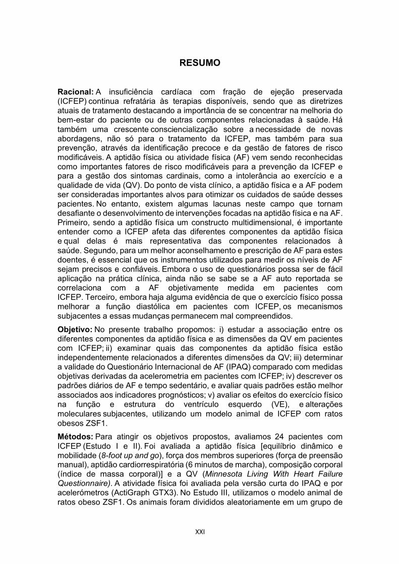
RESUMO
Racional:A insuficiência cardíaca com fração de ejeção preservada (ICFEP)continua refratária às terapias disponíveis, sendo que as diretrizes atuais de tratamento destacando a importância de se concentrar na melhoria do bem-estar do paciente ou de outras componentes relacionadas à saúde.Há também uma crescenteconsciencialização sobre anecessidade de novas abordagens, não só para o tratamento da ICFEP, mas também para sua prevenção, através da identificação precoce e da gestão de fatores de risco modificáveis.A aptidão física ou atividade física (AF) vem sendo reconhecidas como importantes fatores de risco modificáveis para a prevenção da ICFEP e para a gestão dos sintomas cardinais, como a intolerância ao exercício e a qualidade de vida (QV).Do ponto de vista clínico, a aptidão física e a AF podem ser consideradas importantes alvos para otimizar os cuidados de saúde desses pacientes.No entanto, existem algumas lacunas neste campo que tornam desafiante o desenvolvimento deintervenções focadas na aptidão física e na AF. Primeiro, sendo a aptidão física um constructo multidimensional, é importante entender como a ICFEP afeta das diferentes componentes da aptidão física equal delas é mais representativa das componentes relacionados à saúde.Segundo, para um melhor aconselhamento e prescrição de AF para estes doentes, é essencial que os instrumentos utilizados para medir os níveis de AF sejam precisos e confiáveis.Embora o uso de questionários possa ser de fácil aplicação na prática clínica, ainda não se sabe se a AF auto reportada se correlaciona com a AF objetivamente medida em pacientes com ICFEP.Terceiro, emborahaja alguma evidência de que o exercício físico possa melhorar a função diastólica em pacientes com ICFEP,os mecanismos subjacentes a essasmudançaspermanecem mal compreendidos.Objetivo:No presente trabalho propomos: i) estudar a associação entre os diferentes componentes da aptidão física e as dimensões da QV em pacientes com ICFEP;ii) examinar quais das componentes da aptidão física estão independentemente relacionados a diferentes dimensões da QV;iii) determinar a validade do Questionário Internacional de AF (IPAQ) comparado com medidas objetivas derivadas da acelerometria em pacientes com ICFEP;iv) descrever os padrões diários de AF e tempo sedentário, e avaliar quais padrões estão melhor associados aos indicadores prognósticos;v) avaliar osefeitos do exercício físico na função e estrutura do ventrículo esquerdo (VE), ealterações molecularessubjacentes, utilizando um modelo animal de ICFEP com ratos obesos ZSF1.Métodos:Para atingir os objetivos propostos, avaliamos 24 pacientes com ICFEP(Estudo I e II).Foi avaliada a aptidão física [equilíbrio dinâmico e mobilidade (8-foot up and go), força dos membros superiores (força de preensão manual), aptidão cardiorrespiratória (6 minutos de marcha), composição corporal (índice de massa corporal)] e a QV (Minnesota Living With Heart Failure Questionnaire).A atividade física foi avaliada pela versão curta do IPAQ e por acelerómetros (ActiGraph GTX3).No Estudo III, utilizamos o modelo animal de ratos obeso ZSF1.Os animais foram divididos aleatoriamente em um grupo de
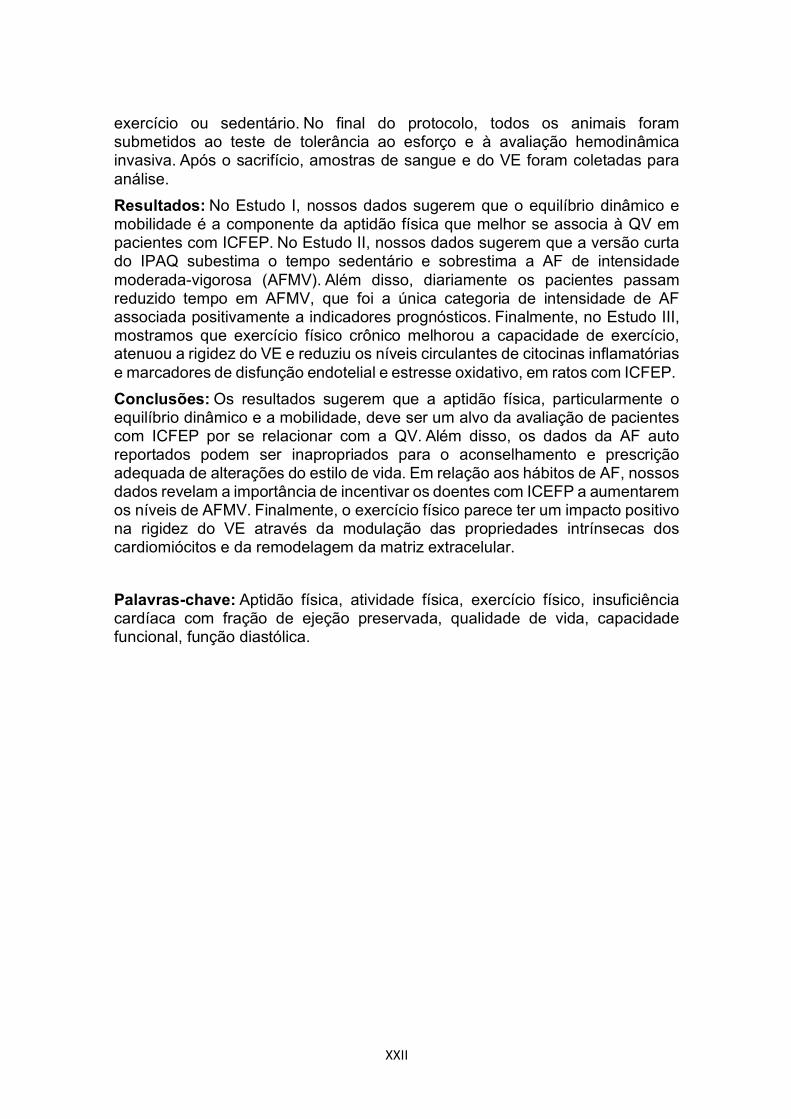
XXII
exercício ou sedentário.No final do protocolo, todos os animais foram submetidos ao teste de tolerância ao esforço e à avaliação hemodinâmica invasiva.Após o sacrifício, amostras de sangue e do VE foram coletadas para análise.Resultados:No Estudo I, nossos dados sugerem que o equilíbrio dinâmico e mobilidade é a componente da aptidão física que melhor se associa à QV em pacientes com ICFEP.No Estudo II, nossos dados sugerem que a versão curta do IPAQ subestima o tempo sedentário e sobrestima a AF de intensidade moderada-vigorosa (AFMV).Além disso, diariamente os pacientes passam reduzido tempo em AFMV, que foi a única categoria de intensidade de AF associada positivamente a indicadores prognósticos.Finalmente, no Estudo III, mostramos que exercício físico crônico melhorou a capacidade de exercício, atenuou a rigidez do VE e reduziu os níveis circulantes de citocinas inflamatórias e marcadores de disfunção endotelial e estresse oxidativo, em ratos com ICFEP.Conclusões:Os resultados sugerem que a aptidão física, particularmente o equilíbrio dinâmico e a mobilidade, deve ser um alvo da avaliação de pacientes com ICFEP por se relacionar com a QV.Além disso, os dados da AF auto reportados podem ser inapropriados para o aconselhamento e prescrição adequada de alterações do estilo de vida.Em relação aos hábitos de AF, nossos dados revelam a importância de incentivar os doentes com ICEFP a aumentarem os níveis de AFMV.Finalmente, o exercício físico parece ter um impacto positivo na rigidez do VE através da modulação das propriedades intrínsecas dos cardiomiócitos e da remodelagem da matriz extracelular. Palavras-chave:Aptidão física, atividade física, exercício físico, insuficiência cardíaca com fração de ejeção preservada, qualidade de vida, capacidade funcional, função diastólica.
XXIII
ABSTRACT Rational: Heart failure with preserved ejection fraction (HFpEF) continues to be refractory to available therapies, with current treatment guidelines highlighting the importance of focusing on the improvement of patient’s well-being or other health-related outcomes. There is also an increasing awareness of the need for novel approaches not only for the treatment of HFpEF but also for its prevention through the early identification and management of potential modifiable contributing risk factors. Physical fitness or physical activity (PA) are becoming recognized as key modifiable factors for the prevention of HFpEF and management of cardinal symptoms such as exercise intolerance and quality of life (QoL). From a clinical perspective, physical fitness and PA may be considered important targets if we aim to maximize the health care of these patients. However, there are some gaps in this field that may challenge the effectiveness of physical fitness and PA based-interventions. First, because physical fitness is a multicomponent construct, it is important to understand how this syndrome affects the different components, and which of them is better representative of health-related outcomes. Second, in order to provide tailored counselling and prescription to HFpEF patients, it is crucial that the instruments that we use to measure PA levels are accurate and reliable. While the use of questionnaires may be easy to apply in the clinical practice, it remains to be confirmed if self-reported and objectively measured PA is correlated in HFpEF. Third, while there is some evidence that exercise training can improve diastolic function in HFpEF patients, the mechanisms underlying these changes remain poorly comprehend. Purpose: In the current work, we propose to: i) study the association between different components of physical fitness and the dimensions of QoL in HFpEF patients; ii) examine which of the physical fitness components are independently related to different dimensions of QoL; iii) to determine the validity of the International Physical Activity Questionnaire (IPAQ) against objective measures from accelerometry in HFpEF patients; iv) to describe the patterns of daily PA and sedentary time and assess which is better associated with prognostic indicators; v) evaluate the effects of exercise training on LV function and structure, and underlying molecular changes, using the ZFF1 obese animal model of HFpEF. Methods: In order to accomplish the proposed aims, we evaluated 24 HFpEF patients (Study I and II). Patients were assessed for physical fitness [dynamic balance and mobility (8-feet-up-and go test), upper body strength (handgrip strength), cardiorespiratory fitness (CRF) (6-minute-walking test), body composition (body mass index)] and for QoL (Minnesota Living With Heart Failure Questionnaire). Physical activity was assessed through the IPAQ short version and triaxial accelerometry (ActiGraph GTX3). In order to evaluate the effects of exercise training on LV function and structure, and underlying molecular changes (Study III), we used the ZSF1 obese animal model. Animals were randomly divided in a training or sedentary group. At the end of the protocol, all animals were submitted to exercise tolerance test, and invasive hemodynamic evaluation. After sacrifice, blood and left ventricular samples were collected for analysis.
XXIV
Results: In Study I, our data suggests that dynamic balance and mobility is the only physical fitness component that better capture QoL in HFpEF patients. In Study II, our data suggests that the IPAQ short version underestimates sedentary time and over-estimates MVPA. In addition, patients spent only a minority of their time involved in moderate-to-vigorous PA, which was the only PA pattern positively associated with prognostic indicators. Finally, in Study III, we show that chronic exercise training improved exercise capacity, attenuated LV stiffness and reduced circulating levels of inflammatory cytokines and markers of endothelial dysfunction and oxidative stress in rats with HFpEF. Conclusions: Our data suggests that physical fitness, particularly dynamic balance and mobility, should be evaluated in HFpEF patients, once that it is associated with QoL. Also, physical activity data gathered solely by self-reported instruments may lead biased counselling and prescription. Regarding to PA patterns, our data points for the importance of recommending HFpEF to more engaged in MVPA. Finally, exercise training seems to positively impact left ventricular stiffness by modulating both cardiomyocyte’s intrinsic proprieties and extracellular matrix remodelling. Keywords: Physical fitness, physical activity, exercise training, heart failure with preserved ejection fraction, quality of life, functional capacity, diastolic function.
XXV
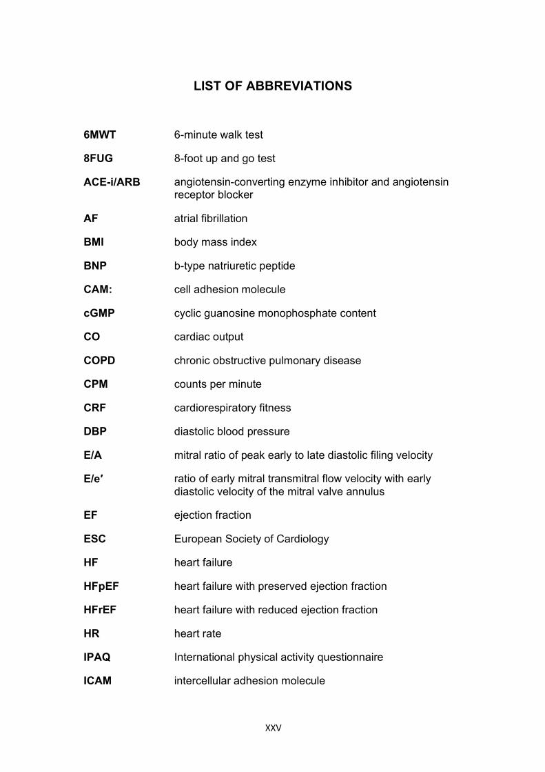
LIST OF ABBREVIATIONS
6MWT 6-minute walk test
8FUG 8-foot up and go test
ACE-i/ARB angiotensin-converting enzyme inhibitor and angiotensin receptor blocker
AF atrial fibrillation
BMI body mass index
BNP b-type natriuretic peptide
CAM: cell adhesion molecule
cGMP cyclic guanosine monophosphate content
CO cardiac output
COPD chronic obstructive pulmonary disease
CPM counts per minute
CRF cardiorespiratory fitness
DBP diastolic blood pressure
E/A mitral ratio of peak early to late diastolic filing velocity
E/e′ ratio of early mitral transmitral flow velocity with early diastolic velocity of the mitral valve annulus
EF ejection fraction
ESC European Society of Cardiology
HF heart failure
HFpEF heart failure with preserved ejection fraction
HFrEF heart failure with reduced ejection fraction
HR heart rate
IPAQ International physical activity questionnaire
ICAM intercellular adhesion molecule
XXVI
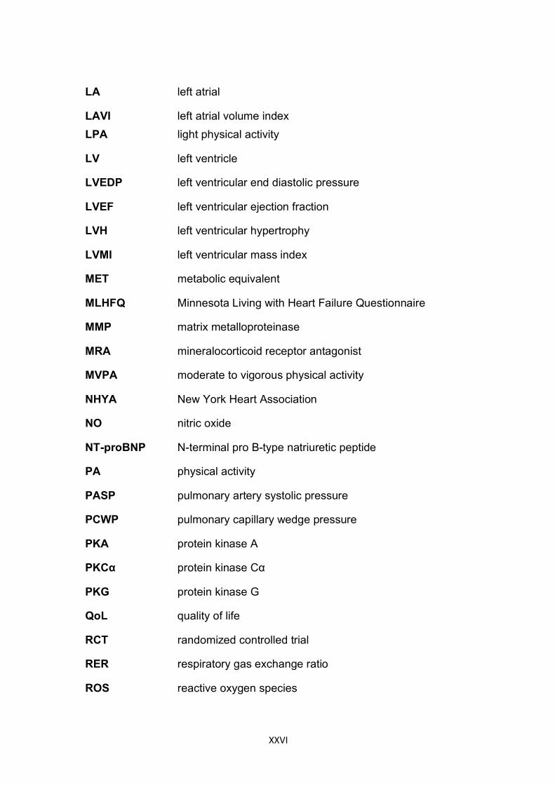
LA left atrial
LAVI LPA
left atrial volume index light physical activity
LV left ventricle
LVEDP left ventricular end diastolic pressure
LVEF left ventricular ejection fraction
LVH left ventricular hypertrophy
LVMI left ventricular mass index
MET metabolic equivalent
MLHFQ Minnesota Living with Heart Failure Questionnaire
MMP matrix metalloproteinase
MRA mineralocorticoid receptor antagonist
MVPA moderate to vigorous physical activity
NHYA New York Heart Association
NO nitric oxide
NT-proBNP N-terminal pro B-type natriuretic peptide
PA physical activity
PASP pulmonary artery systolic pressure
PCWP pulmonary capillary wedge pressure
PKA protein kinase A
PKCα protein kinase Cα
PKG protein kinase G
QoL quality of life
RCT randomized controlled trial
RER respiratory gas exchange ratio
ROS reactive oxygen species
XXVII
RV right ventricular
SBP systolic blood pressure
sGC soluble guanylate cyclase
SV stroke volume
TGF-ß transforming growth factor β
TIMP tissue inhibitors of metalloproteinases
TNF-α tumour necrosis factor alfa
TR tricuspid regurgitation
VCAM vascular cell adhesion molecules
VE minute ventilation
VE/VCO2 ventilatory equivalents for carbon dioxide output
VE/VO2: ventilatory equivalents for oxygen
VO2 oxygen consumption

XXVIII

CRISTINE SCHMIDT | HEART FAILURE WITH PRESERVED EJECTION FRACTION: FROM PROGNOSIS TO CARDIAC FUNCTION |
1
CHAPTER I
GENERAL INTRODUCTION

CRISTINE SCHMIDT | HEART FAILURE WITH PRESERVED EJECTION FRACTION: FROM PROGNOSIS TO CARDIAC FUNTION |
2
CRISTINE SCHMIDT | HEART FAILURE WITH PRESERVED EJECTION FRACTION: FROM PROGNOSIS TO CARDIAC FUNTION |
3
1. OVERVIEW OF HEART FAILURE
Heart failure (HF) is a clinical syndrome caused by impaired cardiac
structure and/or function, resulting in a reduced cardiac output and/or elevated
intracardiac pressures at rest or during stress, which leads to exercise intolerance
(Ponikowski et al., 2016). It affects approximately 26 million people worldwide,
and accounts for an estimated annual health care cost of $31 billion (Mozaffarian
et al., 2016). In Portugal, the EPICA study reported a prevalence of HF ranging
from 7.6% in the 60–69-year-old group to 16.1% in patients >80 years (Ceia et
al., 2002). In addition, it is estimated that the prevalence of HF in Portugal will
increase by around 7% in 2018, 30% in 2035 and 33% in 2060 (Fonseca et al.,
2018). Despite the evident progresses in the treatment of cardiovascular disease
over the past decades, the incidence and prevalence of HF have not decreased,
survival has slightly improved, and morbidity remains excessively high (Meta-
analysis Global Group in Chronic Heart Failure, 2012). One of the challenges and
barriers in successfully treating HF is the phenotypic heterogeneity found on this
condition (e.g. different pathophysiology, disease severity and response to
therapy) (Abbate et al., 2015).
Heart failure is usually associated with reduced left ventricular ejection
fraction (LVEF) (HFrEF), but it is now recognized that HF also encompasses a
wide range of patients with normal LVEF, defined as HF with preserved ejection
fraction (HFpEF) (Ponikowski et al., 2016). Heart failure with preserved ejection
fraction represents approximately 50% of patients with HF, and it is the main form
of HF in adults aged above 65 years old (Dunlay et al., 2017). It is estimated that
by 2020, the relative prevalence of HFpEF and HFrEF are predicted to reach 69%
and 31%, respectively (Steinberg et al., 2012). Patients with HFpEF exhibit a
long-term prognosis similar to HFrEF, with a five-year survival of less than 50%
(Shah et al., 2017). In addition, while there are a few pharmacologic and non-
pharmacological therapies demonstrated to improve survival and reduce HF
hospitalization in HFrEF, the scenario is less satisfactory for patients with HFpEF
as, up to date, no recognized therapies have shown important reductions in
morbidity or mortality (Nanayakkara et al., 2018).
CRISTINE SCHMIDT | HEART FAILURE WITH PRESERVED EJECTION FRACTION: FROM PROGNOSIS TO CARDIAC FUNTION |
4
Given the increasing prevalence of HFpEF, there is an urgent need of
clinical and translational research to better characterize this population and to
identify effective strategies for the prevention and management of HFpEF.
2. HEART FAILURE WITH PRESERVED EJECTION FRACTION 2.1. Definition and diagnosis criterion
Heart failure with preserved ejection fraction is a clinical syndrome
characterized by normal ejection fraction at the expense of increased left
ventricular filling pressures (Andersson & Vasan, 2014). The diagnosis of HFpEF remains defiant, once that signs and symptoms
are often non-specific and do not help to distinguish between HF and other clinical
conditions presenting similar manifestations such as in obese individuals, in the
elderly and in patients with chronic lung disease (Ponikowski et al., 2016). Typical
symptoms of HF are breathlessness, ankle swelling, fatigue and reduced
exercise tolerance, which may be accompanied by signs such as elevated jugular
venous pressure, pulmonary crackles and peripheral oedema (Ponikowski et al.,
2016). Patients are usually classified into subgroups based on exercise limitation
and their symptoms using the New York Heart Association (NYHA) functional
class. Functional capacity is classified with objective assessment (class A to D),
while patient’s symptoms are based on how much they are limited during physical
activity (class I to IV) (Dolgin, 1994).
According to current European Society of Cardiology (ESC)
recommendations (Ponikowski et al., 2016), the diagnosis of HFpEF requires the
following conditions to be fulfilled: i) signs and/or symptoms of HF (mentioned
above); ii) preserved ejection fraction (defined as LVEF >50%); iii) elevated levels
of natriuretic peptides (BNP >35 pg/mL and/or N-terminal pro B-type natriuretic
peptide (NT-proBNP) >125 pg/mL); and objective evidence of other cardiac
functional and structural alterations underlying HF. Finally, in case of uncertainty,
a stress test or invasively measured elevated left ventricle (LV) filling pressure
may be needed to confirm the diagnosis.
Key structural alterations include a left atrial volume index (LAVI) >34 mL/m2
or a left ventricular mass index (LVMI) ≥115 g/m2 for males and ≥95 g/m2 for
CRISTINE SCHMIDT | HEART FAILURE WITH PRESERVED EJECTION FRACTION: FROM PROGNOSIS TO CARDIAC FUNTION |
5
females. Key functional alterations are an E/e′ ≥13 (ratio of early transmitral
diastolic flow velocity to tissue Doppler early mitral annular diastolic velocity) and
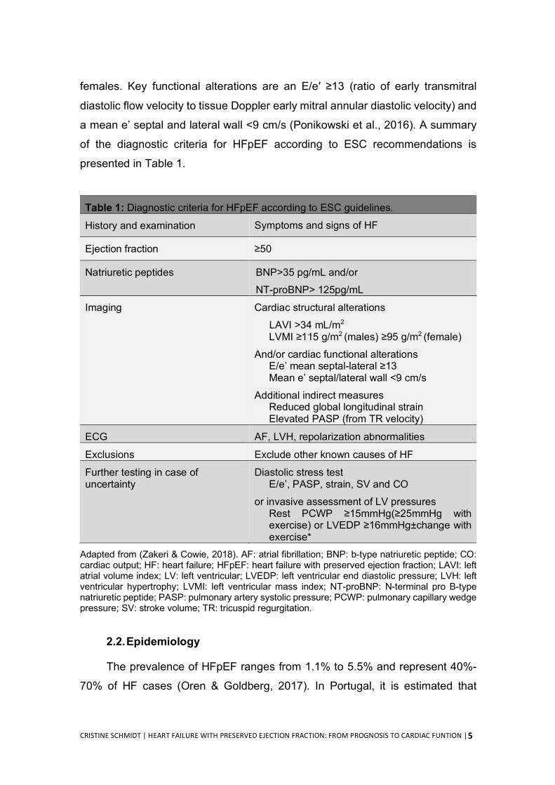
a mean e’ septal and lateral wall <9 cm/s (Ponikowski et al., 2016). A summary
of the diagnostic criteria for HFpEF according to ESC recommendations is
presented in Table 1.
Table 1: Diagnostic criteria for HFpEF according to ESC guidelines.
History and examination Symptoms and signs of HF
Ejection fraction ≥50
Natriuretic peptides BNP>35 pg/mL and/or NT-proBNP> 125pg/mL
Imaging Cardiac structural alterations LAVI >34 mL/m2
LVMI ≥115 g/m2 (males) ≥95 g/m2 (female) And/or cardiac functional alterations
E/e’ mean septal-lateral ≥13 Mean e’ septal/lateral wall <9 cm/s
Additional indirect measures Reduced global longitudinal strain Elevated PASP (from TR velocity)
ECG AF, LVH, repolarization abnormalities
Exclusions Exclude other known causes of HF
Further testing in case of uncertainty
Diastolic stress test E/e’, PASP, strain, SV and CO
or invasive assessment of LV pressures Rest PCWP ≥15mmHg(≥25mmHg with exercise) or LVEDP ≥16mmHg±change with exercise*
Adapted from (Zakeri & Cowie, 2018). AF: atrial fibrillation; BNP: b-type natriuretic peptide; CO: cardiac output; HF: heart failure; HFpEF: heart failure with preserved ejection fraction; LAVI: left atrial volume index; LV: left ventricular; LVEDP: left ventricular end diastolic pressure; LVH: left ventricular hypertrophy; LVMI: left ventricular mass index; NT-proBNP: N-terminal pro B-type natriuretic peptide; PASP: pulmonary artery systolic pressure; PCWP: pulmonary capillary wedge pressure; SV: stroke volume; TR: tricuspid regurgitation.
2.2. Epidemiology
The prevalence of HFpEF ranges from 1.1% to 5.5% and represent 40%-
70% of HF cases (Oren & Goldberg, 2017). In Portugal, it is estimated that
CRISTINE SCHMIDT | HEART FAILURE WITH PRESERVED EJECTION FRACTION: FROM PROGNOSIS TO CARDIAC FUNTION |
6
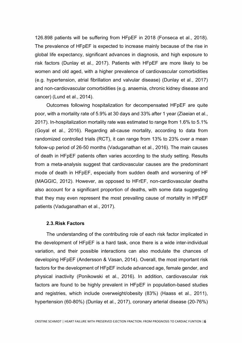
126.898 patients will be suffering from HFpEF in 2018 (Fonseca et al., 2018).
The prevalence of HFpEF is expected to increase mainly because of the rise in
global life expectancy, significant advances in diagnosis, and high exposure to
risk factors (Dunlay et al., 2017). Patients with HFpEF are more likely to be
women and old aged, with a higher prevalence of cardiovascular comorbidities
(e.g. hypertension, atrial fibrillation and valvular disease) (Dunlay et al., 2017)
and non-cardiovascular comorbidities (e.g. anaemia, chronic kidney disease and
cancer) (Lund et al., 2014).
Outcomes following hospitalization for decompensated HFpEF are quite
poor, with a mortality rate of 5.9% at 30 days and 33% after 1 year (Ziaeian et al.,
2017). In-hospitalization mortality rate was estimated to range from 1.6% to 5.1%
(Goyal et al., 2016). Regarding all-cause mortality, according to data from
randomized controlled trials (RCT), it can range from 13% to 23% over a mean
follow-up period of 26-50 months (Vaduganathan et al., 2016). The main causes
of death in HFpEF patients often varies according to the study setting. Results
from a meta-analysis suggest that cardiovascular causes are the predominant
mode of death in HFpEF, especially from sudden death and worsening of HF
(MAGGIC, 2012). However, as opposed to HFrEF, non-cardiovascular deaths
also account for a significant proportion of deaths, with some data suggesting
that they may even represent the most prevailing cause of mortality in HFpEF
patients (Vaduganathan et al., 2017).
2.3. Risk Factors
The understanding of the contributing role of each risk factor implicated in
the development of HFpEF is a hard task, once there is a wide inter-individual
variation, and their possible interactions can also modulate the chances of
developing HFpEF (Andersson & Vasan, 2014). Overall, the most important risk
factors for the development of HFpEF include advanced age, female gender, and
physical inactivity (Ponikowski et al., 2016). In addition, cardiovascular risk
factors are found to be highly prevalent in HFpEF in population-based studies
and registries, which include overweight/obesity (83%) (Haass et al., 2011),
hypertension (60-80%) (Dunlay et al., 2017), coronary arterial disease (20-76%)
CRISTINE SCHMIDT | HEART FAILURE WITH PRESERVED EJECTION FRACTION: FROM PROGNOSIS TO CARDIAC FUNTION |
7
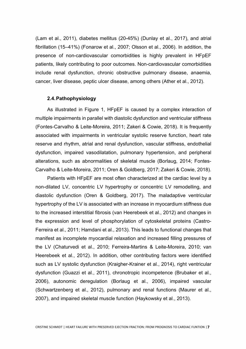
(Lam et al., 2011), diabetes mellitus (20-45%) (Dunlay et al., 2017), and atrial
fibrillation (15–41%) (Fonarow et al., 2007; Olsson et al., 2006). In addition, the
presence of non-cardiovascular comorbidities is highly prevalent in HFpEF
patients, likely contributing to poor outcomes. Non-cardiovascular comorbidities
include renal dysfunction, chronic obstructive pulmonary disease, anaemia,
cancer, liver disease, peptic ulcer disease, among others (Ather et al., 2012).
2.4. Pathophysiology
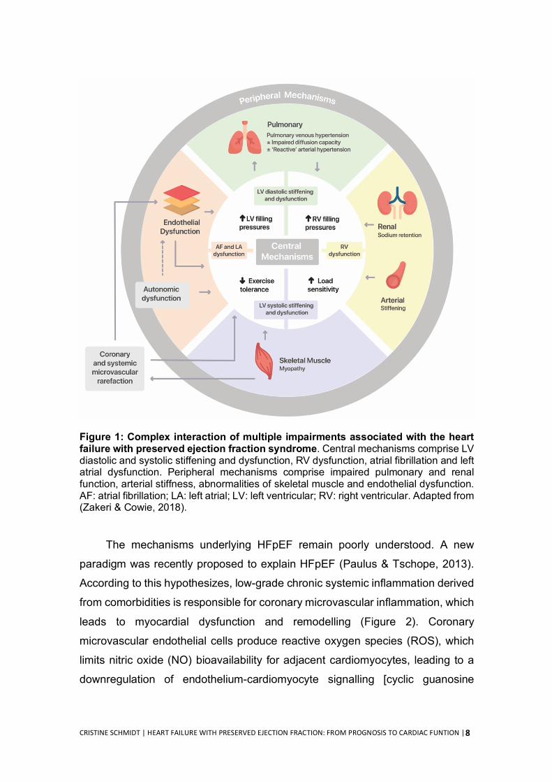
As illustrated in Figure 1, HFpEF is caused by a complex interaction of
multiple impairments in parallel with diastolic dysfunction and ventricular stiffness
(Fontes-Carvalho & Leite-Moreira, 2011; Zakeri & Cowie, 2018). It is frequently
associated with impairments in ventricular systolic reserve function, heart rate
reserve and rhythm, atrial and renal dysfunction, vascular stiffness, endothelial
dysfunction, impaired vasodilatation, pulmonary hypertension, and peripheral
alterations, such as abnormalities of skeletal muscle (Borlaug, 2014; Fontes-
Carvalho & Leite-Moreira, 2011; Oren & Goldberg, 2017; Zakeri & Cowie, 2018).
Patients with HFpEF are most often characterized at the cardiac level by a
non-dilated LV, concentric LV hypertrophy or concentric LV remodelling, and
diastolic dysfunction (Oren & Goldberg, 2017). The maladaptive ventricular
hypertrophy of the LV is associated with an increase in myocardium stiffness due
to the increased interstitial fibrosis (van Heerebeek et al., 2012) and changes in
the expression and level of phosphorylation of cytoskeletal proteins (Castro-
Ferreira et al., 2011; Hamdani et al., 2013). This leads to functional changes that
manifest as incomplete myocardial relaxation and increased filling pressures of
the LV (Chaturvedi et al., 2010; Ferreira-Martins & Leite-Moreira, 2010; van
Heerebeek et al., 2012). In addition, other contributing factors were identified
such as LV systolic dysfunction (Kraigher-Krainer et al., 2014), right ventricular
dysfunction (Guazzi et al., 2011), chronotropic incompetence (Brubaker et al.,
2006), autonomic deregulation (Borlaug et al., 2006), impaired vascular
(Schwartzenberg et al., 2012), pulmonary and renal functions (Maurer et al.,
2007), and impaired skeletal muscle function (Haykowsky et al., 2013).
CRISTINE SCHMIDT | HEART FAILURE WITH PRESERVED EJECTION FRACTION: FROM PROGNOSIS TO CARDIAC FUNTION |
8
Figure 1: Complex interaction of multiple impairments associated with the heart failure with preserved ejection fraction syndrome. Central mechanisms comprise LV diastolic and systolic stiffening and dysfunction, RV dysfunction, atrial fibrillation and left atrial dysfunction. Peripheral mechanisms comprise impaired pulmonary and renal function, arterial stiffness, abnormalities of skeletal muscle and endothelial dysfunction. AF: atrial fibrillation; LA: left atrial; LV: left ventricular; RV: right ventricular. Adapted from (Zakeri & Cowie, 2018).
The mechanisms underlying HFpEF remain poorly understood. A new
paradigm was recently proposed to explain HFpEF (Paulus & Tschope, 2013).
According to this hypothesizes, low-grade chronic systemic inflammation derived
from comorbidities is responsible for coronary microvascular inflammation, which
leads to myocardial dysfunction and remodelling (Figure 2). Coronary
microvascular endothelial cells produce reactive oxygen species (ROS), which
limits nitric oxide (NO) bioavailability for adjacent cardiomyocytes, leading to a
downregulation of endothelium-cardiomyocyte signalling [cyclic guanosine
CRISTINE SCHMIDT | HEART FAILURE WITH PRESERVED EJECTION FRACTION: FROM PROGNOSIS TO CARDIAC FUNTION |
9
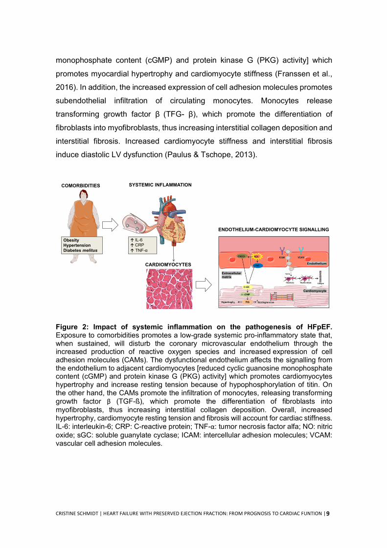
monophosphate content (cGMP) and protein kinase G (PKG) activity] which
promotes myocardial hypertrophy and cardiomyocyte stiffness (Franssen et al.,
2016). In addition, the increased expression of cell adhesion molecules promotes
subendothelial infiltration of circulating monocytes. Monocytes release
transforming growth factor β (TFG- β), which promote the differentiation of
fibroblasts into myofibroblasts, thus increasing interstitial collagen deposition and
interstitial fibrosis. Increased cardiomyocyte stiffness and interstitial fibrosis
induce diastolic LV dysfunction (Paulus & Tschope, 2013).
Figure 2: Impact of systemic inflammation on the pathogenesis of HFpEF. Exposure to comorbidities promotes a low-grade systemic pro-inflammatory state that, when sustained, will disturb the coronary microvascular endothelium through the increased production of reactive oxygen species and increased expression of cell adhesion molecules (CAMs). The dysfunctional endothelium affects the signalling from the endothelium to adjacent cardiomyocytes [reduced cyclic guanosine monophosphate content (cGMP) and protein kinase G (PKG) activity] which promotes cardiomyocytes hypertrophy and increase resting tension because of hypophosphorylation of titin. On the other hand, the CAMs promote the infiltration of monocytes, releasing transforming growth factor β (TGF-ß), which promote the differentiation of fibroblasts into myofibroblasts, thus increasing interstitial collagen deposition. Overall, increased hypertrophy, cardiomyocyte resting tension and fibrosis will account for cardiac stiffness. IL-6: interleukin-6; CRP: C-reactive protein; TNF-α: tumor necrosis factor alfa; NO: nitric oxide; sGC: soluble guanylate cyclase; ICAM: intercellular adhesion molecules; VCAM: vascular cell adhesion molecules.
COMORBIDITIES
CARDIOMYOCYTES
ENDOTHELIUM-CARDIOMYOCYTE SIGNALLING
Endothelium
Cardiomyocyte
Extracellular matrix
↑ IL-6↑ CRP↑ TNF-α
ObesityHypertensionDiabetes melitus
SYSTEMIC INFLAMMATION
CRISTINE SCHMIDT | HEART FAILURE WITH PRESERVED EJECTION FRACTION: FROM PROGNOSIS TO CARDIAC FUNTION |
10
2.5. Treatment
According to 2016 ESC guidelines, no therapy has been consistently proven
to reduce morbidity or mortality in patients with HFpEF (Ponikowski et al., 2016).
However, since these patients are often elders, highly symptomatic, and
perceiving a poor quality of life (Fukuta et al., 2016), current treatment ESC
guidelines highlight the importance of aiming to alleviate symptoms and improve
patients well-being (Ponikowski et al., 2016). Patients should be screened for
cardiovascular and non-cardiovascular comorbidities, which if present, should be
managed with interventions that have been shown to improve symptoms, well-
being or other health-related outcomes, without exacerbating HF (Ponikowski et
al., 2016).
Because HFpEF continues to be refractory to available therapies, there is
an urgent need for novel approaches not only for the treatment and management
of HFpEF but also for its prevention. Indeed, some authors are now arguing that
the attention needs to be directed to the early steps of this syndrome, by the
screening and management of the potential modifiable contributing factors,
ultimately to prevent HFpEF (Bobenko et al., 2018; Kondamudi et al., 2017). In
this sense, growing body of evidence is also highlighting the value of physical
activity (PA)/exercise training programs in the prevention of HFpEF and in the
management of cardinal symptoms such as exercise intolerance and quality of
life (Chan et al., 2016). Indeed, the current ESC guidelines highlight the
importance of PA/exercise training to HFpEF management (Ponikowski et al.,
2016).
3. THE ROLE OF PHYSICAL ACTIVITY IN HFPEF 3.1. Definition of physical activity and sedentary time
Physical activity is usually defined as any body movement produced by
skeletal muscles that require energy expenditure (Caspersen et al., 1985). It is
commonly categorized according to four different contexts: occupational,
household, transport and leisure-time or recreational PA (Physical Activity
Guidelines Advisory Committee Scientific Report, 2018). In addition, PA can be
CRISTINE SCHMIDT | HEART FAILURE WITH PRESERVED EJECTION FRACTION: FROM PROGNOSIS TO CARDIAC FUNTION |
11
classified by its intensity, such as light [1.5-3 metabolic equivalents (METs)],
moderate (3-6 METs), and vigorous or very vigorous intensity (≥6METs) (Riebe
et al., 2018). By comparison, exercise training is defined as a sub-component of
PA that is characterized to be a deliberated practice, planned, structured, and
repetitive, and is designed with the specific purpose of improving or maintaining
physical fitness, physical performance, or health (Caspersen et al., 1985).
Regarding sedentary time, it is defined as any waking activity characterized by a
low level of energy expenditure (≤1.5 METs) while sitting, reclining, or lying
(Tremblay et al., 2017).
3.2. Measurement of physical activity and sedentary time
As PA is a multidimensional practice, valid measurements in free-living
individuals can be a challenging task, regardless the population. There are
several methods for measuring PA, and each one has its own particular
limitations and strengths. These methods include subjective (e.g. questionnaire)
and objective instruments (e.g. accelerometer). Questionnaires are a simple
instrument, easily-administered, appropriate to use in large samples for research
purposes, and is cost-effective (Besson et al., 2010). In addition, it allows
identifying the context in which PA is performed (e.g. occupational and leisure-
time or recreational) (Besson et al., 2010). However, the assessment of PA by
questionnaires is based on self-reports, and therefore, most often biased due to
social desirability, inaccurate memory, and the inability to capture the absolute
level of PA intensity (Prince et al., 2008). Accelerometers are motion sensors
specially designed to assess PA and related energy expenditure. Measurements
with accelerometers provide accurate and reliable information about total PA, as
well as about the intensities and time spent at each PA intensity in everyday life
activities (Cheung et al., 2011; Gorman et al., 2014). Furthermore, the device size
is small and easy to wear. Unfortunately, accelerometers are expensive and
challenging because of the large volume of data that generate and/or the required
expertise to the data management, which make it difficult to use in large
epidemiologic studies (Troiano, 2005).
CRISTINE SCHMIDT | HEART FAILURE WITH PRESERVED EJECTION FRACTION: FROM PROGNOSIS TO CARDIAC FUNTION |
12
Sedentary behaviour or sedentary time can also be ascertained from PA
questionnaires or accelerometry, respectively. Lower sedentary time is usually
related to higher levels of PA in many individuals, however, these variables are
not necessarily correlated. In fact, it is somewhat common to find individuals who
perform higher levels of PA but, concomitantly, also accumulate higher levels of
sedentary time (e.g. sedentary job) (Thorp et al., 2011).
3.3. Physical activity in the prevention of HFpEF
Several studies are supporting the notion that to control the growing burden
of HFpEF, we have to change the focus for primary prevention and identify
modifiable risk factors that can be targeted. Lifestyle risk factors, such as low PA,
sedentary time, and low cardiorespiratory fitness (CRF) are becoming
increasingly recognized as major risk factors for most chronic diseases, including
HF (Booth et al., 2012; Djousse et al., 2009; Kenchaiah et al., 2009). Indeed, a
number of community-based studies have demonstrated an inverse association
between PA levels and risk of HF. The Women’s Health Initiative, an
observational study with a cohort of 84.537 participants, demonstrated that a
relatively high levels of PA was associated with decreased HF risk (Agha et al.,
2014). Similar results were found in a prospective cohort study using data from
20.900 men, where adherence to healthy lifestyle factors was associated with a
lower lifetime risk of HF (Djousse et al., 2009). In addition, physical inactivity
(defined as not meeting the recommended amount of health-related PA within a
week) was also related to increased risk for HF (He et al., 2001). Young and
colleagues examined the correlation between PA and prolonged sedentary time
on risk of HF in 82.695 men followed for 10 years (Young et al., 2014). The
investigators found that apart from higher PA, lower sedentary time was
associated with reduced risk of HF incidence, and these associations had
independent contributions (Young et al., 2014).
While it remains to be clarified which HF phenotype is the mostly affected,
some reports suggest that PA and sedentary time may be more strongly
implicated in the development of HFpEF than in HFrEF. A recent study with a
pooled analysis from three large cohorts demonstrated an inverse relationship
CRISTINE SCHMIDT | HEART FAILURE WITH PRESERVED EJECTION FRACTION: FROM PROGNOSIS TO CARDIAC FUNTION |
13
between leisure-time PA and HF risk, where lower levels of PA were associated
with higher risk of HFpEF but not HFrEF (Pandey et al., 2017). Additionally, in an
elderly cohort from the Framingham Heart Study, lower PA levels were more
strongly associated with risk of HFpEF (Kraigher-Krainer et al., 2013).
Furthermore, corroborating this notion of HF phenotype specificity, it was shown
a strong dose-dependent inverse association between lifetime doses of exercise
training and LV compliance and distensibility, which are hallmark features of
HFpEF (Bhella et al., 2014). Finally, there is evidence that sedentary lifestyle
(prolonged bed rest) is also associated with many of the underlying cardiac and
skeletal muscle abnormalities often present in HFpEF (Dorfman et al., 2008;
Irimia et al., 2017). Thus, it seems that PA and/or sedentary time are important
modifiable aspects to be targeted to prevent HFpEF. In addition to that major key
point, it needs to be highlighted that the above-mentioned studies support their
conclusions on questionnaire-derived information, which often overestimate PA
(Lee et al., 2011), and thus do not allow to extract precise data for PA
prescription. To overcome this important limitation, objectively measures of PA,
such as those derived from accelerometry, need to be explored. Because the use
of accelerometers in large cohort studies is not feasible, an alternative approach
may be the evaluation of physical fitness since it can be relatively easily assessed
and is strongly related to PA. Recent work from the Cooper Centre Longitudinal
Study showed that in midlife, lower CRF, which is one component of physical
fitness, is more powerfully associated with a marked increase in the risk for HF
than acute myocardial infarction in older age (Berry et al., 2013). In addition, lower
CRF in midlife was related to a larger degree of diastolic dysfunction and LV
remodelling, important hallmarks of HFpEF (Brinker et al., 2014). Similar risk
patterns were recently observed by Pandey and co-workers, where lower CRF in
young adulthood was strongly associated with subclinical diastolic filling
impairment 20 years later (Pandey et al., 2017a). Finally, CRF was inversely
associated with relative wall thickness and LVMi in adults, both hallmarks of
HFpEF (Lam et al., 2010). Collectively, these studies suggest that higher levels
of PA, less time spent in sedentary behaviours and a greater CRF are strongly
associated with a reduced risk of HFpEF.
CRISTINE SCHMIDT | HEART FAILURE WITH PRESERVED EJECTION FRACTION: FROM PROGNOSIS TO CARDIAC FUNTION |
14
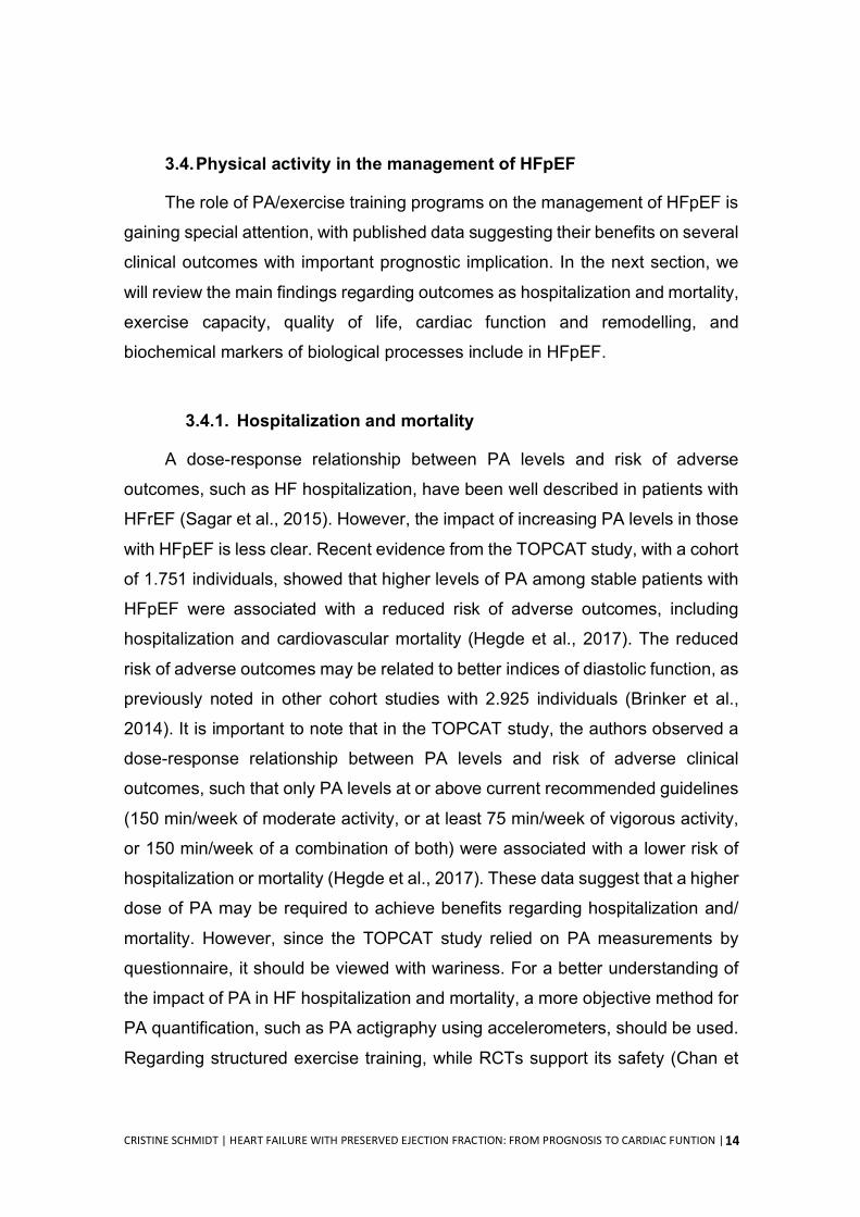
3.4. Physical activity in the management of HFpEF
The role of PA/exercise training programs on the management of HFpEF is
gaining special attention, with published data suggesting their benefits on several
clinical outcomes with important prognostic implication. In the next section, we
will review the main findings regarding outcomes as hospitalization and mortality,
exercise capacity, quality of life, cardiac function and remodelling, and
biochemical markers of biological processes include in HFpEF.
3.4.1. Hospitalization and mortality
A dose-response relationship between PA levels and risk of adverse
outcomes, such as HF hospitalization, have been well described in patients with
HFrEF (Sagar et al., 2015). However, the impact of increasing PA levels in those
with HFpEF is less clear. Recent evidence from the TOPCAT study, with a cohort
of 1.751 individuals, showed that higher levels of PA among stable patients with
HFpEF were associated with a reduced risk of adverse outcomes, including
hospitalization and cardiovascular mortality (Hegde et al., 2017). The reduced
risk of adverse outcomes may be related to better indices of diastolic function, as
previously noted in other cohort studies with 2.925 individuals (Brinker et al.,
2014). It is important to note that in the TOPCAT study, the authors observed a
dose-response relationship between PA levels and risk of adverse clinical
outcomes, such that only PA levels at or above current recommended guidelines
(150 min/week of moderate activity, or at least 75 min/week of vigorous activity,
or 150 min/week of a combination of both) were associated with a lower risk of
hospitalization or mortality (Hegde et al., 2017). These data suggest that a higher
dose of PA may be required to achieve benefits regarding hospitalization and/
mortality. However, since the TOPCAT study relied on PA measurements by
questionnaire, it should be viewed with wariness. For a better understanding of
the impact of PA in HF hospitalization and mortality, a more objective method for
PA quantification, such as PA actigraphy using accelerometers, should be used.
Regarding structured exercise training, while RCTs support its safety (Chan et
CRISTINE SCHMIDT | HEART FAILURE WITH PRESERVED EJECTION FRACTION: FROM PROGNOSIS TO CARDIAC FUNTION |
15
al., 2016), the effect on mortality or hospitalization is largely unknown and future
studies need to specifically address these endpoints.
3.4.2. Exercise capacity
Several studies demonstrated significant improvements in exercise capacity
with PA and exercise training interventions in patients with HFpEF. A multicentre
study from the NEAT-HFpEF trial evaluated daily PA levels using accelerometers
in 110 patients with HFpEF (Snipelisky et al., 2017). The authors reported a
significant association between PA levels and exercise capacity assessed
through the 6-minute walk test (6MWT), where patients with lower levels of daily
PA had lower exercise tolerance. This was corroborated in the multicentre ALDO-
DHF trial with 442 ambulatory HFpEF patients, where the total amount of PA
(METs/week) was positively correlated with submaximal exercise capacity
(Bobenko et al., 2018). Indeed, HFpEF patients have a significant reduction in
exercise capacity (Dhakal et al., 2015), which can lead to early fatigue and
reduction in the overall volume of daily activity. In comparison with healthy
individuals at risk of HF, and HFrEF patients, it seems that patients with HFpEF
have the lower volume of objectively measured daily PA (Yavari et al., 2017).
Moreover, a positive effect of exercise training on exercise capacity was
also shown by a meta-analysis addressing large HFpEF trials (Chan et al., 2016).
From the 8 studies included in the final analysis, 5 showed a significant
improvement in peak oxygen consumption (VO2peak) (+2.08 ml.kg-1.min-1; 15%
increase), which is above the clinically meaningful change (1 ml.kg-1.min-1 or
10%) in VO2peak for patients with HF (Kitzman, 2011). In addition, 4 studies
reported a significant increase in walking distance on the 6MWT (+32.1 meters).
Of note, these improvements were obtained with different types of exercise. Just
to mention some, Haykowsky and colleagues performed 4 months of endurance
exercise training in HFpEF patients and found an improvement of 2.3 ml.kg-1.min-
1 on VO2 peak (Haykowsky et al., 2012). In addition, a multicentre RCT compared
3 months of combined endurance and strength training with usual care alone in
64 clinically stable patients with HFPEF. Peak VO2 increased with exercise
training (from 16.1±4.9 ml.kg-1.min-1 to 18.7±5.4 ml.kg-1.min-1; p<0.001), while it
CRISTINE SCHMIDT | HEART FAILURE WITH PRESERVED EJECTION FRACTION: FROM PROGNOSIS TO CARDIAC FUNTION |
16
remained unchanged with usual care alone (Edelmann et al., 2011). Finally,
aerobic interval training was also effective to significantly improve VO2 peak in
HFpEF patients (Fu et al., 2016) as well as to improve exercise tolerance above
the threshold considered clinically relevant (Alves et al., 2012). Interestingly, a
recent meta-analysis showed that the single use of standard cardiovascular
medications failed to improve exercise capacity, while its combination with
exercise training was effective to increase VO2 peak and the performance in
6MWT (Fukuta et al., 2016).
The physiological mechanisms underlying these improvements in exercise
capacity in HFpEF patients are poorly comprehended. Poor exercise response
was shown to be due to a reduction in both cardiac output and arteriovenous
oxygen content difference (Houstis et al., 2018). Therefore, by knowing the
effects of exercise training in other clinical settings, it seems logical to
hypothesize that training would improve exercise capacity by influencing both
factors. However, current data supports that the greater VO2peak found in HFpEF
patients after training is paralleled by an increase in oxygen extraction but not by
cardiac output (Fu et al., 2016; Haykowsky et al., 2012). Nevertheless, it needs
to be highlighted that the number of studies assessing cardiac hemodynamic
after training is very low and the number of recruited patients is reduced (Fu et
al., 2016; Haykowsky et al., 2012). Moreover, these studies were not designed to
test the most effective dose to potentially improve hemodynamic factors, and it
may be the case that cardiac responsiveness to training is reduced and requires
prolonged (several months to years) and/or more intensity of training programs
as recently suggested for healthy seniors (Bhella et al., 2014). Indeed, it was
shown that competitive master athletes demonstrated improved ventricular
compliance (lower LV chamber stiffness constants) and distensibility (greater
LVEDVi) compared to that in casual exercisers and sedentary subjects (Bhella et
al., 2014). Finally, because HFpEF patients are so heterogeneous, it may be the
case that the magnitude of responsiveness of each factor to training is dependent
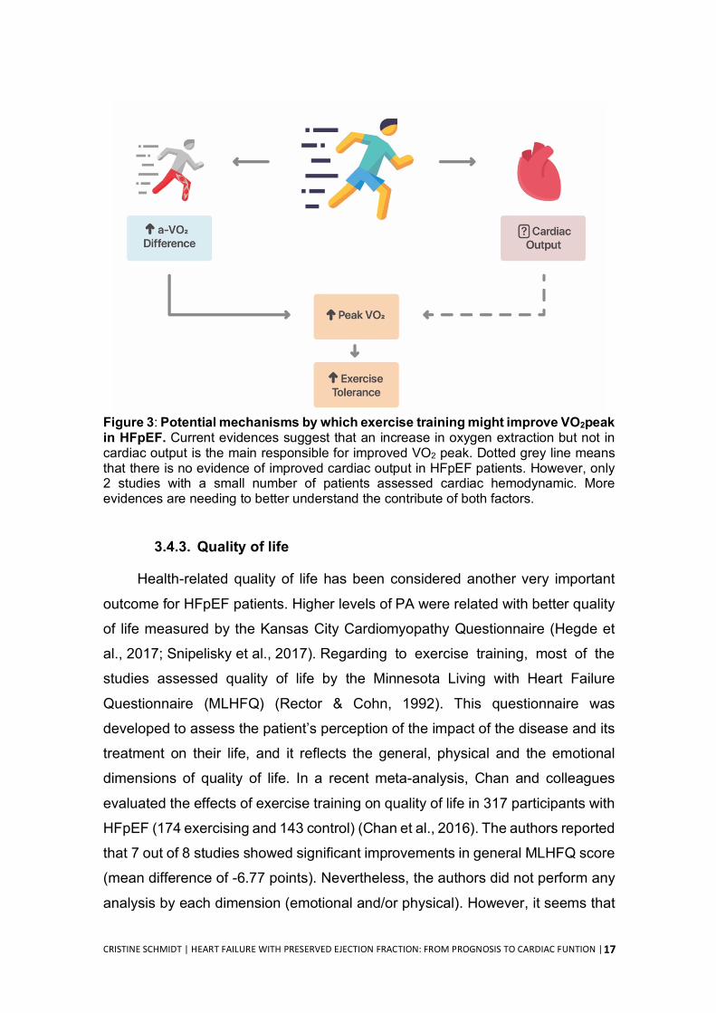
on the patient’s personal profile of defects (Houstis et al., 2018). Figure 3
summarizes how these factors could cooperate to improve exercise capacity in
HFpEF.
CRISTINE SCHMIDT | HEART FAILURE WITH PRESERVED EJECTION FRACTION: FROM PROGNOSIS TO CARDIAC FUNTION |
17
Figure 3: Potential mechanisms by which exercise training might improve VO2peak in HFpEF. Current evidences suggest that an increase in oxygen extraction but not in cardiac output is the main responsible for improved VO2 peak. Dotted grey line means that there is no evidence of improved cardiac output in HFpEF patients. However, only 2 studies with a small number of patients assessed cardiac hemodynamic. More evidences are needing to better understand the contribute of both factors.
3.4.3. Quality of life
Health-related quality of life has been considered another very important
outcome for HFpEF patients. Higher levels of PA were related with better quality
of life measured by the Kansas City Cardiomyopathy Questionnaire (Hegde et
al., 2017; Snipelisky et al., 2017). Regarding to exercise training, most of the
studies assessed quality of life by the Minnesota Living with Heart Failure
Questionnaire (MLHFQ) (Rector & Cohn, 1992). This questionnaire was
developed to assess the patient’s perception of the impact of the disease and its
treatment on their life, and it reflects the general, physical and the emotional
dimensions of quality of life. In a recent meta-analysis, Chan and colleagues
evaluated the effects of exercise training on quality of life in 317 participants with
HFpEF (174 exercising and 143 control) (Chan et al., 2016). The authors reported
that 7 out of 8 studies showed significant improvements in general MLHFQ score
(mean difference of -6.77 points). Nevertheless, the authors did not perform any
analysis by each dimension (emotional and/or physical). However, it seems that
CRISTINE SCHMIDT | HEART FAILURE WITH PRESERVED EJECTION FRACTION: FROM PROGNOSIS TO CARDIAC FUNTION |
18
the large majority of the studies using the MLHFQ (and reporting the dimensions)
showed an improvement only in the physical dimension of quality of life
(Edelmann et al., 2011; Kitzman et al., 2010). This may be explained by the
attenuating effect of exercise training over exercise intolerance and fatigue,
increasing patients’ ability to cope with the demands of daily tasks, and
consequently improving the perceived quality of life in the physical dimension (Fu
et al., 2016). Finally, Smart et al. (2007) showed that 16 weeks of exercise
training only significantly improved the emotional dimension in HFpEF patients
(Smart et al., 2007). Taken together, these evidences suggest that exercise
training programs are effective strategies to induce positive effects on general
quality of life.
3.4.4. Cardiac function and remodelling
Despite the overall beneficial effects of increasing levels of PA in HFpEF
patients, it remains poorly understood if it can also modulate cardiac function and
remodeling. Some preliminary clues to this aspect are provided by a recent study
showing that HFpEF patients engaged in lower levels of daily PA (objectively
measured by accelerometry) presented higher NT-proBNP levels, more
concentric left ventricular remodeling (higher relative wall thickness), larger LAVi,
and a tendency to have higher E/e′ (Snipelisky et al., 2017). Contrasting findings
where shown by Bobenko and collogues, who did not found association between
E/e´ or LAVi and PA levels in HFpEF patients. However, since in the last study
PA levels were based on self-report, the results should be viewed with wariness
(Bobenko et al., 2018).
Regarding to the impact of exercise training on diastolic function in HFpEF,
early meta-analysis suggested that exercise training was not associated with
significant changes in diastolic function (Pandey et al., 2015). This lack of
evidence could be due to the different standard measures of diastolic function
(E/A ratio and deceleration time) between studies included in the meta-analysis.
Moreover, the E/e’ is considered a more specific non-invasive indicator of
diastolic function, once that it is a less load dependent index of LV relaxation and
less influenced by heart rate or age (Pearson et al., 2017). However, studies
CRISTINE SCHMIDT | HEART FAILURE WITH PRESERVED EJECTION FRACTION: FROM PROGNOSIS TO CARDIAC FUNTION |
19
using this parameter were scarce at that time (n=1) and thus were exclude from
meta-analysis. In this sense, a more recent meta-analysis of 8 studies, with 317
participants, showed that exercise training programs significantly improve
diastolic function (changes on E/e’, E/A and/or deceleration time) (Chan et al.,
2016). These results differ from the meta-analysis aforementioned because it
included more studies that evaluated E/e’. Future studies using invasive
assessment (catheterization) of cardiovascular properties at rest and during
exercise may provide a more sensitive analysis of alterations induced by exercise
training (Borlaug & Kass, 2009). In the meantime, improvement of diastolic
function with exercise training was also demonstrated in pre-clinical studies
(Hidalgo et al., 2014; Slater et al., 2017).
Regarding the underlying mechanisms, so far, the literature does not
elucidate how exercise training could improve diastolic function in HFpEF
patients. Edelmann and colleagues demonstrated that exercise training improves
LV diastolic function and atrial reverse remodelling without change in LV mass,
suggesting a switch from pathologic into a more physiologic hypertrophy
(Edelmann et al., 2011). Indeed, exercise training has the ability to
counterbalance the structural and functional cardiac changes induced by
cardiovascular diseases, contributing to phenotypical changes of pathological
cardiac hypertrophy into physiological cardiac hypertrophy (Fernandes et al.,
2015). In addition, exercise training was associated with a significant reduction in
procollagen type I plasma levels, suggesting that improvement in diastolic
function may be associated with reduced collagen turnover (Edelmann et al.,
2011). NT-proBNP is the gold standard marker for myocyte stress and the
decreases in NT-proBNP levels were associated with reduced mortality and
morbidity rates in HFpEF patients (Anand et al., 2011). While exercise training
was capable to significantly reduce the circulating concentrations of NT-proBNP
(Conraads et al., 2004) or BNP (Nakanishi et al., 2017) in HFrEF patients, no
significant changes have been noted in patients with HFpEF (Edelmann et al.,
2011; Kitzman et al., 2010). However, it is important to highlight that levels of
NTproBNP (Edelmann et al., 2011) or BPN (Kitzman et al., 2010) in these studies
were below the levels that were reported in acutely decompensated HFpEF.
CRISTINE SCHMIDT | HEART FAILURE WITH PRESERVED EJECTION FRACTION: FROM PROGNOSIS TO CARDIAC FUNTION |
20
Finally, whether exercise training can modulate LV compliance and distensibility
in HFpEF remains to be explored, but these diastolic variables were recently
shown to be modulated by exercise training in healthy middle age individuals
(Howden et al., 2018) and in healthy seniors (Bhella et al., 2014).
Further insights of the molecular mechanisms underlying exercise-induced
benefits in HFpEF are provided by animal models. Pre-clinical studies of HFpEF
suggest that exercise-induced improvement on diastolic function are linked to
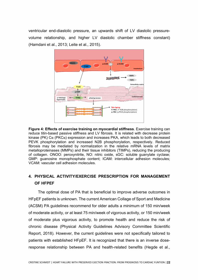
decreased myocardial stiffness (Figure 4). A recent work evaluated the effect of
free-wheel running exercise on cardiac stiffness (extracellular matrix and titin-
based stiffness) in a genetic mouse model (TtnΔIAjxn), where the I-A junction of
titin is removed resulting in increased diastolic stiffness and reduced exercise
tolerance (Slater et al., 2017). The data revealed that intrinsic myocardial
stiffness was reduced in the running group, which was associated with reduced
titin-based passive stiffness in a phosphorylation-dependent manner, as no effect
was detected on extracellular matrix (Slater et al., 2017). The authors observed
that the reduction in passive stiffness was mainly related to both decreased PEVK
phosphorylation and increased N2B phosphorylation (Slater et al., 2017). The
phosphorylation of PEVK element by protein kinase Cα (PKCα) increases titin
stiffness, while phosphorylation of N2B element by protein kinase A (PKA) or
protein kinase G (PKG), decreases titin stiffness (Kotter et al., 2013). Voluntary
exercise in genetically engineered mice with HFpEF symptoms (IG KO mice),
was shown to decrease PKCα expression level in the LV, lowering titin-based
stiffness and improving diastolic filling (Hidalgo et al., 2014). In addition, Slater
and colleagues reported that voluntary exercise in TtnΔIAjxn mice increases titin
phosphorylation by PKA, leading to diminished titin stiffness and improved
diastolic function (Slater et al., 2017). These studies suggest that reduced
passive tension induced by exercise appears to be explained mainly by
modification of cardiac titin phosphorylation status, rather than by titin isoform
expression. Indeed, these experimental studies did not found changes in the ratio
of the N2B or N2BA titin isoforms (Hidalgo, Saripalli, & Granzier, 2014; Slater et
al., 2017).
CRISTINE SCHMIDT | HEART FAILURE WITH PRESERVED EJECTION FRACTION: FROM PROGNOSIS TO CARDIAC FUNTION |
21
At the level of extracellular matrix, treadmill exercise training was shown to
attenuate diastolic impairment by reducing fibrosis in the Yucatan miniature swine
model of HFpEF (aortic-banded to produce concentric LV hypertrophy) (Marshall
et al., 2013). The reduction of fibrosis was associated with normalization in the
relative mRNA levels of matrix metalloproteinases (MMPs) (MMP-2 and MMP-9),
and their tissue inhibitors (TIMPs) (TIMP-1 and TIMP-4). Modification on the
collagen degradation system was shown to be an important contributor to
alterations on extracellular matrix, which can lead to diastolic dysfunction
(Westermann et al., 2011).
While the upstream modulators of this beneficial effects are also far from
being comprehended, it can be hypothesized that the systemic effects promoted
by exercise training may interfere with the cascade of events leading to cardiac
stiffness and HF development. Indeed, previous data from different clinical
conditions provides evidence that exercise training has anti-inflammatory (Smart
& Steele, 2011) and anti-oxidative proprieties (Sties et al., 2018) and improves
endothelial function (Pearson & Smart, 2017), all of which are thought to be
implicated in the pathophysiology of HFpEF (Paulus & Tschope, 2013).
Despite these molecular insights provided by pre-clinical studies, it is
important to highlight that none of these animal models fully mimic the spectrum
of changes found in humans with HFpEF. In addition, in humans, HFpEF is a
condition typically associated to underlying comorbidities and exercise
intolerance, while development of these conditions is rather rare in most animal
models (Lourenco et al., 2018). In an attempt to surpass these limitations, a new
HFpEF model using the obese ZSF1 rat has been proposed (Hamdani et al.,
2013). It is considered a robust model as these animals display hypertension,
obesity, type 2 diabetes, insulin resistance, hyperinsulinemia,
hypertriglyceridemia and hypercholesterolaemia (Hamdani et al., 2013). Over
time, this cardiometabolic risk model develops the main features found in humans
diagnosed with HFpEF: i) reduced exercise tolerance and VO2peak, ii) preserved
systolic function (LVEF, LV maximum rate of pressure rise, and the slope of linear
end-systolic pressure-volume relationship for indexed volumes) and iii) diastolic
dysfunction (higher E/e’, increased left atrial area, prolonged tau, elevated left
CRISTINE SCHMIDT | HEART FAILURE WITH PRESERVED EJECTION FRACTION: FROM PROGNOSIS TO CARDIAC FUNTION |
22
ventricular end-diastolic pressure, an upwards shift of LV diastolic pressure-
volume relationship, and higher LV diastolic chamber stiffness constant)
(Hamdani et al., 2013; Leite et al., 2015).
Figure 4: Effects of exercise training on myocardial stiffness. Exercise training can reduce titin-based passive stiffness and LV fibrosis. It is related with decrease protein kinase (PK) Cα (PKCα) expression and increases PKA, which leads to both decreased PEVK phosphorylation and increased N2B phosphorylation, respectively. Reduced fibrosis may be mediated by normalization in the relative mRNA levels of matrix metalloproteinases (MMPs) and their tissue inhibitors (TIMPs), reducing the producing of collagen. ONOO: peroxynitrite, NO: nitric oxide, sGC: soluble guanylate cyclase; GMP: guanosine monophosphate content; ICAM: intercellular adhesion molecules; VCAM: vascular cell adhesion molecules.
4. PHYSICAL ACTIVITY/EXERCISE PRESCRIPTION FOR MANAGEMENT OF HFPEF
The optimal dose of PA that is beneficial to improve adverse outcomes in
HFpEF patients is unknown. The current American Collage of Sport and Medicine
(ACSM) PA guidelines recommend for older adults a minimum of 150 min/week
of moderate activity, or at least 75 min/week of vigorous activity, or 150 min/week
of moderate plus vigorous activity, to promote health and reduce the risk of
chronic disease (Physical Activity Guidelines Advisory Committee Scientific
Report, 2018). However, the current guidelines were not specifically tailored to
patients with established HFpEF. It is recognized that there is an inverse dose-
response relationship between PA and health-related benefits (Hegde et al.,
ONOO-
↑sGC
↓GMP
↓ICAM ↓VCAM↑NO
↑PKG↓ Pathologic Hypertrophy
↓Resting tension
Titin Spring↑ PKA ( ↑ N2B phosphorylation) ↓ PKC (↓PEVK phosphorylation)
MMPs
TIMPs
Endothelium
Cardiomyocytes
Extracellular matrix
CRISTINE SCHMIDT | HEART FAILURE WITH PRESERVED EJECTION FRACTION: FROM PROGNOSIS TO CARDIAC FUNTION |
23
2017). Indeed, recent findings shown that only the highest PA intensities
measured by accelerometers were associated with maximal exercise capacity
(Bobenko et al., 2018). However, as Yavari and colleagues observed, for the
majority of patients with HFpEF it is difficult to achieve the minimum
recommended amount of moderate or vigorous PA per week (Yavari et al., 2017).
In this sense, it is important to highlight that the ACSM PA guidelines make clear
that any increase in the amount of PA, regardless the intensity, translates into
health benefits (Physical Activity Guidelines Advisory Committee Scientific
Report, 2018). In the clinical context, patients should be encouraged to increase
the daily total time spent at moderate or vigorous PA and progressively increase
the frequency and duration at this intensity.
Regarding exercise training, although ESC guidelines recommend that
HFpEF patients should perform properly designed exercise training (Ponikowski
et al., 2016), there is no specific recommendations regarding the type, doses or
intensity of exercise that can be followed to optimize the management these
patients. While this work was not designed to systematically review the literature
in order to be able to suggest which is the most effective exercise training
program, it seems that the type of exercise is an important “ingredient” to
positively modulate the main clinical outcomes in HFpEF. In fact, combined
endurance and resistance training for 3 months (2-3 days/week) (Edelmann et
al., 2011), as well 6 months (2–3/week) (Nolte et al., 2014) were shown to
improved functional capacity, quality of life and diastolic function in HFpEF
patients. In addition, aerobic interval training performed 3 times per week for 12
weeks (Fu et al., 2016) or 6 months (Alves et al., 2012) was also able to improve
functional capacity and diastolic function. Quality of life was also improved by the
former cited study (Fu et al., 2016). Conversely, moderate aerobic exercise
training, performed 3 times per week for 16 weeks (Haykowsky et al., 2012;
Kitzman et al., 2010; Smart et al., 2012) or 1 year (Fujimoto et al., 2012) was not
effective to improve diastolic function in HFpEF patients. In addition, inspiratory
muscle training for 12 weeks was able to improve exercise capacity and quality
of life, but no changes on diastolic function was observed (Palau et al., 2014). In
summary, combined exercise training or aerobic interval training seems to be the
CRISTINE SCHMIDT | HEART FAILURE WITH PRESERVED EJECTION FRACTION: FROM PROGNOSIS TO CARDIAC FUNTION |
24
better type of exercise to improve exercise capacity, quality of life and diastolic
function in HFpEF. These types of exercise training may be particularly important
for older age or frail patients who may not be able to tolerate continuous aerobic
exercise. Three times per week, for 12 weeks was shown to be sufficient to
induce these benefits. However, the necessary amount of exercise training to
maintain these benefits is unknown.
CRISTINE SCHMIDT | HEART FAILURE WITH PRESERVED EJECTION FRACTION: FROM PROGNOSIS TO CARDIAC FUNTION |
25
REFERENCES Abbate, A., Arena, R., Abouzaki, N., Van Tassell, B. W., Canada, J., Shah, K.,
Biondi-Zoccai, G., & Voelkel, N. F. (2015). Heart failure with preserved ejection fraction: refocusing on diastole. Int J Cardiol, 179, 430-440.
Agha, G., Loucks, E. B., Tinker, L. F., Waring, M. E., Michaud, D. S., Foraker, R. E., Li, W., Martin, L. W., Greenland, P., Manson, J. E., & Eaton, C. B. (2014). Healthy lifestyle and decreasing risk of heart failure in women: the Women's Health Initiative observational study. J Am Coll Cardiol, 64(17), 1777-1785.
Alves, A. J., Ribeiro, F., Goldhammer, E., Rivlin, Y., Rosenschein, U., Viana, J. L., Duarte, J. A., Sagiv, M., & Oliveira, J. (2012). Exercise training improves diastolic function in heart failure patients. Med Sci Sports Exerc, 44(5), 776-785.
Anand, I. S., Rector, T. S., Cleland, J. G., Kuskowski, M., McKelvie, R. S., Persson, H., McMurray, J. J., Zile, M. R., Komajda, M., Massie, B. M., & Carson, P. E. (2011). Prognostic value of baseline plasma amino-terminal pro-brain natriuretic peptide and its interactions with irbesartan treatment effects in patients with heart failure and preserved ejection fraction: findings from the I-PRESERVE trial. Circ Heart Fail, 4(5), 569-577.
Andersson, C., & Vasan, R. S. (2014). Epidemiology of heart failure with preserved ejection fraction. Heart Fail Clin, 10(3), 377-388.
Ather, S., Chan, W., Bozkurt, B., Aguilar, D., Ramasubbu, K., Zachariah, A. A., Wehrens, X. H., & Deswal, A. (2012). Impact of noncardiac comorbidities on morbidity and mortality in a predominantly male population with heart failure and preserved versus reduced ejection fraction. J Am Coll Cardiol, 59(11), 998-1005.
Berry, J. D., Pandey, A., Gao, A., Leonard, D., Farzaneh-Far, R., Ayers, C., DeFina, L., & Willis, B. (2013). Physical fitness and risk for heart failure and coronary artery disease. Circ Heart Fail, 6(4), 627-634.
Besson, H., Brage, S., Jakes, R. W., Ekelund, U., & Wareham, N. J. (2010). Estimating physical activity energy expenditure, sedentary time, and physical activity intensity by self-report in adults. Am J Clin Nutr, 91(1), 106-114.
Bhella, P. S., Hastings, J. L., Fujimoto, N., Shibata, S., Carrick-Ranson, G., Palmer, M. D., Boyd, K. N., Adams-Huet, B., & Levine, B. D. (2014). Impact of lifelong exercise "dose" on left ventricular compliance and distensibility. J Am Coll Cardiol, 64(12), 1257-1266.
Bobenko, A., Bartels, I., Munch, M., Trippel, T., Lindhorst, R., Nolte, K., Herrmann-Lingen, C., Halle, M., Duvinage, A., Dungen, H. D., Gelbrich, G., Tschope, C., Hasenfuss, G., Wachter, R., Pieske, B., & Edelmann, F. (2018). Amount or intensity? Potential targets of exercise interventions in patients with heart failure with preserved ejection fraction. ESC Heart Fail, 5(1), 53-62.
CRISTINE SCHMIDT | HEART FAILURE WITH PRESERVED EJECTION FRACTION: FROM PROGNOSIS TO CARDIAC FUNTION |
26
Booth, F. W., Roberts, C. K., & Laye, M. J. (2012). Lack of exercise is a major cause of chronic diseases. Compr Physiol, 2(2), 1143-1211.
Borlaug, B. A. (2014). The pathophysiology of heart failure with preserved ejection fraction. Nat Rev Cardiol, 11(9), 507-515.
Borlaug, B. A., & Kass, D. A. (2009). Invasive hemodynamic assessment in heart failure. Heart Fail Clin, 5(2), 217-228.
Borlaug, B. A., Melenovsky, V., Russell, S. D., Kessler, K., Pacak, K., Becker, L. C., & Kass, D. A. (2006). Impaired chronotropic and vasodilator reserves limit exercise capacity in patients with heart failure and a preserved ejection fraction. Circulation, 114(20), 2138-2147.
Brinker, S. K., Pandey, A., Ayers, C. R., Barlow, C. E., DeFina, L. F., Willis, B. L., Radford, N. B., Farzaneh-Far, R., de Lemos, J. A., Drazner, M. H., & Berry, J. D. (2014). Association of cardiorespiratory fitness with left ventricular remodeling and diastolic function: the Cooper Center Longitudinal Study. JACC Heart Fail, 2(3), 238-246.
Brubaker, P. H., Joo, K. C., Stewart, K. P., Fray, B., Moore, B., & Kitzman, D. W. (2006). Chronotropic incompetence and its contribution to exercise intolerance in older heart failure patients. J Cardiopulm Rehabil, 26(2), 86-89.
Caspersen, C. J., Powell, K. E., & Christenson, G. M. (1985). Physical activity, exercise, and physical fitness: definitions and distinctions for health-related research. Public Health Rep, 100(2), 126-131.
Castro-Ferreira, R., Fontes-Carvalho, R., Falcao-Pires, I., & Leite-Moreira, A. F. (2011). The role of titin in the modulation of cardiac function and its pathophysiological implications. Arq Bras Cardiol, 96(4), 332-339.
Ceia, F., Fonseca, C., Mota, T., Morais, H., Matias, F., de Sousa, A., Oliveira, A., & Investigators, E. (2002). Prevalence of chronic heart failure in Southwestern Europe: the EPICA study. Eur J Heart Fail, 4(4), 531-539.
Chan, E., Giallauria, F., Vigorito, C., & Smart, N. A. (2016). Exercise training in heart failure patients with preserved ejection fraction: a systematic review and meta-analysis. Monaldi Arch Chest Dis, 86(1-2), 759.
Chaturvedi, R. R., Herron, T., Simmons, R., Shore, D., Kumar, P., Sethia, B., Chua, F., Vassiliadis, E., & Kentish, J. C. (2010). Passive stiffness of myocardium from congenital heart disease and implications for diastole. Circulation, 121(8), 979-988.
Cheung, V. H., Gray, L., & Karunanithi, M. (2011). Review of accelerometry for determining daily activity among elderly patients. Arch Phys Med Rehabil, 92(6), 998-1014.
Conraads, V. M., Beckers, P., Vaes, J., Martin, M., Van Hoof, V., De Maeyer, C., Possemiers, N., Wuyts, F. L., & Vrints, C. J. (2004). Combined endurance/resistance training reduces NT-proBNP levels in patients with chronic heart failure. Eur Heart J, 25(20), 1797-1805.
CRISTINE SCHMIDT | HEART FAILURE WITH PRESERVED EJECTION FRACTION: FROM PROGNOSIS TO CARDIAC FUNTION |
27
Dhakal, B. P., Malhotra, R., Murphy, R. M., Pappagianopoulos, P. P., Baggish, A. L., Weiner, R. B., Houstis, N. E., Eisman, A. S., Hough, S. S., & Lewis, G. D. (2015). Mechanisms of exercise intolerance in heart failure with preserved ejection fraction: the role of abnormal peripheral oxygen extraction. Circ Heart Fail, 8(2), 286-294.
Djousse, L., Driver, J. A., & Gaziano, J. M. (2009). Relation between modifiable lifestyle factors and lifetime risk of heart failure. JAMA, 302(4), 394-400.
Dolgin, M. (1994). Nomenclature and criteria for diagnosis of diseases of the heart and great vessels. The Criteria Committee of the New York Heart Association (9 ed.). Boston: Little, Brown & Co.
Dorfman, T. A., Rosen, B. D., Perhonen, M. A., Tillery, T., McColl, R., Peshock, R. M., & Levine, B. D. (2008). Diastolic suction is impaired by bed rest: MRI tagging studies of diastolic untwisting. J Appl Physiol (1985), 104(4), 1037-1044.
Dunlay, S. M., Roger, V. L., & Redfield, M. M. (2017). Epidemiology of heart failure with preserved ejection fraction. Nat Rev Cardiol, 14(10), 591-602.
Edelmann, F., Gelbrich, G., Dungen, H. D., Frohling, S., Wachter, R., Stahrenberg, R., Binder, L., Topper, A., Lashki, D. J., Schwarz, S., Herrmann-Lingen, C., Loffler, M., Hasenfuss, G., Halle, M., & Pieske, B. (2011). Exercise training improves exercise capacity and diastolic function in patients with heart failure with preserved ejection fraction: results of the Ex-DHF (Exercise training in Diastolic Heart Failure) pilot study. J Am Coll Cardiol, 58(17), 1780-1791.
Fernandes, T., Barauna, V. G., Negrao, C. E., Phillips, M. I., & Oliveira, E. M. (2015). Aerobic exercise training promotes physiological cardiac remodeling involving a set of microRNAs. Am J Physiol Heart Circ Physiol, 309(4), H543-552.
Ferreira-Martins, J., & Leite-Moreira, A. F. (2010). Physiologic basis and pathophysiologic implications of the diastolic properties of the cardiac muscle. J Biomed Biotechnol, 2010, 807084.
Franssen, C., Chen, S., Unger, A., Korkmaz, H. I., De Keulenaer, G. W., Tschope, C., Leite-Moreira, A. F., Musters, R., Niessen, H. W., Linke, W. A., Paulus, W. J., & Hamdani, N. (2016). Myocardial Microvascular Inflammatory Endothelial Activation in Heart Failure With Preserved Ejection Fraction. JACC Heart Fail, 4(4), 312-324
Fonarow, G. C., Stough, W. G., Abraham, W. T., Albert, N. M., Gheorghiade, M., Greenberg, B. H., O'Connor, C. M., Sun, J. L., Yancy, C. W., Young, J. B., Investigators, O.-H., & Hospitals. (2007). Characteristics, treatments, and outcomes of patients with preserved systolic function hospitalized for heart failure: a report from the OPTIMIZE-HF Registry. J Am Coll Cardiol, 50(8), 768-777.
Fonseca, C., Bras, D., Araujo, I., & Ceia, F. (2018). Heart failure in numbers: Estimates for the 21st century in Portugal. Rev Port Cardiol, 37(2), 97-104.
CRISTINE SCHMIDT | HEART FAILURE WITH PRESERVED EJECTION FRACTION: FROM PROGNOSIS TO CARDIAC FUNTION |
28
Fu, T. C., Yang, N. I., Wang, C. H., Cherng, W. J., Chou, S. L., Pan, T. L., & Wang, J. S. (2016). Aerobic Interval Training Elicits Different Hemodynamic Adaptations Between Heart Failure Patients with Preserved and Reduced Ejection Fraction. Am J Phys Med Rehabil, 95(1), 15-27.
Fujimoto, N., Prasad, A., Hastings, J. L., Bhella, P. S., Shibata, S., Palmer, D., & Levine, B. D. (2012). Cardiovascular effects of 1 year of progressive endurance exercise training in patients with heart failure with preserved ejection fraction. Am Heart J, 164(6), 869-877.
Fukuta, H., Goto, T., Wakami, K., & Ohte, N. (2016). Effects of drug and exercise intervention on functional capacity and quality of life in heart failure with preserved ejection fraction: A meta-analysis of randomized controlled trials. Eur J Prev Cardiol, 23(1), 78-85.
Gorman, E., Hanson, H. M., Yang, P. H., Khan, K. M., Liu-Ambrose, T., & Ashe, M. C. (2014). Accelerometry analysis of physical activity and sedentary behavior in older adults: a systematic review and data analysis. Eur Rev Aging Phys Act, 11, 35-49.
Goyal, P., Almarzooq, Z. I., Horn, E. M., Karas, M. G., Sobol, I., Swaminathan, R. V., Feldman, D. N., Minutello, R. M., Singh, H. S., Bergman, G. W., Wong, S. C., & Kim, L. K. (2016). Characteristics of Hospitalizations for Heart Failure with Preserved Ejection Fraction. Am J Med, 129(6), 635 e615-626.
Guazzi, M., Vicenzi, M., Arena, R., & Guazzi, M. D. (2011). Pulmonary hypertension in heart failure with preserved ejection fraction: a target of phosphodiesterase-5 inhibition in a 1-year study. Circulation, 124(2), 164-174.
Haass, M., Kitzman, D. W., Anand, I. S., Miller, A., Zile, M. R., Massie, B. M., & Carson, P. E. (2011). Body mass index and adverse cardiovascular outcomes in heart failure patients with preserved ejection fraction: results from the Irbesartan in Heart Failure with Preserved Ejection Fraction (I-PRESERVE) trial. Circ Heart Fail, 4(3), 324-331.
Hamdani, N., Franssen, C., Lourenco, A., Falcao-Pires, I., Fontoura, D., Leite, S., Plettig, L., Lopez, B., Ottenheijm, C. A., Becher, P. M., Gonzalez, A., Tschope, C., Diez, J., Linke, W. A., Leite-Moreira, A. F., & Paulus, W. J. (2013). Myocardial titin hypophosphorylation importantly contributes to heart failure with preserved ejection fraction in a rat metabolic risk model. Circ Heart Fail, 6(6), 1239-1249.
Haykowsky, M. J., Brubaker, P. H., Morgan, T. M., Kritchevsky, S., Eggebeen, J., & Kitzman, D. W. (2013). Impaired aerobic capacity and physical functional performance in older heart failure patients with preserved ejection fraction: role of lean body mass. J Gerontol A Biol Sci Med Sci, 68(8), 968-975.
Haykowsky, M. J., Brubaker, P. H., Stewart, K. P., Morgan, T. M., Eggebeen, J., & Kitzman, D. W. (2012). Effect of endurance training on the determinants of peak exercise oxygen consumption in elderly patients with stable
CRISTINE SCHMIDT | HEART FAILURE WITH PRESERVED EJECTION FRACTION: FROM PROGNOSIS TO CARDIAC FUNTION |
29
compensated heart failure and preserved ejection fraction. J Am Coll Cardiol, 60(2), 120-128.
He, J., Ogden, L. G., Bazzano, L. A., Vupputuri, S., Loria, C., & Whelton, P. K. (2001). Risk factors for congestive heart failure in US men and women: NHANES I epidemiologic follow-up study. Arch Intern Med, 161(7), 996-1002.
Hegde, S. M., Claggett, B., Shah, A. M., Lewis, E. F., Anand, I., Shah, S. J., Sweitzer, N. K., Fang, J. C., Pitt, B., Pfeffer, M. A., & Solomon, S. D. (2017). Physical Activity and Prognosis in the TOPCAT Trial (Treatment of Preserved Cardiac Function Heart Failure With an Aldosterone Antagonist). Circulation, 136(11), 982-992.
Hidalgo, C., Saripalli, C., & Granzier, H. L. (2014). Effect of exercise training on post-translational and post-transcriptional regulation of titin stiffness in striated muscle of wild type and IG KO mice. Arch Biochem Biophys, 552-553, 100-107.
Houstis, N. E., Eisman, A. S., Pappagianopoulos, P. P., Wooster, L., Bailey, C. S., Wagner, P. D., & Lewis, G. D. (2018). Exercise Intolerance in Heart Failure With Preserved Ejection Fraction: Diagnosing and Ranking Its Causes Using Personalized O2 Pathway Analysis. Circulation, 137(2), 148-161.
Howden, E. J., Sarma, S., Lawley, J. S., Opondo, M., Cornwell, W., Stoller, D., Urey, M. A., Adams-Huet, B., & Levine, B. D. (2018). Reversing the Cardiac Effects of Sedentary Aging in Middle Age-A Randomized Controlled Trial: Implications For Heart Failure Prevention. Circulation, 137(15), 1549-1560.
Irimia, J. M., Guerrero, M., Rodriguez-Miguelez, P., Cadefau, J. A., Tesch, P. A., Cusso, R., & Fernandez-Gonzalo, R. (2017). Metabolic adaptations in skeletal muscle after 84 days of bed rest with and without concurrent flywheel resistance exercise. J Appl Physiol (1985), 122(1), 96-103.
Kenchaiah, S., Sesso, H. D., & Gaziano, J. M. (2009). Body mass index and vigorous physical activity and the risk of heart failure among men. Circulation, 119(1), 44-52.
Kitzman, D. W. (2011). Exercise training in heart failure with preserved ejection fraction: beyond proof-of-concept. J Am Coll Cardiol, 58(17), 1792-1794.
Kitzman, D. W., Brubaker, P. H., Morgan, T. M., Stewart, K. P., & Little, W. C. (2010). Exercise training in older patients with heart failure and preserved ejection fraction: a randomized, controlled, single-blind trial. Circ Heart Fail, 3(6), 659-667.
Kondamudi, N., Haykowsky, M., Forman, D. E., Berry, J. D., & Pandey, A. (2017). Exercise Training for Prevention and Treatment of Heart Failure. Prog Cardiovasc Dis, 60(1), 115-120.
Kotter, S., Gout, L., Von Frieling-Salewsky, M., Muller, A. E., Helling, S., Marcus, K., Dos Remedios, C., Linke, W. A., & Kruger, M. (2013). Differential
CRISTINE SCHMIDT | HEART FAILURE WITH PRESERVED EJECTION FRACTION: FROM PROGNOSIS TO CARDIAC FUNTION |
30
changes in titin domain phosphorylation increase myofilament stiffness in failing human hearts. Cardiovasc Res, 99(4), 648-656.
Kraigher-Krainer, E., Lyass, A., Massaro, J. M., Lee, D. S., Ho, J. E., Levy, D., Kannel, W. B., & Vasan, R. S. (2013). Association of physical activity and heart failure with preserved vs. reduced ejection fraction in the elderly: the Framingham Heart Study. Eur J Heart Fail, 15(7), 742-746.
Kraigher-Krainer, E., Shah, A. M., Gupta, D. K., Santos, A., Claggett, B., Pieske, B., Zile, M. R., Voors, A. A., Lefkowitz, M. P., Packer, M., McMurray, J. J., Solomon, S. D., & Investigators, P. (2014). Impaired systolic function by strain imaging in heart failure with preserved ejection fraction. J Am Coll Cardiol, 63(5), 447-456.
Lam, C. S., Donal, E., Kraigher-Krainer, E., & Vasan, R. S. (2011). Epidemiology and clinical course of heart failure with preserved ejection fraction. Eur J Heart Fail, 13(1), 18-28.
Lam, C. S., Grewal, J., Borlaug, B. A., Ommen, S. R., Kane, G. C., McCully, R. B., & Pellikka, P. A. (2010). Size, shape, and stamina: the impact of left ventricular geometry on exercise capacity. Hypertension, 55(5), 1143-1149.
Lee, P. H., Macfarlane, D. J., Lam, T. H., & Stewart, S. M. (2011). Validity of the International Physical Activity Questionnaire Short Form (IPAQ-SF): a systematic review. Int J Behav Nutr Phys Act, 8, 115.
Leite, S., Oliveira-Pinto, J., Tavares-Silva, M., Abdellatif, M., Fontoura, D., Falcao-Pires, I., Leite-Moreira, A. F., & Lourenco, A. P. (2015). Echocardiography and invasive hemodynamics during stress testing for diagnosis of heart failure with preserved ejection fraction: an experimental study. Am J Physiol Heart Circ Physiol, 308(12), H1556-1563.
Lourenco, A. P., Leite-Moreira, A. F., Balligand, J. L., Bauersachs, J., Dawson, D., de Boer, R. A., de Windt, L. J., Falcao-Pires, I., Fontes-Carvalho, R., Franz, S., Giacca, M., Hilfiker-Kleiner, D., Hirsch, E., Maack, C., Mayr, M., Pieske, B., Thum, T., Tocchetti, C. G., Brutsaert, D. L., & Heymans, S. (2018). An integrative translational approach to study heart failure with preserved ejection fraction: a position paper from the Working Group on Myocardial Function of the European Society of Cardiology. Eur J Heart Fail, 20(2), 216-227.
Lund, L. H., Donal, E., Oger, E., Hage, C., Persson, H., Haugen-Lofman, I., Ennezat, P. V., Sportouch-Dukhan, C., Drouet, E., Daubert, J. C., Linde, C., & KaRen, I. (2014). Association between cardiovascular vs. non-cardiovascular co-morbidities and outcomes in heart failure with preserved ejection fraction. Eur J Heart Fail, 16(9), 992-1001.
MAGGIC, Meta-analysis Global Group in Chronic Heart Failure. (2012). The survival of patients with heart failure with preserved or reduced left ventricular ejection fraction: an individual patient data meta-analysis. Eur Heart J, 33(14), 1750-1757.
Marshall, K. D., Muller, B. N., Krenz, M., Hanft, L. M., McDonald, K. S., Dellsperger, K. C., & Emter, C. A. (2013). Heart failure with preserved
CRISTINE SCHMIDT | HEART FAILURE WITH PRESERVED EJECTION FRACTION: FROM PROGNOSIS TO CARDIAC FUNTION |
31
ejection fraction: chronic low-intensity interval exercise training preserves myocardial O2 balance and diastolic function. J Appl Physiol (1985), 114(1), 131-147.
Maurer, M. S., Burkhoff, D., Fried, L. P., Gottdiener, J., King, D. L., & Kitzman, D. W. (2007). Ventricular structure and function in hypertensive participants with heart failure and a normal ejection fraction: the Cardiovascular Health Study. J Am Coll Cardiol, 49(9), 972-981.
Meta-analysis Global Group in Chronic Heart Failure, M. (2012). The survival of patients with heart failure with preserved or reduced left ventricular ejection fraction: an individual patient data meta-analysis. Eur Heart J, 33(14), 1750-1757.
Mozaffarian, D., Benjamin, E. J., Go, A. S., Arnett, D. K., Blaha, M. J., Cushman, M., Das, S. R., de Ferranti, S., Despres, J. P., Fullerton, H. J., Howard, V. J., Huffman, M. D., Isasi, C. R., Jimenez, M. C., Judd, S. E., Kissela, B. M., Lichtman, J. H., Lisabeth, L. D., Liu, S., Mackey, R. H., Magid, D. J., McGuire, D. K., Mohler, E. R., 3rd, Moy, C. S., Muntner, P., Mussolino, M. E., Nasir, K., Neumar, R. W., Nichol, G., Palaniappan, L., Pandey, D. K., Reeves, M. J., Rodriguez, C. J., Rosamond, W., Sorlie, P. D., Stein, J., Towfighi, A., Turan, T. N., Virani, S. S., Woo, D., Yeh, R. W., & Turner, M. B. (2016). Heart Disease and Stroke Statistics-2016 Update: A Report From the American Heart Association. Circulation, 133(4), e38-360.
Nakanishi, M., Nakao, K., Kumasaka, L., Arakawa, T., Fukui, S., Ohara, T., Yanase, M., Noguchi, T., Yasuda, S., & Goto, Y. (2017). Improvement in Exercise Capacity by Exercise Training Associated With Favorable Clinical Outcomes in Advanced Heart Failure With High B-Type Natriuretic Peptide Level. Circ J, 81(9), 1307-1314.
Nanayakkara, S., Patel, H. C., & Kaye, D. M. (2018). Hospitalisation in Patients With Heart Failure With Preserved Ejection Fraction. Clin Med Insights Cardiol, 12, 1179546817751609.
Nolte, K., Herrmann-Lingen, C., Wachter, R., Gelbrich, G., Dungen, H. D., Duvinage, A., Hoischen, N., von Oehsen, K., Schwarz, S., Hasenfuss, G., Halle, M., Pieske, B., & Edelmann, F. (2015). Effects of exercise training on different quality of life dimensions in heart failure with preserved ejection fraction: the Ex-DHF-P trial. Eur J Prev Cardiol, 22(5), 582-593.
Nolte, K., Schwarz, S., Gelbrich, G., Mensching, S., Siegmund, F., Wachter, R., Hasenfuss, G., Dungen, H. D., Herrmann-Lingen, C., Halle, M., Pieske, B., & Edelmann, F. (2014). Effects of long-term endurance and resistance training on diastolic function, exercise capacity, and quality of life in asymptomatic diastolic dysfunction vs. heart failure with preserved ejection fraction. ESC Heart Fail, 1(1), 59-74.
Olsson, L. G., Swedberg, K., Ducharme, A., Granger, C. B., Michelson, E. L., McMurray, J. J., Puu, M., Yusuf, S., Pfeffer, M. A., & Investigators, C. (2006). Atrial fibrillation and risk of clinical events in chronic heart failure with and without left ventricular systolic dysfunction: results from the
CRISTINE SCHMIDT | HEART FAILURE WITH PRESERVED EJECTION FRACTION: FROM PROGNOSIS TO CARDIAC FUNTION |
32
Candesartan in Heart failure-Assessment of Reduction in Mortality and morbidity (CHARM) program. J Am Coll Cardiol, 47(10), 1997-2004.
Oren, O., & Goldberg, S. (2017). Heart Failure with Preserved Ejection Fraction: Diagnosis and Management. Am J Med, 130(5), 510-516.
Palau, P., Dominguez, E., Nunez, E., Schmid, J. P., Vergara, P., Ramon, J. M., Mascarell, B., Sanchis, J., Chorro, F. J., & Nunez, J. (2014). Effects of inspiratory muscle training in patients with heart failure with preserved ejection fraction. Eur J Prev Cardiol, 21(12), 1465-1473.
Pandey, A., LaMonte, M., Klein, L., Ayers, C., Psaty, B. M., Eaton, C. B., Allen, N. B., de Lemos, J. A., Carnethon, M., Greenland, P., & Berry, J. D. (2017). Relationship Between Physical Activity, Body Mass Index, and Risk of Heart Failure. J Am Coll Cardiol, 69(9), 1129-1142.
Pandey, A., Parashar, A., Kumbhani, D., Agarwal, S., Garg, J., Kitzman, D., Levine, B., Drazner, M., & Berry, J. (2015). Exercise training in patients with heart failure and preserved ejection fraction: meta-analysis of randomized control trials. Circ Heart Fail, 8(1), 33-40.
Paulus, W. J., & Tschope, C. (2013). A novel paradigm for heart failure with preserved ejection fraction: comorbidities drive myocardial dysfunction and remodeling through coronary microvascular endothelial inflammation. J Am Coll Cardiol, 62(4), 263-271.
Pearson, M. J., Mungovan, S. F., & Smart, N. A. (2017). Effect of exercise on diastolic function in heart failure patients: a systematic review and meta-analysis. Heart Fail Rev, 22(2), 229-242.
Pearson, M. J., & Smart, N. A. (2017). Effect of exercise training on endothelial function in heart failure patients: A systematic review meta-analysis. Int J Cardiol, 231, 234-243.
Physical Activity Guidelines Advisory Committee Scientific Report. (2018). 2018 Physical Activity Guidelines Advisory Committee.
Ponikowski, P., Voors, A. A., Anker, S. D., Bueno, H., Cleland, J. G. F., Coats, A. J. S., Falk, V., Gonzalez-Juanatey, J. R., Harjola, V. P., Jankowska, E. A., Jessup, M., Linde, C., Nihoyannopoulos, P., Parissis, J. T., Pieske, B., Riley, J. P., Rosano, G. M. C., Ruilope, L. M., Ruschitzka, F., Rutten, F. H., van der Meer, P., & Group, E. S. C. S. D. (2016). 2016 ESC Guidelines for the diagnosis and treatment of acute and chronic heart failure: The Task Force for the diagnosis and treatment of acute and chronic heart failure of the European Society of Cardiology (ESC)Developed with the special contribution of the Heart Failure Association (HFA) of the ESC. Eur Heart J, 37(27), 2129-2200.
Prince, S. A., Adamo, K. B., Hamel, M. E., Hardt, J., Connor Gorber, S., & Tremblay, M. (2008). A comparison of direct versus self-report measures for assessing physical activity in adults: a systematic review. Int J Behav Nutr Phys Act, 5, 56.
CRISTINE SCHMIDT | HEART FAILURE WITH PRESERVED EJECTION FRACTION: FROM PROGNOSIS TO CARDIAC FUNTION |
33
Rector, T. S., & Cohn, J. N. (1992). Assessment of patient outcome with the Minnesota Living with Heart Failure questionnaire: reliability and validity during a randomized, double-blind, placebo-controlled trial of pimobendan. Pimobendan Multicenter Research Group. Am Heart J, 124(4), 1017-1025.
Riebe, D., Ehrman, J. K., Liguori, G., & Magal, M. (2018). ACSM's guidelines for exercise testing and prescription. Philadelphia: Wolters Kluwer.
Rikli, R. E., & Jones, C. J. (2013). Development and validation of criterion-referenced clinically relevant fitness standards for maintaining physical independence in later years. Gerontologist, 53(2), 255-267.
Sagar, V. A., Davies, E. J., Briscoe, S., Coats, A. J., Dalal, H. M., Lough, F., Rees, K., Singh, S., & Taylor, R. S. (2015). Exercise-based rehabilitation for heart failure: systematic review and meta-analysis. Open Heart, 2(1), e000163.
Schwartzenberg, S., Redfield, M. M., From, A. M., Sorajja, P., Nishimura, R. A., & Borlaug, B. A. (2012). Effects of vasodilation in heart failure with preserved or reduced ejection fraction implications of distinct pathophysiologies on response to therapy. J Am Coll Cardiol, 59(5), 442-451.
Shah, K. S., Xu, H., Matsouaka, R. A., Bhatt, D. L., Heidenreich, P. A., Hernandez, A. F., Devore, A. D., Yancy, C. W., & Fonarow, G. C. (2017). Heart Failure With Preserved, Borderline, and Reduced Ejection Fraction. J Am Coll Cardiol, 70(20), 2476-2486.
Slater, R. E., Strom, J. G., & Granzier, H. (2017). Effect of exercise on passive myocardial stiffness in mice with diastolic dysfunction. J Mol Cell Cardiol, 108, 24-33.
Smart, N., Haluska, B., Jeffriess, L., & Marwick, T. H. (2007). Exercise training in systolic and diastolic dysfunction: effects on cardiac function, functional capacity, and quality of life. Am Heart J, 153(4), 530-536.
Smart, N. A., & Steele, M. (2011). The effect of physical training on systemic proinflammatory cytokine expression in heart failure patients: a systematic review. Congest Heart Fail, 17(3), 110-114.
Smart, N. A., Haluska, B., Jeffriess, L., & Leung, D. (2012). Exercise training in heart failure with preserved systolic function: a randomized controlled trial of the effects on cardiac function and functional capacity. Congest Heart Fail, 18(6), 295-301.
Snipelisky, D., Kelly, J., Levine, J. A., Koepp, G. A., Anstrom, K. J., McNulty, S. E., Zakeri, R., Felker, G. M., Hernandez, A. F., Braunwald, E., & Redfield, M. M. (2017). Accelerometer-Measured Daily Activity in Heart Failure With Preserved Ejection Fraction: Clinical Correlates and Association With Standard Heart Failure Severity Indices. Circ Heart Fail, 10(6), e003878.
Steinberg, B. A., Zhao, X., Heidenreich, P. A., Peterson, E. D., Bhatt, D. L., Cannon, C. P., Hernandez, A. F., Fonarow, G. C., Get With the Guidelines Scientific Advisory, C., & Investigators. (2012). Trends in patients
CRISTINE SCHMIDT | HEART FAILURE WITH PRESERVED EJECTION FRACTION: FROM PROGNOSIS TO CARDIAC FUNTION |
34
hospitalized with heart failure and preserved left ventricular ejection fraction: prevalence, therapies, and outcomes. Circulation, 126(1), 65-75.
Sties, S. W., Andreato, L. V., de Carvalho, T., Gonzales, A. I., Angarten, V. G., Ulbrich, A. Z., de Mara, L. S., Netto, A. S., da Silva, E. L., & Andrade, A. (2018). Influence of exercise on oxidative stress in patients with heart failure. Heart Fail Rev, 23(2), 225-235.
The World Health Organization Quality of Life Assessment Group. (1998). The World Health Organization Quality of Life Assessment (WHOQOL): development and general psychometric properties. Soc Sci Med, 46(12), 1569-1585.
Thorp, A. A., Owen, N., Neuhaus, M., & Dunstan, D. W. (2011). Sedentary behaviors and subsequent health outcomes in adults a systematic review of longitudinal studies, 1996-2011. Am J Prev Med, 41(2), 207-215.
Tremblay, M. S., Aubert, S., Barnes, J. D., Saunders, T. J., Carson, V., Latimer-Cheung, A. E., Chastin, S. F. M., Altenburg, T. M., Chinapaw, M. J. M., & Participants, S. T. C. P. (2017). Sedentary Behavior Research Network (SBRN) - Terminology Consensus Project process and outcome. Int J Behav Nutr Phys Act, 14(1), 75.
Troiano, R. P. (2005). A timely meeting: objective measurement of physical activity. Med Sci Sports Exerc, 37(11 Suppl), S487-489.
Vaduganathan, M., Michel, A., Hall, K., Mulligan, C., Nodari, S., Shah, S. J., Senni, M., Triggiani, M., Butler, J., & Gheorghiade, M. (2016). Spectrum of epidemiological and clinical findings in patients with heart failure with preserved ejection fraction stratified by study design: a systematic review. Eur J Heart Fail, 18(1), 54-65.
Vaduganathan, M., Patel, R. B., Michel, A., Shah, S. J., Senni, M., Gheorghiade, M., & Butler, J. (2017). Mode of Death in Heart Failure With Preserved Ejection Fraction. J Am Coll Cardiol, 69(5), 556-569.
van Heerebeek, L., Hamdani, N., Falcao-Pires, I., Leite-Moreira, A. F., Begieneman, M. P., Bronzwaer, J. G., van der Velden, J., Stienen, G. J., Laarman, G. J., Somsen, A., Verheugt, F. W., Niessen, H. W., & Paulus, W. J. (2012). Low myocardial protein kinase G activity in heart failure with preserved ejection fraction. Circulation, 126(7), 830-839.
Westermann, D., Lindner, D., Kasner, M., Zietsch, C., Savvatis, K., Escher, F., von Schlippenbach, J., Skurk, C., Steendijk, P., Riad, A., Poller, W., Schultheiss, H. P., & Tschope, C. (2011). Cardiac inflammation contributes to changes in the extracellular matrix in patients with heart failure and normal ejection fraction. Circ Heart Fail, 4(1), 44-52.
Yavari, M., Haykowsky, M. J. F., Savu, A., Kaul, P., Dyck, J. R. B., Haennel, R. G., & Alberta, H. I. (2017). Volume and Patterns of Physical Activity Across the Health and Heart Failure Continuum. Can J Cardiol, 33(11), 1465-1471.
Young, D. R., Reynolds, K., Sidell, M., Brar, S., Ghai, N. R., Sternfeld, B., Jacobsen, S. J., Slezak, J. M., Caan, B., & Quinn, V. P. (2014). Effects of
CRISTINE SCHMIDT | HEART FAILURE WITH PRESERVED EJECTION FRACTION: FROM PROGNOSIS TO CARDIAC FUNTION |
35
physical activity and sedentary time on the risk of heart failure. Circ Heart Fail, 7(1), 21-27.
Zakeri, R., & Cowie, M. R. (2018). Heart failure with preserved ejection fraction: controversies, challenges and future directions. Heart, 104(5), 377-384.
Ziaeian, B., Heidenreich, P. A., Xu, H., DeVore, A. D., Matsouaka, R. A., Hernandez, A. F., Bhatt, D. L., Yancy, C. W., & Fonarow, G. C. (2017). Race/Ethnic Differences in Outcomes Among Hospitalized Medicare Patients With Heart Failure and Preserved Ejection Fraction. JACC Heart Fail, 5(7), 483-493.

CRISTINE SCHMIDT | HEART FAILURE WITH PRESERVED EJECTION FRACTION: FROM PROGNOSIS TO CARDIAC FUNTION |
36
CRISTINE SCHMIDT | HEART FAILURE WITH PRESERVED EJECTION FRACTION: FROM PROGNOSIS TO CARDIAC FUNTION |
37
CHAPTER II
GAPS AND AIMS

CRISTINE SCHMIDT | HEART FAILURE WITH PRESERVED EJECTION FRACTION: FROM PROGNOSIS TO CARDIAC FUNTION |
38
CRISTINE SCHMIDT | HEART FAILURE WITH PRESERVED EJECTION FRACTION: FROM PROGNOSIS TO CARDIAC FUNTION |
39
From the above brief review, some gaps in the literature were identified,
which leverage the original studies encompassing this thesis. First, previous
evidence shows that lower levels of physical fitness are associated with poor
quality of life in patients with HFpEF (Kitzman et al., 2010; Nolte et al., 2015), but,
up to date, only CRF was associated with a better quality of life, specifically with
the physical dimension (Edelmann et al., 2011; Kitzman et al., 2010). However,
physical fitness is multicomponent construct (e.g. dynamic balance and mobility,
muscular fitness, CRF and body composition) (Rikli & Jones, 2013) and quality
of life is multidimensional (e.g. general, emotional and physical) (The World
Health Organization Quality of Life Assessment Group, 1998). Therefore, it
remains unknown whether other physical fitness components impact on
dimensions of quality of life, in HFpEF patients. Better clarification of this issue
might have important clinical implications in the design of specific interventional
programs for HFpEF patients by targeting the physical fitness component that
mostly impacts quality of life. Therefore, the first aim of the thesis was:
STUDY 1:
a) to examine the association between different components of
physical fitness (CRF, upper body strength, dynamic balance
and mobility, and body composition) and the dimensions of
quality of life (total, physical and emotional) in HFpEF patients;
b) to examine which of the physical fitness components are
independently related to different dimensions of quality of life in
this specific population.
Second, in HFpEF patients there is a lack of studies describing and
comparing daily PA levels assessed by questionnaire and accelerometers. There
is a growing recognition of the important role that PA can have on the
management of HFpEF (Hegde et al., 2017). From a clinical point of view
accurate and reliable methods to assess PA are needed to provide tailored
counselling and prescription. Therefore, the second aim of the thesis was:
CRISTINE SCHMIDT | HEART FAILURE WITH PRESERVED EJECTION FRACTION: FROM PROGNOSIS TO CARDIAC FUNTION |
40
STUDY 2:
a) to determine the validity of the International Physical Activity
Questionnaire (IPAQ) against objective measures from triaxial
accelerometry in HFpEF patients;
b) to describe patterns of daily PA and sedentary time measured by
accelerometry;
c) to study the association of objective PA measures with
prognostic indicators.
Third, although there is some evidence that exercise training can improve
diastolic function in HFpEF patients, the mechanisms underlying this
improvement are still unknown. In the impossibility to obtain human cardiac
biopsies, pre-clinical models are the preferred choice to address this issue.
However, given the complex pathophysiology underlying HFpEF, none of the
current models fully mimic the human phenotype. The ZSF1 obese animal model
was shown to present several features of HFpEF (Hamdani et al., 2013; Leite et
al., 2015), placing this model one step ahead towards clinical translation
(Lourenco et al., 2018). However, to our best knowledge, there are no studies
evaluating the impact of exercise training in HFpEF using this model.
Understanding the exercise-induced adaptations may hold promise as potential
targets and open the door to novel therapeutic approaches aimed at restoring
diastolic function. Therefore, the third aim of the thesis was:
STUDY 3:
a) to examine the effects of exercise training on diastolic function in
the ZSF1 obese animal model of HFpEF;
b) to analyse structural and molecular changes induced by exercise
training.
To accomplish the first and second aims, a cross-sectional study with
HFpEF patients was conducted in a Portuguese public hospital (Centro
Hospitalar do Porto - Hospital de Santo Antonio, Porto). Patients were recruited

CRISTINE SCHMIDT | HEART FAILURE WITH PRESERVED EJECTION FRACTION: FROM PROGNOSIS TO CARDIAC FUNTION |
41
from November 2016 to September 2017. All data from each patient was collated
in the same day. The obtained data are presented in Study I and Study II in the
next section. To accomplish the third aim, a low-intensity exercise training
protocol using the ZSF1 obese animal model was conducted, which gave origin
to our Study III.
CRISTINE SCHMIDT | HEART FAILURE WITH PRESERVED EJECTION FRACTION: FROM PROGNOSIS TO CARDIAC FUNTION |
42
REFERENCES Edelmann, F., Gelbrich, G., Dungen, H. D., Frohling, S., Wachter, R.,
Stahrenberg, R., Binder, L., Topper, A., Lashki, D. J., Schwarz, S., Herrmann-Lingen, C., Loffler, M., Hasenfuss, G., Halle, M., & Pieske, B. (2011). Exercise training improves exercise capacity and diastolic function in patients with heart failure with preserved ejection fraction: results of the Ex-DHF (Exercise training in Diastolic Heart Failure) pilot study. J Am Coll Cardiol, 58(17), 1780-1791.
Hamdani, N., Franssen, C., Lourenco, A., Falcao-Pires, I., Fontoura, D., Leite, S., Plettig, L., Lopez, B., Ottenheijm, C. A., Becher, P. M., Gonzalez, A., Tschope, C., Diez, J., Linke, W. A., Leite-Moreira, A. F., & Paulus, W. J. (2013). Myocardial titin hypophosphorylation importantly contributes to heart failure with preserved ejection fraction in a rat metabolic risk model. Circ Heart Fail, 6(6), 1239-1249.
Hegde, S. M., Claggett, B., Shah, A. M., Lewis, E. F., Anand, I., Shah, S. J., Sweitzer, N. K., Fang, J. C., Pitt, B., Pfeffer, M. A., & Solomon, S. D. (2017). Physical Activity and Prognosis in the TOPCAT Trial (Treatment of Preserved Cardiac Function Heart Failure With an Aldosterone Antagonist). Circulation, 136(11), 982-992.
Kitzman, D. W., Brubaker, P. H., Morgan, T. M., Stewart, K. P., & Little, W. C. (2010). Exercise training in older patients with heart failure and preserved ejection fraction: a randomized, controlled, single-blind trial. Circ Heart Fail, 3(6), 659-667.
Leite, S., Oliveira-Pinto, J., Tavares-Silva, M., Abdellatif, M., Fontoura, D., Falcao-Pires, I., Leite-Moreira, A. F., & Lourenco, A. P. (2015). Echocardiography and invasive hemodynamics during stress testing for diagnosis of heart failure with preserved ejection fraction: an experimental study. Am J Physiol Heart Circ Physiol, 308(12), H1556-1563.
Lourenco, A. P., Leite-Moreira, A. F., Balligand, J. L., Bauersachs, J., Dawson, D., de Boer, R. A., de Windt, L. J., Falcao-Pires, I., Fontes-Carvalho, R., Franz, S., Giacca, M., Hilfiker-Kleiner, D., Hirsch, E., Maack, C., Mayr, M., Pieske, B., Thum, T., Tocchetti, C. G., Brutsaert, D. L., & Heymans, S. (2018). An integrative translational approach to study heart failure with preserved ejection fraction: a position paper from the Working Group on Myocardial Function of the European Society of Cardiology. Eur J Heart Fail, 20(2), 216-227.
Nolte, K., Herrmann-Lingen, C., Wachter, R., Gelbrich, G., Dungen, H. D., Duvinage, A., Hoischen, N., von Oehsen, K., Schwarz, S., Hasenfuss, G., Halle, M., Pieske, B., & Edelmann, F. (2015). Effects of exercise training on different quality of life dimensions in heart failure with preserved ejection fraction: the Ex-DHF-P trial. Eur J Prev Cardiol, 22(5), 582-593.
CRISTINE SCHMIDT | HEART FAILURE WITH PRESERVED EJECTION FRACTION: FROM PROGNOSIS TO CARDIAC FUNTION |
43
Rikli, R. E., & Jones, C. J. (2013). Development and validation of criterion-referenced clinically relevant fitness standards for maintaining physical independence in later years. Gerontologist, 53(2), 255-267.
The World Health Organization Quality of Life Assessment Group. (1998). The World Health Organization Quality of Life Assessment (WHOQOL): development and general psychometric properties. Soc Sci Med, 46(12), 1569-1585.

CRISTINE SCHMIDT | HEART FAILURE WITH PRESERVED EJECTION FRACTION: FROM PROGNOSIS TO CARDIAC FUNTION |
44
CRISTINE SCHMIDT | HEART FAILURE WITH PRESERVED EJECTION FRACTION: FROM PROGNOSIS TO CARDIAC FUNTION |
45
CHAPTER III
ORIGINAL STUDIES

CRISTINE SCHMIDT | HEART FAILURE WITH PRESERVED EJECTION FRACTION: FROM PROGNOSIS TO CARDIAC FUNTION |
46
CRISTINE SCHMIDT | HEART FAILURE WITH PRESERVED EJECTION FRACTION: FROM PROGNOSIS TO CARDIAC FUNTION |
47
STUDY I
DYNAMIC BALANCE AND MOBILITY EXPLAINS QUALITY OF LIFE IN HEART
FAILURE WITH PRESERVED EJECTION FRACTION, OUTPERFORMING ALL
THE OTHER COMPONENTS OF PHYSICAL FITNESS

CRISTINE SCHMIDT | HEART FAILURE WITH PRESERVED EJECTION FRACTION: FROM PROGNOSIS TO CARDIAC FUNTION |
48
CRISTINE SCHMIDT | HEART FAILURE WITH PRESERVED EJECTION FRACTION: FROM PROGNOSIS TO CARDIAC FUNTION |
49
ABSTRACT
Rational: Physical fitness is an important determinant of quality of life (QoL) in heart failure with preserved ejection fraction (HFpEF) patients. However, physical fitness and QoL are multicomponent and multidimensional, respectively, remaining unknown how the different components of physical fitness relate with the specific dimensions of QoL in HFpEF patients. Aim: To evaluate the association between different components of physical fitness and dimensions of QoL in HFpEF patients, and, to examine which physical fitness components are independently related to different dimensions of QoL. Methods: Patients with HFpEF (n=24; stable and well-medicated) were recruited and assessed for physical fitness [dynamic balance and mobility (8-feet-up-and go test), upper body strength (handgrip strength), cardiorespiratory fitness (CRF) (6-minute-walking test) and body composition (body mass index)] and for QoL (Minnesota Living With Heart Failure Questionnaire). The dimensions of QoL were total score, emotional and physical. Partial correlation was used to verify the association between physical fitness components and dimensions of QoL. The determination of independent predictors dimensions of QoL was computed through stepwise multivariate linear regression analysis. Results: Both CRF and dynamic balance and mobility are significantly associated with total score and physical dimensions of QoL (p<0.05), but only dynamic balance and mobility was concomitantly associated with the emotional dimension (r=0.597; p=0.004). Dynamic balance and mobility was independently associated with total score (b=0.651; r2=0.424; p=0.001), physical (b=0.570; r2=0.324; p=0.04) and emotional (b=0.611; r2=0.373 p=0.002) dimensions of QoL. Upper body strength, CRF, body composition were not independent predictors of any dimensions of QoL (p>0.05). Conclusion: Our data suggests that dynamic balance and mobility better capture QoL than the commonly measured in clinical practice CRF. Whether interventions specifically targeting dynamic balance and mobility differently impacts on QoL is still unknown. Keywords: physical fitness, quality of life, dynamic balance and mobility, HFpEF.

CRISTINE SCHMIDT | HEART FAILURE WITH PRESERVED EJECTION FRACTION: FROM PROGNOSIS TO CARDIAC FUNTION |
50
CRISTINE SCHMIDT | HEART FAILURE WITH PRESERVED EJECTION FRACTION: FROM PROGNOSIS TO CARDIAC FUNTION |
51
INTRODUTION
Heart failure with preserved ejection fraction (HFpEF) accounts for half of
all HF population in the developed world (Dunlay et al., 2017). The most common
manifestation of the disease is exercise intolerance, which impacts on patients’
ability to cope with activities of daily life and reduces their quality of life (QoL)
(Edelmann et al., 2011b). Furthermore, QoL is related with poor outcomes such
as higher frequency of hospital readmission and higher mortality rates
(Rodriguez-Artalejo et al., 2005). Despite its high prevalence and poor prognosis,
HFpEF remains a disease with no approved therapy that improves survival
(Holland et al., 2011). Therefore, current recommendations for the treatment of
these patients highlight the importance to focus on effective therapies capable to
alleviate symptoms and meaningfully improve QoL (Ponikowski et al., 2016).
Reduced levels of physical fitness are associated with poor QoL in patients
with HFpEF (Kitzman et al., 2010; Nolte et al., 2015). Importantly, exercise
training was shown to improve physical fitness, together with an amelioration in
symptoms and QoL (Kitzman et al., 2010; Nolte et al., 2015). Because physical
fitness and QoL are mutually related, targeting physical fitness with exercise
training programs may be an effective strategy to accomplish the management
recommendations of patients with HFpEF (Ponikowski et al., 2016). However,
physical fitness is multicomponent construct (e.g. dynamic balance and mobility,
muscular fitness, cardiorespiratory fitness (CRF) and body composition) (Rikli &
Jones, 2013) and, in parallel, QoL is multidimensional (e.g. general, emotional
and physical) (The World Health Organization Quality of Life Assessment Group,
1998). Until now, it remains unknown how the different components of physical
fitness relate with the specific dimensions of QoL in HFpEF patients. To date, it
was only demonstrated that higher CRF is associated with a better QoL, mainly
the physical dimension (Edelmann et al., 2011a; Kitzman et al., 2010). However,
the influence of other physical fitness components on QoL dimensions is
relatively unknown. Therefore, the clarification of this issue might have important
clinical implications in the design of specific interventional programs for HFpEF
patients targeting the physical fitness component that most impacts QoL in
general or in one of its depressed dimensions.
CRISTINE SCHMIDT | HEART FAILURE WITH PRESERVED EJECTION FRACTION: FROM PROGNOSIS TO CARDIAC FUNTION |
52
Therefore, the aims of the present study are twofold: i) to evaluate the
association between different components of physical fitness (CRP, upper body
strength, dynamic balance and mobility, and body composition) and the
dimensions of QoL (total, physical and emotional) in HFpEF patients, and ii) to
examine which of the physical fitness components are independently related to
different dimensions of QoL in this specific population.
CRISTINE SCHMIDT | HEART FAILURE WITH PRESERVED EJECTION FRACTION: FROM PROGNOSIS TO CARDIAC FUNTION |
53
MATERIAL AND METHODS
Study design, recruitment and participants
This is a cross-sectional study conducted in a Portuguese public hospital
(Centro Hospitalar do Porto - Hospital de Santo Antonio, Porto). Inclusion criteria
was diagnosis of HFpEF according to the European Society of Cardiology
guidelines (McMurray et al., 2012). Patients were excluded if they presented with
unstable angina, acute coronary syndrome as primary diagnosis, symptomatic
severe aortic stenosis, acute pulmonary embolus, acute myocarditis,
uncompensated heart failure, uncontrolled hypertension, complex ventricular
arrhythmias, severe renal dysfunction, severe chronic obstructive pulmonary
disease, medical or orthopaedic conditions that precluded independent
ambulation and exercise testing.
Patients who potentially were eligible to participate in the study were
identified from the clinical files of the hospital’s cardiology department. A total of
30 patients were invited through phone calls by a cardiologist. From those, 24
patients (17 women and 7 men) accepted to take part in the study. The study was
approved by the Ethics Committee of Centro Hospitalar do Porto - Hospital de
Santo Antonio (N/S: 2015.125). All procedures were conducted according to the
declaration of Helsinki, and participants signed informed consent to participate
Data were collected from November 2016 to September 2017 in a single
day in the hospital.
Data collection
Blood pressure A trained researcher performed blood pressure measurements after
10 minutes resting in seated position. Blood pressure was assessed (Colin, BP
8 800; Critikron, Inc., USA) in the left arm and systolic blood pressure and
diastolic blood pressure were computed as the average of 3 readings. Additional
readings were performed when differences between readings exceeded 5 mmHg
(Mancia et al., 2014).
CRISTINE SCHMIDT | HEART FAILURE WITH PRESERVED EJECTION FRACTION: FROM PROGNOSIS TO CARDIAC FUNTION |
54
Blood collection and biochemical determinations
Peripheral venous blood (15 mL) was collected to EDTA tube. The EDTA
tubes were immediately placed on ice and allowed to clot for 30 minutes before
centrifugation for 15 minutes at 1000xg. The plasma was aliquoted and stored at
-80ºC for biochemical analysis. Brain Natriuretic Peptide (BNP) was quantified in
a certificated laboratory by chemiluminescent microparticle immunoassay
(ARCHITECT BNP).
Anthropometric and body composition measures Body height (cm) was measured standing upright against a stadiometer
(Holtain Ltd., Crymmych, UK) (Lohman, 1988). Patients were bare or stocking
feet. Weight (kg), body mass index (BMI; kg/m2), fat mass (%) and free fat mass
(kg) were measured with patients lightly dressed, using an electronic segmental
body composition analyser (Tanita, BC-418, Tokyo, Japan). Waist circumference
(cm) was measured at the midpoint between the lowest rib and the iliac crest at
the end of normal expiration (Riley et al., 2016). Obesity was determined as BMI
equal or higher than 30 kg/m2 (WHO, 2000).
Functional classification Patients were classified by the physician into subgroups based on their
symptoms using the New York Heart Association (NYHA) functional class.
Patients symptoms is based on how much they are limited during physical activity
(class I to IV) (Dolgin, 1994).
Echocardiography Evaluation Supine transthoracic echocardiography was performed using a
cardiovascular ultrasound Vivid E95 ® (GE Healthcare). All quantitative
echocardiographic measurements were performed by a single reader blinded to
the results of the other evaluations, using a computerized off-line analysis station.
Peak early diastolic tissue velocity was measured at the septal and lateral mitral
annulus. Mitral inflow velocity was assessed by pulsed wave Doppler from the
apical 4-chamber view, positioning the sample volume at the tip of the mitral
CRISTINE SCHMIDT | HEART FAILURE WITH PRESERVED EJECTION FRACTION: FROM PROGNOSIS TO CARDIAC FUNTION |
55
leaflets. E/e’ ratio was calculated as E wave divided by e’ velocities. LV mass
was estimated from LV linear dimensions and indexed to body surface area as
recommended by ESC guidelines (Lang et al., 2015). LV hypertrophy was
defined as LV mass indexed to body surface area (LVMi) >115 g/m2 in men or
>95 g/m2 in women. LV volumes were estimated by the modified Simpson
method using the apical 4- and 2-chamber views, and LVEF was derived from
volumes in the standard manner. LA volume was estimated by the method of
disks using apical 4- and 2-chamber views at an end-systolic frame preceding
mitral valve opening and was indexed to body surface area to derive LA volume
index.
Physical Fitness
Dynamic balance and mobility
Dynamic balance and mobility was assessed with the 8-foot up and go
(8FUG) test (Jones, 2002). Patient starts the evaluation in the seated position.
After a signal, patient must stand up, walk 8 feet (2,44m), turned around a cone,
and return back to the initial position as fast as possible (Jones, 2002). Patients
had two attempts. Time (in seconds) to complete each trial was measured with a
stopwatch and the result was the shorter trial (Jones, 2002).
Upper body strength
Grip strength (kg) was isometrically measured using a Lafayette Instrument
Hand dynamometer (Model 78010, 78011, Indiana, USA). Both arms were
measured 3 times while patients were seated, with shoulder adducted and
neutrally rotated, the elbow flexed at 90°, and the forearm and wrist in neutral
position. The average between attempts was used as final score for each arm
(MacDermid et al., 2015).
Cardiorespiratory fitness with pulmonary gas exchange assessment
Cardiorespiratory fitness was assessed by the 6-minute walk test (6MWT)
in a 25-m-long unobstructed corridor. Participants were instructed to walk the
maximal distance in 6 minutes time. Resting stops were allowed when patients
CRISTINE SCHMIDT | HEART FAILURE WITH PRESERVED EJECTION FRACTION: FROM PROGNOSIS TO CARDIAC FUNTION |
56
feel to be necessary. The 6MWT was performed wearing a portable gas analyser
(K4b2, Cosmed, Rome, Italy) and a heart rate monitor (Polar Electro Oy,
Kempele, Finland). Oxygen uptake (VO2; mL.min-1.kg-1) and heart rate (HR; bpm)
were measured directly and continuously. In addition, minute ventilation (VE;
L.min-1), absolute and relative VO2, carbon dioxide output (VCO2; mL.min-1),
ventilatory equivalents for oxygen (VE/VO2), ventilatory equivalents for carbon
dioxide output (VE/VCO2) and respiratory gas exchange ratio (RER) were
collected. Respiratory and HR samplings were collected in a breath-by-breath
and beat-to-beat basis, respectively, and then, data were averaged over 5-s
intervals. Data was calculated as the averaged of measures taken in test total
duration (6 minutes).
Health-related quality of life Health-related QoL was performed by interview through the Minnesota
Living With Heart Failure Questionnaire (MLWHFQ). The MLHFQ encompasses
21 questions, whose purpose is to determine how disease affects the physical,
psychological and socioeconomic conditions of the patients during the previous
month (Rector & Cohn, 1992). The questions compass symptoms and signs
relevant to disease, levels of physical activity, work, social interaction, sexual
activity, and emotions. The MLHFQ total score range from 0 to 105 (no
impairment to maximum impairment). Two other scores can be determined: the
physical dimension (8 items, 0–40), and the emotional dimension (5 items, 0–25).
Higher MLHFQ score mean worse QoL. Answers options ranges from 0 (none)
to 5 (very much), where 0 represented no limitation and 105 represented maximal
limitation.
Statistical analyses
Data normality was verified by Shapiro Wilk test. Not normally distributed
variables were transformed into natural logarithm (weight, fat mass, free fat mass,
8FUG, MLHFQ total score, MLHFQ physical and MLHFQ emotional) for
subsequent analysis and then transformed back to the original scale for the
purpose of clarity. Data are expressed as mean ± standard deviation. Categorical
CRISTINE SCHMIDT | HEART FAILURE WITH PRESERVED EJECTION FRACTION: FROM PROGNOSIS TO CARDIAC FUNTION |
57
data are reported as absolute values and percentages. Between gender
comparisons were performed by independent t-test and chi-square test as
appropriated. Pearson’s correlation was used to analyse the relationship between
physical fitness components (dynamic balance and mobility, upper body strength,
CRF and BMI) to verify collinearity between variables (r>0.75). Partial correlation
(adjusted for age, gender and NYHA class) was used to assess the association
between physical fitness components and dimensions of QoL. Multivariate linear
regression analysis, with stepwise selection of variables was performed to
determine the relationship between QoL dimensions with age, gender, NYHA
functional class and physical fitness components, that were identified as
potentials independent predictors of QoL. Statistical analysis was performed
using the IBM SPSS 24 software (SPSS, USA), and the statistical significance
was set at p <0.05.
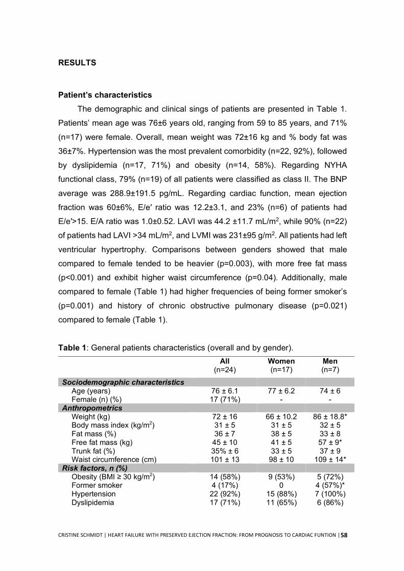
CRISTINE SCHMIDT | HEART FAILURE WITH PRESERVED EJECTION FRACTION: FROM PROGNOSIS TO CARDIAC FUNTION |
58
RESULTS
Patient’s characteristics The demographic and clinical sings of patients are presented in Table 1.
Patients’ mean age was 76±6 years old, ranging from 59 to 85 years, and 71%
(n=17) were female. Overall, mean weight was 72±16 kg and % body fat was
36±7%. Hypertension was the most prevalent comorbidity (n=22, 92%), followed
by dyslipidemia (n=17, 71%) and obesity (n=14, 58%). Regarding NYHA
functional class, 79% (n=19) of all patients were classified as class II. The BNP
average was 288.9±191.5 pg/mL. Regarding cardiac function, mean ejection
fraction was 60±6%, E/e′ ratio was 12.2±3.1, and 23% (n=6) of patients had
E/e′>15. E/A ratio was 1.0±0.52. LAVI was 44.2 ±11.7 mL/m2, while 90% (n=22)
of patients had LAVI >34 mL/m2, and LVMI was 231±95 g/m2. All patients had left
ventricular hypertrophy. Comparisons between genders showed that male
compared to female tended to be heavier (p=0.003), with more free fat mass
(p<0.001) and exhibit higher waist circumference (p=0.04). Additionally, male
compared to female (Table 1) had higher frequencies of being former smoker’s
(p=0.001) and history of chronic obstructive pulmonary disease (p=0.021)
compared to female (Table 1).
Table 1: General patients characteristics (overall and by gender). All
(n=24) Women (n=17)
Men (n=7)
Sociodemographic characteristics Age (years) 76 ± 6.1 77 ± 6.2 74 ± 6 Female (n) (%) 17 (71%) - -
Anthropometrics Weight (kg) 72 ± 16 66 ± 10.2 86 ± 18.8* Body mass index (kg/m2) 31 ± 5 31 ± 5 32 ± 5 Fat mass (%) 36 ± 7 38 ± 5 33 ± 8 Free fat mass (kg) 45 ± 10 41 ± 5 57 ± 9* Trunk fat (%) 35% ± 6 33 ± 5 37 ± 9 Waist circumference (cm) 101 ± 13 98 ± 10 109 ± 14*
Risk factors, n (%) Obesity (BMI ≥ 30 kg/m2) 14 (58%) 9 (53%) 5 (72%) Former smoker 4 (17%) 0 4 (57%)* Hypertension 22 (92%) 15 (88%) 7 (100%) Dyslipidemia 17 (71%) 11 (65%) 6 (86%)
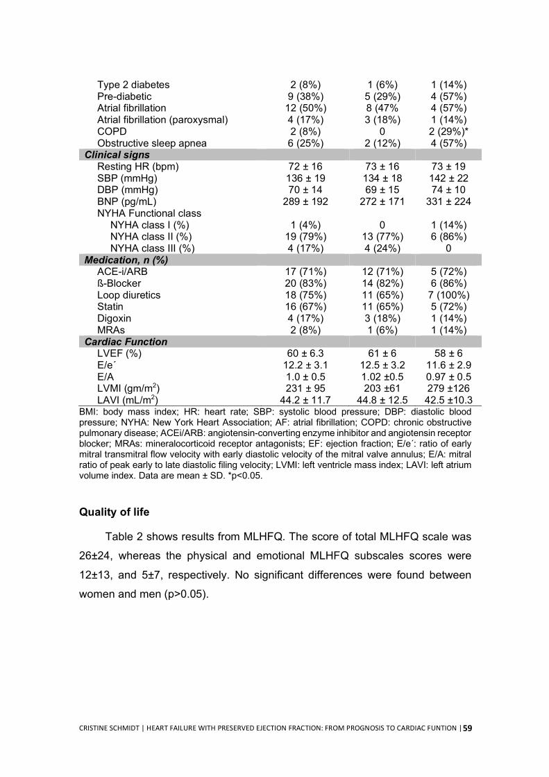
CRISTINE SCHMIDT | HEART FAILURE WITH PRESERVED EJECTION FRACTION: FROM PROGNOSIS TO CARDIAC FUNTION |
59
Type 2 diabetes 2 (8%) 1 (6%) 1 (14%) Pre-diabetic 9 (38%) 5 (29%) 4 (57%) Atrial fibrillation 12 (50%) 8 (47% 4 (57%) Atrial fibrillation (paroxysmal) 4 (17%) 3 (18%) 1 (14%) COPD 2 (8%) 0 2 (29%)* Obstructive sleep apnea 6 (25%) 2 (12%) 4 (57%)
Clinical signs Resting HR (bpm) 72 ± 16 73 ± 16 73 ± 19 SBP (mmHg) 136 ± 19 134 ± 18 142 ± 22 DBP (mmHg) 70 ± 14 69 ± 15 74 ± 10 BNP (pg/mL) 289 ± 192 272 ± 171 331 ± 224 NYHA Functional class
NYHA class I (%) 1 (4%) 0 1 (14%) NYHA class II (%) 19 (79%) 13 (77%) 6 (86%) NYHA class III (%) 4 (17%) 4 (24%) 0
Medication, n (%) ACE-i/ARB 17 (71%) 12 (71%) 5 (72%) ß-Blocker 20 (83%) 14 (82%) 6 (86%) Loop diuretics 18 (75%) 11 (65%) 7 (100%) Statin 16 (67%) 11 (65%) 5 (72%) Digoxin 4 (17%) 3 (18%) 1 (14%) MRAs 2 (8%) 1 (6%) 1 (14%)
Cardiac Function LVEF (%) 60 ± 6.3 61 ± 6 58 ± 6 E/e´ 12.2 ± 3.1 12.5 ± 3.2 11.6 ± 2.9 E/A 1.0 ± 0.5 1.02 ±0.5 0.97 ± 0.5 LVMI (gm/m2) 231 ± 95 203 ±61 279 ±126 LAVI (mL/m2) 44.2 ± 11.7 44.8 ± 12.5 42.5 ±10.3
BMI: body mass index; HR: heart rate; SBP: systolic blood pressure; DBP: diastolic blood pressure; NYHA: New York Heart Association; AF: atrial fibrillation; COPD: chronic obstructive pulmonary disease; ACEi/ARB: angiotensin-converting enzyme inhibitor and angiotensin receptor blocker; MRAs: mineralocorticoid receptor antagonists; EF: ejection fraction; E/e´: ratio of early mitral transmitral flow velocity with early diastolic velocity of the mitral valve annulus; E/A: mitral ratio of peak early to late diastolic filing velocity; LVMI: left ventricle mass index; LAVI: left atrium volume index. Data are mean ± SD. *p<0.05.
Quality of life
Table 2 shows results from MLHFQ. The score of total MLHFQ scale was
26±24, whereas the physical and emotional MLHFQ subscales scores were
12±13, and 5±7, respectively. No significant differences were found between
women and men (p>0.05).
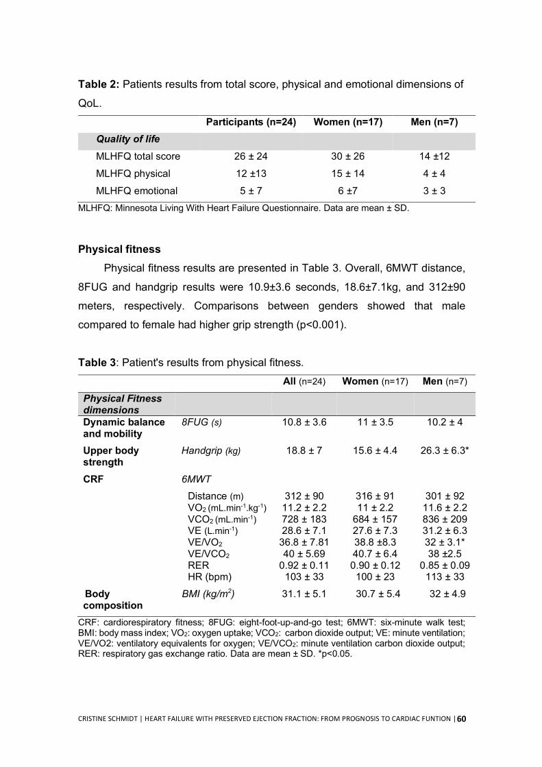
CRISTINE SCHMIDT | HEART FAILURE WITH PRESERVED EJECTION FRACTION: FROM PROGNOSIS TO CARDIAC FUNTION |
60
Table 2: Patients results from total score, physical and emotional dimensions of
QoL. Participants (n=24) Women (n=17) Men (n=7) Quality of life MLHFQ total score 26 ± 24 30 ± 26 14 ±12
MLHFQ physical 12 ±13 15 ± 14 4 ± 4
MLHFQ emotional 5 ± 7 6 ±7 3 ± 3 MLHFQ: Minnesota Living With Heart Failure Questionnaire. Data are mean ± SD.
Physical fitness
Physical fitness results are presented in Table 3. Overall, 6MWT distance,
8FUG and handgrip results were 10.9±3.6 seconds, 18.6±7.1kg, and 312±90
meters, respectively. Comparisons between genders showed that male
compared to female had higher grip strength (p<0.001).
Table 3: Patient's results from physical fitness. All (n=24) Women (n=17) Men (n=7) Physical Fitness dimensions
Dynamic balance and mobility
8FUG (s) 10.8 ± 3.6 11 ± 3.5 10.2 ± 4
Upper body strength
Handgrip (kg) 18.8 ± 7 15.6 ± 4.4 26.3 ± 6.3*
CRF
6MWT Distance (m) 312 ± 90 316 ± 91 301 ± 92 VO2 (mL.min-1.kg-1) 11.2 ± 2.2 11 ± 2.2 11.6 ± 2.2 VCO2 (mL.min-1) 728 ± 183 684 ± 157 836 ± 209 VE (L.min-1) 28.6 ± 7.1 27.6 ± 7.3 31.2 ± 6.3 VE/VO2 36.8 ± 7.81 38.8 ±8.3 32 ± 3.1* VE/VCO2 40 ± 5.69 40.7 ± 6.4 38 ±2.5 RER 0.92 ± 0.11 0.90 ± 0.12 0.85 ± 0.09 HR (bpm) 103 ± 33 100 ± 23 113 ± 33
Body composition
BMI (kg/m2) 31.1 ± 5.1 30.7 ± 5.4 32 ± 4.9
CRF: cardiorespiratory fitness; 8FUG: eight-foot-up-and-go test; 6MWT: six-minute walk test; BMI: body mass index; VO2: oxygen uptake; VCO2: carbon dioxide output; VE: minute ventilation; VE/VO2: ventilatory equivalents for oxygen; VE/VCO2: minute ventilation carbon dioxide output; RER: respiratory gas exchange ratio. Data are mean ± SD. *p<0.05.
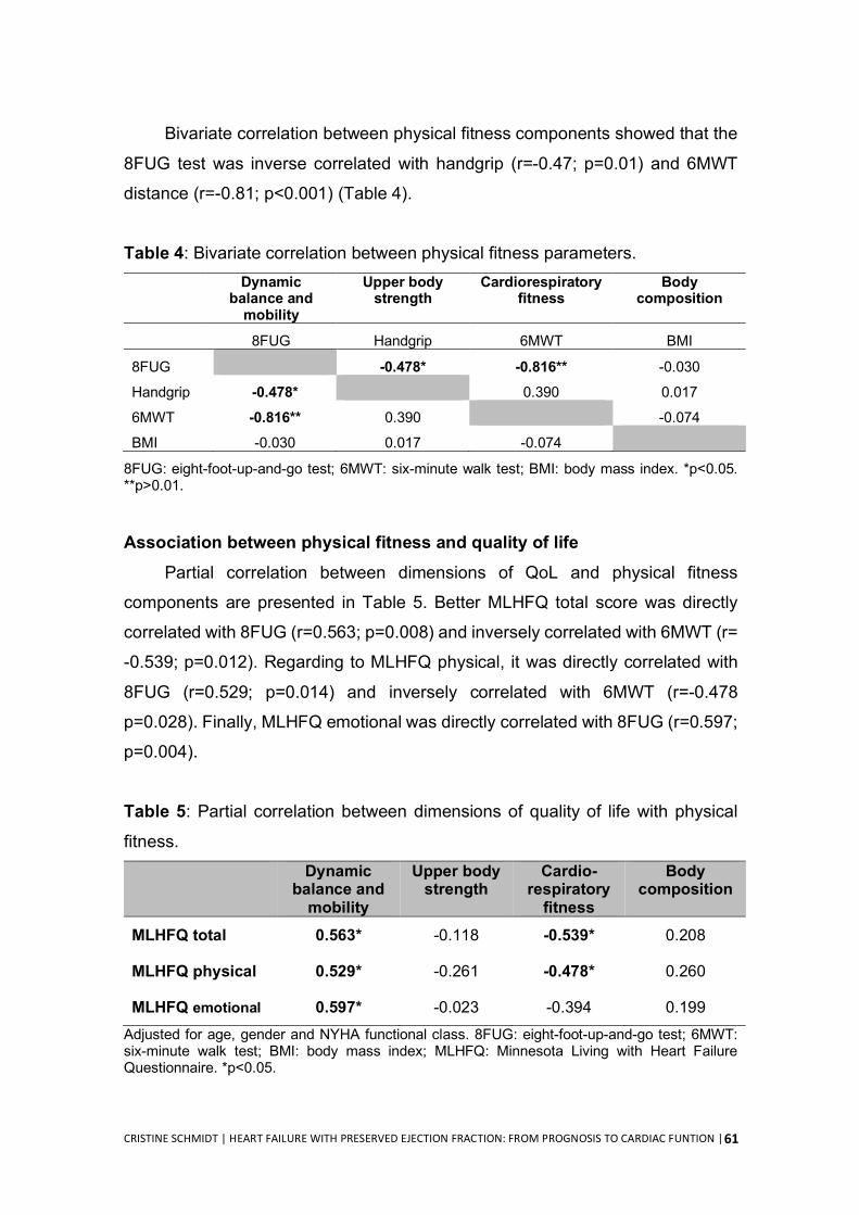
CRISTINE SCHMIDT | HEART FAILURE WITH PRESERVED EJECTION FRACTION: FROM PROGNOSIS TO CARDIAC FUNTION |
61
Bivariate correlation between physical fitness components showed that the
8FUG test was inverse correlated with handgrip (r=-0.47; p=0.01) and 6MWT
distance (r=-0.81; p<0.001) (Table 4).
Table 4: Bivariate correlation between physical fitness parameters. Dynamic
balance and mobility
Upper body strength
Cardiorespiratory fitness
Body composition
8FUG Handgrip 6MWT BMI
8FUG -0.478* -0.816** -0.030
Handgrip -0.478* 0.390 0.017
6MWT -0.816** 0.390 -0.074
BMI -0.030 0.017 -0.074
8FUG: eight-foot-up-and-go test; 6MWT: six-minute walk test; BMI: body mass index. *p<0.05. **p>0.01.
Association between physical fitness and quality of life
Partial correlation between dimensions of QoL and physical fitness
components are presented in Table 5. Better MLHFQ total score was directly
correlated with 8FUG (r=0.563; p=0.008) and inversely correlated with 6MWT (r=
-0.539; p=0.012). Regarding to MLHFQ physical, it was directly correlated with
8FUG (r=0.529; p=0.014) and inversely correlated with 6MWT (r=-0.478
p=0.028). Finally, MLHFQ emotional was directly correlated with 8FUG (r=0.597;
p=0.004). Table 5: Partial correlation between dimensions of quality of life with physical
fitness. Dynamic
balance and mobility
Upper body strength
Cardio-respiratory
fitness
Body composition
MLHFQ total 0.563* -0.118 -0.539* 0.208
MLHFQ physical 0.529* -0.261 -0.478* 0.260
MLHFQ emotional 0.597* -0.023 -0.394 0.199 Adjusted for age, gender and NYHA functional class. 8FUG: eight-foot-up-and-go test; 6MWT: six-minute walk test; BMI: body mass index; MLHFQ: Minnesota Living with Heart Failure Questionnaire. *p<0.05.
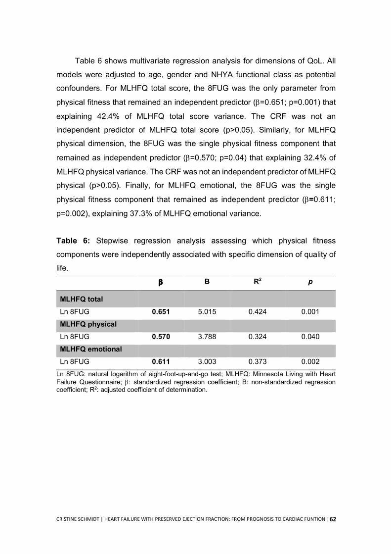
CRISTINE SCHMIDT | HEART FAILURE WITH PRESERVED EJECTION FRACTION: FROM PROGNOSIS TO CARDIAC FUNTION |
62
Table 6 shows multivariate regression analysis for dimensions of QoL. All
models were adjusted to age, gender and NHYA functional class as potential
confounders. For MLHFQ total score, the 8FUG was the only parameter from
physical fitness that remained an independent predictor (b=0.651; p=0.001) that
explaining 42.4% of MLHFQ total score variance. The CRF was not an
independent predictor of MLHFQ total score (p>0.05). Similarly, for MLHFQ
physical dimension, the 8FUG was the single physical fitness component that
remained as independent predictor (b=0.570; p=0.04) that explaining 32.4% of
MLHFQ physical variance. The CRF was not an independent predictor of MLHFQ
physical (p>0.05). Finally, for MLHFQ emotional, the 8FUG was the single
physical fitness component that remained as independent predictor (b=0.611;
p=0.002), explaining 37.3% of MLHFQ emotional variance. Table 6: Stepwise regression analysis assessing which physical fitness
components were independently associated with specific dimension of quality of
life. b B R2 p
MLHFQ total
Ln 8FUG 0.651 5.015 0.424 0.001
MLHFQ physical Ln 8FUG 0.570 3.788 0.324 0.040
MLHFQ emotional Ln 8FUG 0.611 3.003 0.373 0.002
Ln 8FUG: natural logarithm of eight-foot-up-and-go test; MLHFQ: Minnesota Living with Heart Failure Questionnaire; b: standardized regression coefficient; B: non-standardized regression coefficient; R2: adjusted coefficient of determination.
CRISTINE SCHMIDT | HEART FAILURE WITH PRESERVED EJECTION FRACTION: FROM PROGNOSIS TO CARDIAC FUNTION |
63
DISCUSSION
The data provided by our study indicates that physical fitness is positively
correlated with QoL in HFpEF patients. In addition, dynamic balance and mobility
was the unique physical fitness component that was independently associated
with QoL total score, and physical and emotional dimensions. These findings
suggest that this specific component of physical fitness outperforms CRF in
capturing HFpEF patients’ QoL. In addition, it highlights the need to study
interventions targeting specifically these fitness components to enhance QoL
gains.
Despite the high prevalence and poor prognostic of HFpEF, evidence-
based therapies to convincingly reduce morbidity or mortality remains to be
developed (Holland et al., 2011). These patients are often characterized by poor
quality of life and current treatment guidelines highlight the importance of aiming
to improve patients well-being (Ponikowski et al., 2016). Physical fitness is
multicomponent construct (Rikli & Jones, 2013) and several studies shows it is a
major determinant of QoL in HFpEF (Kitzman et al., 2010; Nolte et al., 2015). Our
results corroborate this finding, once we observed that QoL total score strongly
correlated with physical fitness (e.g. dynamic balance and mobility, and CRF) in
HFpEF patients.
Because physical fitness might influence QoL, strategies targeting physical
fitness might potentially improve QoL, independently of further health benefits
(Pandey et al., 2015). A recent meta-analysis showed that the combination of
endurance exercise training together with cardiovascular drugs provide a
clinically relevant improvement in both exercise capacity and QoL in HFpEF
patients (Fukuta et al., 2016). However, physical fitness and QoL are
multicomponent and multidimensional, respectively, and it is crucial to ascertain
which dimension/component is better related to each other to maximize possible
QoL improvements.
Previous studies have shown that CRF is mainly associated with the
physical dimension, but not necessarily with the total score or emotional
dimension of QoL (Edelmann et al., 2011a; Kitzman et al., 2010). We observed
that CRF (assessed by 6MWT) and dynamic balance and mobility (assessed by
CRISTINE SCHMIDT | HEART FAILURE WITH PRESERVED EJECTION FRACTION: FROM PROGNOSIS TO CARDIAC FUNTION |
64
8FUG) were both associated with physical dimensions of QoL. Moreover,
dynamic balance and mobility was the only physical fitness component
associated with emotional dimension of QoL, while upper body strength
(assessed by handgrip) and body composition were not associated with any
dimension. In addition, multivariate analysis revealed that the dynamic balance
and mobility was the only physical fitness component independently associated
with all dimensions of QoL, explaining 42% of variance in the total score QoL,
32% of physical dimension and 37% of emotional dimension of QoL. Thus, of all
the components of physical fitness, dynamic balance and mobility seems to be
the one that better capture QoL in HFpEF patients.
Collectively, our data suggest that improving the specific physical fitness
component of dynamic balance and mobility, will eventually proportionate the
greatest improvement in QoL. 8FUG reflects the specific demands of activities
such as standing up from a seated position, walking short distances, turning,
stopping and sitting down (Wall et al., 2000). This might be explained by the wide
range of physical abilities, including lower body strength, dynamic balance,
walking ability, agility and gait speed (Rikli & Jones, 2013) involved in the 8FUG.
These abilities are also required in the normal daily tasks of an independent and
autonomous life, especially in older people (Mlinac & Feng, 2016). Future studies
(e.g. longitudinal training programs) should assess if an exercise training program
focused on enhancing motor abilities (e.g. dynamic balance and mobility) will
improve the physical and emotional components of QoL in HFpEF in comparison
to current standard ones.
CRISTINE SCHMIDT | HEART FAILURE WITH PRESERVED EJECTION FRACTION: FROM PROGNOSIS TO CARDIAC FUNTION |
65
STUDY LIMITATIONS
The small sample size and the cross-sectional design of our study limits the
generalization of our results. Despite that, our sample gathers the usual clinical
features of HFpEF population reported in large studies (Ponikowski et al., 2016)
with higher prevalence of elderly women and higher prevalence of comorbidities.
Further prospective cohort studies with a larger sample size are needed to
strengthen or refute our conclusions that dynamic balance and mobility is more
discriminative to capturing HFpEF patients' QoL.
CONCLUSION
Overall, our findings indicate that both CRF and dynamic balance and
mobility are directly associated with total score and physical dimensions of QoL
in patients with HFpEF, but only dynamic balance and mobility was concomitantly
associated with the emotional dimension. Multivariate analyses revealed that
dynamic balance and mobility outperforms CRF in capturing HFpEF patients'
QoL.
CRISTINE SCHMIDT | HEART FAILURE WITH PRESERVED EJECTION FRACTION: FROM PROGNOSIS TO CARDIAC FUNTION |
66
REFERENCES Dolgin, M. (1994). Nomenclature and criteria for diagnosis of diseases of the
heart and great vessels. The Criteria Committee of the New York Heart Association (9 ed.). Boston: Little, Brown & Co.
Dunlay, S. M., Roger, V. L., & Redfield, M. M. (2017). Epidemiology of heart failure with preserved ejection fraction. Nat Rev Cardiol, 14(10), 591-602.
Edelmann, F., Gelbrich, G., Dungen, H. D., Frohling, S., Wachter, R., Stahrenberg, R., Binder, L., Topper, A., Lashki, D. J., Schwarz, S., Herrmann-Lingen, C., Loffler, M., Hasenfuss, G., Halle, M., & Pieske, B. (2011a). Exercise training improves exercise capacity and diastolic function in patients with heart failure with preserved ejection fraction: results of the Ex-DHF (Exercise training in Diastolic Heart Failure) pilot study. J Am Coll Cardiol, 58(17), 1780-1791.
Edelmann, F., Stahrenberg, R., Polzin, F., Kockskamper, A., Dungen, H. D., Duvinage, A., Binder, L., Kunde, J., Scherer, M., Gelbrich, G., Hasenfuss, G., Pieske, B., Wachter, R., & Herrmann-Lingen, C. (2011b). Impaired physical quality of life in patients with diastolic dysfunction associates more strongly with neurohumoral activation than with echocardiographic parameters: quality of life in diastolic dysfunction. Am Heart J, 161(4), 797-804.
Fukuta, H., Goto, T., Wakami, K., & Ohte, N. (2016). Effects of drug and exercise intervention on functional capacity and quality of life in heart failure with preserved ejection fraction: A meta-analysis of randomized controlled trials. Eur J Prev Cardiol, 23(1), 78-85.
Holland, D. J., Kumbhani, D. J., Ahmed, S. H., & Marwick, T. H. (2011). Effects of treatment on exercise tolerance, cardiac function, and mortality in heart failure with preserved ejection fraction. A meta-analysis. J Am Coll Cardiol, 57(16), 1676-1686.
Jones, C. J., & Rikli, R. E. (2002). Measuring functional. The Journal on active aging, 1, 24-30.
Kitzman, D. W., Brubaker, P. H., Morgan, T. M., Stewart, K. P., & Little, W. C. (2010). Exercise training in older patients with heart failure and preserved ejection fraction: a randomized, controlled, single-blind trial. Circ Heart Fail, 3(6), 659-667.
Lang, R. M., Badano, L. P., Mor-Avi, V., Afilalo, J., Armstrong, A., Ernande, L., Flachskampf, F. A., Foster, E., Goldstein, S. A., Kuznetsova, T., Lancellotti, P., Muraru, D., Picard, M. H., Rietzschel, E. R., Rudski, L., Spencer, K. T., Tsang, W., & Voigt, J. U. (2015). Recommendations for cardiac chamber quantification by echocardiography in adults: an update from the American Society of Echocardiography and the European Association of Cardiovascular Imaging. Eur Heart J Cardiovasc Imaging, 16(3), 233-270.
CRISTINE SCHMIDT | HEART FAILURE WITH PRESERVED EJECTION FRACTION: FROM PROGNOSIS TO CARDIAC FUNTION |
67
Lohman, T. G., A.F. Roche, and R. Martorell. (1988). Anthropometric Standardization Reference Manual. Champaign, IL: Human Kinetics Books.
MacDermid, J., Solomon, G., & Valdes, K. U. (2015). Clinical Assessment Recommendations: American Society of Hand Therapists.
Mancia, G., Grassi, G., & Redon, J. (2014). Manual of Hypertension of the European Society of Hypertension (Second ed.): CRC Press.
McMurray, J. J., Adamopoulos, S., Anker, S. D., Auricchio, A., Böhm, M., Dickstein, K., Falk, V., Filippatos, G., Fonseca, C., & Gomez-Sanchez, M. A. (2012). ESC Guidelines for the diagnosis and treatment of acute and chronic heart failure 2012: The Task Force for the Diagnosis and Treatment of Acute and Chronic Heart Failure 2012 of the European Society of Cardiology. Developed in collaboration with the Heart Failure Association (HFA) of the ESC. Eur J Heart Fail, 14(8), 803-869.
Mlinac, M. E., & Feng, M. C. (2016). Assessment of Activities of Daily Living, Self-Care, and Independence. Arch Clin Neuropsychol, 31(6), 506-516.
Nolte, K., Herrmann-Lingen, C., Wachter, R., Gelbrich, G., Dungen, H. D., Duvinage, A., Hoischen, N., von Oehsen, K., Schwarz, S., Hasenfuss, G., Halle, M., Pieske, B., & Edelmann, F. (2015). Effects of exercise training on different quality of life dimensions in heart failure with preserved ejection fraction: the Ex-DHF-P trial. Eur J Prev Cardiol, 22(5), 582-593.
Pandey, A., Parashar, A., Kumbhani, D., Agarwal, S., Garg, J., Kitzman, D., Levine, B., Drazner, M., & Berry, J. (2015). Exercise training in patients with heart failure and preserved ejection fraction: meta-analysis of randomized control trials. Circ Heart Fail, 8(1), 33-40.
Ponikowski, P., Voors, A. A., Anker, S. D., Bueno, H., Cleland, J. G. F., Coats, A. J. S., Falk, V., Gonzalez-Juanatey, J. R., Harjola, V. P., Jankowska, E. A., Jessup, M., Linde, C., Nihoyannopoulos, P., Parissis, J. T., Pieske, B., Riley, J. P., Rosano, G. M. C., Ruilope, L. M., Ruschitzka, F., Rutten, F. H., van der Meer, P., & Group, E. S. C. S. D. (2016). 2016 ESC Guidelines for the diagnosis and treatment of acute and chronic heart failure: The Task Force for the diagnosis and treatment of acute and chronic heart failure of the European Society of Cardiology (ESC)Developed with the special contribution of the Heart Failure Association (HFA) of the ESC. Eur Heart J, 37(27), 2129-2200.
Rector, T. S., & Cohn, J. N. (1992). Assessment of patient outcome with the Minnesota Living with Heart Failure questionnaire: reliability and validity during a randomized, double-blind, placebo-controlled trial of pimobendan. Pimobendan Multicenter Research Group. Am Heart J, 124(4), 1017-1025.
Rikli, R. E., & Jones, C. J. (2013). Development and validation of criterion-referenced clinically relevant fitness standards for maintaining physical independence in later years. Gerontologist, 53(2), 255-267.
Riley, L., Guthold, R., Cowan, M., Savin, S., Bhatti, L., Armstrong, T., & Bonita, R. (2016). The World Health Organization STEPwise Approach to
CRISTINE SCHMIDT | HEART FAILURE WITH PRESERVED EJECTION FRACTION: FROM PROGNOSIS TO CARDIAC FUNTION |
68
Noncommunicable Disease Risk-Factor Surveillance: Methods, Challenges, and Opportunities. Am J Public Health, 106(1), 74-78.
Rodriguez-Artalejo, F., Guallar-Castillon, P., Pascual, C. R., Otero, C. M., Montes, A. O., Garcia, A. N., Conthe, P., Chiva, M. O., Banegas, J. R., & Herrera, M. C. (2005). Health-related quality of life as a predictor of hospital readmission and death among patients with heart failure. Arch Intern Med, 165(11), 1274-1279.
The World Health Organization Quality of Life Assessment Group. (1998). The World Health Organization Quality of Life Assessment (WHOQOL): development and general psychometric properties. Soc Sci Med, 46(12), 1569-1585.
Wall, J. C., Bell, C., Campbell, S., & Davis, J. (2000). The Timed Get-up-and-Go test revisited: measurement of the component tasks. J Rehabil Res Dev, 37(1), 109-113.
WHO. (2000). Obesity: preventing and managing the global epidemic. Report of a WHO consultation. World Health Organ Tech Rep Ser, 894, 1-253.
CRISTINE SCHMIDT | HEART FAILURE WITH PRESERVED EJECTION FRACTION: FROM PROGNOSIS TO CARDIAC FUNTION |
69
STUDY II
COMPARISON OF QUESTIONNAIRE AND ACCELEROMETER-BASED
ASSESSMENTS OF PHYSICAL ACTIVITY IN PATIENTS WITH HEART FAILURE WITH PRESERVED EJECTION FRACTION: CLINICAL AND
PROGNOSTIC IMPLICATIONS

CRISTINE SCHMIDT | HEART FAILURE WITH PRESERVED EJECTION FRACTION: FROM PROGNOSIS TO CARDIAC FUNTION |
70
CRISTINE SCHMIDT | HEART FAILURE WITH PRESERVED EJECTION FRACTION: FROM PROGNOSIS TO CARDIAC FUNTION |
71
ABSTRACT
Rational: Heart failure with preserved ejection fraction (HFpEF) accounts for approximately 50% of heart failure cases and is associated with poor outcome. Higher physical activity (PA) levels are associated with greater quality of life and lower risk of hospitalization and mortality. Purpose: The study aims: i) to compare daily PA levels and sedentary time evaluated by the International Physical Activity Questionnaire (IPAQ) and by a triaxial accelerometer in HFpEF patients; ii) to describe daily PA patterns and sedentary time in HFpEF patients based in objective measurements; and iii) to observe the association between prognostic indicators and PA, and sedentary time measurements. Methods: This is a cross-sectional study with 24 stable and well-medicated HFpEF patients. Physical activity was assessed through the IPAQ short version and triaxial accelerometry (ActiGraph GTX3). Time spent in moderate-to-vigorous PA (MVPA) from IPAQ was computed as self-reported walking and MVPA. MVPA from acceletrometry was set as ≥2752 counts/minute. Prognostic indicators were: distance on the 6-minute-walking test (6MWT), oxygen consumption (VO2) during the test, quality of life, brain natriuretic peptide plasma level, and E/e´ ratio. Results: Sample mean age was 76±4 years. Compared to accelerometry, IPAQ underestimated sedentary time (253±156 vs. 392±104 min/day, p=0.001) and overestimated MVPA (44±56 vs 19.3±26. min/day, p<0.001). Using IPAQ, 59% of patients met international PA by international guidelines, while using accelerometer-derived data, 27% of patients actually met the guidelines, decreasing to zero when considering the number of 10min-MVPA bouts. On average, accelerometer-derived data showed that HFpEF patients spent 50% (392±104 min/day) of their waking time in sedentary behaviours and 2.5% (19.3±26 min/day) in MVPA. Of measured surrogate prognostic markers, functional capacity (6MWT, r=0.652, p=0.04; VO2, r=0.512=, p=0.02) and quality of life (r=-0.490, p=0.04) were correlated with MVPA. Conclusion: The IPAQ short version underestimated sedentary time and over-estimated MVPA in patients with HFpEF. Using accelerometer-derived data, HFpEF patients spent only a minority of their time involved in MVPA, which was the only PA pattern positively associated with prognostic indicators. Our results highlight the importance of replacing some time spent in sedentary activities by MVPA in HFpEF throughout the day. Key-words: physical activity, IPAQ, accelerometer, HFpEF, prognostic indicators.

CRISTINE SCHMIDT | HEART FAILURE WITH PRESERVED EJECTION FRACTION: FROM PROGNOSIS TO CARDIAC FUNTION |
72

CRISTINE SCHMIDT | HEART FAILURE WITH PRESERVED EJECTION FRACTION: FROM PROGNOSIS TO CARDIAC FUNTION |
73
INTRODUCTION
Epidemiologic evidence indicates that heart failure (HF) prevalence has
been increasing in the last decade, with heart failure with preserved ejection
fraction (HFpEF) accounting up to half of HF population (Oktay et al., 2013). The
growing prevalence of HFpEF is attributed to life-expectancy expansion, better
diagnostic awareness therapy (Glezeva & Baugh, 2014), and increasing
prevalence of major risk factors (e.g. obesity, diabetes, hypertension and atrial
fibrillation) (Ather et al., 2012). Consequently, it is expected that HFpEF will
become the most common cause of hospitalization in older adults over the
coming years (Nanayakkara et al., 2018). Since HFpEF continues to be resistant
to current therapies, primary prevention strategies might be useful to control its
growing burden at the population level (Pandey et al., 2017a). Therefore, in
addition to understanding the natural history and pathophysiology of HFpEF, it
will be important to identify modifiable risk factors that can be targeted.
Lifestyle risk factors such as low physical activity (PA) and high sedentary
time are recognized as primary factors for most chronic diseases, including HF
(Booth et al., 2012). While it remains to be clarified which HF phenotypes is most
affected, recent observations have more strongly implicated low PA in the
development of HFpEF (Pandey et al., 2017b). An inverse relationship between
leisure-time PA and HF risk was recently demonstrate, where lower levels of PA
were associated with higher risk of HFpEF, but not HF with reduced ejection
fraction (Pandey et al., 2017). In addition, evidence shows that a sedentary
lifestyle (e.g. prolonged bed rest) is associated with many underlying cardiac and
skeletal muscle abnormalities often present in HFpEF (Dorfman et al., 2008;
Irimia et al., 2017). For those with established HFpEF, lower levels PA have been
associated with poor quality of life (QoL) and worse clinical outcomes such as
functional class (NYHA) (Snipelisky et al., 2017), hospitalisation rate, and
mortality (Hegde et al., 2017). Overall, these observational studies suggest that
PA and/or sedentary time are important treatment targets to improve clinical
outcomes in HFpEF.
Regardless the population, daily PA assessment is a challenging task.
There are different methods (e.g. self-report methods such as questionnaires or
CRISTINE SCHMIDT | HEART FAILURE WITH PRESERVED EJECTION FRACTION: FROM PROGNOSIS TO CARDIAC FUNTION |
74
objective methods such as accelerometry), and each one has its own limitations
and strengths. Despite of it, accelerometry seems to be superior than
questionnaires in terms of accuracy, reliability, representatively of daily activities
and related energy expenditure (Skender et al., 2016). Additionally, in clinical
settings (e.g. diagnosis, prognosis, or prescription of therapeutic interventions)
accelerometry might represent a challenge compared to questionnaires, which
are better in terms of costs and practicality. However, correlation between self-
report and objective PA measurements in the large majority of studies is poor
(Lee et al., 2011). Additionally, there are no data comparing objective PA
measurements (e.g. accelerometers) and questionnaire in HFpEF patients.
Therefore, this study aimed: i) to determine the validity of the International
Physical Activity Questionnaire (IPAQ) against objective measures from triaxial
accelerometry in HFpEF patients; ii) to describe daily PA patterns and sedentary
time in HFpEF patients based in objective measurements, and iii) to observe the
association between prognostic indicators and PA and sedentary time
measurements.
CRISTINE SCHMIDT | HEART FAILURE WITH PRESERVED EJECTION FRACTION: FROM PROGNOSIS TO CARDIAC FUNTION |
75
MATERIAL AND METHODS Study design, recruitment and participants
This is a cross-sectional study conducted in a Portuguese public hospital
(Centro Hospitalar do Porto - Hospital de Santo Antonio, Porto). Inclusion criteria
was diagnosis of HFpEF according to the European Society of Cardiology
guidelines (Ponikowski et al., 2016). Patients were excluded if they presented
with unstable angina, acute coronary syndrome as primary diagnosis,
symptomatic severe aortic stenosis, acute pulmonary embolus, acute
myocarditis, uncompensated heart failure, uncontrolled hypertension, complex
ventricular arrhythmias, severe renal dysfunction, severe chronic obstructive
pulmonary disease, medical or orthopaedic conditions that precluded
independent ambulation and exercise testing.
Patients who potentially were eligible to participate in the study were
identified from the clinical files of the hospital’s cardiology department. A total of
30 patients were invited through phone calls by a cardiologist. From those, 24
patients (17 female and 7 male) accepted to take part in the study, but only 22
completed data acquisition. The study was approved by the Ethics Committee of
Centro Hospitalar do Porto - Hospital de Santo Antonio (N/S: 2015.125). All
procedures were conducted according to the declaration of Helsinki, and
participants signed informed consent to participate.
Data were collected from November 2016 to September 2017 in a single
day in the hospital. Data collection
Blood pressure A trained researcher performed blood pressure measurements after
10 minutes resting in seated position. Blood pressure was assessed (Colin, BP
8 800; Critikron, Inc., USA) in the left arm. Systolic blood pressure (SBP) and
Diastolic blood pressure (DBP) were computed as the average of 3 readings.
Additional measurements were performed when differences between readings
exceeded 5 mmHg (Mancia et al., 2014).
CRISTINE SCHMIDT | HEART FAILURE WITH PRESERVED EJECTION FRACTION: FROM PROGNOSIS TO CARDIAC FUNTION |
76
Blood collection and biochemical determinations
Peripheral venous blood (15 mL) was collected to EDTA tube. The EDTA
tubes were immediately placed on ice and allowed to clot for 30 minutes before
centrifugation for 15 minutes at 1000xg. The plasma was aliquoted and stored at
-80ºC for biochemical analysis. Brain Natriuretic Peptide (BNP) was quantified in
a certificated laboratory by chemiluminescent microparticle immunoassay
(ARCHITECT BNP).
Anthropometric Measures Body height (cm) was measured standing upright against a stadiometer
(Holtain Ltd., Crymmych, UK) (Lohman, 1988). Patients were bare or stocking
feet. Weight (kg), body mass index (BMI; kg/m2), fat mass (%) and free fat mass
(kg) were measured with patients lightly dressed, using an electronic segmental
body composition analyser (Tanita, BC-418, Tokyo, Japan). Waist circumference
(cm) was measured at the midpoint between the lowest rib and the iliac crest at
the end of normal expiration (Riley et al., 2016). Obesity was determined as BMI
equal or higher than 30 kg/m2 (WHO, 2000).
Self-Reported Physical Activity Self-reported physical activity was assessed with the short version of the
International Physical Activity Questionnaire (IPAQ-SF) (Craig et al., 2003),
through personal interview. The IPAQ contains seven items to estimate PA
frequency and duration during the previous seven days. Additional questions on
sedentariness were also asked. The IPAQ focus on vigorous and moderate
physical activity intensities and walking periods lasting at least 10 minutes on
week days. Additionally, the questionnaire asked about “sitting time” on week and
weekend days. Frequency of activity was measured in days, and duration in
hours and minutes. Total weekly PA was estimated using the instrument's scoring
protocol by weighting time spent in each activity intensity with its estimated
metabolic equivalent (MET) energy expenditure (Ipaq Research Committee,
2005). IPAQ scoring protocol assigns the values of 3.3 METs to walking, 4.0
CRISTINE SCHMIDT | HEART FAILURE WITH PRESERVED EJECTION FRACTION: FROM PROGNOSIS TO CARDIAC FUNTION |
77
METs to moderate, and 8.0 METs vigorous activity (Ipaq Research Committee,
2005). Meeting physical activity guidelines was defined as accumulating ≥150
minutes per week of moderate-intensity physical activity (MVPA) (≥3 METs)
(Haskell et al., 2007). Thus, to estimate MVPA data, we merged the reported
activities ≥3 METs (walking + moderate + vigorous PA).
Accelerometer-assessed physical activity Daily PA was measured using a triaxial accelerometer (Actigraph GT3X,
Pensacola, FL, USA). The triaxial activity monitor measures acceleration in three
individual orthogonal planes (vertical, antero-posterior and medio-lateral) and
provides activity counts as a composite vector magnitude (VM) of these three
axes (Sasaki et al., 2011). The VM is the square root of quadrate of the three
separate dimensional axes [(x2+y2+z2)1/2] (Crouter et al., 2012). Participants were
instructed to wear the accelerometer over the right hip for eight consecutive days,
except while sleeping, bathing and water-based activities. The accelerometer
was programmed to record triaxial data at a frequency of 30 Hz and 1 second
length epochs. ActiLife software (Actigraph, Florida, USA, version 6.9) was used
to process the accelerometer data. Data were downloaded and integrated into
60-second epochs. Non-wear time was defined as 90 consecutive minutes of
zero counts, with an allowance of 2-minutes of nonzero counts provided there
were 30-minute consecutive zero count windows up and downstream (Choi et al.,
2011). Non-wear time was excluded from the analysis. Patients with valid data
were those having a minimum of 4 days with at least 10 hours/day of wear-time.
The output of the Actigraph was given in counts per minute (CPM) derived from
VM. The average minutes/day spent at different categories of PA intensity was
determined according to cut point that relate PA to counts/min: sedentary time
(<200 counts/min) (Aguilar-Farias et al., 2014), light PA (LPA) (200-2751
counts/min) and moderate and vigorous PA (MVPA) (>2752 counts/min) (Santos-
Lozano et al., 2013). Meeting international PA guidelines was defined as ≥150
minutes/week of MVPA from accelerometry. This definition was applied
considering: i) only MVPA that occurred in bouts of ≥10 minutes’ long (episodes
of continuous MVPA lasting for at least 10 minutes) as specified by the guidelines
CRISTINE SCHMIDT | HEART FAILURE WITH PRESERVED EJECTION FRACTION: FROM PROGNOSIS TO CARDIAC FUNTION |
78
(Haskell et al., 2007), and ii) without bout length restriction (e.g. considering all
MVPA including MVPA occurring in bouts shorter than 10 minutes’ duration).
Functional classification Patients were classified by the physician into subgroups based on their
symptoms using the New York Heart Association (NYHA) functional class.
Patients symptoms is based on how much they are limited during physical activity
(class I to IV) (Dolgin, 1994).
Echocardiography Evaluation Supine transthoracic echocardiography was performed using a
cardiovascular ultrasound Vivid E95 ® (GE Healthcare). All quantitative
echocardiographic measurements were performed by a single reader blinded to
the results of the other evaluations, using a computerized off-line analysis station.
Peak early diastolic tissue velocity was measured at the septal and lateral mitral
annulus. Mitral inflow velocity was assessed by pulsed wave Doppler from the
apical 4-chamber view, positioning the sample volume at the tip of the mitral
leaflets. E/e’ ratio was calculated as E wave divided by e’ velocities. LV mass
was estimated from LV linear dimensions and indexed to body surface area as
recommended by ESC guidelines (Lang et al., 2015). LV hypertrophy was
defined as LV mass indexed to body surface area (LV mass index) >115 g/m2 in
men or >95 g/m2 in women. LV volumes were estimated by the modified Simpson
method using the apical 4- and 2-chamber views, and LVEF was derived from
volumes in the standard manner. LA volume was estimated by the method of
disks using apical 4- and 2-chamber views at an end-systolic frame preceding
mitral valve opening and was indexed to body surface area to derive LA volume
index.
Cardiorespiratory fitness with pulmonary gas exchange analysis Cardiorespiratory fitness was assessed by the 6-minute walk test (6MWT)
in a 25-m-long unobstructed corridor. Participants were instructed to walk the
maximal distance in 6 minutes time. Resting stops were allowed when patients
CRISTINE SCHMIDT | HEART FAILURE WITH PRESERVED EJECTION FRACTION: FROM PROGNOSIS TO CARDIAC FUNTION |
79
feel to be necessary. The 6MWT was performed wearing a portable gas analyser
(K4b2, Cosmed, Rome, Italy). Oxygen uptake (VO2; mL.min-1.kg-1) was measured
directly and continuously. Respiratory samplings were collected in a breath-by-
breath, and then, data was averaged over 5-s intervals. Data was calculated as
the averaged of measures taken in test total duration (6 minutes).
Health-related quality of life Health-related QoL was performed by interview through the Minnesota
Living With Heart Failure Questionnaire (MLWHFQ). The MLHFQ encompasses
21 questions, whose purpose is to determine how disease affects the physical,
psychological and socioeconomic conditions of the patients during the previous
month (Rector & Cohn, 1992). The questions compass symptoms and signs
relevant to disease, levels of PA, work, social interaction, sexual activity, and
emotions. The MLHFQ total score range from 0 to 105 (no impairment to
maximum impairment). Two other scores can be determined: the physical
dimension (8 items, 0–40), and the emotional dimension (5 items, 0–25). Higher
MLHFQ score mean worse QoL. Answers options ranges from 0 (none) to 5 (very
much), where 0 represented no limitation and 105 represented maximal
limitation.
Statistical analyses Statistical analysis was performed using the IBM SPSS 24 software (SPSS,
USA). Normal data distribution was examined by the Shapiro Wilk test. Non-
normal data was transformed with the square root (IPAQ-MVPA,
IPAQ_METs/day, accelerometer-MVPA, accelerometer-10min-bouts-MVPA,
MLHFQ total score, MLHFQ physical and MLHFQ emotional) or its natural
logarithm (weight, fat mass, free fat mass, accelerometer total hours and
accelerometer total days), for subsequent analysis and then transformed back to
the original scale for the purpose of clarity. Categorical data are reported as
absolute values and percentages. Between gender and age comparisons were
performed by independent t-test and chi-square test as appropriated. Cut point
to age analyses was defined by median, and it was set as 76 years old. Siting
CRISTINE SCHMIDT | HEART FAILURE WITH PRESERVED EJECTION FRACTION: FROM PROGNOSIS TO CARDIAC FUNTION |
80
time and walk+moderate+vigorous PA (MVPA) from IPAQ were compared with
sedentary time, total MVPA and 10 min-bouts of MVPA derived from GT3X
accelerometer, using paired t-test. Partial correlation was used to analyse the
relationship between the variables derived from the two methods adjusted by
gender and age. The strength and limits of agreement between the two methods
was assessed using the Bland–Altman technique (Bland & Altman, 1986).
Statistical significance was established for p <0.05.
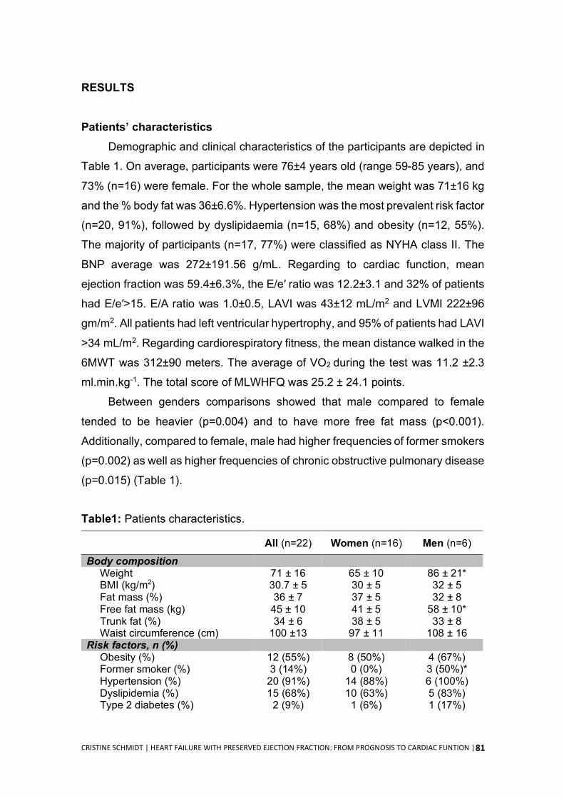
CRISTINE SCHMIDT | HEART FAILURE WITH PRESERVED EJECTION FRACTION: FROM PROGNOSIS TO CARDIAC FUNTION |
81
RESULTS
Patients’ characteristics Demographic and clinical characteristics of the participants are depicted in
Table 1. On average, participants were 76±4 years old (range 59-85 years), and
73% (n=16) were female. For the whole sample, the mean weight was 71±16 kg
and the % body fat was 36±6.6%. Hypertension was the most prevalent risk factor
(n=20, 91%), followed by dyslipidaemia (n=15, 68%) and obesity (n=12, 55%).
The majority of participants (n=17, 77%) were classified as NYHA class II. The
BNP average was 272±191.56 g/mL. Regarding to cardiac function, mean
ejection fraction was 59.4±6.3%, the E/e′ ratio was 12.2±3.1 and 32% of patients
had E/e′>15. E/A ratio was 1.0±0.5, LAVI was 43±12 mL/m2 and LVMI 222±96
gm/m2. All patients had left ventricular hypertrophy, and 95% of patients had LAVI
>34 mL/m2. Regarding cardiorespiratory fitness, the mean distance walked in the
6MWT was 312±90 meters. The average of VO2 during the test was 11.2 ±2.3
ml.min.kg-1. The total score of MLWHFQ was 25.2 ± 24.1 points.
Between genders comparisons showed that male compared to female
tended to be heavier (p=0.004) and to have more free fat mass (p<0.001).
Additionally, compared to female, male had higher frequencies of former smokers
(p=0.002) as well as higher frequencies of chronic obstructive pulmonary disease
(p=0.015) (Table 1).
Table1: Patients characteristics.
All (n=22) Women (n=16) Men (n=6) Body composition
Weight 71 ± 16 65 ± 10 86 ± 21* BMI (kg/m2) 30.7 ± 5 30 ± 5 32 ± 5 Fat mass (%) 36 ± 7 37 ± 5 32 ± 8 Free fat mass (kg) 45 ± 10 41 ± 5 58 ± 10* Trunk fat (%) 34 ± 6 38 ± 5 33 ± 8 Waist circumference (cm) 100 ±13 97 ± 11 108 ± 16
Risk factors, n (%) Obesity (%) 12 (55%) 8 (50%) 4 (67%) Former smoker (%) 3 (14%) 0 (0%) 3 (50%)* Hypertension (%) 20 (91%) 14 (88%) 6 (100%) Dyslipidemia (%) 15 (68%) 10 (63%) 5 (83%) Type 2 diabetes (%) 2 (9%) 1 (6%) 1 (17%)
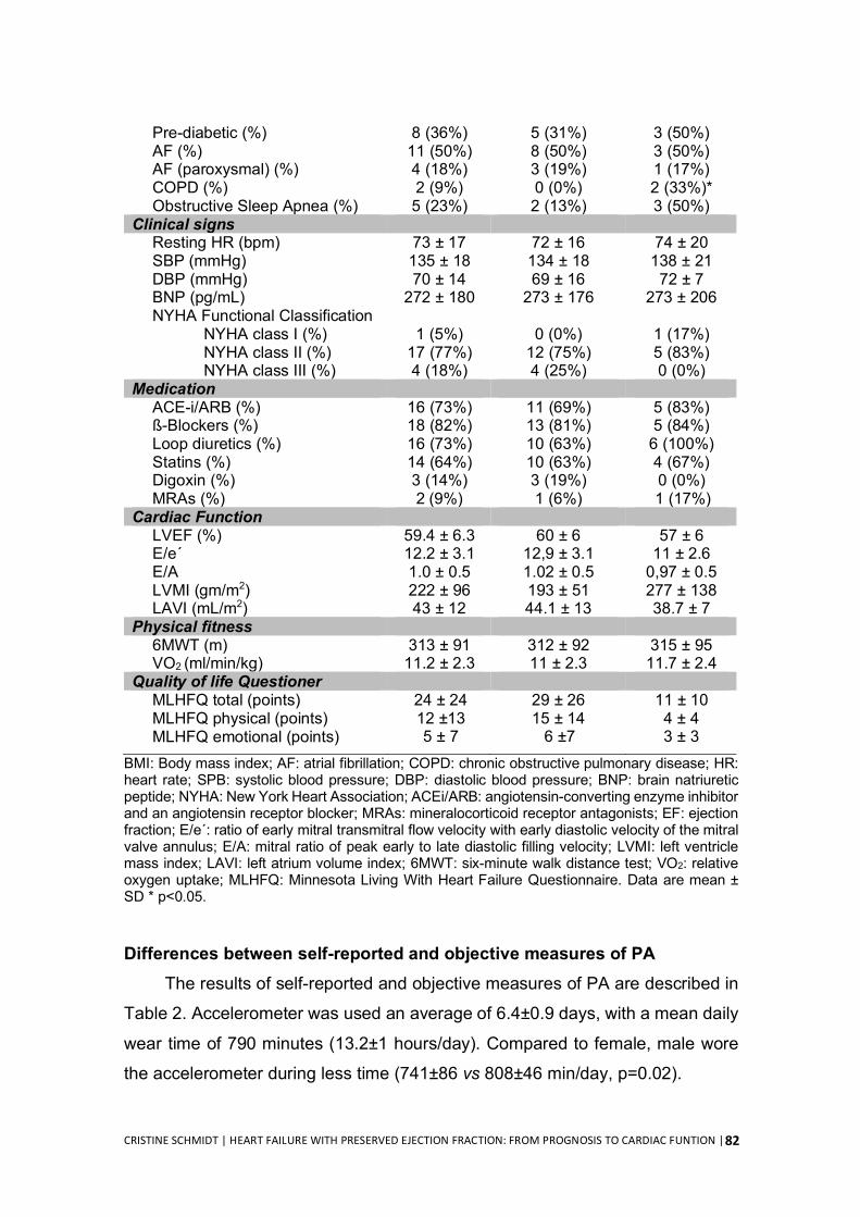
CRISTINE SCHMIDT | HEART FAILURE WITH PRESERVED EJECTION FRACTION: FROM PROGNOSIS TO CARDIAC FUNTION |
82
Pre-diabetic (%) 8 (36%) 5 (31%) 3 (50%) AF (%) 11 (50%) 8 (50%) 3 (50%) AF (paroxysmal) (%) 4 (18%) 3 (19%) 1 (17%) COPD (%) 2 (9%) 0 (0%) 2 (33%)* Obstructive Sleep Apnea (%) 5 (23%) 2 (13%) 3 (50%)
Clinical signs Resting HR (bpm) 73 ± 17 72 ± 16 74 ± 20 SBP (mmHg) 135 ± 18 134 ± 18 138 ± 21 DBP (mmHg) 70 ± 14 69 ± 16 72 ± 7 BNP (pg/mL) 272 ± 180 273 ± 176 273 ± 206 NYHA Functional Classification
NYHA class I (%) 1 (5%) 0 (0%) 1 (17%) NYHA class II (%) 17 (77%) 12 (75%) 5 (83%) NYHA class III (%) 4 (18%) 4 (25%) 0 (0%)
Medication ACE-i/ARB (%) 16 (73%) 11 (69%) 5 (83%) ß-Blockers (%) 18 (82%) 13 (81%) 5 (84%) Loop diuretics (%) 16 (73%) 10 (63%) 6 (100%) Statins (%) 14 (64%) 10 (63%) 4 (67%) Digoxin (%) 3 (14%) 3 (19%) 0 (0%) MRAs (%) 2 (9%) 1 (6%) 1 (17%)
Cardiac Function LVEF (%) 59.4 ± 6.3 60 ± 6 57 ± 6 E/e´ 12.2 ± 3.1 12,9 ± 3.1 11 ± 2.6 E/A 1.0 ± 0.5 1.02 ± 0.5 0,97 ± 0.5 LVMI (gm/m2) 222 ± 96 193 ± 51 277 ± 138 LAVI (mL/m2) 43 ± 12 44.1 ± 13 38.7 ± 7
Physical fitness 6MWT (m) 313 ± 91 312 ± 92 315 ± 95 VO2 (ml/min/kg) 11.2 ± 2.3 11 ± 2.3 11.7 ± 2.4
Quality of life Questioner MLHFQ total (points) 24 ± 24 29 ± 26 11 ± 10 MLHFQ physical (points) 12 ±13 15 ± 14 4 ± 4 MLHFQ emotional (points) 5 ± 7 6 ±7 3 ± 3
BMI: Body mass index; AF: atrial fibrillation; COPD: chronic obstructive pulmonary disease; HR: heart rate; SPB: systolic blood pressure; DBP: diastolic blood pressure; BNP: brain natriuretic peptide; NYHA: New York Heart Association; ACEi/ARB: angiotensin-converting enzyme inhibitor and an angiotensin receptor blocker; MRAs: mineralocorticoid receptor antagonists; EF: ejection fraction; E/e´: ratio of early mitral transmitral flow velocity with early diastolic velocity of the mitral valve annulus; E/A: mitral ratio of peak early to late diastolic filling velocity; LVMI: left ventricle mass index; LAVI: left atrium volume index; 6MWT: six-minute walk distance test; VO2: relative oxygen uptake; MLHFQ: Minnesota Living With Heart Failure Questionnaire. Data are mean ± SD * p<0.05.
Differences between self-reported and objective measures of PA
The results of self-reported and objective measures of PA are described in
Table 2. Accelerometer was used an average of 6.4±0.9 days, with a mean daily
wear time of 790 minutes (13.2±1 hours/day). Compared to female, male wore
the accelerometer during less time (741±86 vs 808±46 min/day, p=0.02).
CRISTINE SCHMIDT | HEART FAILURE WITH PRESERVED EJECTION FRACTION: FROM PROGNOSIS TO CARDIAC FUNTION |
83
Mean total activity volume from IPAQ was 152±183 MET/min/day, with older
patients compared to younger, reporting lower activity volume (232±71 vs 72±114
MET/min/day, respectively; p=0.03). From accelerometry, mean total activity
volume was 550±239 CPM.
Mean sedentary time was significantly lower in self-reported measurement
(253±156 min.day-1) compared to accelerometry (392±104 min.day-1, p=0.001).
No significant differences were found regarding to gender and age (p>0.05).
Considering MVPA, self-reported daily time compared to accelerometer was
higher (44±56 vs. 19.3±26 min/day, respectively; p<0.001). Applying the ≥10min-
bouts at MVPA criterion, the discrepancy between measurements was even
higher (IPAQ= 44±56 min/day; Accelerometry= 1.1±2.4 min/day, p<0.001). From
accelerometry data, it was possible to verify that only 5 patients accomplished
with at least one bout of MVPA per week. Gender comparison showed that female
compared to male, spent less time in 10 min-bouts of MVPA per day (0.2±0.8 vs
3.4±7, respectively; p=0.01). Analysis by age showed that older patients spent
significantly less time in MVPA (34 ± 31 vs 5±3, p=0.002) and did not perform
10min-bouts of MVPA.
Regarding to LPA, comparisons are not possible because it is measured
only via accelerometry. Mean time in LPA was 379±128 min/day. Gender
comparisons show that male compared to female spent less time in LPA
(423±111 vs 260±91, respectively; p=0.005), while no significant difference was
found comparing Youngers and elders (p>0.05).
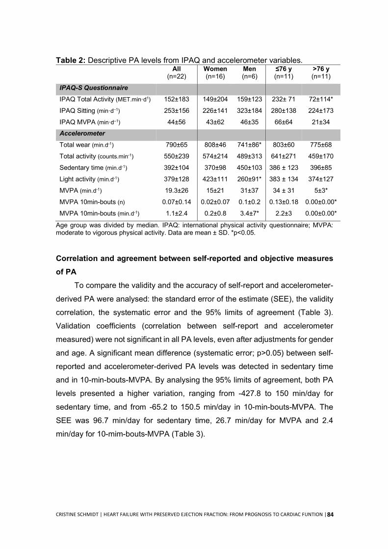
CRISTINE SCHMIDT | HEART FAILURE WITH PRESERVED EJECTION FRACTION: FROM PROGNOSIS TO CARDIAC FUNTION |
84
Table 2: Descriptive PA levels from IPAQ and accelerometer variables. All
(n=22) Women (n=16)
Men (n=6)
≤76 y (n=11)
>76 y (n=11)
IPAQ-S Questionnaire
IPAQ Total Activity (MET.min·d1) 152±183 149±204 159±123 232± 71 72±114*
IPAQ Sitting (min·d–1) 253±156 226±141 323±184 280±138 224±173
IPAQ MVPA (min·d–1) 44±56 43±62 46±35 66±64 21±34
Accelerometer
Total wear (min.d-1) 790±65 808±46 741±86* 803±60 775±68
Total activity (counts.min-1) 550±239 574±214 489±313 641±271 459±170
Sedentary time (min.d-1) 392±104 370±98 450±103 386 ± 123 396±85
Light activity (min.d-1) 379±128 423±111 260±91* 383 ± 134 374±127
MVPA (min.d-1) 19.3±26 15±21 31±37 34 ± 31 5±3*
MVPA 10min-bouts (n) 0.07±0.14 0.02±0.07 0.1±0.2 0.13±0.18 0.00±0.00*
MVPA 10min-bouts (min.d-1) 1.1±2.4 0.2±0.8 3.4±7* 2.2±3 0.00±0.00*
Age group was divided by median. IPAQ: international physical activity questionnaire; MVPA: moderate to vigorous physical activity. Data are mean ± SD. *p<0.05.
Correlation and agreement between self-reported and objective measures of PA
To compare the validity and the accuracy of self-report and accelerometer-
derived PA were analysed: the standard error of the estimate (SEE), the validity
correlation, the systematic error and the 95% limits of agreement (Table 3).
Validation coefficients (correlation between self-report and accelerometer
measured) were not significant in all PA levels, even after adjustments for gender
and age. A significant mean difference (systematic error; p>0.05) between self-
reported and accelerometer-derived PA levels was detected in sedentary time
and in 10-min-bouts-MVPA. By analysing the 95% limits of agreement, both PA
levels presented a higher variation, ranging from -427.8 to 150 min/day for
sedentary time, and from -65.2 to 150.5 min/day in 10-min-bouts-MVPA. The
SEE was 96.7 min/day for sedentary time, 26.7 min/day for MVPA and 2.4
min/day for 10-mim-bouts-MVPA (Table 3).
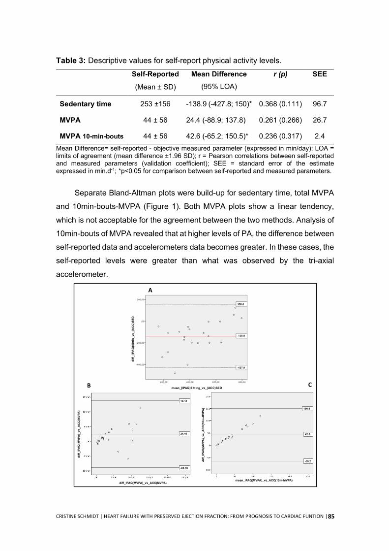
CRISTINE SCHMIDT | HEART FAILURE WITH PRESERVED EJECTION FRACTION: FROM PROGNOSIS TO CARDIAC FUNTION |
85
Table 3: Descriptive values for self-report physical activity levels. Self-Reported
(Mean ± SD)
Mean Difference (95% LOA)
r (p) SEE
Sedentary time 253 ±156 -138.9 (-427.8; 150)* 0.368 (0.111) 96.7
MVPA 44 ± 56 24.4 (-88.9; 137.8) 0.261 (0.266) 26.7
MVPA 10-min-bouts 44 ± 56 42.6 (-65.2; 150.5)* 0.236 (0.317) 2.4 Mean Difference= self-reported - objective measured parameter (expressed in min/day); LOA = limits of agreement (mean difference ±1.96 SD); r = Pearson correlations between self-reported and measured parameters (validation coefficient); SEE = standard error of the estimate expressed in min.d-1; *p<0.05 for comparison between self-reported and measured parameters.
Separate Bland-Altman plots were build-up for sedentary time, total MVPA
and 10min-bouts-MVPA (Figure 1). Both MVPA plots show a linear tendency,
which is not acceptable for the agreement between the two methods. Analysis of
10min-bouts of MVPA revealed that at higher levels of PA, the difference between
self-reported data and accelerometers data becomes greater. In these cases, the
self-reported levels were greater than what was observed by the tri-axial
accelerometer.
137.8
-88.93
24.45
diff_
IPA
Q(M
VPA
)_vs
_AC
C(M
VPA
)
diff_IPAQ(MVPA)_vs_ACC(MVPA)
CB
A
diff_IPAQ(MVPA)_vs_ACC(10m-MVPA)
mean_IPAQ(MVPA)_vs_ACC(10m-MVPA)
150.5
-65.2
42.6
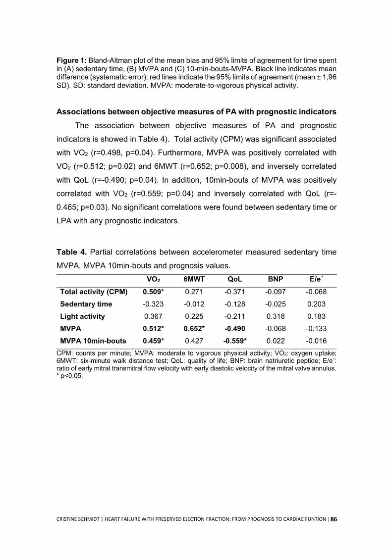
CRISTINE SCHMIDT | HEART FAILURE WITH PRESERVED EJECTION FRACTION: FROM PROGNOSIS TO CARDIAC FUNTION |
86
Figure 1: Bland-Altman plot of the mean bias and 95% limits of agreement for time spent in (A) sedentary time, (B) MVPA and (C) 10-min-bouts-MVPA. Black line indicates mean difference (systematic error); red lines indicate the 95% limits of agreement (mean ± 1,96 SD). SD: standard deviation. MVPA: moderate-to-vigorous physical activity.
Associations between objective measures of PA with prognostic indicators The association between objective measures of PA and prognostic
indicators is showed in Table 4). Total activity (CPM) was significant associated
with VO2 (r=0.498, p=0.04). Furthermore, MVPA was positively correlated with
VO2 (r=0.512; p=0.02) and 6MWT (r=0.652; p=0.008), and inversely correlated
with QoL (r=-0.490; p=0.04). In addition, 10min-bouts of MVPA was positively
correlated with VO2 (r=0.559; p=0.04) and inversely correlated with QoL (r=-
0.465; p=0.03). No significant correlations were found between sedentary time or
LPA with any prognostic indicators.
Table 4. Partial correlations between accelerometer measured sedentary time
MVPA, MVPA 10min-bouts and prognosis values. VO2 6MWT QoL BNP E/e´ Total activity (CPM) 0.509* 0.271 -0.371 -0.097 -0.068 Sedentary time -0.323 -0.012 -0.128 -0.025 0.203 Light activity 0.367 0.225 -0.211 0.318 0.183 MVPA 0.512* 0.652* -0.490 -0.068 -0.133 MVPA 10min-bouts 0.459* 0.427 -0.559* 0.022 -0.016
CPM: counts per minute; MVPA: moderate to vigorous physical activity; VO2: oxygen uptake; 6MWT: six-minute walk distance test; QoL: quality of life; BNP: brain natriuretic peptide; E/e´: ratio of early mitral transmitral flow velocity with early diastolic velocity of the mitral valve annulus. * p<0.05.
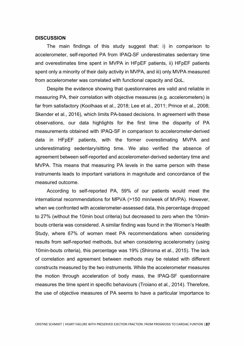
CRISTINE SCHMIDT | HEART FAILURE WITH PRESERVED EJECTION FRACTION: FROM PROGNOSIS TO CARDIAC FUNTION |
87
DISCUSSION The main findings of this study suggest that: i) in comparison to
accelerometer, self-reported PA from IPAQ-SF underestimates sedentary time
and overestimates time spent in MVPA in HFpEF patients, ii) HFpEF patients
spent only a minority of their daily activity in MVPA, and iii) only MVPA measured
from accelerometer was correlated with functional capacity and QoL.
Despite the evidence showing that questionnaires are valid and reliable in
measuring PA, their correlation with objective measures (e.g. accelerometers) is
far from satisfactory (Koolhaas et al., 2018; Lee et al., 2011; Prince et al., 2008;
Skender et al., 2016), which limits PA-based decisions. In agreement with these
observations, our data highlights for the first time the disparity of PA
measurements obtained with IPAQ-SF in comparison to accelerometer-derived
data in HFpEF patients, with the former overestimating MVPA and
underestimating sedentary/sitting time. We also verified the absence of
agreement between self-reported and accelerometer-derived sedentary time and
MVPA. This means that measuring PA levels in the same person with these
instruments leads to important variations in magnitude and concordance of the
measured outcome.
According to self-reported PA, 59% of our patients would meet the
international recommendations for MPVA (>150 min/week of MVPA). However,
when we confronted with accelerometer-assessed data, this percentage dropped
to 27% (without the 10min bout criteria) but decreased to zero when the 10min-
bouts criteria was considered. A similar finding was found in the Women’s Health
Study, where 67% of women meet PA recommendations when considering
results from self-reported methods, but when considering accelerometry (using
10min-bouts criteria), this percentage was 19% (Shiroma et al., 2015). The lack
of correlation and agreement between methods may be related with different
constructs measured by the two instruments. While the accelerometer measures
the motion through acceleration of body mass, the IPAQ-SF questionnaire
measures the time spent in specific behaviours (Troiano et al., 2014). Therefore,
the use of objective measures of PA seems to have a particular importance to
CRISTINE SCHMIDT | HEART FAILURE WITH PRESERVED EJECTION FRACTION: FROM PROGNOSIS TO CARDIAC FUNTION |
88
avoid bias in populations with limited physical function and limited past
knowledge and experience on regular PA (Prince et al., 2008).
Given the growing recognition of the impact of PA levels in the prognosis of
HFpEF patients, a rigorous characterization of their patterns is crucial for
prescribing tailored life-style changes. In our study, descriptive accelerometer
data shows that HFpEF patients spent 50% of recorded time in sedentary
behaviours, 47.5% at LPA and only 2.5% at MVPA (0.1% with 10min-bout
criteria). Similar patterns were recently observed by Yavari and colleagues,
where HFpEF patients reported to spent most of their waking time in sedentary
behaviours while their daily activity was mainly comprised of LPA, and just a few
minutes in MVPA (Yavari et al., 2017). Overall, these patterns of PA are similar
to those described for adults older than 60 years, where sedentary time
accounted for approximately for 60% of their waking time, LPA for 40% and 4%
for MVPA (Spittaels et al., 2012). Although the patterns of time spent in each
category are similar, our results show that the proportion of time spent in LPA
was almost 10% higher, while sedentary time was 10% lower. While the fact that
our patients were in optimal medical control may have accounted for that,
differences in the type of accelerometers and cut-points also may have influenced
the results. In fact, we used the GT3X triaxial accelerometer with the low
frequency extension filter, which was shown to be more sensitive to capture
slower movements, translating into decreased sedentary time and increase time
in all PA intensities (Migueles et al., 2017). In addition, although specific cut-
points for GT3X triaxial accelerometer have not been validated in HFpEF
patients, we used cut-points for older adults validated with the GT3X, which we
believe are more representative for our population.
With respect to prognosis, it was recently shown in the NEAT-HFpEF trial
that accelerometer-derived total daily PA in HFpEF patients was associated with
better 6MWT, NYHA functional class, QoL, and NT-proBNP levels (Snipelisky et
al., 2017). Our study adds the novelty that only time spent in MVPA was
significantly associated with important clinical outcomes as VO2, 6MWT and QoL,
but not with E/e´ or BNP levels. Thus, it seems that intensity is a requirement for
significantly impacting the patient’s prognosis. Corroborating this hypothesis, it
CRISTINE SCHMIDT | HEART FAILURE WITH PRESERVED EJECTION FRACTION: FROM PROGNOSIS TO CARDIAC FUNTION |
89
was recently shown a dose-response relationship between MVPA and risk of
hospitalization or mortality in HFpEF patients (Hegde et al., 2017). In addition, it
should be noted that only a minority of patients achieved the weekly
recommendations of MVPA (without the 10min-bouts criteria). While any increase
in the amount of PA may translate into some health benefits (Physical Activity
Guidelines Advisory Committee Scientific Report, 2018), our data suggests that
patients should be educated about the importance of reducing their sedentary
time and engage in more MVPA for greater benefits.
CRISTINE SCHMIDT | HEART FAILURE WITH PRESERVED EJECTION FRACTION: FROM PROGNOSIS TO CARDIAC FUNTION |
90
STUDY LIMITATIONS
Some limitations should be acknowledged. The small sample size limits
inferences regarding the agreement between PA measurements from IPAQ-SF
questionnaire and PA measures derived from triaxial accelerometer. This report
has a cross-sectional design, which limits the establishment of causal inferences
regarding PA and its influence on prognostic indicators, and the sample size
preclude the determination of predictive models. Moreover, the sample in this
study does not reflect all stages of the disease course.
CONCLUSION
Our results suggest that IPAQ-SF, compared to objectively measured PA,
underestimates sedentary time and over-estimates time spent in MVPA in
patients with HFpEF, limiting its use to support accurate recommendations.
Accelerometer-derived data shows that HFpEF patients spent only a minority of
their time involved in MVPA, which was the only PA pattern positively associated
with prognostic indicators, highlighting the importance of reducing sedentary time
and performing MVPA throughout the day.
CRISTINE SCHMIDT | HEART FAILURE WITH PRESERVED EJECTION FRACTION: FROM PROGNOSIS TO CARDIAC FUNTION |
91
REFERENCES Aguilar-Farias, N., Brown, W. J., & Peeters, G. M. (2014). ActiGraph GT3X+ cut-
points for identifying sedentary behaviour in older adults in free-living environments. J Sci Med Sport, 17(3), 293-299.
Ather, S., Chan, W., Bozkurt, B., Aguilar, D., Ramasubbu, K., Zachariah, A. A., Wehrens, X. H., & Deswal, A. (2012). Impact of noncardiac comorbidities on morbidity and mortality in a predominantly male population with heart failure and preserved versus reduced ejection fraction. J Am Coll Cardiol, 59(11), 998-1005.
Bland, J. M., & Altman, D. G. (1986). Statistical methods for assessing agreement between two methods of clinical measurement. Lancet, 1(8476), 307-310.
Booth, F. W., Roberts, C. K., & Laye, M. J. (2012). Lack of exercise is a major cause of chronic diseases. Compr Physiol, 2(2), 1143-1211.
Choi, L., Liu, Z., Matthews, C. E., & Buchowski, M. S. (2011). Validation of accelerometer wear and nonwear time classification algorithm. Med Sci Sports Exerc, 43(2), 357-364.
Craig, C. L., Marshall, A. L., Sjostrom, M., Bauman, A. E., Booth, M. L., Ainsworth, B. E., Pratt, M., Ekelund, U., Yngve, A., Sallis, J. F., & Oja, P. (2003). International physical activity questionnaire: 12-country reliability and validity. Med Sci Sports Exerc, 35(8), 1381-1395.
Crouter, S. E., Horton, M., & Bassett, D. R., Jr. (2012). Use of a two-regression model for estimating energy expenditure in children. Med Sci Sports Exerc, 44(6), 1177-1185.
Dolgin, M. (1994). Nomenclature and criteria for diagnosis of diseases of the heart and great vessels. The Criteria Committee of the New York Heart Association (9 ed.). Boston: Little, Brown & Co.
Glezeva, N., & Baugh, J. A. (2014). Role of inflammation in the pathogenesis of heart failure with preserved ejection fraction and its potential as a therapeutic target. Heart Fail Rev, 19(5), 681-694.
Haskell, W. L., Lee, I. M., Pate, R. R., Powell, K. E., Blair, S. N., Franklin, B. A., Macera, C. A., Heath, G. W., Thompson, P. D., Bauman, A., American College of Sports, M., & American Heart, A. (2007). Physical activity and public health: updated recommendation for adults from the American College of Sports Medicine and the American Heart Association. Circulation, 116(9), 1081-1093.
Hegde, S. M., Claggett, B., Shah, A. M., Lewis, E. F., Anand, I., Shah, S. J., Sweitzer, N. K., Fang, J. C., Pitt, B., Pfeffer, M. A., & Solomon, S. D. (2017). Physical Activity and Prognosis in the TOPCAT Trial (Treatment of Preserved Cardiac Function Heart Failure With an Aldosterone Antagonist). Circulation, 136(11), 982-992.
CRISTINE SCHMIDT | HEART FAILURE WITH PRESERVED EJECTION FRACTION: FROM PROGNOSIS TO CARDIAC FUNTION |
92
Ipaq Research Committee. (2005). Guidelines for data processing and analysis of the International Physical Activity Questionnaire (IPAQ)-short and long forms. http://www.ipaq.ki.se.
Koolhaas, C. M., van Rooij, F. J., Cepeda, M., Tiemeier, H., Franco, O. H., & Schoufour, J. D. (2018). Physical activity derived from questionnaires and wrist-worn accelerometers: comparability and the role of demographic, lifestyle, and health factors among a population-based sample of older adults. Clin Epidemiol, 10, 1-16.
Lang, R. M., Badano, L. P., Mor-Avi, V., Afilalo, J., Armstrong, A., Ernande, L., Flachskampf, F. A., Foster, E., Goldstein, S. A., Kuznetsova, T., Lancellotti, P., Muraru, D., Picard, M. H., Rietzschel, E. R., Rudski, L., Spencer, K. T., Tsang, W., & Voigt, J. U. (2015). Recommendations for cardiac chamber quantification by echocardiography in adults: an update from the American Society of Echocardiography and the European Association of Cardiovascular Imaging. Eur Heart J Cardiovasc Imaging, 16(3), 233-270.
Lee, P. H., Macfarlane, D. J., Lam, T. H., & Stewart, S. M. (2011). Validity of the International Physical Activity Questionnaire Short Form (IPAQ-SF): a systematic review. Int J Behav Nutr Phys Act, 8, 115.
Lohman, T. G., A.F. Roche, and R. Martorell. (1988). Anthropometric Standardization Reference Manual. Champaign, IL: Human Kinetics Books.
Mancia, G., Grassi, G., & Redon, J. (2014). Manual of Hypertension of the European Society of Hypertension (Second ed.): CRC Press.
Migueles, J. H., Cadenas-Sanchez, C., Ekelund, U., Delisle Nystrom, C., Mora-Gonzalez, J., Lof, M., Labayen, I., Ruiz, J. R., & Ortega, F. B. (2017). Accelerometer Data Collection and Processing Criteria to Assess Physical Activity and Other Outcomes: A Systematic Review and Practical Considerations. Sports Med, 47(9), 1821-1845.
Nanayakkara, S., Patel, H. C., & Kaye, D. M. (2018). Hospitalisation in Patients With Heart Failure With Preserved Ejection Fraction. Clin Med Insights Cardiol, 12, 1179546817751609.
Oktay, A. A., Rich, J. D., & Shah, S. J. (2013). The emerging epidemic of heart failure with preserved ejection fraction. Curr Heart Fail Rep, 10(4), 401-410.
Pandey, A., Allen, N. B., Ayers, C., Reis, J. P., Moreira, H. T., Sidney, S., Rana, J. S., Jacobs, D. R., Jr., Chow, L. S., de Lemos, J. A., Carnethon, M., & Berry, J. D. (2017a). Fitness in Young Adulthood and Long-Term Cardiac Structure and Function: The CARDIA Study. JACC Heart Fail, 5(5), 347-355.
Pandey, A., LaMonte, M., Klein, L., Ayers, C., Psaty, B. M., Eaton, C. B., Allen, N. B., de Lemos, J. A., Carnethon, M., Greenland, P., & Berry, J. D. (2017b). Relationship Between Physical Activity, Body Mass Index, and Risk of Heart Failure. J Am Coll Cardiol, 69(9), 1129-1142.
Physical Activity Guidelines Advisory Committee Scientific Report. (2018). 2018 Physical Activity Guidelines Advisory Committee.
CRISTINE SCHMIDT | HEART FAILURE WITH PRESERVED EJECTION FRACTION: FROM PROGNOSIS TO CARDIAC FUNTION |
93
Ponikowski, P., Voors, A. A., Anker, S. D., Bueno, H., Cleland, J. G. F., Coats, A. J. S., Falk, V., Gonzalez-Juanatey, J. R., Harjola, V. P., Jankowska, E. A., Jessup, M., Linde, C., Nihoyannopoulos, P., Parissis, J. T., Pieske, B., Riley, J. P., Rosano, G. M. C., Ruilope, L. M., Ruschitzka, F., Rutten, F. H., van der Meer, P., & Group, E. S. C. S. D. (2016). 2016 ESC Guidelines for the diagnosis and treatment of acute and chronic heart failure: The Task Force for the diagnosis and treatment of acute and chronic heart failure of the European Society of Cardiology (ESC)Developed with the special contribution of the Heart Failure Association (HFA) of the ESC. Eur Heart J, 37(27), 2129-2200.
Prince, S. A., Adamo, K. B., Hamel, M. E., Hardt, J., Connor Gorber, S., & Tremblay, M. (2008). A comparison of direct versus self-report measures for assessing physical activity in adults: a systematic review. Int J Behav Nutr Phys Act, 5, 56.
Rector, T. S., & Cohn, J. N. (1992). Assessment of patient outcome with the Minnesota Living with Heart Failure questionnaire: reliability and validity during a randomized, double-blind, placebo-controlled trial of pimobendan. Pimobendan Multicenter Research Group. Am Heart J, 124(4), 1017-1025.
Riley, L., Guthold, R., Cowan, M., Savin, S., Bhatti, L., Armstrong, T., & Bonita, R. (2016). The World Health Organization STEPwise Approach to Noncommunicable Disease Risk-Factor Surveillance: Methods, Challenges, and Opportunities. Am J Public Health, 106(1), 74-78.
Santos-Lozano, A., Santin-Medeiros, F., Cardon, G., Torres-Luque, G., Bailon, R., Bergmeir, C., Ruiz, J. R., Lucia, A., & Garatachea, N. (2013). Actigraph GT3X: validation and determination of physical activity intensity cut points. Int J Sports Med, 34(11), 975-982.
Sasaki, J. E., John, D., & Freedson, P. S. (2011). Validation and comparison of ActiGraph activity monitors. J Sci Med Sport, 14(5), 411-416.
Shiroma, E. J., Cook, N. R., Manson, J. E., Buring, J. E., Rimm, E. B., & Lee, I. M. (2015). Comparison of Self-Reported and Accelerometer-Assessed Physical Activity in Older Women. PLoS One, 10(12), e0145950.
Skender, S., Ose, J., Chang-Claude, J., Paskow, M., Bruhmann, B., Siegel, E. M., Steindorf, K., & Ulrich, C. M. (2016). Accelerometry and physical activity questionnaires - a systematic review. BMC Public Health, 16, 515.
Snipelisky, D., Kelly, J., Levine, J. A., Koepp, G. A., Anstrom, K. J., McNulty, S. E., Zakeri, R., Felker, G. M., Hernandez, A. F., Braunwald, E., & Redfield, M. M. (2017). Accelerometer-Measured Daily Activity in Heart Failure With Preserved Ejection Fraction: Clinical Correlates and Association With Standard Heart Failure Severity Indices. Circ Heart Fail, 10(6), e003878.
Spittaels, H., Van Cauwenberghe, E., Verbestel, V., De Meester, F., Van Dyck, D., Verloigne, M., Haerens, L., Deforche, B., Cardon, G., & De Bourdeaudhuij, I. (2012). Objectively measured sedentary time and physical activity time across the lifespan: a cross-sectional study in four age groups. Int J Behav Nutr Phys Act, 9, 149.
CRISTINE SCHMIDT | HEART FAILURE WITH PRESERVED EJECTION FRACTION: FROM PROGNOSIS TO CARDIAC FUNTION |
94
Troiano, R. P., McClain, J. J., Brychta, R. J., & Chen, K. Y. (2014). Evolution of accelerometer methods for physical activity research. Br J Sports Med, 48(13), 1019-1023.
WHO. (2000). Obesity: preventing and managing the global epidemic. Report of a WHO consultation. World Health Organ Tech Rep Ser, 894, 1-253.
Yavari, M., Haykowsky, M. J. F., Savu, A., Kaul, P., Dyck, J. R. B., Haennel, R. G., & Alberta, H. I. (2017). Volume and Patterns of Physical Activity Across the Health and Heart Failure Continuum. Can J Cardiol, 33(11), 1465-1471.
CRISTINE SCHMIDT | HEART FAILURE WITH PRESERVED EJECTION FRACTION: FROM PROGNOSIS TO CARDIAC FUNTION |
95
STUDY III
CHRONIC EXERCISE TRAINING REDUCES LEFT VENTRICLE STIFFNESS
IN AN ANIMAL MODEL OF HEART FAILURE WITH PRESERVED EJECTION
FRACTION

CRISTINE SCHMIDT | HEART FAILURE WITH PRESERVED EJECTION FRACTION: FROM PROGNOSIS TO CARDIAC FUNTION |
96
CRISTINE SCHMIDT | HEART FAILURE WITH PRESERVED EJECTION FRACTION: FROM PROGNOSIS TO CARDIAC FUNTION |
97
ABSTRACT
Rational: In heart failure with preserved ejection fraction (HFpEF), microvascular endothelial inflammation is responsible for cardiomyocyte stiffness and interstitial fibrosis which leads to diastolic left ventricle (LV) dysfunction. Nonetheless, HFpEF optimal treatment remains widely undefined. Purpose: To assess the impact of exercise training at the level of cardiac function and characterize underlying cellular and molecular changes. Methods: Nine-week old ZSF1 obese rats (Ob n=20) were divided into two groups: sedentary (ObSED, n=10) and exercised (ObEX, n=10). At the 16th week of life, the ObEX group was submitted to treadmill exercise training for 4 weeks, 5 days per week, 60 min per day, at a speed of 20m/min. At the end of the protocol (20th week), all animals performed a maximal oxygen consumption (VO2max) test, hemodynamic evaluation, and samples were collected for analysis. Plasma proteins were analysed by ELISA. Samples from LV were collated for: i) histological analysis (cross-sectional area (CSA), collagen content) and ii) passive tension analysis in skinned cardiomyocytes. Results: Exercise training improved VO2max (21±2.5 vs 17±1.8 mL.Kg-1.min-1, p=0.005). The end-diastolic pressure volume relationship had a trend to decrease in ObEX that, together with the decreased passive tension presented in skinned cardiomyocytes (p<0.05) and reduced collagen/muscle ratio (p<0.05), suggests a reduction in myocardial stiffness. No significant differences were observed between the groups in CSA. In addition, exercised animals showed reduced levels of several circulating biomarkers, such as markers of inflammation, endothelial dysfunction, oxidative stress, neurohumoral activation and extracellular matrix remodelling (p<0.05). Conclusion: Chronic exercise training improved exercise capacity and attenuated LV diastolic function and in rats with HFpEF. These improvements were associated with decreased cardiomyocyte passive tension and extracellular matrix fibroses. Moreover, exercise training reduced circulating levels of inflammatory cytokines and markers of endothelial dysfunction, oxidative stress, neurohumoral activation and extracellular matrix remodelling. Key-words: exercise training, LV stiffness, HFpEF, circulating biomarkers.

CRISTINE SCHMIDT | HEART FAILURE WITH PRESERVED EJECTION FRACTION: FROM PROGNOSIS TO CARDIAC FUNTION |
98
CRISTINE SCHMIDT | HEART FAILURE WITH PRESERVED EJECTION FRACTION: FROM PROGNOSIS TO CARDIAC FUNTION |
99
INTRODUCTION
Heart Failure (HF) with preserved ejection fraction (HFpEF) is a global
clinical problem, accounting for more than 50% of all HF cases (Dunlay et al.,
2017). Its prevalence has strikingly grown due to increasing prevalence of risk
factors such as obesity, hypertension and diabetes in an ageing population
(Dunlay et al., 2017). Heart Failure with preserved ejection fraction is responsible
for frequent hospitalizations, high consumption of resources and poor prognosis,
and no treatment has yet been shown to reduce morbidity or mortality (Owan et
al., 2006; Ponikowski et al., 2016). Ventricular diastolic dysfunction is a central
feature of HFpEF, caused by impaired relaxation and increased diastolic stiffness
(Redfield, 2016). This is mediated, at least in part, by extracellular matrix
remodeling (van Heerebeek et al., 2012) and changes in the expression and
phosphorylation of cytoskeletal proteins (Hamdani et al., 2013).
Exercise training is emerging as an important ally for the clinical
management of health-related outcomes in HFpEF patients. Different meta-
analysis of randomized clinical trials shows that exercise training, in comparison
to usual care, improves functional capacity, cardiorespiratory fitness and quality
of life (Fukuta et al., 2016; Pandey et al., 2015). It has been proposed that non-
cardiac peripheral adaptations are the major contributors (Haykowsky & Kitzman,
2014). However, some clinical (Dieberg et al., 2015; Pearson et al., 2017) and
pre-clinical studies (Hidalgo et al., 2014; Marshall et al., 2013) support the
hypothesis that exercise training may also improve diastolic function. Whether
this will translate into a greater peak cardiac output remains unknown.
Understanding the pathways that mediate these cardiac benefits could yield
novel therapeutic approaches based on agents that mimic or potentiate the
physiological benefits of exercise. In this sense, it was already shown that chronic
free-wheel running improved cardiac stiffness in a genetic mouse model of
HFpEF, an effect that was mediated by the reduction on titin-based passive
stiffness in a phosphorylation-dependent manner (Hidalgo et al., 2014; Slater et
al., 2017). Using the Yucatan miniature swine model of HFpEF, Marshal and
collaborators also showed that treadmill exercise training attenuated diastolic
impairment by reducing extracellular matrix (ECM) fibrosis (Marshall et al., 2013).
CRISTINE SCHMIDT | HEART FAILURE WITH PRESERVED EJECTION FRACTION: FROM PROGNOSIS TO CARDIAC FUNTION |
100
Despite these molecular insights, it is important to highlight that none of
these animal models fully mimic the spectrum of changes found in humans with
HFpEF (Ponikowski et al., 2016). Recently, our group contributed to surpass
these limitations by validating the ZSF1 obese animal model for HFpEF (Hamdani
et al., 2013). The ZSF1 obese animals exhibit several features of human HFpEF
such as the presence of comorbidities, exercise intolerance, and diastolic
dysfunction with preserved systolic function (Hamdani et al., 2013, Leite et al.,
2015), placing this model one step ahead towards clinical translation (Lourenco
et al., 2018). Therefore, by using this animal model of HFpEF, we aimed to
assess the impact of exercise training at the level of cardiac function and
characterize underlying cellular and molecular changes.
CRISTINE SCHMIDT | HEART FAILURE WITH PRESERVED EJECTION FRACTION: FROM PROGNOSIS TO CARDIAC FUNTION |
101
METHODS Animal model
The animal protocol was approved by the ethics committee of Faculty of
Medicine of Porto and was conducted in accordance with the Guide for the Care
and Use of Laboratory Animals published by the National Institutes of Health (NIH
publication no. 85-23, revised 1996).
Nine-week-old rats ZSF1 obese (Zucker diabetic fatty/Spontaneously
hypertensive heart failure F1 hybrid rats) (ZSF1 Ob, n=17) were obtained from
Charles River (Barcelona, Spain). Animals were housed in groups of 2 animals
per cage, maintained on a 12-hour light-dark cycle, at a room temperature of
22°C, fed with Purina Diet (#5008) and water ad libitum. Rats were allowed 1
week of adaptation to laboratory conditions before experiments. At 16th week of
life, the ZSF1 Ob rats were randomly divided into two groups, obese sedentary
(ObSED, n=7; normal activity at the cage) and obese exercised (ObEx, n=10;
aerobic exercise training). Exercise training protocol consisted of running on the
treadmill (76-0896, PanLab Harvard Apparatus) 5 days per week, 1 hour per day,
at a speed of 20m/min, for 4 weeks. The sedentary group was submitted to a
stationary treadmill in order to minimize environmental confounders among the
animals not subjected to the exercise protocol. The protocol was interrupted 48
hours before terminal evaluation and sample collection to avoid confounding with
acute effects of the last training session.
Peak effort testing with oxygen consumption determination
At 20th week, animals underwent an exercise tolerance test with oxygen
consumption (VO2) measurement (LE8700C, OxyletPro System, PanLab
Harvard Apparatus). Initially, animals were adapted to treadmill at 9m/min for 3
minutes, followed by a rapid effort escalation to assess peak oxygen consumption
(VO2peak). Velocity was changed to 18m/min and then stepped up 3m/min at
1min intervals. Treadmill was stopped whenever animals were unable/unwilling
to maintain pace, as defined by incapability to come off the back of the treadmill
lane for >3 seconds. Gas concentrations (O2 and CO2), speed and covered
CRISTINE SCHMIDT | HEART FAILURE WITH PRESERVED EJECTION FRACTION: FROM PROGNOSIS TO CARDIAC FUNTION |
102
distance were continuously recorded. Data analysis was performed with the aid
of software (Metabolism V2.2.01, Panlab Harvard Apparatus®).
Hemodynamic evaluation Forty-eight hours after exercise testing, we performed terminal
hemodynamic evaluation. After sedation (100 μg.kg-1 and 5 mg.kg-1
intraperitoneal fentanyl and midazolam, respectively), animals were anesthetized
by inhalation of a mixture of sevoflurane (2.5–3% sevoflurane; Penlon Sigma
Delta) and oxygen, intubated for mechanical ventilation (TOPO, Kent Scientific)
and placed over a heating pad (body temperature was maintained at 38°C). A
peripheral venous catheter was introduced in the right femoral vein for
intravenous warm Ringer’s solution infusion (8 mL.kg-1.h-1; NE-1000, New Era
Pump Systems). A left thoracotomy allowed assessing the heart for insertion of
a pressure-volume catheter in the LV through the apex (SPR-838 Millar
Instruments, respectively). A probe (Transonics) was placed around the
ascending aorta that allowed CO measurement (Active Redirection Transit Time
Flowmeter, Triton Technology). Signals were continuously acquired (MPVS 300,
Millar Instruments), recorded at 1000 Hz (ML880 PowerLab 16/30,
ADinstruments), and analysed (PVAN 3.5, Millar Instruments). Recordings were
obtained at suspended end-expiration. The LV catheter was advanced to record
systemic arterial pressure. Parallel conductance was assessed with hypertonic
saline. After euthanasia (100 mg.kg-1 intravenous pentobarbital), blood (4mL)
was collected for storage (-80ºC) and for volume calibration (910–1048, Millar
instruments). LV volumes were varied using transient inferior vena cava
constrictions by adjusting a sling around the inferior vena cava. Blood was used
for volume calibration in standard wells. Heart, LV+septum, lung, skeletal muscle
(gastrocnemius, soleus and tibialis anterior) were collected, weighed and
immediately snap frozen on liquid nitrogen, and stored at 80oC. Tibia length was
measured and then used to normalizations. Body surface area was defined as
9.1*(weight)2/3 (Hamdani et al., 2013).
CRISTINE SCHMIDT | HEART FAILURE WITH PRESERVED EJECTION FRACTION: FROM PROGNOSIS TO CARDIAC FUNTION |
103
Biomarkers quantification Blood samples were obtained through RV cannulation, were collected in
EDTA tubes after hemodynamic evaluation. Samples were centrifuged at 5000
rpm for 15 minutes at 4°C. Serum was subsequently separated and frozen.
Before the quantification, serum was centrifuged at 20 000 rpm for 15 minutes at
4°C for lipid separation. Quantitative enzyme immunoassays (ELISA) of
interleukin- 6 (IL-6), IL-10, intercellular adhesion molecule-1 (ICAM-1)/CD54,
metalloproteinase (MMP)-2, MMP-9, tissue inhibitor of metalloproteinase-1
(TIMP-1) (R&D Systems, #R6000B, #R1000, #RIC100, #MMP200, #RMP900,
#RTM100, respectively), NT-pro-BNP, malondialdehyde (MDA), high sensitivity
C-Reactive Protein (hs-CRP), TNF-alpha, vascular adhesion molecule-1 (VCAM-
1) (MyBioSource, #MBS704791, #MBS727531, #MBS281245, #MBS824520,
#MBS2502676 respectively) and protein carbonyl content (Abcam, #ab126287)
were measured according to the manufacturer’s instructions. Results were
analysed using an ELISA plate reader (UVM-340, ASYS Hitech GmbH). A
calibration curve was constructed by plotting the absorbance values at 450nm
(with specific correction, according to manufacturer’s protocol) and
concentrations of unknown samples were determined.
Histological studies
Slices of LV tissue were submersed in a fixative solution of 4%
paraformaldehyde by diffusion for 24 hours and subsequently dehydrated with
graded ethanol and included in paraffin blocks. Xilene was used in the transition
between dehydration and impregnation. LV blocks were embedded in the upright
position in order to distinguish the endocardium, midwall, and the epicardium of
the LV free wall in cross sections. Serial sections (5 μm of thickness) of paraffin
blocks were cut by a microtome (Minot type microtome (RM2125RTS, Leica,
Nussloch, Germany) and mounted on silane-coated slides. The slides were
dewaxed in xylene and hydrated through graded alcohols finishing in phosphate
buffered saline solution prepared by dissolving Na2HPO4 (1.44 g), KH2PO4
(0.24 g), NaCl (8 g), KCl (0.2 g) and adjusting pH to 7.2. Deparaffinized sections
from LV were stained for haematoxylin-eosin, performed by immersing slides in
CRISTINE SCHMIDT | HEART FAILURE WITH PRESERVED EJECTION FRACTION: FROM PROGNOSIS TO CARDIAC FUNTION |
104
Mayer’s haematoxylin solution for 3–4 min followed by immersion in 1% eosin
solution for 7 min, dehydration with graded alcohols through xylene, and mounted
with DPX. Stained sections of slides were digitally photographed (Olympus XC30
Digital colour Camera). Cardiomyocytes surface area (CSA) was measured using
imaging software (cell^B, Olympus). We analysed 5 random animals per group,
10 photos per animal (magnification of X400) and only nuclei-centred
cardiomyocytes were considered for analysis. In order to determine the amount
of cardiac fibrosis, slides were immersed in picrosirius red for 2h, washed with
acid water (0.05% acetic acid) and mounted after de-hydration as described
before (Falcao-Pires et al., 2011b). Digital images (magnification of X400) were
analysed, using image analysis software (Image-Pro Plus version 6.0, Media
Cybernetics Inc.), from 5 random animals per group, 10 photos per animal.
Isolated cardiomyocyte Force measurements were performed in single and mechanically isolated
cardiomyocytes as previously described (Borbely et al., 2005; Falcao-Pires et al.,
2011a). Myocardial samples (10-15 mg wet weight) from rat hearts subjected to
the exercise protocol described above were defrosted in cold relaxing solution,
mechanically disrupted, and incubated for 5 minutes in relaxing solution
supplemented with 0.2% Triton X-100, in order to remove membrane’s structures.
Afterwards, single cardiomyocytes were attached between the force transducer
and the piezoelectric motor with glue. Passive tension was measured at
sarcomere lengths ranging between 1.8 µm to 2.3µm (0.1 µm step increases) at
15°C. Force values were normalized for myocyte cross-sectional area.
Statistical Analysis
The Shapiro-Wilk or Kolmogorov–Smirnov test was performed to check
normality of the data. Between-group comparisons of normal data were
performed with independent sample t-test. Non-normal data were analysed with
nonparametric tests, namely the Mann-Whitney U Test. Data are expressed as
means ± SD. Statistical analysis was performed using the GrapPad Prism 8 and
the statistical significance was set at p <0.05.
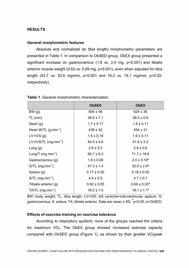
CRISTINE SCHMIDT | HEART FAILURE WITH PRESERVED EJECTION FRACTION: FROM PROGNOSIS TO CARDIAC FUNTION |
105
RESULTS
General morphometric features
Absolute and normalized (to tibia length) morphometric parameters are
presented in Table 1. In comparison to ObSED group, ObEX group presented a
significant increase on gastrocnemius (1.8 vs. 2.0 mg, p<0.001) and tibialis
anterior muscle weight (0.62 vs. 0.69 mg, p<0.001), even when adjusted for tibia
length (43.7 vs. 52.6 mg/mm, p˂0.001 and 16.2 vs. 18.1 mg/mm, p=0.02,
respectively).
Table 1. General morphometric characterization.
ObSED ObEX BW (g) 604 ± 49 624 ± 36 TL (mm) 38.5 ± 1.1 38.5 ± 0.8 Heart (g) 1.7 ± 0.17 1.8 ± 0.11
Heart W/TL (g.mm-1) 438 ± 42 454 ± 31
LV+IVS (g) 1.5 ± 0.18 1.6 ± 0.11
LV+IVS/TL (mg.mm-1) 40.5 ± 4.6 41.6 ± 3.2
Lung (g) 2.6 ± 0.3 2.8 ± 0.6
Lung/T (mg.mm-1) 66.7 ± 8.2 71.7 ± 16.6
Gastrocnemius (g) 1.8 ± 0.08 2.0 ± 0.10* G/TL (mg.mm-1) 47.3 ± 1.4 52.6 ± 2.4*
Soleus (g) 0.17 ± 0.02 0.18 ± 0.02
S/TL (mg.mm-1) 4.4 ± 0.5 4.7 ± 0.7
Tibialis anterior (g) 0.62 ± 0.05 0.69 ± 0.03* TA/TL (mg.mm-1) 16.2 ± 1.5 18.1 ± 1.1*
BW: body weight; TL: tibia length; LV+IVS: left ventricle+interventricular septum; G: gastrocnemius; S: soleus; TA: tibialis anterior. Data are mean ± SD. *p<0.05 vs ObSED. Effects of exercise training on exercise tolerance
According to respiratory quotient, none of the groups reached the criteria
for maximum VO2. The ObEX group showed increased exercise capacity
compared with ObSED group (Figure 1), as shown by their greater VO2peak
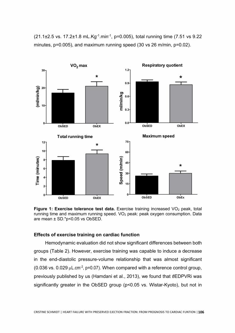
CRISTINE SCHMIDT | HEART FAILURE WITH PRESERVED EJECTION FRACTION: FROM PROGNOSIS TO CARDIAC FUNTION |
106
(21.1±2.5 vs. 17.2±1.8 mL.Kg-1.min-1, p=0.005), total running time (7.51 vs 9.22
minutes, p=0.005), and maximum running speed (30 vs 26 m/min, p=0.02).
Figure 1: Exercise tolerance test data. Exercise training increased VO2 peak, total running time and maximum running speed. VO2 peak: peak oxygen consumption. Data are mean ± SD.*p<0.05 vs ObSED.
Effects of exercise training on cardiac function
Hemodynamic evaluation did not show significant differences between both
groups (Table 2). However, exercise training was capable to induce a decrease
in the end-diastolic pressure-volume relationship that was almost significant
(0.036 vs. 0.029 µL.cm-2, p=0.07). When compared with a reference control group,
previously published by us (Hamdani et al., 2013), we found that βEDPVRi was
significantly greater in the ObSED group (p<0.05 vs. Wistar-Kyoto), but not in
ObSED ObEX0.0
0.3
0.6
0.9
1.2Respiratory quotient
*
ml/m
in/k
g
(ml/m
in/k
g)
ObSED ObEX0
10
20
30
*
VO2 max
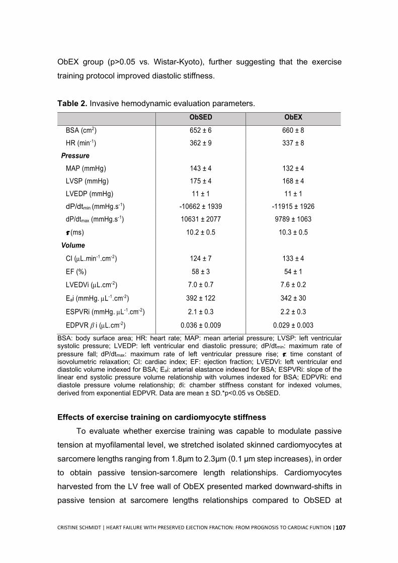
CRISTINE SCHMIDT | HEART FAILURE WITH PRESERVED EJECTION FRACTION: FROM PROGNOSIS TO CARDIAC FUNTION |
107
ObEX group (p>0.05 vs. Wistar-Kyoto), further suggesting that the exercise
training protocol improved diastolic stiffness.
Table 2. Invasive hemodynamic evaluation parameters. ObSED ObEX
BSA (cm2) 652 ± 6 660 ± 8
HR (min-1) 362 ± 9 337 ± 8
Pressure
MAP (mmHg) 143 ± 4 132 ± 4
LVSP (mmHg) 175 ± 4 168 ± 4
LVEDP (mmHg) 11 ± 1 11 ± 1
dP/dtmin (mmHg.s-1) -10662 ± 1939 -11915 ± 1926
dP/dtmax (mmHg.s-1) 10631 ± 2077 9789 ± 1063
t (ms) 10.2 ± 0.5 10.3 ± 0.5
Volume
CI (µL.min-1.cm-2) 124 ± 7 133 ± 4
EF (%) 58 ± 3 54 ± 1
LVEDVi (µL.cm-2) 7.0 ± 0.7 7.6 ± 0.2
Eai (mmHg. µL-1.cm-2) 392 ± 122 342 ± 30
ESPVRi (mmHg. µL-1.cm-2) 2.1 ± 0.3 2.2 ± 0.3
EDPVR b i (µL.cm-2) 0.036 ± 0.009 0.029 ± 0.003
BSA: body surface area; HR: heart rate; MAP: mean arterial pressure; LVSP: left ventricular systolic pressure; LVEDP: left ventricular end diastolic pressure; dP/dtmin: maximum rate of pressure fall; dP/dtmax: maximum rate of left ventricular pressure rise; t: time constant of isovolumetric relaxation; CI: cardiac index; EF: ejection fraction; LVEDVi: left ventricular end diastolic volume indexed for BSA; Eai: arterial elastance indexed for BSA; ESPVRi: slope of the linear end systolic pressure volume relationship with volumes indexed for BSA; EDPVRi: end diastole pressure volume relationship; βi: chamber stiffness constant for indexed volumes, derived from exponential EDPVR. Data are mean ± SD.*p<0.05 vs ObSED.
Effects of exercise training on cardiomyocyte stiffness To evaluate whether exercise training was capable to modulate passive
tension at myofilamental level, we stretched isolated skinned cardiomyocytes at
sarcomere lengths ranging from 1.8µm to 2.3µm (0.1 µm step increases), in order
to obtain passive tension-sarcomere length relationships. Cardiomyocytes
harvested from the LV free wall of ObEX presented marked downward-shifts in
passive tension at sarcomere lengths relationships compared to ObSED at
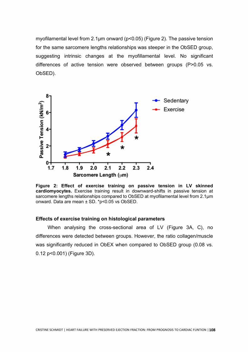
CRISTINE SCHMIDT | HEART FAILURE WITH PRESERVED EJECTION FRACTION: FROM PROGNOSIS TO CARDIAC FUNTION |
108
myofilamental level from 2.1µm onward (p<0.05) (Figure 2). The passive tension
for the same sarcomere lengths relationships was steeper in the ObSED group,
suggesting intrinsic changes at the myofillamental level. No significant
differences of active tension were observed between groups (P>0.05 vs.
ObSED).
Figure 2: Effect of exercise training on passive tension in LV skinned cardiomyocytes. Exercise training result in downward-shifts in passive tension at sarcomere lengths relationships compared to ObSED at myofilamental level from 2.1µm onward. Data are mean ± SD. *p<0.05 vs ObSED.
Effects of exercise training on histological parameters When analysing the cross-sectional area of LV (Figure 3A, C), no
differences were detected between groups. However, the ratio collagen/muscle
was significantly reduced in ObEX when compared to ObSED group (0.08 vs.
0.12 p<0.001) (Figure 3D).
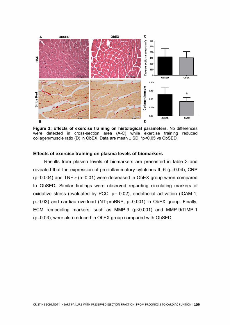
CRISTINE SCHMIDT | HEART FAILURE WITH PRESERVED EJECTION FRACTION: FROM PROGNOSIS TO CARDIAC FUNTION |
109
Figure 3: Effects of exercise training on histological parameters. No differences were detected in cross-section area (A-C) while exercise training reduced collagen/muscle ratio (D) in ObEX. Data are mean ± SD. *p<0.05 vs ObSED.
Effects of exercise training on plasma levels of biomarkers
Results from plasma levels of biomarkers are presented in table 3 and
revealed that the expression of pro-inflammatory cytokines IL-6 (p=0.04), CRP
(p=0.004) and TNF-α (p=0.01) were decreased in ObEX group when compared
to ObSED. Similar findings were observed regarding circulating markers of
oxidative stress (evaluated by PCC; p= 0.02), endothelial activation (ICAM-1;
p=0.03) and cardiac overload (NT-proBNP, p=0.001) in ObEX group. Finally,
ECM remodeling markers, such as MMP-9 (p<0.001) and MMP-9/TIMP-1
(p=0.03), were also reduced in ObEX group compared with ObSED.
B
ObSEDSi
rius
Red
ObEXH
&E
ObSED ObEX0.00
0.05
0.10
0.15
0.20
*
Collagen/muscle
ObSED ObEX0
150
300
450
600
750
900
Cro
ss-s
ectio
na a
rea
(µm2 )
A C
D
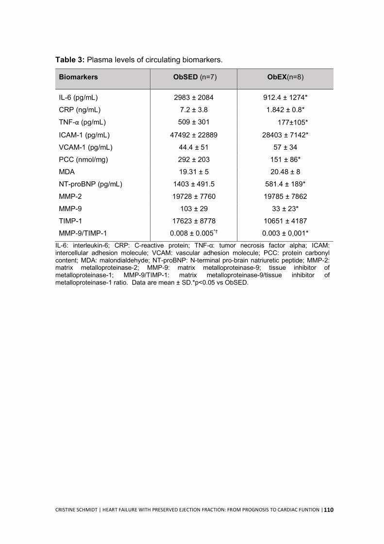
CRISTINE SCHMIDT | HEART FAILURE WITH PRESERVED EJECTION FRACTION: FROM PROGNOSIS TO CARDIAC FUNTION |
110
Table 3: Plasma levels of circulating biomarkers.
Biomarkers ObSED (n=7) ObEX(n=8)
IL-6 (pg/mL) 2983 ± 2084 912.4 ± 1274*
CRP (ng/mL) 7.2 ± 3.8 1.842 ± 0.8*
TNF-α (pg/mL) 509 ± 301 177±105*
ICAM-1 (pg/mL) 47492 ± 22889 28403 ± 7142*
VCAM-1 (pg/mL) 44.4 ± 51 57 ± 34
PCC (nmol/mg) 292 ± 203 151 ± 86*
MDA 19.31 ± 5 20.48 ± 8
NT-proBNP (pg/mL) 1403 ± 491.5 581.4 ± 189*
MMP-2 19728 ± 7760 19785 ± 7862
MMP-9 103 ± 29 33 ± 23*
TIMP-1 17623 ± 8778 10651 ± 4187
MMP-9/TIMP-1 0.008 ± 0.005*† 0.003 ± 0,001* IL-6: interleukin-6; CRP: C-reactive protein; TNF-α: tumor necrosis factor alpha; ICAM: intercellular adhesion molecule; VCAM: vascular adhesion molecule; PCC: protein carbonyl content; MDA: malondialdehyde; NT-proBNP: N-terminal pro-brain natriuretic peptide; MMP-2: matrix metalloproteinase-2; MMP-9: matrix metalloproteinase-9; tissue inhibitor of metalloproteinase-1; MMP-9/TIMP-1: matrix metalloproteinase-9/tissue inhibitor of metalloproteinase-1 ratio. Data are mean ± SD.*p<0.05 vs ObSED.
CRISTINE SCHMIDT | HEART FAILURE WITH PRESERVED EJECTION FRACTION: FROM PROGNOSIS TO CARDIAC FUNTION |
111
DISCUSSION
The present study demonstrates beneficial effects of moderate exercise
training in the ZSF1 rat model of HFpEF. We showed that exercise training: i)
improved exercise capacity, ii) attenuated LV stiffness, and iii) reduced circulating
biomarkers of inflammation, endothelial activation, oxidative stress,
neurohumoral activation and ECM remodeling.
Exercise intolerance is a primary symptom in patients with HFpEF,
manifested by early fatigue and dyspnea, which is a strong determinant of
prognosis and reduced quality of life (Upadhya et al., 2015). While drugs
interventions have failed to improve exercise capacity and quality of life in HFpEF
patients, exercise training has been shown to improve both outcomes (Fukuta et
al., 2016). Using a previously validated animal model of HFpEF (Hamdani et al.,
2013), the present study corroborates that exercise training improves exercise
capacity in HFpEF. Although the improvement on exercise capacity by exercise
training is well recognized, the physiological mechanisms underlying these
changes in HFpEF are poorly understood. Current published data suggests that
the greater VO2peak found in HFpEF patients after a training program is mainly
because of non-cardiac peripheral adaptations, as no study has yet shown to
improve cardiac output (Fu et al., 2016; Haykowsky et al., 2012). However, both
clinical (Dieberg et al., 2015; Pearson et al., 2017) and pre-clinical studies
(Hidalgo et al., 2014; Marshall et al., 2013) support the hypothesis that exercise
training improves diastolic function in HFpEF setting, which may also account for
a greater exercise response. Consistently, our data showed that exercise training
modulates LV compliance and distensibility in HFpEF animals. We found a
borderline-significant decrease in end-diastolic pressure-volume relationship,
which was accompanied by reduced cardiomyocyte stiffness. In addition,
exercised animals showed lower circulating levels of NT-proBNP, suggesting
reduced wall stress, possibly due to a less stiffer heart (Bordbar et al., 2012).
Whether exercise training can modulate LV compliance and distensibility in
HFpEF remains to be explored, but these diastolic variables were shown to be
modulated by exercise training in healthy middle age individuals (Howden et al.,
2018) and in healthy seniors (Bhella et al., 2014).
CRISTINE SCHMIDT | HEART FAILURE WITH PRESERVED EJECTION FRACTION: FROM PROGNOSIS TO CARDIAC FUNTION |
112
So far, the literature does not elucidate how exercise training could improve
diastolic function in HFpEF patients. Edelmann and colleagues demonstrated
that exercise training improved LV diastolic function and promoted atrial reverse
remodelling without change in LV mass, suggesting a switch from pathologic to
physiologic hypertrophy (Edelmann et al., 2011). In addition, exercise training
was associated with a significant reduction in procollagen type I plasma levels,
suggesting that improvement in diastolic function may be associated with
reduced collagen turnover (Edelmann et al., 2011). Our data strengthens the
hypothesis that exercise training enhances cardiac function by decreasing
cardiomyocyte passive tension and LV fibrosis. We observed a downward-shift
in passive tension at sarcomere lengths relationships from LV cardiomyocytes of
ObEX rats comparing to those extracted from ObSED. These findings confirm an
increase in LV compliance promoted, at least in part, by intrinsic cardiomyocyte
changes. This is supported by previous data showing that chronic voluntary
running decreases titin-based passive stiffness in a phosphorylation-dependent
manner in mice with diastolic dysfunction (Slater et al., 2017) (Kotter et al., 2013).
In addition, it is also possible that the improved LV compliance could be due to
adaptations at the ECM level, as we found that exercise training attenuated LV
collagen formation and reduced systemic levels of MMP9 and MM9/TIMP1 ratio.
Similarly, treadmill exercise training was shown to attenuate diastolic impairment
by reducing fibrosis in the Yucatan miniature swine model of HFpEF (aortic-
banded to produce concentric LV hypertrophy) (Marshall et al., 2013). Our data
suggests that the overall beneficial effect of exercise on cardiac function might
be due to both cardiomyocyte’s intrinsic and extrinsic mechanisms.
It has been proposed that myocardial remodelling and dysfunction in HFpEF
is triggered by low-grade chronic systemic inflammation induced by comorbidities
(Paulus & Tschope, 2013). This persistent inflammatory state causes coronary
microvascular endothelial inflammation and oxidative damage, leading to lower
nitric oxide bioavailability and PKG activity, thus contributing to LV stiffness by
inducing hypertrophy and hypophosphorylation of titin (Franssen et al., 2016;
Paulus & Tschope, 2013). In addition, endothelial activation of cell adhesion
molecules by inflammation would favours the migration of monocytes into the
CRISTINE SCHMIDT | HEART FAILURE WITH PRESERVED EJECTION FRACTION: FROM PROGNOSIS TO CARDIAC FUNTION |
113
subendothelium and stimulates the conversion of fibroblasts to myofibroblasts by
release transforming growth factor β (TGF-β). This would also contribute to
cardiac stiffness, as myofibroblasts are responsible for depositing collagen in the
interstitial space. The majority of these features have also been shown to be
present in the ZSF1 obese animal model (Hamdani et al., 2013; Leite-Moreira et
al., 2018). In the current work, we found that ObEX animals had reduced levels
of circulating inflammatory cytokines (CRP, IL-6 and TNF-α), oxidative stress
(PCC), and endothelial activation (ICAM-1). Thus, we hypothesized that these
systemic effects promoted by exercise training were capable to interfere with the
cascade of events leading to increased diastolic left ventricular stiffness and heart
failure development.
CRISTINE SCHMIDT | HEART FAILURE WITH PRESERVED EJECTION FRACTION: FROM PROGNOSIS TO CARDIAC FUNTION |
114
CONCLUSION
Our findings suggest that moderate exercise training improves exercise
capacity and attenuates LV diastolic stiffness in the ZSF1 obese rat model of
HFpEF. This was paralleled by reduced circulating levels of inflammatory
cytokines and markers of endothelial dysfunction and oxidative stress. These
results provide novel mechanistic insight into the benefits of training in a clinically
meaningful translational animal model of HFpEF.
CRISTINE SCHMIDT | HEART FAILURE WITH PRESERVED EJECTION FRACTION: FROM PROGNOSIS TO CARDIAC FUNTION |
115
REFERENCES Bhella, P. S., Hastings, J. L., Fujimoto, N., Shibata, S., Carrick-Ranson, G.,
Palmer, M. D., Boyd, K. N., Adams-Huet, B., & Levine, B. D. (2014). Impact of lifelong exercise "dose" on left ventricular compliance and distensibility. J Am Coll Cardiol, 64(12), 1257-1266.
Borbely, A., van der Velden, J., Papp, Z., Bronzwaer, J. G., Edes, I., Stienen, G. J., & Paulus, W. J. (2005). Cardiomyocyte stiffness in diastolic heart failure. Circulation, 111(6), 774-781.
Bordbar, S., Bigi, M. A., Aslani, A., Rahimi, E., & Ahmadi, N. (2012). Effect of endurance and strength exercise on release of brain natriuretic peptide. J Cardiovasc Dis Res, 3(1), 22-25.
Dieberg, G., Ismail, H., Giallauria, F., & Smart, N. A. (2015). Clinical outcomes and cardiovascular responses to exercise training in heart failure patients with preserved ejection fraction: a systematic review and meta-analysis. J Appl Physiol (1985), 119(6), 726-733.
Dunlay, S. M., Roger, V. L., & Redfield, M. M. (2017). Epidemiology of heart failure with preserved ejection fraction. Nat Rev Cardiol, 14(10), 591-602.
Edelmann, F., Gelbrich, G., Dungen, H. D., Frohling, S., Wachter, R., Stahrenberg, R., Binder, L., Topper, A., Lashki, D. J., Schwarz, S., Herrmann-Lingen, C., Loffler, M., Hasenfuss, G., Halle, M., & Pieske, B. (2011). Exercise training improves exercise capacity and diastolic function in patients with heart failure with preserved ejection fraction: results of the Ex-DHF (Exercise training in Diastolic Heart Failure) pilot study. J Am Coll Cardiol, 58(17), 1780-1791.
Falcao-Pires, I., Hamdani, N., Borbely, A., Gavina, C., Schalkwijk, C. G., van der Velden, J., van Heerebeek, L., Stienen, G. J., Niessen, H. W., Leite-Moreira, A. F., & Paulus, W. J. (2011a). Diabetes mellitus worsens diastolic left ventricular dysfunction in aortic stenosis through altered myocardial structure and cardiomyocyte stiffness. Circulation, 124(10), 1151-1159.
Falcao-Pires, I., Palladini, G., Goncalves, N., van der Velden, J., Moreira-Goncalves, D., Miranda-Silva, D., Salinaro, F., Paulus, W. J., Niessen, H. W., Perlini, S., & Leite-Moreira, A. F. (2011b). Distinct mechanisms for diastolic dysfunction in diabetes mellitus and chronic pressure-overload. Basic Res Cardiol, 106(5), 801-814.
Franssen, C., Chen, S., Unger, A., Korkmaz, H. I., De Keulenaer, G. W., Tschope, C., Leite-Moreira, A. F., Musters, R., Niessen, H. W., Linke, W. A., Paulus, W. J., & Hamdani, N. (2016). Myocardial Microvascular Inflammatory Endothelial Activation in Heart Failure With Preserved Ejection Fraction. JACC Heart Fail, 4(4), 312-324.
Fu, T. C., Yang, N. I., Wang, C. H., Cherng, W. J., Chou, S. L., Pan, T. L., & Wang, J. S. (2016). Aerobic Interval Training Elicits Different Hemodynamic
CRISTINE SCHMIDT | HEART FAILURE WITH PRESERVED EJECTION FRACTION: FROM PROGNOSIS TO CARDIAC FUNTION |
116
Adaptations Between Heart Failure Patients with Preserved and Reduced Ejection Fraction. Am J Phys Med Rehabil, 95(1), 15-27.
Fukuta, H., Goto, T., Wakami, K., & Ohte, N. (2016). Effects of drug and exercise intervention on functional capacity and quality of life in heart failure with preserved ejection fraction: A meta-analysis of randomized controlled trials. Eur J Prev Cardiol, 23(1), 78-85.
Hamdani, N., Franssen, C., Lourenco, A., Falcao-Pires, I., Fontoura, D., Leite, S., Plettig, L., Lopez, B., Ottenheijm, C. A., Becher, P. M., Gonzalez, A., Tschope, C., Diez, J., Linke, W. A., Leite-Moreira, A. F., & Paulus, W. J. (2013). Myocardial titin hypophosphorylation importantly contributes to heart failure with preserved ejection fraction in a rat metabolic risk model. Circ Heart Fail, 6(6), 1239-1249.
Haykowsky, M. J., Brubaker, P. H., Stewart, K. P., Morgan, T. M., Eggebeen, J., & Kitzman, D. W. (2012). Effect of endurance training on the determinants of peak exercise oxygen consumption in elderly patients with stable compensated heart failure and preserved ejection fraction. J Am Coll Cardiol, 60(2), 120-128.
Haykowsky, M. J., & Kitzman, D. W. (2014). Exercise physiology in heart failure and preserved ejection fraction. Heart Fail Clin, 10(3), 445-452.
Hidalgo, C., Saripalli, C., & Granzier, H. L. (2014). Effect of exercise training on post-translational and post-transcriptional regulation of titin stiffness in striated muscle of wild type and IG KO mice. Arch Biochem Biophys, 552-553, 100-107.
Howden, E. J., Sarma, S., Lawley, J. S., Opondo, M., Cornwell, W., Stoller, D., Urey, M. A., Adams-Huet, B., & Levine, B. D. (2018). Reversing the Cardiac Effects of Sedentary Aging in Middle Age-A Randomized Controlled Trial: Implications For Heart Failure Prevention. Circulation, 137(15), 1549-1560.
Kotter, S., Gout, L., Von Frieling-Salewsky, M., Muller, A. E., Helling, S., Marcus, K., Dos Remedios, C., Linke, W. A., & Kruger, M. (2013). Differential changes in titin domain phosphorylation increase myofilament stiffness in failing human hearts. Cardiovasc Res, 99(4), 648-656.
Lourenco, A. P., Leite-Moreira, A. F., Balligand, J. L., Bauersachs, J., Dawson, D., de Boer, R. A., de Windt, L. J., Falcao-Pires, I., Fontes-Carvalho, R., Franz, S., Giacca, M., Hilfiker-Kleiner, D., Hirsch, E., Maack, C., Mayr, M., Pieske, B., Thum, T., Tocchetti, C. G., Brutsaert, D. L., & Heymans, S. (2018). An integrative translational approach to study heart failure with preserved ejection fraction: a position paper from the Working Group on Myocardial Function of the European Society of Cardiology. Eur J Heart Fail, 20(2), 216-227.
Leite, S., Oliveira-Pinto, J., Tavares-Silva, M., Abdellatif, M., Fontoura, D., Falcao-Pires, I., Leite-Moreira, A. F., & Lourenco, A. P. (2015). Echocardiography and invasive hemodynamics during stress testing for diagnosis of heart failure with preserved ejection fraction: an experimental study. Am J Physiol Heart Circ Physiol, 308(12), H1556-1563.
CRISTINE SCHMIDT | HEART FAILURE WITH PRESERVED EJECTION FRACTION: FROM PROGNOSIS TO CARDIAC FUNTION |
117
Leite-Moreira, A. M., Almeida-Coelho, J., Neves, J. S., Pires, A. L., Ferreira-Martins, J., Castro-Ferreira, R., Ladeiras-Lopes, R., Conceicao, G., Miranda-Silva, D., Rodrigues, P., Hamdani, N., Herwig, M., Falcao-Pires, I., Paulus, W. J., Linke, W. A., Lourenco, A. P., & Leite-Moreira, A. F. (2018). Stretch-induced compliance: a novel adaptive biological mechanism following acute cardiac load. Cardiovasc Res, 114(5), 656-667.
Marshall, K. D., Muller, B. N., Krenz, M., Hanft, L. M., McDonald, K. S., Dellsperger, K. C., & Emter, C. A. (2013). Heart failure with preserved ejection fraction: chronic low-intensity interval exercise training preserves myocardial O2 balance and diastolic function. J Appl Physiol (1985), 114(1), 131-147.
Owan, T. E., Hodge, D. O., Herges, R. M., Jacobsen, S. J., Roger, V. L., & Redfield, M. M. (2006). Trends in prevalence and outcome of heart failure with preserved ejection fraction. N Engl J Med, 355(3), 251-259.
Pandey, A., Parashar, A., Kumbhani, D., Agarwal, S., Garg, J., Kitzman, D., Levine, B., Drazner, M., & Berry, J. (2015). Exercise training in patients with heart failure and preserved ejection fraction: meta-analysis of randomized control trials. Circ Heart Fail, 8(1), 33-40.
Paulus, W. J., & Tschope, C. (2013). A novel paradigm for heart failure with preserved ejection fraction: comorbidities drive myocardial dysfunction and remodeling through coronary microvascular endothelial inflammation. J Am Coll Cardiol, 62(4), 263-271.
Pearson, M. J., Mungovan, S. F., & Smart, N. A. (2017). Effect of exercise on diastolic function in heart failure patients: a systematic review and meta-analysis. Heart Fail Rev, 22(2), 229-242.
Ponikowski, P., Voors, A. A., Anker, S. D., Bueno, H., Cleland, J. G. F., Coats, A. J. S., Falk, V., Gonzalez-Juanatey, J. R., Harjola, V. P., Jankowska, E. A., Jessup, M., Linde, C., Nihoyannopoulos, P., Parissis, J. T., Pieske, B., Riley, J. P., Rosano, G. M. C., Ruilope, L. M., Ruschitzka, F., Rutten, F. H., van der Meer, P., & Group, E. S. C. S. D. (2016). 2016 ESC Guidelines for the diagnosis and treatment of acute and chronic heart failure: The Task Force for the diagnosis and treatment of acute and chronic heart failure of the European Society of Cardiology (ESC)Developed with the special contribution of the Heart Failure Association (HFA) of the ESC. Eur Heart J, 37(27), 2129-2200.
Redfield, M. M. (2016). Heart Failure with Preserved Ejection Fraction. N Engl J Med, 375(19), 1868-1877.
Slater, R. E., Strom, J. G., & Granzier, H. (2017). Effect of exercise on passive myocardial stiffness in mice with diastolic dysfunction. J Mol Cell Cardiol, 108, 24-33.
Upadhya, B., Haykowsky, M. J., Eggebeen, J., & Kitzman, D. W. (2015). Exercise intolerance in heart failure with preserved ejection fraction: more than a heart problem. J Geriatr Cardiol, 12(3), 294-304.
van Heerebeek, L., Hamdani, N., Falcao-Pires, I., Leite-Moreira, A. F., Begieneman, M. P., Bronzwaer, J. G., van der Velden, J., Stienen, G. J., Laarman, G. J., Somsen, A., Verheugt, F. W., Niessen, H. W., & Paulus, W. J. (2012). Low myocardial protein kinase G activity in heart failure with preserved ejection fraction. Circulation, 126(7), 830-839.

CRISTINE SCHMIDT | HEART FAILURE WITH PRESERVED EJECTION FRACTION: FROM PROGNOSIS TO CARDIAC FUNTION |
118
CRISTINE SCHMIDT | HEART FAILURE WITH PRESERVED EJECTION FRACTION: FROM PROGNOSIS TO CARDIAC FUNTION |
119
CHAPTER IV
GENERAL DISCUSSION

CRISTINE SCHMIDT | HEART FAILURE WITH PRESERVED EJECTION FRACTION: FROM PROGNOSIS TO CARDIAC FUNTION |
120
CRISTINE SCHMIDT | HEART FAILURE WITH PRESERVED EJECTION FRACTION: FROM PROGNOSIS TO CARDIAC FUNTION |
121
Methodological discussion
The rationale underlying this work is grounded in several premises and
knowledge gaps. The first premise is that HFpEF continues to be refractory to
available therapies, with current treatment ESC guidelines highlighting the
importance of aiming to alleviate symptoms and specifically targeting patient’s
well-being or other health-related outcomes (Ponikowski et al., 2016). The
second premise is the awareness of an urgent need for novel approaches not
only for the treatment and management of HFpEF but also for its prevention by
the screening and management of the potential modifiable contributing risk
factors, ultimately to prevent HFpEF (Bobenko et al., 2018; Kondamudi et al.,
2017). The third premise is that physical fitness or physical activity (PA) are
recognized as key modifiable factors for the prevention of HFpEF and
management of cardinal symptoms such as exercise intolerance and quality of
life, which are the main targets currently identified by the ESC guidelines (Chan
et al., 2016) (Kitzman et al., 2010; Nolte et al., 2015). From a clinical perspective,
this imposes some challenges that need to be tackled to maximize the health
care, which leads us to the knowledge gaps that we identified in our literature
review and thus substantiate the aims of our study. First, it must be considered
that physical fitness is a multicomponent construct and apart from
cardiorespiratory fitness (CRF), we do not know how HFpEF affects the other
components or which of these is better representative of health-related outcomes
such as quality of life. To appraise this issue in Study I, we selected four tests
from the physical fitness test battery for seniors proposed by Rikli and Jones (Rikli
& Jones, 2013). We choose the 8-foot up and go test (8FUG) to assess dynamic
balance and mobility, the 6-minute walk test (6MWT) to assess CRF, the handgrip
strength test to assess upper body strength, and body mass index to assess body
composition. These tests are widely used and representative of the different
physical fitness components, there are reference standard values for
comparison, they are easy to apply on the clinical setting and they are highly
related to the daily activity demands of the senior population, which is the most
representative population segment affected by HFpEF. The current approach
strengthens this study once that previews studies in HFpEF patients only
CRISTINE SCHMIDT | HEART FAILURE WITH PRESERVED EJECTION FRACTION: FROM PROGNOSIS TO CARDIAC FUNTION |
122
evaluated one or two physical fitness components (CRF and body composition)
(Edelmann et al., 2011; Kitzman et al., 2010).
Similarly to physical fitness, quality of life is also a multidimensional
construct, and therefore can also be assessed according to different dimensions
(e.g. general, emotional and physical) (The World Health Organization Quality of
Life Assessment Group, 1998). The most frequent instruments used to assess
health-related quality of life in patients with HFpEF are The 36-Item Short-Form
Health Survey (SF-36) and the Minnesota Living With Heart Failure
Questionnaire (MLHFQ) (Edelmann et al., 2011; Kitzman et al., 2013; Nolte et
al., 2015). In Study I, we choose the MLHFQ because it encompasses the
different dimensions of QoL (such as general, physical and the emotional) and it
was specifically designed and validated to assess patient’s perception of the
impact of HF signs and symptoms on their life (Rector & Cohn, 1992).
A second identified gap is related to the methods and instruments used to
assess PA, the quality and robustness of the information they provide and their
usefulness in clinical context. In order to provide tailored counselling and
prescription to HFpEF patients, it is crucial that the instruments to measure PA
levels are accurate and reliable. In epidemiological studies encompassing large
samples as well as in clinical settings, questionnaires are often used to assess
and monitor PA levels. However, in healthy and unhealthy populations, PA data
measured by questionnaires are not strongly associated with those obtained with
objective measurement instruments, for instance those resulting from
accelerometer evaluation (Lee et al., 2011). In addition, the evaluation of PA by
questionnaires does not allow to obtain information on the entire spectrum of
physical activities performed in the everyday life (Prince et al., 2008). This is a
major flaw to the knowledge of the real PA patterns of HFpEF patients and thus
limits its use for counselling and control of PA in clinical settings. Moreover, PA
assessment by questionnaires are based on recall, and this is probably one of
the main reasons to the observed weak-to- mild associations with the data
gathered from more objective instruments. Indeed, for many subjects and
specially for the advanced aged ones it is not easy to have an accurate and
reliable perception of the amount and intensity of PA they performed (Cheung et
CRISTINE SCHMIDT | HEART FAILURE WITH PRESERVED EJECTION FRACTION: FROM PROGNOSIS TO CARDIAC FUNTION |
123
al., 2011). Beyond the aforementioned, there is no specific PA questionnaire
validated to assess PA in HFpEF patients. On its turn, accelerometers provide
accurate and reliable information regarding daily PA levels and the intensities at
which it is performed (Gorman et al., 2014), although their use involves some
financial cost and requires expertise to manage the provided data (Troiano,
2005). In order to supply the need of a reliable and valid instrument to assess PA
levels in the clinical practice for HFpEF patients, we compared PA levels from
IPAQ (short form) with accelerometer-derived data (Actigraph GT3X). The short
form of IPAQ was selected because it was validated against accelerometer
measurements in 12 countries with several populations (Craig et al., 2003), and
was developed by the World Health Organization to guide policy development
related to health-enhancing physical activity (http://www.ipaq.ki.se). Regarding
the accelerometer, we opted for the Actigraph GT3X, which has been used in
hundreds of large-scale studies (Martin et al., 2014; Shiroma et al., 2013), but
was never used to study and assess PA in HFpEF patients. This specific
accelerometer was selected because it has an activity monitor that can capture
body accelerations in three different planes of motion, such as vertical, antero-
posterior and medio-lateral (Zisko et al., 2015). The device provides activity
counts as a composite vector magnitude (VM) of these three axes (Sasaki et al.,
2011). By measuring motion in three different planes, the GT3X accelerometer
has the ability of quantify PA levels more accurately than common uniaxial
accelerometers (Kelly et al., 2013). While for the above-mentioned reasons we
believe that PA assessed through triaxial accelerometry strengthens our study, it
posed a methodological challenge as there are no cut-point validated to define
PA intensities in HFpEF patients. In order to overcome that issue, we opted to
select cut-points previously validated in older adults using the same device
(Santos-Lozano et al., 2013), once the clinical population included in our study
was mostly elderly (76±6 years). Therefore, we can compare the obtained results
with an aged matched reference population. However, we must recognize that
these cut-points were developed in a laboratory setting during treadmill walking
and not in free-living conditions (Santos-Lozano et al., 2013). This is a major
issue to consider once cut-points determined in laboratory settings were
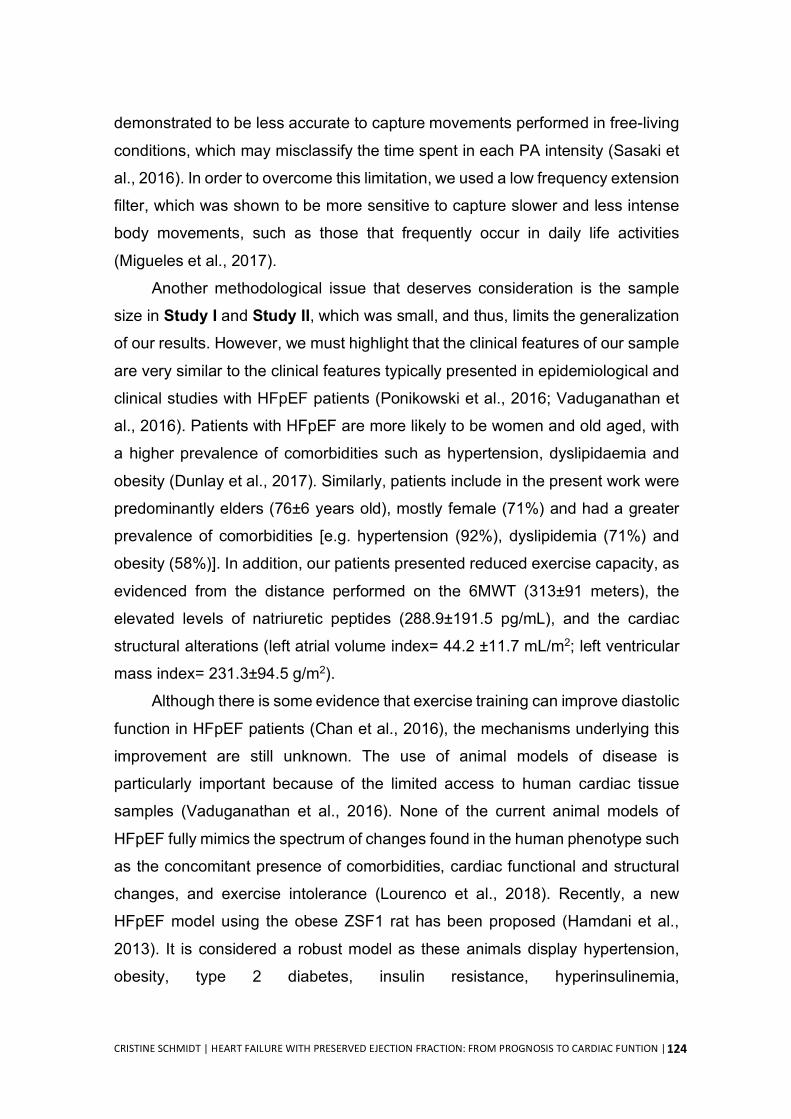
CRISTINE SCHMIDT | HEART FAILURE WITH PRESERVED EJECTION FRACTION: FROM PROGNOSIS TO CARDIAC FUNTION |
124
demonstrated to be less accurate to capture movements performed in free-living
conditions, which may misclassify the time spent in each PA intensity (Sasaki et
al., 2016). In order to overcome this limitation, we used a low frequency extension
filter, which was shown to be more sensitive to capture slower and less intense
body movements, such as those that frequently occur in daily life activities
(Migueles et al., 2017).
Another methodological issue that deserves consideration is the sample
size in Study I and Study II, which was small, and thus, limits the generalization
of our results. However, we must highlight that the clinical features of our sample
are very similar to the clinical features typically presented in epidemiological and
clinical studies with HFpEF patients (Ponikowski et al., 2016; Vaduganathan et
al., 2016). Patients with HFpEF are more likely to be women and old aged, with
a higher prevalence of comorbidities such as hypertension, dyslipidaemia and
obesity (Dunlay et al., 2017). Similarly, patients include in the present work were
predominantly elders (76±6 years old), mostly female (71%) and had a greater
prevalence of comorbidities [e.g. hypertension (92%), dyslipidemia (71%) and
obesity (58%)]. In addition, our patients presented reduced exercise capacity, as
evidenced from the distance performed on the 6MWT (313±91 meters), the
elevated levels of natriuretic peptides (288.9±191.5 pg/mL), and the cardiac
structural alterations (left atrial volume index= 44.2 ±11.7 mL/m2; left ventricular
mass index= 231.3±94.5 g/m2).
Although there is some evidence that exercise training can improve diastolic
function in HFpEF patients (Chan et al., 2016), the mechanisms underlying this
improvement are still unknown. The use of animal models of disease is
particularly important because of the limited access to human cardiac tissue
samples (Vaduganathan et al., 2016). None of the current animal models of
HFpEF fully mimics the spectrum of changes found in the human phenotype such
as the concomitant presence of comorbidities, cardiac functional and structural
changes, and exercise intolerance (Lourenco et al., 2018). Recently, a new
HFpEF model using the obese ZSF1 rat has been proposed (Hamdani et al.,
2013). It is considered a robust model as these animals display hypertension,
obesity, type 2 diabetes, insulin resistance, hyperinsulinemia,
CRISTINE SCHMIDT | HEART FAILURE WITH PRESERVED EJECTION FRACTION: FROM PROGNOSIS TO CARDIAC FUNTION |
125
hypertriglyceridemia and hypercholesterolaemia (Hamdani et al., 2013). Over
time, this cardiometabolic risk model develops the main features found in humans
diagnosed with HFpEF: i) reduced exercise tolerance, ii) preserved systolic
function (LVEF, LVdP/dtmax, and the slope of ESPVR) and iii) diastolic dysfunction
(higher E/e’, prolonged tau, elevated LVEDP, an upwards shift of LV diastolic
pressure-volume relationship, and higher LV diastolic chamber stiffness
constant) (Hamdani et al., 2013; Leite et al., 2015). Therefore, in Study III, we
used the ZSF1 obese animal model of HFpEF to evaluate the effects of exercise
training on LV function, structure and underlying molecular changes. Given that
ZSF1 rats are obese, present exercise intolerance and impaired cardiac function,
we anticipated that it would be difficult for these animals to complete an exercise
training protocol. In attempt to ensure compliance, an aerobic continuous training
of low-intensity protocol consisting of in running in a treadmill at 20 m/min, 60
min/day, 5 days per week, during a period of 5 weeks was selected.
CRISTINE SCHMIDT | HEART FAILURE WITH PRESERVED EJECTION FRACTION: FROM PROGNOSIS TO CARDIAC FUNTION |
126
Discussion of the main results
The present thesis had three important findings. Data from Study I showed
that physical fitness, particularly dynamic balance and agility, was positively
correlated with all dimensions of quality of life, outperforming all the other
components of physical fitness. Moreover, Study II demonstrated that IPAQ-SF
is not an accurate instrument to assess PA levels in HFpEF patients, and that
MVPA was the single PA intensity category positively associated with functional
capacity and quality of life. Finally, Study III showed that exercise training
improves exercise capacity, modulates LV stiffness and reduces circulating levels
of inflammatory, oxidative stress and endothelial dysfunction markers.
Association between different components of physical fitness and dimensions of quality of life in HFpEF patients As already stated, several studies have shown that physical fitness is an
important determinant of quality of life in HFpEF (Kitzman et al., 2010; Nolte et
al., 2015). Up to date, it was only established that higher levels of CRF are
associated with a better quality of life, mainly the physical dimension (Edelmann
et al., 2011; Kitzman et al., 2010). Our study adds novelty by showing that
dynamic balance and mobility is the single physical fitness component associated
with all dimensions of quality of life (general, physical and emotional). In addition,
our data revealed that dynamic balance and mobility outperforms CRF in
capturing HFpEF patients' quality of life. The capacity of the 8FUG test to better
identify patient’s quality of life might be explained by the wide range of physical
factors that influence the performance in this test (lower body strength, balance,
walking, agility and gait speed) (Rikli & Jones, 2013), which are also analogous
to the required in the normal daily tasks of an independent and autonomous life
(Mlinac & Feng, 2016) and perceived as the most important determinants of
patient’s quality of life. Although CRF assessed by maximal or symptom-limited
graded exercise tests or by the six-minute walk test were proven to be associated
with prognostic indicators and able to discriminate the grade of severity of disease
course, it might be possible that the continuous nature of the exercise might not
be sensitive to show an association with the dimensions of perceived quality of
CRISTINE SCHMIDT | HEART FAILURE WITH PRESERVED EJECTION FRACTION: FROM PROGNOSIS TO CARDIAC FUNTION |
127
life that is probably more influenced by the intermittent physical demands related
of household activities and commuting. In this sense, results from Study I gave
some insights from a new instrument to be used in the clinical setting, since the
underlying capacities are important to monitor HFpEF patients’ quality of life
correlates. Despite that, the 8FUG test has the advantage of being simple to
administer, requiring a limited space, minimal equipment (i.e. markers on the floor
and a chair) and it is easily understand by patients (Wilkinson et al., 2018).
Therefore, we recommend the use of the 8FUG test on clinical practice in
combination with the assessment of patients’ quality of life.
The utility of IPAQ short form to measure PA levels in HFpEF patients Despite the evidence shows that questionnaires are valid and reliable in
measuring PA, their correlation with accelerometers-derived data is far from
satisfactory (Koolhaas et al., 2018; Lee et al., 2011; Prince et al., 2008; Skender
et al., 2016), which limits PA-based decisions in the clinical setting regarding
counselling and recommendations. Our results corroborate previous findings in
the general population, showing that the measures derived from IPAQ-SF
underestimates sedentary time and overestimates time spent in MVPA in HFpEF
patients, and also showed poor agreement between self-reported and
accelerometer-derived PA measures. Therefore, based on our findings, the use
of IPAQ-SF should not be recommended to support accurate recommendations
in this specific population. For clinical purposes, at an individual level, it is
important to have accurate and reliable PA information to assess the risk level
and to prescribe life-style changes. Therefore, the use of accelerometers to
assess daily PA levels in HFpEF patients seems to have a particular importance.
Objective measures can avoid bias related with subjective measures, especially
in populations that have limited physical function and no experience performing
PA. Moreover, self-reported measures depend on recall which can be biased
specially in older patients (Prince et al., 2008). Nevertheless, although
accelerometry is considered a better methodology to assess PA when compared
to questionnaires, it cannot provide comprehensive information in terms of type
and context in which PA occur. In this sense, to provide tailored
CRISTINE SCHMIDT | HEART FAILURE WITH PRESERVED EJECTION FRACTION: FROM PROGNOSIS TO CARDIAC FUNTION |
128
recommendations in the clinical setting, we suggest the use of accelerometers
together with a diary log to better characterize patients’ PA levels.
Patterns of PA and sedentary time in HFpEF patients, and its association with prognostic indicators
Given the growing recognition of the impact of PA levels in the prognosis of
HFpEF patients, a rigorous characterization of their patterns is crucial for
prescribing tailored life-style changes to improve well-being and quality of life,
and to mitigate/attenuate the disease progression. In the study II we observed
that HFpEF patients spent most of their waking time in sedentary behaviours
while their daily activity was mainly comprised of light physical activity, and just a
few minutes in MVPA. Increased sedentary behaviours and decreased time
spend in MVPA are both associated with worse clinical outcomes in HFpEF
patients (Hegde et al., 2017; Snipelisky et al., 2017). Interestingly, our study adds
the novelty that only time spent in MVPA was significantly associated with
important clinical outcomes as VO2, 6MWT performance and QoL, but not with
E/e´ or BNP levels. Thus, it seems that intensity is a requirement for significantly
impacting the patient’s prognosis. Corroborating this hypothesis, it was recently
shown a dose-response relationship between MVPA and risk of hospitalization
or mortality in HFpEF patients (Hegde et al., 2017). Despite that, it was already
shown that any increase in the amount of PA may translate into some health
benefits (Physical Activity Guidelines Advisory Committee Scientific Report,
2018). Taking together, these data emphasise the clinical importance to educate
patients about the importance of engaging in more MVPA throughout the day and
to reduce their time spent in sedentary behaviours in order to achieve higher
health-related benefits.
Effects of exercise training in an animal model of HFpEF
Exercise training is emerging as an important ally for the clinical
management of health-related outcomes in HFpEF patients. However, the
literature thus far cannot elucidate the mechanisms by which exercise training
can induce improvements in HFpEF patients. Current published data suggests
CRISTINE SCHMIDT | HEART FAILURE WITH PRESERVED EJECTION FRACTION: FROM PROGNOSIS TO CARDIAC FUNTION |
129
that the improvements in VO2peak found in HFpEF patients after an exercise
training program are mainly due to non-cardiac peripheral adaptations, as no
study has yet shown to improve peak cardiac output (Fu et al., 2016; Haykowsky
et al., 2012). However, both clinical (Dieberg et al., 2015; Pearson et al., 2017)
and pre-clinical studies (Hidalgo et al., 2014; Marshall et al., 2013) support the
hypothesis that exercise training improves diastolic function in HFpEF setting,
which may also account for an improved acute exercise response. Results from
Study III show that exercise training can improve exercise capacity and
enhances cardiac function by decreasing cardiomyocyte passive tension and by
attenuating LV fibrosis in HFpEF animals. These results provide novel
mechanistic insights into the benefits of regular exercise training in a clinically
translational animal model of HFpEF. Understanding the pathways that mediate
these benefits, and learning how to manipulate them in vivo, could yield novel
therapeutic approaches based on agents that mimic or potentiate the
physiological benefits of exercise. As mentioned in the first chapter (General
Introduction, page 9) low-grade chronic systemic inflammation derived from
comorbidities is responsible for coronary microvascular inflammation, which
might lead to myocardial dysfunction and remodelling. Our results in Study III
suggest that beyond the improvements in cardiomyocyte passive tension and LV
fibrosis, regular exercise training was able to attenuate systemic inflammation
and to diminish circulating markers of oxidative stress and endothelial
dysfunction. Collectively, these results empower the hypothesizes that low-grade
chronic systemic inflammation is the main trigger of HFpEF. Therefore, our
results confirm the pleiotropic effects of exercise training, namely its anti-
inflammatory effect, which might modulate improvements in cardiac function by
interfering in the cascade of events that lead to decreased diastolic left ventricular
stiffness and interstitial fibrosis. Overall, results from Study III suggest that
exercise training may be a particularly powerful adjuvant treatment option to
HFpEF patients, but clinical studies are needed to clarify the best type of training,
frequency and intensity in large populations with HFpEF.
CRISTINE SCHMIDT | HEART FAILURE WITH PRESERVED EJECTION FRACTION: FROM PROGNOSIS TO CARDIAC FUNTION |
130
REFERENCES Bobenko, A., Bartels, I., Munch, M., Trippel, T., Lindhorst, R., Nolte, K.,
Herrmann-Lingen, C., Halle, M., Duvinage, A., Dungen, H. D., Gelbrich, G., Tschope, C., Hasenfuss, G., Wachter, R., Pieske, B., & Edelmann, F. (2018). Amount or intensity? Potential targets of exercise interventions in patients with heart failure with preserved ejection fraction. ESC Heart Fail, 5(1), 53-62.
Chan, E., Giallauria, F., Vigorito, C., & Smart, N. A. (2016). Exercise training in heart failure patients with preserved ejection fraction: a systematic review and meta-analysis. Monaldi Arch Chest Dis, 86(1-2), 759.
Cheung, V. H., Gray, L., & Karunanithi, M. (2011). Review of accelerometry for determining daily activity among elderly patients. Arch Phys Med Rehabil, 92(6), 998-1014.
Craig, C. L., Marshall, A. L., Sjostrom, M., Bauman, A. E., Booth, M. L., Ainsworth, B. E., Pratt, M., Ekelund, U., Yngve, A., Sallis, J. F., & Oja, P. (2003). International physical activity questionnaire: 12-country reliability and validity. Med Sci Sports Exerc, 35(8), 1381-1395.
Dieberg, G., Ismail, H., Giallauria, F., & Smart, N. A. (2015). Clinical outcomes and cardiovascular responses to exercise training in heart failure patients with preserved ejection fraction: a systematic review and meta-analysis. J Appl Physiol (1985), 119(6), 726-733.
Dunlay, S. M., Roger, V. L., & Redfield, M. M. (2017). Epidemiology of heart failure with preserved ejection fraction. Nat Rev Cardiol, 14(10), 591-602.
Edelmann, F., Gelbrich, G., Dungen, H. D., Frohling, S., Wachter, R., Stahrenberg, R., Binder, L., Topper, A., Lashki, D. J., Schwarz, S., Herrmann-Lingen, C., Loffler, M., Hasenfuss, G., Halle, M., & Pieske, B. (2011). Exercise training improves exercise capacity and diastolic function in patients with heart failure with preserved ejection fraction: results of the Ex-DHF (Exercise training in Diastolic Heart Failure) pilot study. J Am Coll Cardiol, 58(17), 1780-1791.
Fu, T. C., Yang, N. I., Wang, C. H., Cherng, W. J., Chou, S. L., Pan, T. L., & Wang, J. S. (2016). Aerobic Interval Training Elicits Different Hemodynamic Adaptations Between Heart Failure Patients with Preserved and Reduced Ejection Fraction. Am J Phys Med Rehabil, 95(1), 15-27.
Gorman, E., Hanson, H. M., Yang, P. H., Khan, K. M., Liu-Ambrose, T., & Ashe, M. C. (2014). Accelerometry analysis of physical activity and sedentary behavior in older adults: a systematic review and data analysis. Eur Rev Aging Phys Act, 11, 35-49.
Hamdani, N., Franssen, C., Lourenco, A., Falcao-Pires, I., Fontoura, D., Leite, S., Plettig, L., Lopez, B., Ottenheijm, C. A., Becher, P. M., Gonzalez, A., Tschope, C., Diez, J., Linke, W. A., Leite-Moreira, A. F., & Paulus, W. J. (2013). Myocardial titin hypophosphorylation importantly contributes to
CRISTINE SCHMIDT | HEART FAILURE WITH PRESERVED EJECTION FRACTION: FROM PROGNOSIS TO CARDIAC FUNTION |
131
heart failure with preserved ejection fraction in a rat metabolic risk model. Circ Heart Fail, 6(6), 1239-1249.
Haykowsky, M. J., Brubaker, P. H., Stewart, K. P., Morgan, T. M., Eggebeen, J., & Kitzman, D. W. (2012). Effect of endurance training on the determinants of peak exercise oxygen consumption in elderly patients with stable compensated heart failure and preserved ejection fraction. J Am Coll Cardiol, 60(2), 120-128.
Hegde, S. M., Claggett, B., Shah, A. M., Lewis, E. F., Anand, I., Shah, S. J., Sweitzer, N. K., Fang, J. C., Pitt, B., Pfeffer, M. A., & Solomon, S. D. (2017). Physical Activity and Prognosis in the TOPCAT Trial (Treatment of Preserved Cardiac Function Heart Failure With an Aldosterone Antagonist). Circulation, 136(11), 982-992.
Hidalgo, C., Saripalli, C., & Granzier, H. L. (2014). Effect of exercise training on post-translational and post-transcriptional regulation of titin stiffness in striated muscle of wild type and IG KO mice. Arch Biochem Biophys, 552-553, 100-107.
Kelly, L. A., McMillan, D. G., Anderson, A., Fippinger, M., Fillerup, G., & Rider, J. (2013). Validity of actigraphs uniaxial and triaxial accelerometers for assessment of physical activity in adults in laboratory conditions. BMC Med Phys, 13(1), 5.
Kitzman, D. W., Brubaker, P. H., Herrington, D. M., Morgan, T. M., Stewart, K. P., Hundley, W. G., Abdelhamed, A., & Haykowsky, M. J. (2013). Effect of endurance exercise training on endothelial function and arterial stiffness in older patients with heart failure and preserved ejection fraction: a randomized, controlled, single-blind trial. J Am Coll Cardiol, 62(7), 584-592.
Kitzman, D. W., Brubaker, P. H., Morgan, T. M., Stewart, K. P., & Little, W. C. (2010). Exercise training in older patients with heart failure and preserved ejection fraction: a randomized, controlled, single-blind trial. Circ Heart Fail, 3(6), 659-667.
Kondamudi, N., Haykowsky, M., Forman, D. E., Berry, J. D., & Pandey, A. (2017). Exercise Training for Prevention and Treatment of Heart Failure. Prog Cardiovasc Dis, 60(1), 115-120.
Koolhaas, C. M., van Rooij, F. J., Cepeda, M., Tiemeier, H., Franco, O. H., & Schoufour, J. D. (2018). Physical activity derived from questionnaires and wrist-worn accelerometers: comparability and the role of demographic, lifestyle, and health factors among a population-based sample of older adults. Clin Epidemiol, 10, 1-16.
Lee, P. H., Macfarlane, D. J., Lam, T. H., & Stewart, S. M. (2011). Validity of the International Physical Activity Questionnaire Short Form (IPAQ-SF): a systematic review. Int J Behav Nutr Phys Act, 8, 115.
Lourenco, A. P., Leite-Moreira, A. F., Balligand, J. L., Bauersachs, J., Dawson, D., de Boer, R. A., de Windt, L. J., Falcao-Pires, I., Fontes-Carvalho, R., Franz, S., Giacca, M., Hilfiker-Kleiner, D., Hirsch, E., Maack, C., Mayr, M., Pieske, B., Thum, T., Tocchetti, C. G., Brutsaert, D. L., & Heymans, S.
CRISTINE SCHMIDT | HEART FAILURE WITH PRESERVED EJECTION FRACTION: FROM PROGNOSIS TO CARDIAC FUNTION |
132
(2018). An integrative translational approach to study heart failure with preserved ejection fraction: a position paper from the Working Group on Myocardial Function of the European Society of Cardiology. Eur J Heart Fail, 20(2), 216-227.
Marshall, K. D., Muller, B. N., Krenz, M., Hanft, L. M., McDonald, K. S., Dellsperger, K. C., & Emter, C. A. (2013). Heart failure with preserved ejection fraction: chronic low-intensity interval exercise training preserves myocardial O2 balance and diastolic function. J Appl Physiol (1985), 114(1), 131-147.
Martin, K. R., Koster, A., Murphy, R. A., Van Domelen, D. R., Hung, M. Y., Brychta, R. J., Chen, K. Y., & Harris, T. B. (2014). Changes in daily activity patterns with age in U.S. men and women: National Health and Nutrition Examination Survey 2003-04 and 2005-06. J Am Geriatr Soc, 62(7), 1263-1271.
Migueles, J. H., Cadenas-Sanchez, C., Ekelund, U., Delisle Nystrom, C., Mora-Gonzalez, J., Lof, M., Labayen, I., Ruiz, J. R., & Ortega, F. B. (2017). Accelerometer Data Collection and Processing Criteria to Assess Physical Activity and Other Outcomes: A Systematic Review and Practical Considerations. Sports Med, 47(9), 1821-1845.
Mlinac, M. E., & Feng, M. C. (2016). Assessment of Activities of Daily Living, Self-Care, and Independence. Arch Clin Neuropsychol, 31(6), 506-516.
Nolte, K., Herrmann-Lingen, C., Wachter, R., Gelbrich, G., Dungen, H. D., Duvinage, A., Hoischen, N., von Oehsen, K., Schwarz, S., Hasenfuss, G., Halle, M., Pieske, B., & Edelmann, F. (2015). Effects of exercise training on different quality of life dimensions in heart failure with preserved ejection fraction: the Ex-DHF-P trial. Eur J Prev Cardiol, 22(5), 582-593.
Pearson, M. J., Mungovan, S. F., & Smart, N. A. (2017). Effect of exercise on diastolic function in heart failure patients: a systematic review and meta-analysis. Heart Fail Rev, 22(2), 229-242.
Physical Activity Guidelines Advisory Committee Scientific Report. (2018). 2018 Physical Activity Guidelines Advisory Committee.
Ponikowski, P., Voors, A. A., Anker, S. D., Bueno, H., Cleland, J. G. F., Coats, A. J. S., Falk, V., Gonzalez-Juanatey, J. R., Harjola, V. P., Jankowska, E. A., Jessup, M., Linde, C., Nihoyannopoulos, P., Parissis, J. T., Pieske, B., Riley, J. P., Rosano, G. M. C., Ruilope, L. M., Ruschitzka, F., Rutten, F. H., van der Meer, P., & Group, E. S. C. S. D. (2016). 2016 ESC Guidelines for the diagnosis and treatment of acute and chronic heart failure: The Task Force for the diagnosis and treatment of acute and chronic heart failure of the European Society of Cardiology (ESC)Developed with the special contribution of the Heart Failure Association (HFA) of the ESC. Eur Heart J, 37(27), 2129-2200.
Prince, S. A., Adamo, K. B., Hamel, M. E., Hardt, J., Connor Gorber, S., & Tremblay, M. (2008). A comparison of direct versus self-report measures
CRISTINE SCHMIDT | HEART FAILURE WITH PRESERVED EJECTION FRACTION: FROM PROGNOSIS TO CARDIAC FUNTION |
133
for assessing physical activity in adults: a systematic review. Int J Behav Nutr Phys Act, 5, 56.
Rector, T. S., & Cohn, J. N. (1992). Assessment of patient outcome with the Minnesota Living with Heart Failure questionnaire: reliability and validity during a randomized, double-blind, placebo-controlled trial of pimobendan. Pimobendan Multicenter Research Group. Am Heart J, 124(4), 1017-1025.
Rikli, R. E., & Jones, C. J. (2013). Development and validation of criterion-referenced clinically relevant fitness standards for maintaining physical independence in later years. Gerontologist, 53(2), 255-267.
Santos-Lozano, A., Santin-Medeiros, F., Cardon, G., Torres-Luque, G., Bailon, R., Bergmeir, C., Ruiz, J. R., Lucia, A., & Garatachea, N. (2013). Actigraph GT3X: validation and determination of physical activity intensity cut points. Int J Sports Med, 34(11), 975-982.
Sasaki, J. E., Hickey, A. M., Staudenmayer, J. W., John, D., Kent, J. A., & Freedson, P. S. (2016). Performance of Activity Classification Algorithms in Free-Living Older Adults. Med Sci Sports Exerc, 48(5), 941-950.
Sasaki, J. E., John, D., & Freedson, P. S. (2011). Validation and comparison of ActiGraph activity monitors. J Sci Med Sport, 14(5), 411-416.
Shiroma, E. J., Freedson, P. S., Trost, S. G., & Lee, I. M. (2013). Patterns of accelerometer-assessed sedentary behavior in older women. JAMA, 310(23), 2562-2563.
Skender, S., Ose, J., Chang-Claude, J., Paskow, M., Bruhmann, B., Siegel, E. M., Steindorf, K., & Ulrich, C. M. (2016). Accelerometry and physical activity questionnaires - a systematic review. BMC Public Health, 16, 515.
Snipelisky, D., Kelly, J., Levine, J. A., Koepp, G. A., Anstrom, K. J., McNulty, S. E., Zakeri, R., Felker, G. M., Hernandez, A. F., Braunwald, E., & Redfield, M. M. (2017). Accelerometer-Measured Daily Activity in Heart Failure With Preserved Ejection Fraction: Clinical Correlates and Association With Standard Heart Failure Severity Indices. Circ Heart Fail, 10(6), e003878.
The World Health Organization Quality of Life Assessment Group. (1998). The World Health Organization Quality of Life Assessment (WHOQOL): development and general psychometric properties. Soc Sci Med, 46(12), 1569-1585.
Troiano, R. P. (2005). A timely meeting: objective measurement of physical activity. Med Sci Sports Exerc, 37(11 Suppl), S487-489.
Vaduganathan, M., Michel, A., Hall, K., Mulligan, C., Nodari, S., Shah, S. J., Senni, M., Triggiani, M., Butler, J., & Gheorghiade, M. (2016). Spectrum of epidemiological and clinical findings in patients with heart failure with preserved ejection fraction stratified by study design: a systematic review. Eur J Heart Fail, 18(1), 54-65.
Wilkinson, T. J., Lemmey, A. B., Clayton, R. J., Jones, J. G., & O’Brien, T. D. (2018). The 8-foot up and go test is the best way to assess physical function
CRISTINE SCHMIDT | HEART FAILURE WITH PRESERVED EJECTION FRACTION: FROM PROGNOSIS TO CARDIAC FUNTION |
134
in the rheumatoid arthritis clinic. Rheumatology Advances in Practice, 2(1), rkx017-rkx017.
Zisko, N., Carlsen, T., Salvesen, O., Aspvik, N. P., Ingebrigtsen, J. E., Wisloff, U., & Stensvold, D. (2015). New relative intensity ambulatory accelerometer thresholds for elderly men and women: the Generation 100 study. BMC Geriatr, 15, 97.
CRISTINE SCHMIDT | HEART FAILURE WITH PRESERVED EJECTION FRACTION: FROM PROGNOSIS TO CARDIAC FUNTION |
135
CHAPTER V
CONCLUSIONS AND FUTURE DIRECTION

CRISTINE SCHMIDT | HEART FAILURE WITH PRESERVED EJECTION FRACTION: FROM PROGNOSIS TO CARDIAC FUNTION |
136
CRISTINE SCHMIDT | HEART FAILURE WITH PRESERVED EJECTION FRACTION: FROM PROGNOSIS TO CARDIAC FUNTION |
137
CONCLUSIONS
Based on the general conclusions of each study that were presented in this
thesis, it is possible to outline the following major conclusions:
• Physical fitness, particularly dynamic balance and mobility, is
associated with all dimensions of quality of life in patients with
HFpEF. In addition, dynamic balance and mobility outperforms
others physical fitness components in capturing HFpEF patients'
quality of life.
• The IPAQ-SF underestimates sedentary time and overestimates
time spent in MVPA in patients with HFpEF. Patients spent only a
minority of their daily time involved in MVPA, which was the only PA
intensity positively associated with functional capacity and quality of
life.
• Moderate exercise training improves exercise capacity and
attenuates left ventricle diastolic stiffness in the ZSF1 obese animal
model of HFpEF. This was paralleled by reduced circulating levels of
inflammatory cytokines and markers of endothelial dysfunction and
oxidative stress.

CRISTINE SCHMIDT | HEART FAILURE WITH PRESERVED EJECTION FRACTION: FROM PROGNOSIS TO CARDIAC FUNTION |
138
CRISTINE SCHMIDT | HEART FAILURE WITH PRESERVED EJECTION FRACTION: FROM PROGNOSIS TO CARDIAC FUNTION |
139
FUTURE DIRECTIONS
Further prospective cohort studies with a larger sample size are needed
to strengthen or refute our conclusions that dynamic balance and mobility is more
discriminative to capture HFpEF patients' quality of life, and to verify the
agreement between PA measurements from the IPAQ-SF and PA measures
derived from triaxial accelerometer.
In addition, experimental studies exploring the molecular changes
underlying the cardiomyocyte’s intrinsic and extrinsic changes induced by
exercise training are fundamental as they may lead to the identification of
therapeutic molecular targets.
Moreover, different exercise training protocols with the ZSF1 animal model
are also required in order to better understanding the dose-response relationship
between exercise training and HFpEF improvements.

CRISTINE SCHMIDT | HEART FAILURE WITH PRESERVED EJECTION FRACTION: FROM PROGNOSIS TO CARDIAC FUNTION |
140

CRISTINE SCHMIDT | HEART FAILURE WITH PRESERVED EJECTION FRACTION: FROM PROGNOSIS TO CARDIAC FUNTION |
141
APPENDICES

CRISTINE SCHMIDT | HEART FAILURE WITH PRESERVED EJECTION FRACTION: FROM PROGNOSIS TO CARDIAC FUNTION |
142
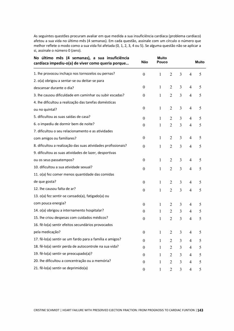
CRISTINE SCHMIDT | HEART FAILURE WITH PRESERVED EJECTION FRACTION: FROM PROGNOSIS TO CARDIAC FUNTION |
143
As seguintes questões procuram avaliar em que medida a sua insuficiência cardíaca (problema cardíaco) afetou a sua vida no último mês (4 semanas). Em cada questão, assinale com um círculo o número que melhor reflete o modo como a sua vida foi afetada (0, 1, 2, 3, 4 ou 5). Se alguma questão não se aplicar a si, assinale o número 0 (zero).
No último mês (4 semanas), a sua insuficiência cardíaca impediu-o(a) de viver como queria porque… 1. lhe provocou inchaço nos tornozelos ou pernas?
2. o(a) obrigou a sentar-se ou deitar-se para
descansar durante o dia?
3. lhe causou dificuldade em caminhar ou subir escadas?
4. lhe dificultou a realização das tarefas domésticas
ou no quintal?
5. dificultou as suas saídas de casa?
6. o impediu de dormir bem de noite?
7. dificultou o seu relacionamento e as atividades
com amigos ou familiares?
8. dificultou a realização das suas atividades profissionais?
9. dificultou as suas atividades de lazer, desportivas
ou os seus passatempos?
10. dificultou a sua atividade sexual?
11. o(a) fez comer menos quantidade das comidas
de que gosta?
12. lhe causou falta de ar?
13. o(a) fez sentir-se cansado(a), fatigado(a) ou
com pouca energia?
14. o(a) obrigou a internamento hospitalar?
15. lhe criou despesas com cuidados médicos?
16. fê-lo(a) sentir efeitos secundários provocados
pela medicação?
17. fê-lo(a) sentir-se um fardo para a família e amigos?
18. fê-lo(a) sentir perda de autocontrole na sua vida?
19. fê-lo(a) sentir-se preocupado(a)?
20. lhe dificultou a concentração ou a memória?
21. fê-lo(a) sentir-se deprimido(a)
Muito Não Pouco Muito
0 1 2 3 4 5
0 1 2 3 4 5
0 1 2 3 4 5
0 1 2 3 4 5
0 1 2 3 4 5 0 1 2 3 4 5
0 1 2 3 4 5
0 1 2 3 4 5
0 1 2 3 4 5
0 1 2 3 4 5
0 1 2 3 4 5
0 1 2 3 4 5
0 1 2 3 4 5 0 1 2 3 4 5
0 1 2 3 4 5
0 1 2 3 4 5
0 1 2 3 4 5 0 1 2 3 4 5
0 1 2 3 4 5 0 1 2 3 4 5
0 1 2 3 4 5

CRISTINE SCHMIDT | HEART FAILURE WITH PRESERVED EJECTION FRACTION: FROM PROGNOSIS TO CARDIAC FUNTION |
144

CRISTINE SCHMIDT | HEART FAILURE WITH PRESERVED EJECTION FRACTION: FROM PROGNOSIS TO CARDIAC FUNTION |
145
Atividade Física Estamos interessados em conhecer os diferentes tipos de atividade física, que as pessoas fazem no seu quotidiano. As questões que lhe vou colocar, referem-se à semana imediatamente anterior, considerando o tempo em que esteve fisicamente ativo/a. Por favor, responda a todas as questões, mesmo que não se considere uma pessoa fisicamente ativa. Vou colocar-lhe questões sobre as atividades desenvolvidas na sua atividade profissional e nas suas deslocações, sobre as atividades referentes aos trabalhos domésticos e às atividades que efetuou no seu tempo livre para recreação ou prática de exercício físico / desporto. Ao responder às seguintes questões considere o seguinte: Actividades físicas vigorosas referem-se a atividades que requerem um esforço físico intenso que fazem ficar com a respiração ofegante. Actividades físicas moderadas referem-se a atividades que requerem esforço físico moderado e tornam a respiração um pouco mais forte que o normal. Ao responder às questões considere apenas as actividades físicas que realize durante pelo menos 10 minutos seguidos
Q.1 Diga-me por favor, nos últimos 7 dias, em quantos dias fez actividades físicas vigorosas, como por exemplo, levantar objetos pesados, cavar, ginástica aeróbica, nadar, jogar futebol, andar de bicicleta a um ritmo rápido?
Dias
Q.2 Nos dias em que pratica actividades físicas vigorosas, quanto tempo em média dedica normalmente a essas actividades?
Horas Minutos (por dia)
Q.3 Diga-me por favor, nos últimos 7 dias, em quantos dias fez actividades físicas moderadas como por exemplo, carregar objectos leves, caçar, trabalhos de carpintaria, andar de bicicleta a um ritmo normal ou ténis de pares? Por favor não inclua o “andar”.
Dias
Q.4 Nos dias em que faz actividades físicas moderadas, quanto tempo em média dedica normalmente a essas actividades?
Horas Minutos (por dia)
Q.5 Diga-me por favor, nos últimos 7 dias, em quantos dias andou pelo menos 10 minutos seguidos?
Dias
Q.6 Quanto tempo no total, despendeu num desses dias, a andar/caminhar?
Horas Minutos (por dia)
Q.7 Diga-me por favor, num dia normal quanto tempo passa sentado? Isto pode incluir o tempo que passa a uma secretária, a visitar amigos, a ler, a estudar ou a ver televisão.
Horas Minutos (por dia)

CRISTINE SCHMIDT | HEART FAILURE WITH PRESERVED EJECTION FRACTION: FROM PROGNOSIS TO CARDIAC FUNTION |
146


![Northumbria Research Linknrl.northumbria.ac.uk/24201/1/Hickey et al 2015... · 7 Table 3: ActiGraph™ intensity cut-points [10], the synthesised count ratio and the derived PRO-Diary™](https://img.pdfslide.us/doc/110x75/5f93343b84cc0a5a0f2ebc73/northumbria-research-et-al-2015-7-table-3-actigrapha-intensity-cut-points.jpg)