Embed Size (px)
Citation preview
Poster Design & Printing by Genigraphics® - 800.790.4001
Sean M. Miller, MDDepartment of Otolaryngology –
Head and Neck Surgery Saint Louis University
email: [email protected]
Objective:• To determine whether a type 2 diabetic mouse model demonstrates hearing loss• To examine cochlear ultrastructure of a type 2 diabetic mouse model for evidence of pathology
Subjects and Methods:The Leprdb/Leprdb (Db/Db) mouse has a point mutation in the leptin receptor which causes polyphagia, hyperglycemia, and obesity; modeling type 2 diabetes mellitus. Db/Db and wild type mice from the parent strain were used. Auditory brainstem response measurements and distortion product otoacoustic emissions were performed followed by analysis of cochlear ultrastructure using scanning and transmission electron microscopy.
Results:The mutant animals demonstrate a statistically significant mid-high frequency hearing loss when compared to age-matched, wild type counterparts. Furthermore, this hearing loss worsens with age. Fine structure analysis reveals evidence of morphological alterations in multiple locations within the cochlea, including loss of neuron density and intracellular inclusions. Scanning electron microscopy reveals no evidence of hair cell loss but some aberrant organization of stereocilia.
Conclusion:This is the first demonstration of hearing loss and ultrastructural changes in a genetically derived type 2 diabetes mellitus mouse model. Use of the Db/Db mouse provides an opportunity to investigate the underlying mechanism of type 2 diabetes otopathology.
Hearing Loss and Cochlear Pathology in a Type 2 Diabetic Mouse ModelSean M. Miller, MD1; Grady Phillips, MS1; Alexis Varvares, Flint Boettcher, PhD1, Michael Anne Gratton, PhD1,
1Department of Otolaryngology – Head and Neck Surgery, Saint Louis University
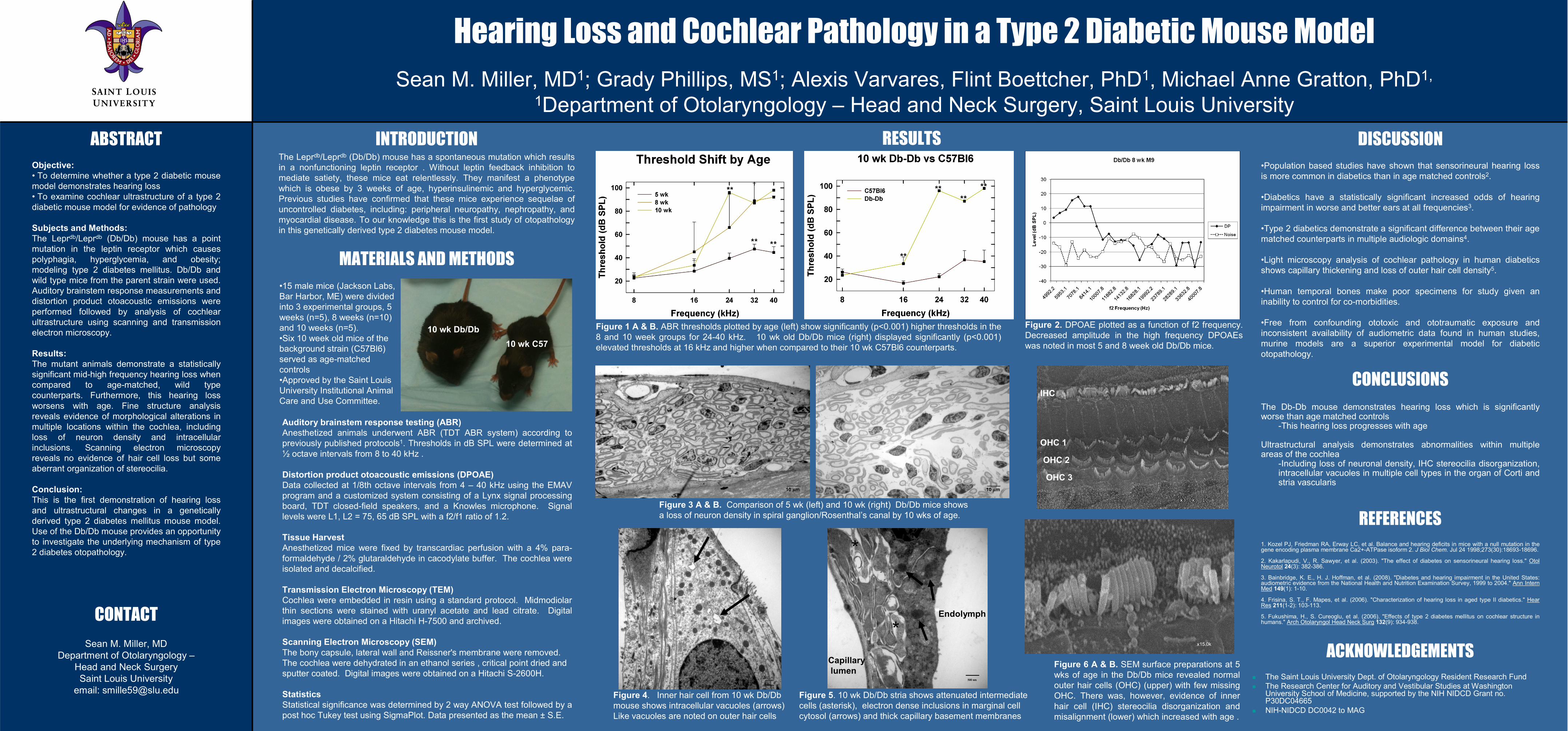
Figure 2. DPOAE plotted as a function of f2 frequency. Decreased amplitude in the high frequency DPOAEs was noted in most 5 and 8 week old Db/Db mice.
•Population based studies have shown that sensorineural hearing loss is more common in diabetics than in age matched controls2.
•Diabetics have a statistically significant increased odds of hearing impairment in worse and better ears at all frequencies3.
•Type 2 diabetics demonstrate a significant difference between their age matched counterparts in multiple audiologic domains4.
•Light microscopy analysis of cochlear pathology in human diabetics shows capillary thickening and loss of outer hair cell density5.
•Human temporal bones make poor specimens for study given an inability to control for co-morbidities.
•Free from confounding ototoxic and ototraumatic exposure and inconsistent availability of audiometric data found in human studies, murine models are a superior experimental model for diabetic otopathology.
The Db-Db mouse demonstrates hearing loss which is significantly worse than age matched controls
-This hearing loss progresses with age
Ultrastructural analysis demonstrates abnormalities within multiple areas of the cochlea
-Including loss of neuronal density, IHC stereocilia disorganization,intracellular vacuoles in multiple cell types in the organ of Corti and stria vascularis
The Leprdb/Leprdb (Db/Db) mouse has a spontaneous mutation which results in a nonfunctioning leptin receptor . Without leptin feedback inhibition to mediate satiety, these mice eat relentlessly. They manifest a phenotype which is obese by 3 weeks of age, hyperinsulinemic and hyperglycemic. Previous studies have confirmed that these mice experience sequelae of uncontrolled diabetes, including: peripheral neuropathy, nephropathy, and myocardial disease. To our knowledge this is the first study of otopathology in this genetically derived type 2 diabetes mouse model.
INTRODUCTION
MATERIALS AND METHODS
1. Kozel PJ, Friedman RA, Erway LC, et al. Balance and hearing deficits in mice with a null mutation in the gene encoding plasma membrane Ca2+-ATPase isoform 2. J Biol Chem. Jul 24 1998;273(30):18693-18696.
2. Kakarlapudi, V., R. Sawyer, et al. (2003). "The effect of diabetes on sensorineural hearing loss." Otol Neurotol 24(3): 382-386.
3. Bainbridge, K. E., H. J. Hoffman, et al. (2008). "Diabetes and hearing impairment in the United States: audiometric evidence from the National Health and Nutrition Examination Survey, 1999 to 2004." Ann Intern Med 149(1): 1-10.
4. Frisina, S. T., F. Mapes, et al. (2006). "Characterization of hearing loss in aged type II diabetics." Hear Res 211(1-2): 103-113.
5. Fukushima, H., S. Cureoglu, et al. (2006). "Effects of type 2 diabetes mellitus on cochlear structure in humans." Arch Otolaryngol Head Neck Surg 132(9): 934-938.
CONCLUSIONS
DISCUSSIONRESULTS
REFERENCES
Figure 1 A & B. ABR thresholds plotted by age (left) show significantly (p<0.001) higher thresholds in the 8 and 10 week groups for 24-40 kHz. 10 wk old Db/Db mice (right) displayed significantly (p<0.001) elevated thresholds at 16 kHz and higher when compared to their 10 wk C57Bl6 counterparts.
ABSTRACT
CONTACT
Figure 6 A & B. SEM surface preparations at 5 wks of age in the Db/Db mice revealed normal outer hair cells (OHC) (upper) with few missing OHC. There was, however, evidence of inner hair cell (IHC) stereocilia disorganization and misalignment (lower) which increased with age .
ACKNOWLEDGEMENTS The Saint Louis University Dept. of Otolaryngology Resident Research Fund The Research Center for Auditory and Vestibular Studies at Washington
University School of Medicine, supported by the NIH NIDCD Grant no. P30DC04665
NIH-NIDCD DC0042 to MAG
Auditory brainstem response testing (ABR)Anesthetized animals underwent ABR (TDT ABR system) according topreviously published protocols1. Thresholds in dB SPL were determined at ½ octave intervals from 8 to 40 kHz .
Distortion product otoacoustic emissions (DPOAE)Data collected at 1/8th octave intervals from 4 – 40 kHz using the EMAV program and a customized system consisting of a Lynx signal processing board, TDT closed-field speakers, and a Knowles microphone. Signal levels were L1, L2 = 75, 65 dB SPL with a f2/f1 ratio of 1.2.
Tissue HarvestAnesthetized mice were fixed by transcardiac perfusion with a 4% para-formaldehyde / 2% glutaraldehyde in cacodylate buffer. The cochlea were isolated and decalcified.
Transmission Electron Microscopy (TEM)Cochlea were embedded in resin using a standard protocol. Midmodiolar thin sections were stained with uranyl acetate and lead citrate. Digital images were obtained on a Hitachi H-7500 and archived.
Scanning Electron Microscopy (SEM)The bony capsule, lateral wall and Reissner's membrane were removed. The cochlea were dehydrated in an ethanol series , critical point dried and sputter coated. Digital images were obtained on a Hitachi S-2600H.
StatisticsStatistical significance was determined by 2 way ANOVA test followed by a post hoc Tukey test using SigmaPlot. Data presented as the mean ± S.E.
•15 male mice (Jackson Labs, Bar Harbor, ME) were divided into 3 experimental groups, 5 weeks (n=5), 8 weeks (n=10) and 10 weeks (n=5). •Six 10 week old mice of the background strain (C57Bl6) served as age-matched controls•Approved by the Saint Louis University Institutional Animal Care and Use Committee.
10 wk Db/Db10 wk C57
Figure 3 A & B. Comparison of 5 wk (left) and 10 wk (right) Db/Db mice shows a loss of neuron density in spiral ganglion/Rosenthal’s canal by 10 wks of age.
x15.0k
_____10 µm
_____10 µm
Figure 4. Inner hair cell from 10 wk Db/Db mouse shows intracellular vacuoles (arrows) Like vacuoles are noted on outer hair cells
Figure 5. 10 wk Db/Db stria shows attenuated intermediate cells (asterisk), electron dense inclusions in marginal cell cytosol (arrows) and thick capillary basement membranes
*
*Capillarylumen
Endolymph
OHC 1
OHC 2
OHC 3
IHC