Embed Size (px)
Citation preview

Healing 48
Wound Healing 49–51
Healing – Fibrosis 52
Healing – Special Situations 53–57
Fracture Healing 58–60
CHAPTER 3
HEALING

Healing is the final stage of the response of tissue to injury.
HEALING
HEALING
48
ANGIOGENESISnew capillary
formatione.g. VEGF
(vascular endothelialgrowth factors)
Damagedepithelialcells
MacrophagesBloodplatelets
GROWTH FACTORS and CYTOKINES
SPECIALISED CELLREGENERATION
e.g. EGF(epidermal
growth factor)
FIBROBLASTACTIVATION
e.g. TGFβ(transforminggrowth factor
beta)
REGENERATION involves TWO PROCESSES:
1. PROLIFERATION of SURVIVING CELLS to replace lost tissue.
2. MIGRATION of SURVIVING CELLS into the vacant space.
The FACTORS which CONTROL healing and repair are complex: they include the
production of a large variety of growth factors.
The capacity of a tissue for REGENERATION depends on its PROLIFERATIVE ABILITY and on the
type and severity of the damage. In particular, regeneration is not possible if the STEM
CELLS are destroyed.
Three broad GROUPS of cells are considered in the context of the cell cycle (p.3).
SPECIALISED TISSUE (REGENERATION) HEALING
FIBROUS TISSUE (SCARRING)
REMOVAL ofDEAD TISSUE
REPLACEMENT by
DAMAGE INFLAMMATION
LABILE CELLSnormally continous turnover(e.g. covering epithelium:bone marrow)
CHANCES OF REGENERATION are EXCELLENT
G2 G1 G0
M
S
PERMANENT CELLS(terminally differentiated)not capable of proliferation(e.g. adult neurones)
HEALING BY SCARRING(no regeneration)
STABLE CELLS – normally littleproliferation but remain capableof more rapid cell divisionfollowing injury (e.g. liver: renaltubular epithelium)
CHANCES OF REGENERATION are GOOD
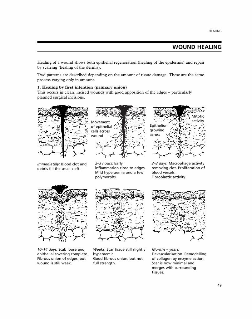
Healing of a wound shows both epithelial regeneration (healing of the epidermis) and repair
by scarring (healing of the dermis).
Two patterns are described depending on the amount of tissue damage. These are the same
process varying only in amount.
1. Healing by first intention (primary union)
This occurs in clean, incised wounds with good apposition of the edges – particularly
planned surgical incisions.
HEALING
WOUND HEALING
49
Months – years:Devascularisation. Remodellingof collagen by enzyme action.Scar is now minimal andmerges with surroundingtissues.
Weeks: Scar tissue still slightlyhyperaemic.Good fibrous union, but notfull strength.
10–14 days: Scab loose andepithelial covering complete.Fibrous union of edges, butwound is still weak.
2–3 days: Macrophage activityremoving clot. Proliferation ofblood vessels.Fibroblastic activity.
2–3 hours: Earlyinflammation close to edges.Mild hyperaemia and a fewpolymorphs.
Mitoticactivity
Epitheliumgrowingacross
Movement of epithelialcells acrosswound
Immediately: Blood clot anddebris fill the small cleft.

HEALING
WOUND HEALING
50
2. Healing by second intention (secondary union)
This occurs in open wounds, particularly when there has been significant loss of tissue,
necrosis or infection.
Early
Fewer cells
Collagen arrangedhorizontally
Capillaries lessprominent
Epithelial coveringcomplete
2 weeks onwards
Granulation tissue is seen in the base of the wound. This tissue consists of newly formed capillaries with fibroblasts and macrophages and occurs in manycircumstances in addition to wounds.
Loose connectivetissue formed byfibroblasts
Surface debrishas been shedEpithelium continues
to grow across
1 week approximately
Contractioncontinuing
New capillary loops bringmacrophages, neutrophilsand fibroblasts
Contraction ofwound size due toaction ofmyofibroblasts atedges
Scab dries out
A single sheet of epithelial cells isbeing pushed between the surfacedebris and the underlying living tissue
Mitotic activityin epithelium
A few days
Acute inflammationstarts at junction ofliving tissue
Cavity fills with bloodand fibrin clot

HEALING
WOUND HEALING
51
2. Keloid
The formation of excess collagen in the form of thick interlacing
bundles which causes marked swelling at the site of the wound is
known as a KELOID. The essential cause is unknown. It is
particularly common in black people.
Wound contraction
Wound contraction, which is beneficial and begins early, is due mainly to the young,
specialised ‘myofibroblasts’ in the granulation tissue exerting a traction effect at the wound
edges. The exposed surface is reduced by gradual regeneration of the surface epithelium.
The remodelling of the collagen continues for many months.
COMPLICATIONS
1. Contracture
Later, CONTRACTURE may cause serious cosmetic
and functional disability, particularly in deep and
extensive skin burns and around joints if muscles
are badly damaged.
Contracture followingburn of neckand jaw

FIBROSIS is the end result of WOUND HEALING, CHRONIC INFLAMMATION and
ORGANISATION.
Formation of fibrous tissue
HEALING
HEALING – FIBROSIS
52
Failure of proper collagen
synthesis with delayed
healing and weak scars.
REMODELLING follows: Action of COLLAGENASE SCAR TISSUE
+ secretion of COLLAGEN
Factors delaying healing
1. Local
INFECTION, a POOR BLOOD SUPPLY, excessive movement and
presence of foreign material DELAY HEALING.
2. General
DEFICIENCY of VITAMIN C
DEFICIENCY of AMINO ACIDS (in malnutrition)
DEFICIENCY of ZINC
EXCESS of ADRENAL GLUCOCORTICOIDS
DEBILITATING CHRONIC DISEASE
⎫⎪⎪⎪⎬⎪⎪⎪⎭
2. Adhesive glyco-proteins –FIBRONECTINSwhich provide ascaffolding andcontribute to theprogress of therepair process
COLLAGENFIBRE
(d) Cross-linking+
polymerisation
cleavage ofterminal peptides
(c)
Secretion to EXTRACELLULAR SITE
1.INTRACELLULAR PRODUCTIONof COLLAGEN precursors.
(a) Hydroxylation of proline andlysine (vit C required)
(b) Triple helix formation.
STIMULUS – growth factor, e.g. TGFβ (see p.48),derived from damaged cells and macrophages.
FIBROCYTES(and primitive stem cells) situated around capillaries and loose connective tissues
Enlarge to become active FIBROBLASTS and active PROTEIN SYNTHESIS begins

INTERNAL SURFACES
The epithelial lining of the gastrointestinal tract regenerates in a similar way to the skin.
HEALING
HEALING – SPECIAL SITUATIONS
53
Contractingscar tissuewhich maycauseseriouseffects dueto stricture,e.g. pyloricstenosis
Restoration tonormal includingreappearance ofspecialised cells
OrganisationSurface cellsbudding downwardto form new glands.
These cells arewithout their
specialised qualities
GranulationtissueMitotic
activityinmucouscells
Cells moving across from edges
Debris
Deep damageSuperficial damage

SOLID EPITHELIAL ORGANS
1. Following gross tissue damage – including supporting tissue (post-necrotic
scarring)
e.g. Kidney
HEALING
HEALING – SPECIAL SITUATIONS
54
Survivingcellsproliferateand movealongreticulinframeworkto thehepaticvenule
Progressive removalof debris
REGENERATION of epithelial cells at first
undifferentiated
RESTORATION to NORMAL
Mitosespresent
Tubuleslined by lowcuboidalepithelium
Surviving cells
Survivingsupportingtissues
Necrotic cellsand debris
Perivenular hepatic cell necrosise.g. Tubular necrosis in kidney
2. Following cell damage with survival of the supporting (reticular) tissues
Progressive removalof dead tissue withorganisation and
COARSE SCARformation
Necrotictissue
Liver

MUSCLE
Muscle fibres of all 3 types – skeletal, cardiac and smooth – have only limited capacity to
regenerate.
When a MASS of muscle tissue is damaged, repair by SCARRING occurs. This is particularly
important in the HEART after infarction.
If the damage affects individual muscle fibres diffusely and with varying severity, then
regeneration of the specialised fibres is possible (e.g. the myocardium may recover
completely from the effects of diphtheria toxin and virus infection).
NERVOUS TISSUE
Central nervous system
Regeneration does not occur when a neurone is lost.
In cases of acute damage, the initial functional loss often exceeds the loss of actual nerve
tissue because of the reactive changes in the surrounding tissue. As these changes diminish,
some function may be restored.
HEALING
HEALING – SPECIAL SITUATIONS
55
Scarring within the CNS is by proliferation of ASTROCYTES and the production of fibrillary
glial acidic protein – a process known as GLIOSIS.
+Establishment of new synapsesby surviving neurones
Internalcapsuleno longeraffected
Small area of necrotic tissue remains (no regeneration);oedema andcongestion nowabsent
ParalysisreducedDays
Weeks
Internalcapsuleaffected
Small areas ofnecrotic tissue(infarction)
Surroundingoedema andcongestion
Hemiplegia

NERVOUS TISSUE (continued)
Peripheral Nerves
When a peripheral nerve is damaged, the axon and its myelin sheath rapidly degenerate
distally. The supporting tissues of the nerve (Schwann cells) degenerate slowly.
Regeneration can occur because the central neurone of which the axon is a peripheral
extension is remote from the site of damage.
A spinal motor nerve is taken as an example.
HEALING
HEALING – SPECIAL SITUATIONS
56
Axon disintegratesMyelin disintegrates Fatty dropletsSchwann cells survive
Loss of Nissl substance (RNA)(chromatolysis)
WALLERIAN DEGENERATIONof distal nerve
Mild degenerativechanges in neurones
Atrophyof musclefibres
Cutting or crushing
Results of damage
Prominent Nissl substance (RNA)
Motorendplates
Muscle fibres
Schwann cellnucleus
MyelinsheathAxonSpinal neurone
Motor impulse
Nerve trunkAnteriorroot
Normal spinal cord

Peripheral Nerves (continued)
Regeneration takes the form of a sprouting of the cut ends of the axons.
HEALING
HEALING – SPECIAL SITUATIONS
57
Poor appositionDistal nerve remnant
disappears
6–12 months
Irregular sprouting ofaxons and proliferationof Schwann cells Severe atrophy
of muscle
Formation ofTRAUMATIC‘NEUROMA’
The best results are seen in crushing injuries
where the sheaths remain in continuity.
Good restorationGood apposition
The results depend on the apposition of the
distal remnant with the sprouting axons.
Sproutingof axons
Growth along the trackof the degenerate nerve(about 1 mm per day)

BONE – Fracture Healing
HEALING
FRACTURE HEALING
58
Fracture site may be almost invisible
6. Final reconstructionMonths later
Lamellarbone
Osteoblasts andosteoclasts active
5. Remodelling of callusDefinitive – weeks into months
Osteoblastic and osteoclasticactivity proceeding
Cortical gap healedby ossification
4. Mature callus– from 3 weeks onwards
Provisional callus bridges the gap – first,osteoid tissue (may include cartilage)then woven bone
Medullary
PeriostealOsteoblastic activity
3. Formation of callus(early bone regeneration) –after 1 week.
Early organisation:capillaries and fibroblasts
Phagocytosis ofdebris and necrotic
tissues
2. Early reaction-inflammatoryFirst 4–5 days
Damage to soft tissues
with haemorrhage
Cortex
Medulla
Necrosis ofends of bonePeriosteum
1. Immediate effects

Events following a fracture (continued)
Complications
HEALING
FRACTURE HEALING
59
Very easily fractured
Mixture of tumourand haematoma –healing inhibited
A common condition is asecondary tumour growingin and destroying the bone
PATHOLOGICAL FRACTURE
When the break occurs at the site of pre-existing disease of the bone, the term ‘pathological
fracture’ is applied.
Penetratinginjury fromoutside
By sharpbone ends
2. InfectionIf the overlying skin is breached in any way, i.e. the fracture is ‘compound’, the risk of
infection is greatly increased; this is an important adverse factor in the healing process.
1. Fat embolism may occur
in fracture of long bones
due to entry of fat from
the marrow cavity into
the torn ends of veins.

HEALING
FRACTURE HEALING
60
There may be interpositionof soft tissue, e.g. muscle
Large irregular callus: slow repair, permanent deformity of bone
Small callus,quick repair
Callus formationinhibited
Fibrous unionSmall callus, goodbone formation
⎫⎬⎭
FACTORS INFLUENCING HEALING OF FRACTURES
ADVERSE FAVOURABLE1. Local factors
(a) Infection See previous(b) Pathological fracture page
(c) Poor apposition and alignment . . . . . . . . . . . . . . . . . . . Good apposition
(d) Continuing movement of bone ends . . . . . . . . . . . . . . Good immobilisation
In extreme cases, a rudimentary joint (pseudoarthrosis) may form
(e) Poor blood supply . . . . . . . . . . . . . . . . . . . . . . . . . Good blood supply
This is largely influenced by the anatomical site In favourable conditionsof the fracture, for example: blood supply is derived (a) Nutrient artery entering remote from the from:
fracture or damaged by fracture (a) periosteal arteries(e.g. scaphoid, femoral head) (b) nutrient artery
(b) Fracture through area devoid of periosteum (c) adjacent soft tissues.(e.g. neck of femur)
(c) Minimal adjacent soft tissue (e.g. tibia).
2. General factors
(a) Old age . . . . . . . . . . . . . . . . . . . . . . . . . Youth(b) Poor nutrition – e.g. famine, . . . . . . . . . . . . . . . . . . . . . Good nutrition – especially
malabsorption leading to lack of protein, calcium, vit D and vit C.protein, calcium, vit D and vit C.