Embed Size (px)
Citation preview
HeadacheintheEmergencyRoom:Non‐vascularheadache
Michael Marmura Thomas Jefferson UniversityThomas Jefferson University
Department of Neurology, Jefferson Headache CenterJune 9, 2015
[email protected]@JeffHeadacheCtr
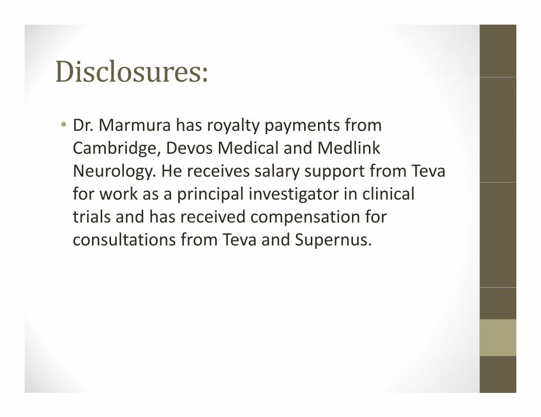
Disclosures:Disclosures:• Dr. Marmura has royalty payments fromDr. Marmura has royalty payments from Cambridge, Devos Medical and Medlink Neurology. He receives salary support from Teva for work as a principal investigator in clinical trials and has received compensation for
lt ti f T d Sconsultations from Teva and Supernus.
Objectives:Objectives:• Distinguish the presentation of acute headache g psecondary to brain tumor in the emergency department from more typical presentations to an outpatient clinic.E l t th l ti hi b t h t i• Evaluate the relationship between hypertensive emergency and acute headache.
• Evaluate potential infectious causes of headache such as pbrain abscess.
• Review the presentation and evaluation of facial pain in h dthe emergency department.
Howcommon is headache in the ED?HowcommonisheadacheintheED?• More common than you think (and not just migraine)• Depends on if a symptom or the chief compliant• In children: viral and respiratory illnesses, concussion/post‐traumatic ventriculoperitoneal shuntconcussion/post‐traumatic, ventriculoperitoneal shunt malfunctions are not rare.
• But SERIOUS life‐threatening causes (aseptic meningitis, subdural oe epidural hematoma, proven VP shunt malfunction, brain abscess, pseudotumor cerebri) are uncommon (< 10% total).uncommon (< 10% total).
• In adults < 50 life‐threatening causes are rare
Kan et al. Headache 2001; Goldstein et al. Cephalalgia 2006
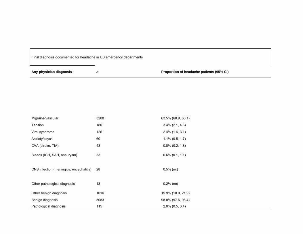
Final diagnosis documented for headache in US emergency departments
Any physician diagnosis n Proportion of headache patients (95% CI)Any physician diagnosis n Proportion of headache patients (95% CI)
Migraine/vascular 3208 63.5% (60.9, 66.1)
Tension 180 3.4% (2.1, 4.6)
Viral syndrome 126 2 4% (1 6 3 1)Viral syndrome 126 2.4% (1.6, 3.1)
Anxiety/psych 60 1.1% (0.5, 1.7)
CVA (stroke, TIA) 43 0.8% (0.2, 1.8)
Bleeds (ICH, SAH, aneurysm) 33 0.6% (0.1, 1.1)
CNS infection (meningitis, encephalitis) 28 0.5% (nc)
Other pathological diagnosis 13 0.2% (nc)
Other benign diagnosis 1016 19.9% (18.0, 21.9)
Benign diagnosis 5083 98.0% (97.6, 98.4)
Pathological diagnosis 115 2.0% (0.5, 3.4)
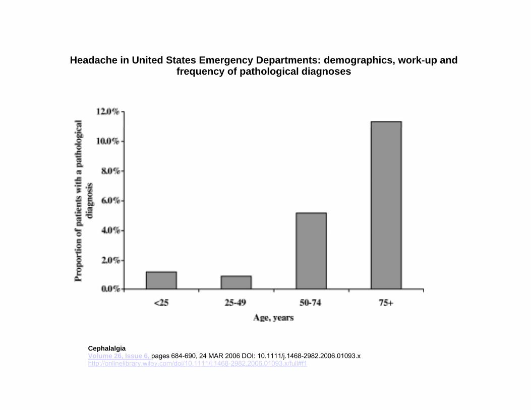
Headache in United States Emergency Departments: demographics, work‐up and frequency of pathological diagnosesq y p g g
CephalalgiaVolume 26, Issue 6, pages 684-690, 24 MAR 2006 DOI: 10.1111/j.1468-2982.2006.01093.xhttp://onlinelibrary.wiley.com/doi/10.1111/j.1468-2982.2006.01093.x/full#f1
Case1:IhaveaheadacheandI’mblind!
23 ld• 23 year old woman• Developed a severe headache while watchingheadache while watching July 4th fireworks
• Now says she can’t see• Now says she can t see anything
Further historyFurtherhistory…• Sudden onset headache• Exam: blind• “I have a craniopharyngioma… could this have something to do with this?”do with this?
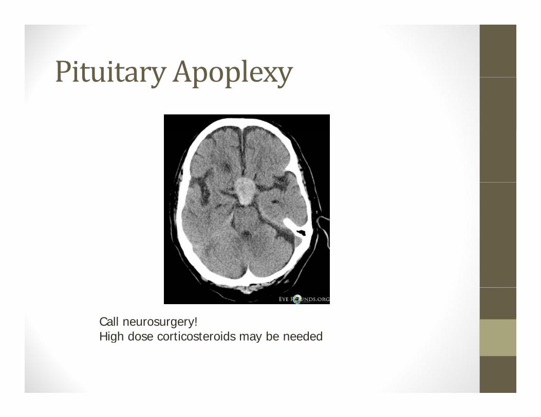
Pituitary ApoplexyPituitaryApoplexy
Call neurosurgery!High dose corticosteroids may be neededHigh dose corticosteroids may be needed
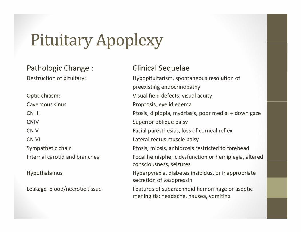
Pituitary ApoplexyPituitaryApoplexyPathologic Change : Clinical SequelaeDestruction of pituitary: Hypopituitarism, spontaneous resolution of
preexisting endocrinopathyOptic chiasm: Visual field defects, visual acuityCavernous sinus Proptosis, eyelid edemaCN III Ptosis, diplopia, mydriasis, poor medial + down gazeCNIV Superior oblique palsyCN V Facial paresthesias loss of corneal reflexCN V Facial paresthesias, loss of corneal reflexCN VI Lateral rectus muscle palsySympathetic chain Ptosis, miosis, anhidrosis restricted to foreheadInternal carotid and branches Focal hemispheric dysfunction or hemiplegia, altered
consciousness, seizuresHypothalamus Hyperpyrexia, diabetes insipidus, or inappropriate
secretion of vasopressinLeakage blood/necrotic tissue Features of subarachnoid hemorrhage or asepticLeakage blood/necrotic tissue Features of subarachnoid hemorrhage or aseptic
meningitis: headache, nausea, vomiting
Case2:EDfollow‐upforpseudotumor• Obese 43 yo school cafeteria worker w progressive headache + papilledema. CT/MRI without gad wnl.
• Opening pressure = 26 cm, headache improved in ED with LP and medicationsand medications
• Headache right sided only, ocular, constant w fluctuations, pressure > throbbing
• Follow‐up with me, improved with topiramate but papilledema did not resolve and OP still high on repeat LP
• Developed worsening headache, now with right ptosis…p g , g p
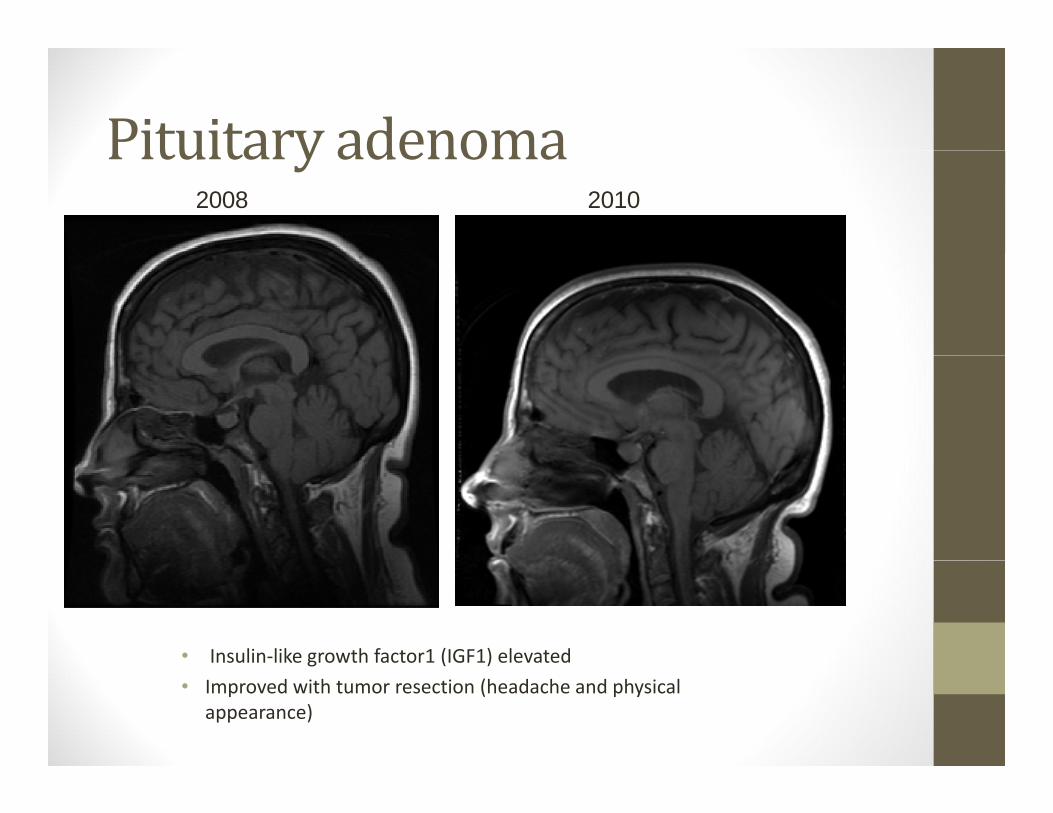
Pituitary adenomaPituitaryadenoma2008 2010
• Insulin‐like growth factor1 (IGF1) elevatedg ( )• Improved with tumor resection (headache and physical
appearance)
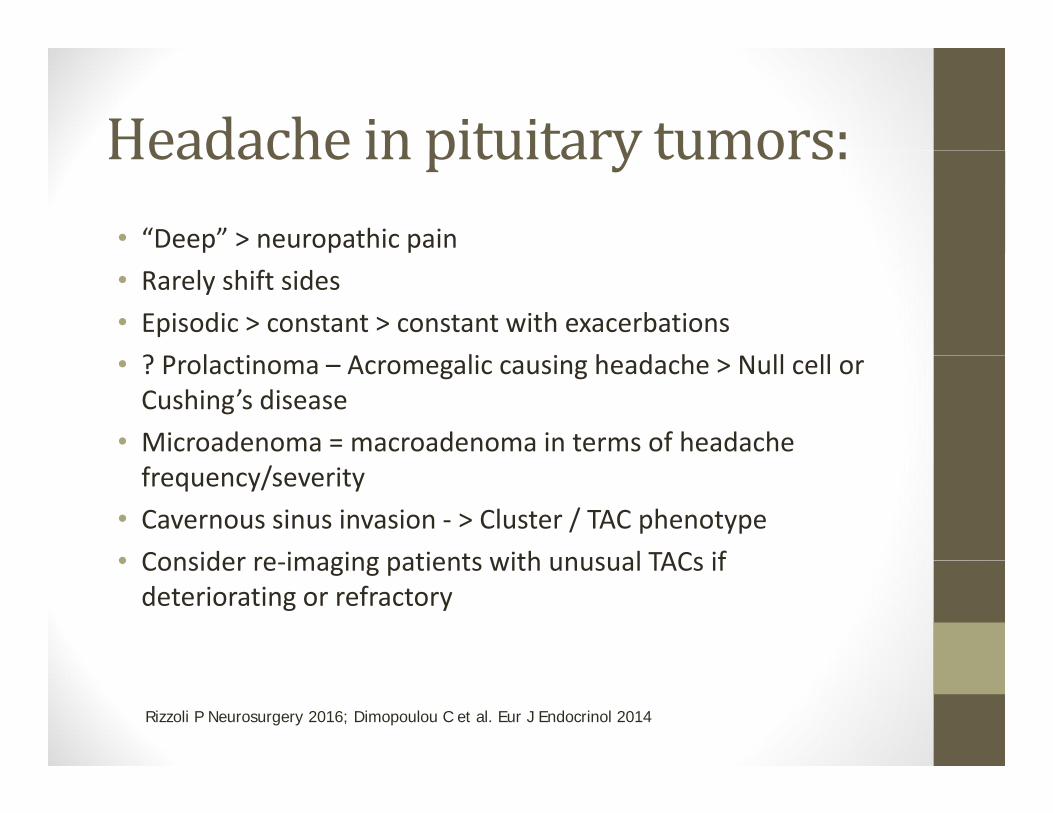
Headache in pituitary tumors:Headacheinpituitarytumors:• “Deep” > neuropathic pain• Rarely shift sides• Episodic > constant > constant with exacerbations? l i A li i h d h N ll ll• ? Prolactinoma – Acromegalic causing headache > Null cell or Cushing’s disease
• Microadenoma = macroadenoma in terms of headache frequency/severity
• Cavernous sinus invasion ‐ > Cluster / TAC phenotype• Consider re imaging patients with unusual TACs if• Consider re‐imaging patients with unusual TACs if deteriorating or refractory
Rizzoli P Neurosurgery 2016; Dimopoulou C et al. Eur J Endocrinol 2014
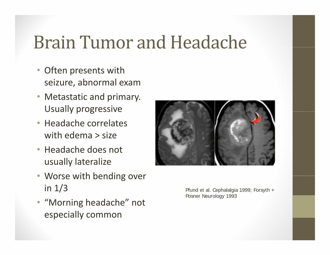
Brain Tumor andHeadacheBrainTumorandHeadache• Often presents with
i b lseizure, abnormal exam• Metastatic and primary. Usually progressiveUsually progressive
• Headache correlates with edema > size
• Headache does not usually lateralize
• Worse with bending over• Worse with bending over in 1/3
• “Morning headache” not Pfund et al. Cephalalgia 1999; Forsyth + Posner Neurology 1993
especially common
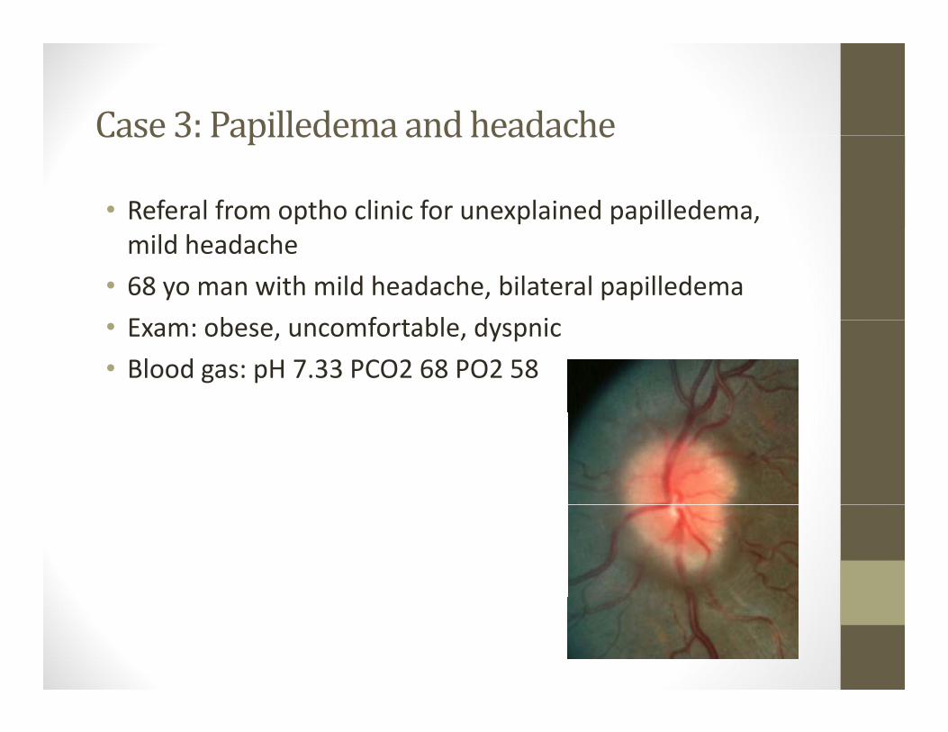
Case 3: Papilledema andheadacheCase3:Papilledemaandheadache
• Referal from optho clinic for unexplained papilledema, mild headache
• 68 yo man with mild headache, bilateral papilledemaE b f t bl d i• Exam: obese, uncomfortable, dyspnic
• Blood gas: pH 7.33 PCO2 68 PO2 58
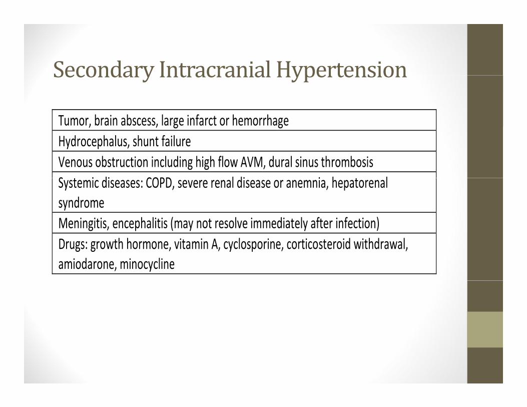
Secondary Intracranial HypertensionSecondaryIntracranialHypertension
Tumor, brain abscess, large infarct or hemorrhage, , g gHydrocephalus, shunt failure Venous obstruction including high flow AVM, dural sinus thrombosis S i di COPD l di i h lSystemic diseases: COPD, severe renal disease or anemnia, hepatorenal syndrome Meningitis, encephalitis (may not resolve immediately after infection)g , p ( y y )Drugs: growth hormone, vitamin A, cyclosporine, corticosteroid withdrawal, amiodarone, minocycline
CarcinomatousmeningitisCarcinomatousmeningitis• Presentation typically consists of headache/neck pain, encephalopathy, cranial nerve palsies
• Occurs in 5% of those with cancer• Leukemia/lymphoma breast lung melanoma• Leukemia/lymphoma, breast, lung, melanoma• Increased opening pressure, CSF protein common• + CSF cytology in > 50%, almost always after 3 LPs.
Case 4: Catastrophic headacheCase4:Catastrophicheadache
• 26 yo healthy medical resident26 yo healthy medical resident• Presents with headache worsening over 2‐3 days• Bilateral headache/pressureBilateral headache/pressure• Exam: Ill‐appearing, BP 206/142, HR 141, otherwise wnlotherwise wnl
• Is the high blood pressure the cause of headache?
Hypertension andHeadacheHypertensionandHeadache• About 5% of patients in ED presenting with headache have hypertensive‐related headache (mean >170 SBP, > 100 DBP)
• Essential hypertension and migraine link?: conflicting results• Mild moderate changes in BP do not cause headache• Mild‐moderate changes in BP do not cause headache• Eclampsia/preeclampsia more strongly linked to migraine during pregnancy
• Treatment with antihypertensives may treat migraine• Hypertensive encephalopathy is part of the spectrum of Reversible posterior leukoencephalopathy syndrome (alsoReversible posterior leukoencephalopathy syndrome (also called PRES)
• High BP WITHOUT acute end‐organ dysfunc on ≠ hypertensive emergencyhypertensive emergency
Dhopesh et al Headache 1979; Adeney and Williams Headache 2006
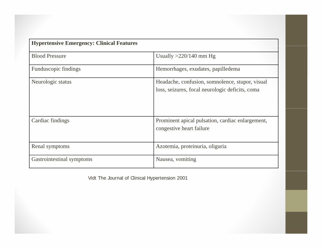
Hypertensive Emergency: Clinical Featuresyp g y
Blood Pressure Usually >220/140 mm Hg
Funduscopic findings Hemorrhages, exudates, papilledema
Neurologic status Headache, confusion, somnolence, stupor, visual loss, seizures, focal neurologic deficits, coma
Cardiac findings Prominent apical pulsation, cardiac enlargement, congestive heart failure
Renal symptoms Azotemia, proteinuria, oliguria
Gastrointestinal symptoms Nausea, vomiting
Vidt The Journal of Clinical Hypertension 2001
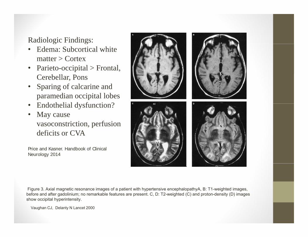
Radiologic Findings:g g• Edema: Subcortical white
matter > Cortex• Parieto-occipital > Frontal,
Cerebellar, Pons• Sparing of calcarine and
paramedian occipital lobesE d th li l d f ti ?• Endothelial dysfunction?
• May cause vasoconstriction, perfusion deficits or CVAdeficits or CVA
Price and Kasner. Handbook of Clinical Neurology 2014
Figure 3. Axial magnetic resonance images of a patient with hypertensive encephalopathyA, B: T1-weighted images, b f d ft d li i k bl f t t C D T2 i ht d (C) d t d it (D) ibefore and after gadolinium; no remarkable features are present. C, D: T2-weighted (C) and proton-density (D) images show occipital hyperintensity.
Vaughan CJ, Delanty N Lancet 2000
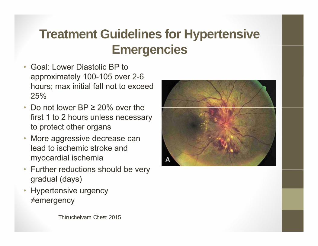
Treatment Guidelines for Hypertensive E iEmergencies
• Goal: Lower Diastolic BP to approximately 100 105 over 2 6approximately 100-105 over 2-6 hours; max initial fall not to exceed 25%
• Do not lower BP ≥ 20% over the• Do not lower BP ≥ 20% over the first 1 to 2 hours unless necessary to protect other organs
• More aggressive decrease can• More aggressive decrease can lead to ischemic stroke and myocardial ischemia
• Further reductions should be very• Further reductions should be very gradual (days)
• Hypertensive urgency ≠emergency≠emergency
Thiruchelvam Chest 2015

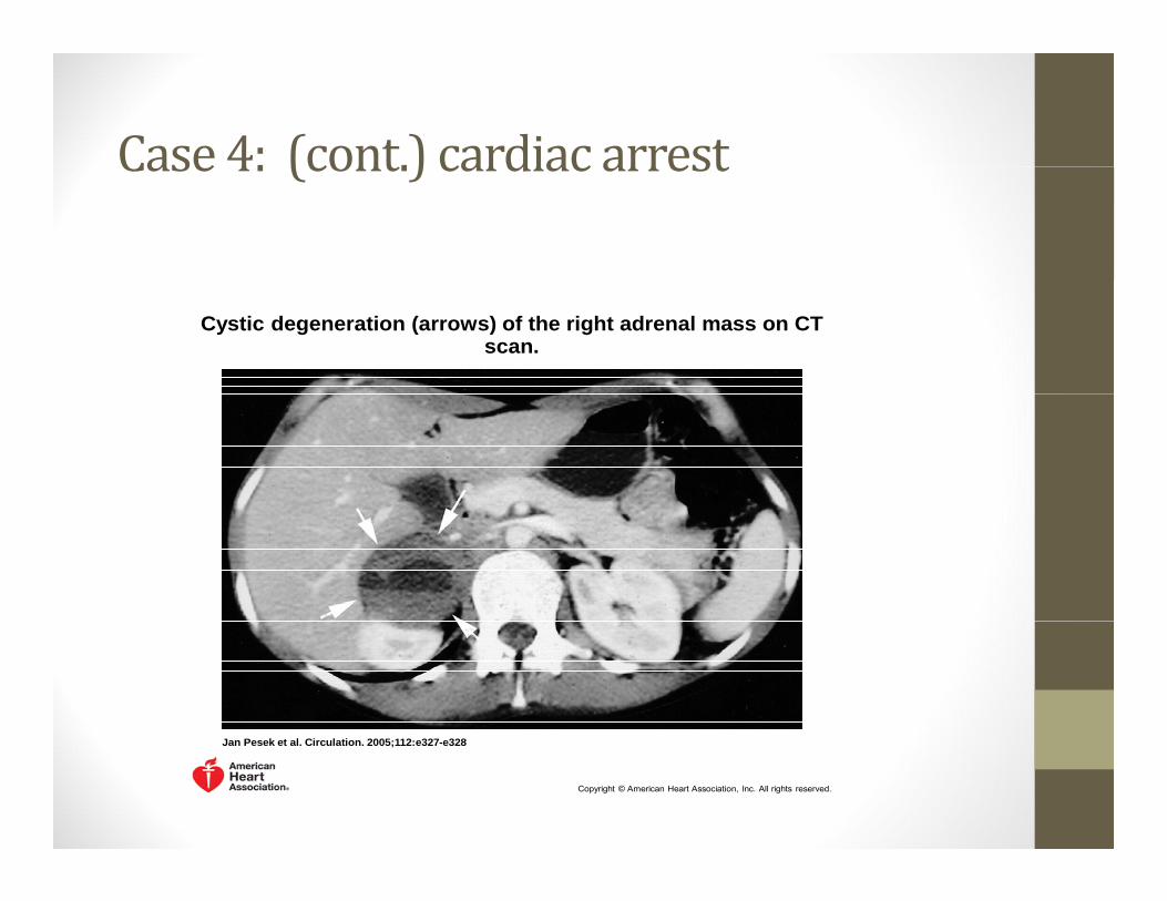
Case 4: (cont.) cardiac arrestCase4:(cont.)cardiacarrest
Cystic degeneration (arrows) of the right adrenal mass on CT scan.
Jan Pesek et al. Circulation. 2005;112:e327-e328
Copyright © American Heart Association, Inc. All rights reserved.
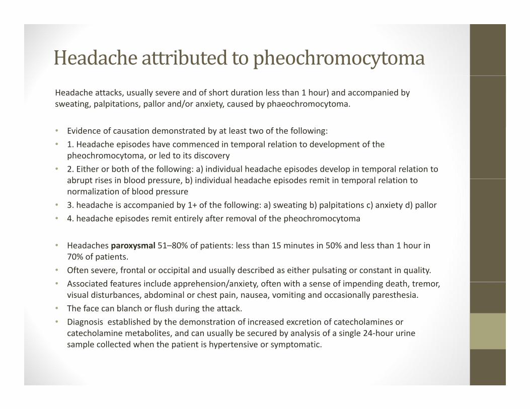
HeadacheattributedtopheochromocytomaHeadache attacks, usually severe and of short duration less than 1 hour) and accompanied by sweating, palpitations, pallor and/or anxiety, caused by phaeochromocytoma.
• Evidence of causation demonstrated by at least two of the following:• 1. Headache episodes have commenced in temporal relation to development of the
pheochromocytoma, or led to its discovery• 2. Either or both of the following: a) individual headache episodes develop in temporal relation to
abrupt rises in blood pressure b) individual headache episodes remit in temporal relation toabrupt rises in blood pressure, b) individual headache episodes remit in temporal relation to normalization of blood pressure
• 3. headache is accompanied by 1+ of the following: a) sweating b) palpitations c) anxiety d) pallor• 4. headache episodes remit entirely after removal of the pheochromocytoma
• Headaches paroxysmal 51–80% of patients: less than 15 minutes in 50% and less than 1 hour in 70% of patients.
• Often severe, frontal or occipital and usually described as either pulsating or constant in quality. A i t d f t i l d h i / i t ft ith f i di d th t• Associated features include apprehension/anxiety, often with a sense of impending death, tremor, visual disturbances, abdominal or chest pain, nausea, vomiting and occasionally paresthesia.
• The face can blanch or flush during the attack.• Diagnosis established by the demonstration of increased excretion of catecholamines or
catecholamine metabolites and can usually be secured by analysis of a single 24‐hour urinecatecholamine metabolites, and can usually be secured by analysis of a single 24‐hour urine sample collected when the patient is hypertensive or symptomatic.
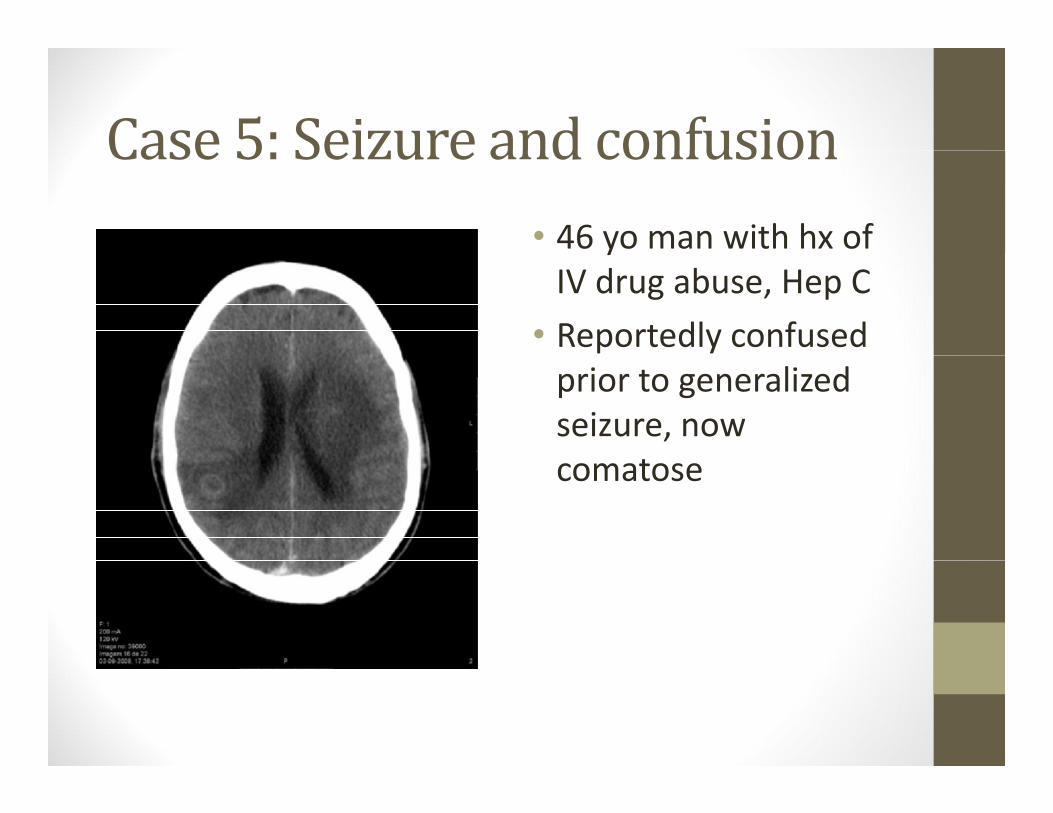
Case 5: Seizure and confusionCase5:Seizureandconfusion• 46 yo man with hx of yIV drug abuse, Hep C
• Reportedly confused prior to generalized seizure, now comatose
Brain Abscess:BrainAbscess:• Organisms: streptococcus, staphylococcus aureus, bacteroides species, enterobacter. Fungal causes include Candida species, aspergillosis and blastomycosis.
• Risk factors: Contiguous area of infection (sinusitis earsRisk factors: Contiguous area of infection (sinusitis, ears, dental), immunosupression
• MRI diffusion /ADC (abscess has higher DWI intensity, lower ADC) d MR t h l di ti i h f tADC) and MR spectroscopy can help distinguish from tumor
• Multiple abscesses in > 10%, Capsular appearance > cerebritis
• Surgical treatment is usually necessary in most cases