Embed Size (px)
Citation preview
1
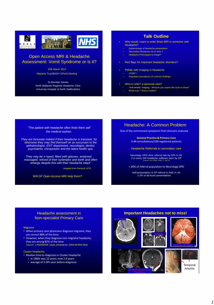
Open Access MRI & Headache
Assessment: Vomit Syndrome or is it?
Dr Brendan Davies
North Midlands Regional Headache Clinic
University Hospital of North Staffordshire
15th March 2012
Migraine Trust/BASH GPwSI Meeting
Talk Outline • Why would I want to order Brian MRI in someone with
Headache?
– Epidemiology of headache presentation
– Secondary Headache not to miss ?
– Headache Phenotypes to image?
• Red flags for important headache disorders?
• Pitfalls with Imaging in Headache
– VOMIT !
– Population prevalence of common findings !
• Who to refer? a personal view?
– ―Anti-emetic‖ imaging - What do you expect the scan to show?
– What scan ? Does it matter?
“The patient with headache often finds them self
the medical orphan.
They are fortunate indeed if their headache is transient, for otherwise they may find themself on an excursion to the opthalmologist, ENT department, neurologist, dentist,
psychiatrist, chiropractor and the latest health spa.
They may be x-rayed, fitted with glasses, analysed, massaged, relived of their turbinates and teeth and often
emerge despite this with their headache intact”
Adapted from Packard 1979
Will GP Open Access MRI help them?
One of the commonest symptoms that clinicians evaluate
General Practice & Primary Care 4.44 consultations/100 registered patients
Headache Referrals to secondary care
Neurology OPD clinic referral rate by GPs in UK
2 in every 100 headache sufferers seen by GP (Latnovic et al. (2006): JNNP;77: 385-87)
≈ 20% of referral population to Neurology OPD
Self-presentation & GP referral to A&E in UK
1-2% of all Acute presentations
Headache: A Common Problem
Migraine • When primary care physicians diagnose migraine, they
are correct 98% of the time • However, when they diagnose non-migraine headache,
they are wrong 82% of the time Source: LANDMARK study (Headache 2004;44:856-864)
Cluster Headache • Median time to diagnosis in Cluster Headache
in 1960s was 22 years; now 2.6 years average of 3 GPs seen before diagnosis
Headache assessment in
Non-specialist Primary Care
Glioma
Important Headaches not to miss!
Acute SAH
Bacterial Meningitis
Temporal
Arteritis Cerebral Venous Sinus
Thrombosis

2
The Scope of the problem Population headache demographics
Prevalence & Incidence
• Migraine 6-18 per 100 SAH 10 per 100, 0000
• Cluster HA 1 per 1,000 Dissection 3-5 per 100,000
• Other TACs Rare Brain Tumour 7 per 100,000
(all types) 25-60% have headache
Isolated headache only 2<16% of tumours
18
6
41
40
5 2.8
0%
10%
20%
30%
40%
50%
Episodic Migraine
Episodic Tension-Type Headache
Chronic Daily Headache
One-year prevalence of common headache disorders
The Primary Headaches
• Migraine
• Tension-Type headache
• Cluster headache
• Trigeminal Autonomic Cephalagias
– Paroxysmal hemicrania
– SUNCT
• Other
– Primary Stabbing headache
– Primary Cough headache
– Primary Exertional headache
– Primary Thunderclap headache
– Primary Sexual headache
– Hypnic headache
– New Daily Persistent Headache
– Hemicrania Continua
©International Headache Society 2003/4ICHD-II. Cephalalgia 2004; 24 (Suppl 1)
INTERNATIONAL CLASSIFICATION
of
HEADACHE DISORDERS
2nd edition
(ICHD-II)
The Primary Headaches
Who to scan? Controversies? • Migraine
• Tension-Type headache
• Cluster headache
• Trigeminal Autonomic Cephalagias
– Paroxysmal hemicrania
– SUNCT
• Other
– Primary Stabbing headache
– Primary Cough headache
– Primary Exertional headache
– Primary Thunderclap headache
– Primary Sexual headache
– Hypnic headache
– New Daily Persistent Headache
– Hemicrania Continua
SCAN
SCAN
SCAN
SCAN ?
SCAN ?
SCAN ?
Classification of secondary
headaches
• Less common than primary headache but epidemiology dependent on epidemiology of underlying cause
• IHS Classification: [Sections 1-4 used for Primary Headaches]
5. Headache attributed to head and/or neck trauma
6. Headache attributed to cranial or cervical vascular disorder - e.g. Stroke, Haemorrhage, Dissection, Venous thrombosis, Arteritis
7. Headache attributed to non-vascular intracranial disorder - e.g. High and Low CSF pressure, Tumours, Chiari malformation
8. Headache attributed to a substance or its withdrawal - e.g. Medication Overuse Headache, withdrawal of caffeine, opiates, oestrogen
9. Headache attributed to infection - e.g. Meningitis, Encephalitis
10.Headache attributed to disorder of homoeostasis - e.g. Sleep apnoea
11.Headache or facial pain attributed to disorder of cranium, neck, eyes, ears, nose, sinuses, teeth, mouth or other facial or cranial structures
- e.g. Cervicogenic, Acute Glaucoma, Sinusitis, TMJ pain
12.Headache attributed to psychiatric disorder - e.g. Somatisation, Psychosis
SCAN
SCAN
SCAN ?
The Spectrum of Migraine
80%
15% 30%
0.01%
Unclear
2- 4%
IHCD II Classification 2004
Cephalagia Vol 24; Suppl 1
< 10% ?
But 20-60% of my clinic patients !!
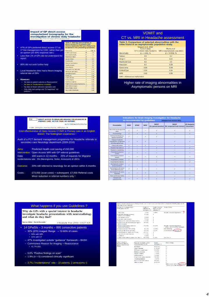
Indications for Brain imaging investigation for Headache
as recommended by guidelines (Adapted from Kernick 2011)
Presentation SIGN5 EFNS6 NICE7 BASH3
(for general practice)
BASH8
(for suspected brain tumour)
US Headache
Consortium9
New onset over the age of 50
years ✓ ✓ — ✓ ✓ ✓
Focal neurology ✓ ✓ — ✓ — ✓
Non-focal neurology (e.g.
cognitive impairment) ✓ — ✓ ✓ — —
Change in headache
characteristics—progressive
(including atypical aura)
✓ ✓ ✓ ✓ ✓ ✓
Change with posture ✓ — ✓ — ✓ —
Headache that wakes up ✓ — — ✓ — ✓
Headache on awakening — — — — — —
Precipitated by physical
exertion ✓ — — — — —
Exacerbated by physical
exertion — — — ✓ — —
Precipitated by Valsalva ✓ — — — — —
Exacerbated by Valsalva — — — ✓ — ✓
Risk factors for cerebral
venous thrombosis ✓ — — — — —
New headache with human
immunodeficiency virus ✓ ✓ — — ✓ —
New headache with cancer
elsewhere ✓ — ✓ ✓ ✓ —
Pulse synchronous tinnitus — — — — — —
Headache with
nausea/vomiting — — — ✓ ✓ ✓
Headache with seizure history — — — ✓ — —
3
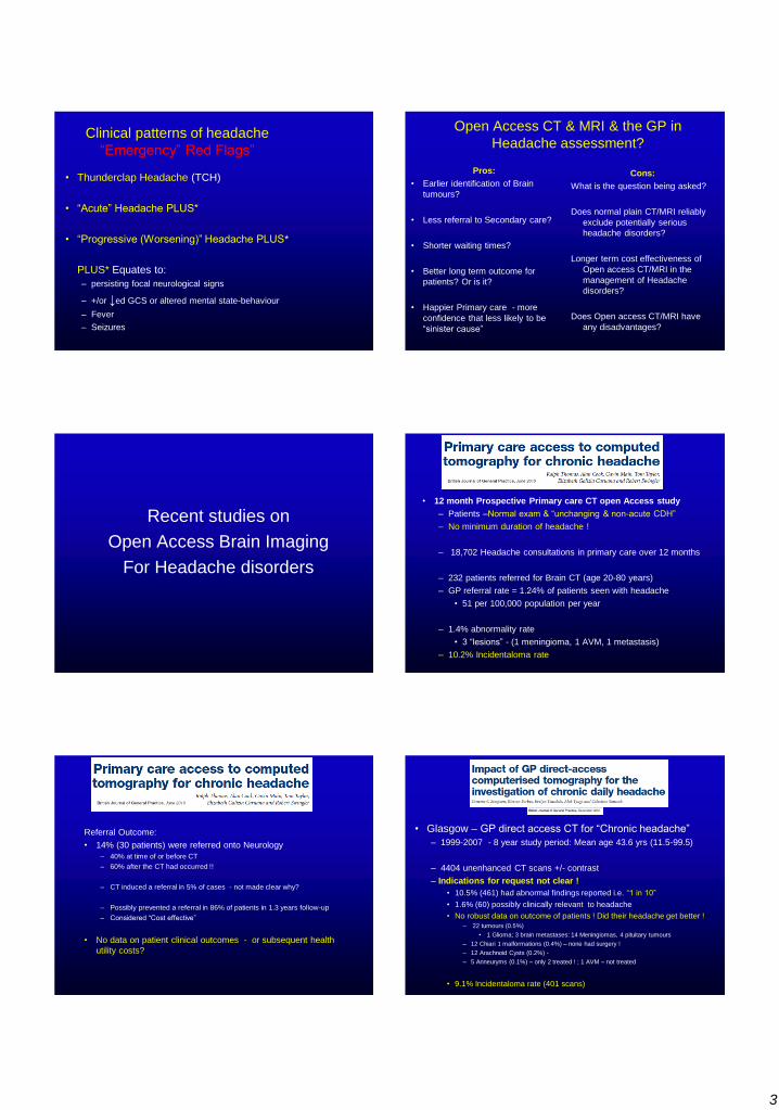
Clinical patterns of headache
―Emergency‖ Red Flags‖
• Thunderclap Headache (TCH)
• ―Acute‖ Headache PLUS*‖
• ―Progressive (Worsening)‖ Headache PLUS*
PLUS* Equates to:
– persisting focal neurological signs
– +/or ↓ed GCS or altered mental state-behaviour
– Fever
– Seizures
Open Access CT & MRI & the GP in
Headache assessment?
Pros:
• Earlier identification of Brain
tumours?
• Less referral to Secondary care?
• Shorter waiting times?
• Better long term outcome for
patients? Or is it?
• Happier Primary care - more
confidence that less likely to be
―sinister cause‖
Cons:
What is the question being asked?
Does normal plain CT/MRI reliably
exclude potentially serious
headache disorders?
Longer term cost effectiveness of
Open access CT/MRI in the
management of Headache
disorders?
Does Open access CT/MRI have
any disadvantages?
Recent studies on
Open Access Brain Imaging
For Headache disorders
• 12 month Prospective Primary care CT open Access study
– Patients –Normal exam & ―unchanging & non-acute CDH‖
– No minimum duration of headache !
– 18,702 Headache consultations in primary care over 12 months
– 232 patients referred for Brain CT (age 20-80 years)
– GP referral rate = 1.24% of patients seen with headache
• 51 per 100,000 population per year
– 1.4% abnormality rate
• 3 ―lesions‖ - (1 meningioma, 1 AVM, 1 metastasis)
– 10.2% Incidentaloma rate
Referral Outcome:
• 14% (30 patients) were referred onto Neurology
– 40% at time of or before CT
– 60% after the CT had occurred !!
– CT induced a referral in 5% of cases - not made clear why?
– Possibly prevented a referral in 86% of patients in 1.3 years follow-up
– Considered ―Cost effective‖
• No data on patient clinical outcomes - or subsequent health
utility costs?
• Glasgow – GP direct access CT for ―Chronic headache‖
– 1999-2007 - 8 year study period: Mean age 43.6 yrs (11.5-99.5)
– 4404 unenhanced CT scans +/- contrast
– Indications for request not clear !
• 10.5% (461) had abnormal findings reported i.e. ―1 in 10‖
• 1.6% (60) possibly clinically relevant to headache
• No robust data on outcome of patients ! Did their headache get better !
– 22 tumours (0.5%)
• 1 Glioma; 3 brain metastases: 14 Meningiomas, 4 pituitary tumours
– 12 Chiari 1 malformations (0.4%) – none had surgery !
– 12 Arachnoid Cysts (0.2%) -
– 5 Anneuryms (0.1%) – only 2 treated ! ; 1 AVM – not treated
• 9.1% Incidentaloma rate (401 scans)
4
• 47% of GPs preferred direct access CT as
1st line management for CDH rather than get
an opinion (20-30% response rate )
• Less than 1% of GPs did not understand the
report.
• 86% did not seek further help
• Local Headache clinic had a Neuro-imaging
referral rate of 29%
• However:
– No data on patient outcome or Reassurance!
– No date on ―Incidentaloma morbidity‖
– No date on future referral to specialist care
– Only data cost savings and ―Dr happiness‖ not
patients !!
VOMIT and
CT vs. MRI in Headache assessment
Higher rate of imaging abnormalities in
Asymptomatic persons on MRI
Simpson et al., 2010
Cost effectiveness of Open Access CT/MR in Primary care in an English
district: The Nottingham experience !
Audit of a PCT demand management programme for Headache referrals to
secondary care Neurology department (2009-2010)
Aims: Predicted Health cost saving of £50,000
Intervention: Open Access MRI with GP referral guidelines
Data: 169 scans in 12 months ; 25% of requests for Migraine
Incidentaloma rate: 3% (Meningioma, Stoke, Annerysm) & UBOs
Outcome: 20% still referred to neurology for an opinion within 6 months
Costs: £73,000 (scan costs) + subsequent £7,000 Referral costs
Minor reduction in referral numbers only !
Indications for Brain imaging investigation for Headache
as recommended by guidelines (Adapted from Kernick 2011)
Presentation SIGN5 EFNS6 NICE7 BASH3
(for general practice)
BASH8
(for suspected brain tumour)
US Headache
Consortium9
New onset over the age of 50
years ✓ ✓ — ✓ ✓ ✓
Focal neurology ✓ ✓ — ✓ — ✓
Non-focal neurology (e.g.
cognitive impairment) ✓ — ✓ ✓ — —
Change in headache
characteristics—progressive
(including atypical aura)
✓ ✓ ✓ ✓ ✓ ✓
Change with posture ✓ — ✓ — ✓ —
Headache that wakes up ✓ — — ✓ — ✓
Headache on awakening — — — — — —
Precipitated by physical
exertion ✓ — — — — —
Exacerbated by physical
exertion — — — ✓ — —
Precipitated by Valsalva ✓ — — — — —
Exacerbated by Valsalva — — — ✓ — ✓
Risk factors for cerebral
venous thrombosis ✓ — — — — —
New headache with human
immunodeficiency virus ✓ ✓ — — ✓ —
New headache with cancer
elsewhere ✓ — ✓ ✓ ✓ —
Pulse synchronous tinnitus — — — — — —
Headache with
nausea/vomiting — — — ✓ ✓ ✓
Headache with seizure history — — — ✓ — —
• 14 GPwSIs – 3 months – 895 consecitive patients
– 30% (270) imaged: Range → 12-60% of cases
• 59% with MRI
• 41% with CT
– 47% investigated outside ―guidance‖ framework – BASH
– Commonest Reason for Imaging = Reassurance
• 41.7% (65)
– 5.6% ―Positive findings on scan‖
– 1.9% (n = 5) considered clinically significant
– 3.7% (―incidentaloma‖ rate – 10 patients; 2 anneuryms !)
What happens if you use Guidelines ?
?
5
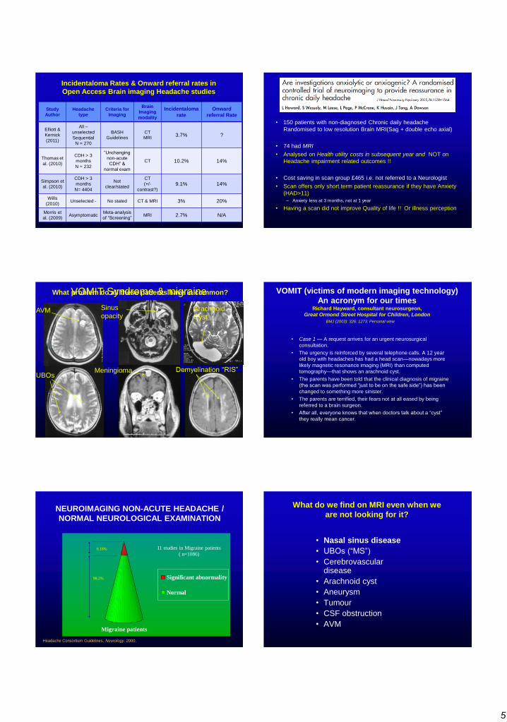
Incidentaloma Rates & Onward referral rates in
Open Access Brain imaging Headache studies
Study
Author
Headache
type
Criteria for
Imaging
Brain
Imaging
modality
Incidentaloma
rate
Onward
referral Rate
Elliott &
Kernick
(2011)
All –
unselected
Sequential
N = 270
BASH
Guidelines
CT
MRI 3.7% ?
Thomas et
al. (2010)
CDH > 3
months
N = 232
―Unchanging
non-acute
CDH‖ &
normal exam
CT 10.2% 14%
Simpson et
al. (2010)
CDH > 3
months
N= 4404
Not
clear/stated
CT
(+/-
contrast?) 9.1% 14%
Wills
(2010) Unselected - No stated CT & MRI 3% 20%
Morris et
al. (2009) Asymptomatic
Meta-analysis
of ―Screening‖ MRI 2.7% N/A
• 150 patients with non-diagnosed Chronic daily headache
Randomised to low resolution Brain MRI(Sag + double echo axial)
• 74 had MRI
• Analysed on Health utility costs in subsequent year and NOT on
Headache impairment related outcomes !!
• Cost saving in scan group £465 i.e. not referred to a Neurologist
• Scan offers only short term patient reassurance if they have Anxiety
(HAD>11)
– Anxiety less at 3 months, not at 1 year
• Having a scan did not improve Quality of life !! Or illness perception
VOMIT Syndrome & migraine
AVM
UBOs
Sinus
opacity
Meningioma
Arachnoid
Cyst
Demyelination ―RIS‖
What problem do all these patients have in common? VOMIT (victims of modern imaging technology)
An acronym for our times Richard Hayward, consultant neurosurgeon,
Great Ormond Street Hospital for Children, London
• Case 1 — A request arrives for an urgent neurosurgical
consultation.
• The urgency is reinforced by several telephone calls. A 12 year
old boy with headaches has had a head scan—nowadays more
likely magnetic resonance imaging (MRI) than computed
tomography—that shows an arachnoid cyst.
• The parents have been told that the clinical diagnosis of migraine
(the scan was performed ―just to be on the safe side‖) has been
changed to something more sinister.
• The parents are terrified, their fears not at all eased by being
referred to a brain surgeon.
• After all, everyone knows that when doctors talk about a ―cyst‖
they really mean cancer.
BMJ (2003): 326: 1273: Personal view
Migraine patients
Significant abnormality
Normal
NEUROIMAGING NON-ACUTE HEADACHE /
NORMAL NEUROLOGICAL EXAMINATION
Headache Consortium Guidelines. Neurology. 2000.
11 studies in Migraine patients
( n=1086) 0.18%
98.2%
What do we find on MRI even when we
are not looking for it?
• Nasal sinus disease
• UBOs (―MS‖)
• Cerebrovascular disease
• Arachnoid cyst
• Aneurysm
• Tumour
• CSF obstruction
• AVM

6
• 19,600 people; 16 studies
• Mean age 11-63 years old
• Prevalence of any incidental brain
finding = 2.7%
No. Needed to scan = 37
• Any Neoplastic finding = 0.7%
(95% CI 0.47-0.98)
No. Needed to scan = 143
• Any Non-Neoplastic finding = 2.0%
(95% CI 1.1-3.1%)
No. Needed to scan = 50
• Higher resolution scan = ↑ prevalence
“Population Epidemiology” of Common
findings on MRI
• Pituitary Adenomas
– 10% of population; symptomatic in 74-90 per 100,000
• Chiari I malformations
– 0.1 % i.e. 1 in 1000 – Tonsillar descent > 3-5 mm
• Cerebral Anneurysms
– 2 -4% ; Rupture rate relates to size & site
• Arterio-venous malformations (AVMs)
– Overall All 2-18 per 100,000
• 1 per 100,000 Brain ICM; 0.5 per 100,000 CVMs; 0.43 per
100,00 for Venous malformations; 0.16 Dural AVMS
• Arachnoid Cysts
– 4% ; 80% Asymptomatic
• UBOs & Incidental WML or ischaemia?
– 7%; Often asymptomatic; Increases with age
My Opinion?
Open access CT & MRI would appear useful
but .......!!!
They do not often diagnose a persons headache
the assessing physician does !!
The diagnosis is in the history – Most of the time !
Imaging should be focussed !!
Secondary Headache disorders when
simple Brain CT/MRI can be normal !
Subarachnoid Haemorrhage
Meningoencephalitis
Cerebral Venous Sinus Thrombosis
Carotid & Vertebral Arterial dissection
Temporal Arteritis
Malignant Hypertension
Head Injury & CSF Hypovolaemia
Why do patients get referred for Brain imaging?
1. Diagnostic clarification & a suspected secondary headache
disorder? & VOMIT syndrome......!!
2. Explanation & Anxiety management
3. Medicolegal concerns
4. Not sure what to do next?
5. Refractory syndromes before ONS implantation
6. ―I just don`t want to see headache patients !‖
7. The SIGN or NICE Guidelines suggest I should ...........
8. The patient & their relatives insisted I did ??
Brain Imaging and the clinical question? What
imaging modality do you request?
• What question are you asking? Atypical primary headache?
Suspected CVST ?
Orthostatic Headache?
Triggered Headache?
Slice thickness & Sequence?
―Excludogram?‖

7
Which Brain imaging investigation? Examples of disorder specific requests for multi-modality imaging
• MRI - Posterior Fossa Pathology
- Trigeminal Autonomic Cephalalgias (TACs)
- TN / SUNCT sequences
- Painful 3rd nerve palsy
+/- Contrast - Intracranial Hypotension (CSF hypovolaemia)
- Cough Headache
• MRV - Immediate Post-partum Headache
or CTV - Papilloedema & normal CT
- New Daily Persistant Headache?
• MRA - Acute Arterial Dissection
or CTA - Painful 3rd nerve palsy (Periorbital pain)
- Recurrent Thunderclap headache
When to Refer patients with Headache
A personal view?
• Same day Referral
– Acute TCH – i.e. Onset <1-5mins
– Acute HA + focal signs +/- seizures
– Progressive HA + Fever + drowsiness
– Progressive HA & Papilloedema
• Urgent-or Semi-urgent Referral:
– Reliably Triggered Headache e.g. Valsalva, Cough
– Headaches with non emergency Red flag symptoms
– Cluster headache or TACs
– Treatment unresponsive Trigeminal Neuralgia
– Headache, Papilloedema & Normal CT,CTV or MRV
– Orthostatic New Daily headache
When to Refer Headache patients to
Specialist (a Headache GPwSI) or Neurologist
A personal view?
• Routine Referral: Categories of patient
– If you don‘t know what to do next .....!
– If the diagnosis is unclear and there is headache related disability
– Primary care treatment refractory headaches
• Ongoing disabling headache symptoms despite at least 2 tolerated evidence
based therapies at adequate dosing/duration
– Unusual Migraine Aura Variants e.g.
• Motor aura / Hemiplegic migraine / Brainstem symptoms – Basilar migraine
– Suspected ‗VOMIT syndrome‖
– Refractory Chronic Migraine, Chronic cluster headache etc.*
– Refractory Analgesic MOH*
Conclusion
Take a better headache history
and remember
―Primum Non Nocere‖
Think about
―SNOOP-TO‖
And if there are no red flags
Make someone‘s happy
Sort out their headache rather than simply
sort out their scan
5th Keele Residential Headache Teaching Course
University of Keele
North Staffordshire
June 15th-17th 2012
Application forms - [email protected] or BASH website
www.bash.org.uk