Embed Size (px)
Citation preview
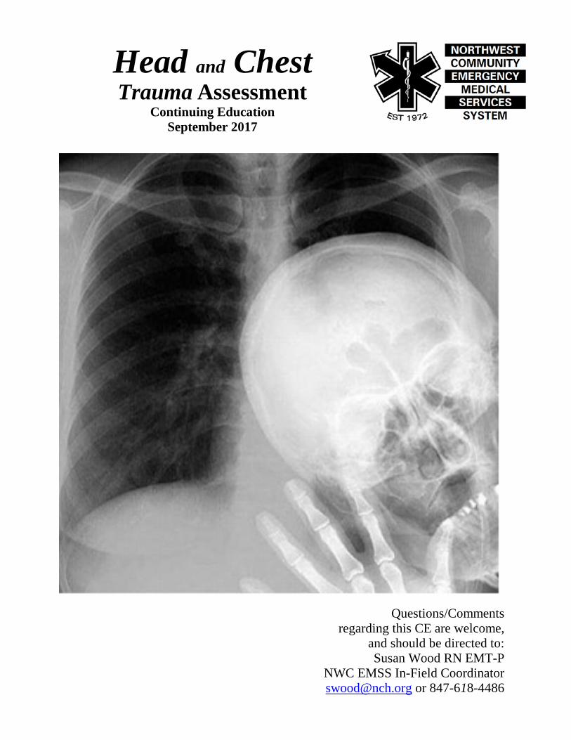
Head and Chest Trauma Assessment
Continuing Education September 2017
Questions/Comments regarding this CE are welcome,
and should be directed to: Susan Wood RN EMT-P
NWC EMSS In-Field Coordinator [email protected] or 847-618-4486
Northwest Community EMSS Continuing Education Head and Chest Trauma Susan Wood, R.N., EMT-P
National EMS Education standard: Epidemiology, pathophysiology, psychosocial impact, presentations, prognosis, and management of (complex depth, comprehensive breadth) patients who are involved in a traumatic mechanism of injury concentrating on head and chest injury.
Assigned readings: This handout; NWC EMSS SOPs
Goal: Upon completion of the class, each participant will independently do the following with a degree of accuracy that meets or exceeds the standards established for their scope of practice: OBJECTIVES:
1. state the incidence, morbidity, and mortality of chest injuries. 2. predict the anatomy and physiology of the organs and structures injured related to chest trauma through a complete
trauma assessment. 3. anticipate chest injuries based on mechanism of injury. 4. sequence the pathogenesis of life-threatening chest injuries. 5. perform the emergency interventions for patients with immediately life-threatening chest injuries. 6. evaluate the pathophysiology of chest wall injuries, including: rib fracture, flail segment, sternal fracture through hands on
assessment. 7. describe the management of patients with chest wall injuries. 8. assemble assessment findings to determine that a patient has a lung injury. 9. assess the management of patients with lung injuries. 10. describe the pathophysiology of cardiac injuries including: pericardial tamponade, blunt cardiac injury, & myocardial
rupture. 11. discuss the assessment findings associated with myocardial injuries. 12. assess the management of patients with myocardial injuries. 13. discuss the assessment findings associated with thoracic vascular injuries. 14. defend the management of patients with thoracic vascular injuries. 15. defend the need for rapid intervention and transport of a pt with chest trauma to the nearest appropriate trauma center. 16. describe the incidence, morbidity, and mortality of head trauma and traumatic brain injury (TBI). 17. Create the initial assessment to be performed on a patient with head injury and state the resuscitative priorities to
achieve airway control, adequate ventilations & gas exchange, and circulatory support. 18. Sequence the steps in performing a neuro exam on a head injured patient with an emphasis on assessing mental
status including GCS, cranial nerve, motor and sensory integrity or deficits. 19. Interpret assessment data to formulate appropriate field impressions associated with head injured patients. 20. Sequence appropriate EMS interventions based on assessment findings and the SOPs including the need for
advanced airway adjuncts; presence of herniation syndrome needing controlled hyperventilation monitored by capnography; IVF resuscitation, and maintaining CPP.
21. Evaluate the pathophysiology, patient presentations, and management of intracranial hemorrhage including epidural, subdural, intracerebral, and subarachnoid hemorrhages.
22. Evaluate the effectiveness of emergency interventions and amend the care plan as indicated by patient responses.
NWC EMSS CE September 2017
Head and Chest Trauma This article was taken from EMS1.com and reprinted with permission from the author. The majority of the continuing educational materials pertaining to brain injury this month came from these materials. Traumatic brain injury: 10 things you
need to know to save lives Proper assessment, treatment, and transport of patients with traumatic brain injury saves lives,
here’s how Sep 3, 2015
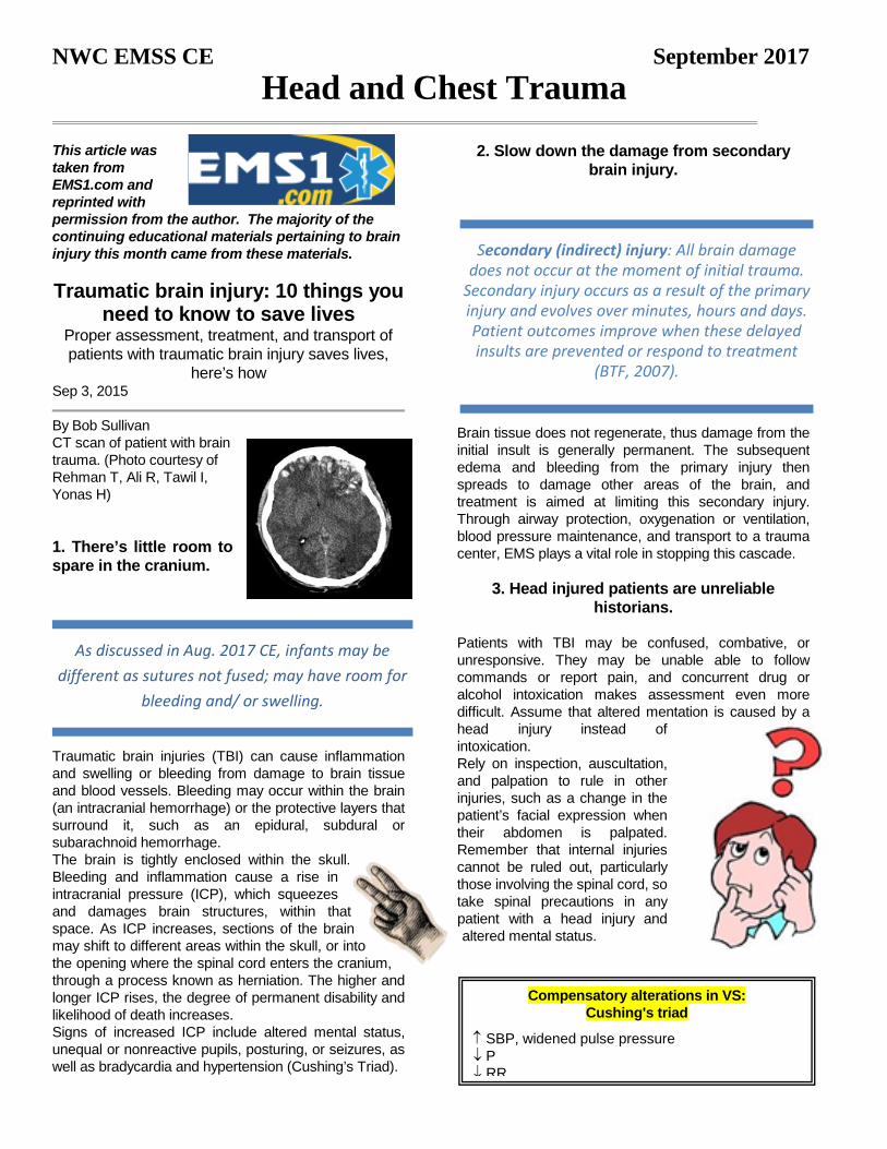
By Bob Sullivan CT scan of patient with brain trauma. (Photo courtesy of Rehman T, Ali R, Tawil I, Yonas H) 1. There’s little room to spare in the cranium.
Traumatic brain injuries (TBI) can cause inflammation and swelling or bleeding from damage to brain tissue and blood vessels. Bleeding may occur within the brain (an intracranial hemorrhage) or the protective layers that surround it, such as an epidural, subdural or subarachnoid hemorrhage. The brain is tightly enclosed within the skull. Bleeding and inflammation cause a rise in intracranial pressure (ICP), which squeezes and damages brain structures, within that space. As ICP increases, sections of the brain may shift to different areas within the skull, or into the opening where the spinal cord enters the cranium, through a process known as herniation. The higher and longer ICP rises, the degree of permanent disability and likelihood of death increases. Signs of increased ICP include altered mental status, unequal or nonreactive pupils, posturing, or seizures, as well as bradycardia and hypertension (Cushing’s Triad).
2. Slow down the damage from secondary
brain injury.
Brain tissue does not regenerate, thus damage from the initial insult is generally permanent. The subsequent edema and bleeding from the primary injury then spreads to damage other areas of the brain, and treatment is aimed at limiting this secondary injury. Through airway protection, oxygenation or ventilation, blood pressure maintenance, and transport to a trauma center, EMS plays a vital role in stopping this cascade.
3. Head injured patients are unreliable historians.
Patients with TBI may be confused, combative, or unresponsive. They may be unable able to follow commands or report pain, and concurrent drug or alcohol intoxication makes assessment even more difficult. Assume that altered mentation is caused by a head injury instead of intoxication. Rely on inspection, auscultation, and palpation to rule in other injuries, such as a change in the patient’s facial expression when their abdomen is palpated. Remember that internal injuries cannot be ruled out, particularly those involving the spinal cord, so take spinal precautions in any patient with a head injury and altered mental status.
Secondary (indirect) injury: All brain damage does not occur at the moment of initial trauma.
Secondary injury occurs as a result of the primary injury and evolves over minutes, hours and days. Patient outcomes improve when these delayed insults are prevented or respond to treatment
(BTF, 2007).
As discussed in Aug. 2017 CE, infants may be different as sutures not fused; may have room for
bleeding and/ or swelling.
Compensatory alterations in VS: Cushing's triad
↑ SBP, widened pulse pressure ↓ P ↓ RR
NWC EMSS Continuing Education page 3 Head and Chest Trauma
4. Sound the alarm at the trauma center
Treatment for head injured patients requires rapid assessment, imaging with a CT scan, and possibly neurosurgery. Time is brain, and the chance of disability or death increases with any delay. Transporting patients to trauma centers that offer neurological
services, and early activation of teams at these centers, streamlines the process to definitive care. Know which hospitals these are, initiate transport as quickly as possible, and alert the hospital to prepare staff and equipment for your arrival. 5. Airway compromise from TBI comes in many
forms. Unconscious head injured patients may lose muscle tone in their jaw, and their tongue may obstruct the airway. The gag reflex may also be compromised in TBI patients, which increases the risk of aspiration from vomit or blood. Clenched teeth, known as trismus, is another common finding that makes position and suctioning airway secretions difficult. Attempt to open the airway with a jaw thrust. Use an oral or nasal airway if the patient will tolerate it, (In NWC EMSS, we choose not to take this risk if a known head injury may likely result in a basilar skull fracture) position the patient on their left side to help prevent aspiration, and suction secretions. Endotracheal intubation is the definitive airway management for TBI, often after sedation and paralysis with RSI. This procedure carries considerable risk, and whether it should be done by ground EMS crews is
controversial. (Again, NWC EMSS only suggests an advanced airway if the patient is unable to maintain their own airway and EMS cannot manage with less invasive means). Remember that the goal for airway
management should be to use the least invasive means to maintain a clear path for oxygenation and ventilation.
6. Hypoxia and hypotension are harmful – even
for a short time.
Irreversible brain damage can occur in TBI patients after only four minutes of anoxia, which can be caused by a compromised airway, altered respiratory patterns from the head injury, or lung injury in multi-system trauma. Use pulse-oximetry, skin color, and respiratory rate to assess adequate oxygenation. Administer oxygen via nasal cannula, non-rebreather mask, or bag valve to maintain a pulse-ox reading of at least 95%. Cerebral oxygen delivery is also compromised by hypotension, which is associated with poor outcomes after even transient episodes. Hypotension caused directly by a head injury is a rare and ominous finding; it is more often caused by shock associated with other injuries. Administer IV fluids and titrate blood pressure to 90 – 100 mmHG systolic in head injured patients who are hypotensive (see red box discussion below).
7. Ventilating too fast reduces blood flow to the brain.
As harmful as hypoxia is in TBI, hypocapnea from excessive ventilation is harmful as well. A higher respiratory rate decreases the amount of CO2 in the blood, which triggers cerebral vasoconstriction and less oxygen delivery. Hyperventilation of head injuries has been associated with poor outcomes. When assisted ventilation is necessary in isolated head injuries, follow your local protocols regarding use of end-tidal CO2 (EtCO2) monitoring for a head-injured patient. Shock from multisystem trauma can also cause a low EtCO2 reading, in which case it should not be used to guide ventilation. If capnography is not available, or if the patient may be in shock from other injuries, ventilate adults at 10 breaths per minute. There is one exception...
Here is where our system differs from the article based on the evidence reviewed. Our SOP instructs that we are to administer IVFs and
titrate to a target SBP of 110-120 (MAP 85-90) or higher. p. 47
NWC EMSS Continuing Education page 4 Head and Chest Trauma
8. Hyperventilation may slow herniation
(Class content has deviated from this part of
the conversation found in the article based on system SOP’s.)
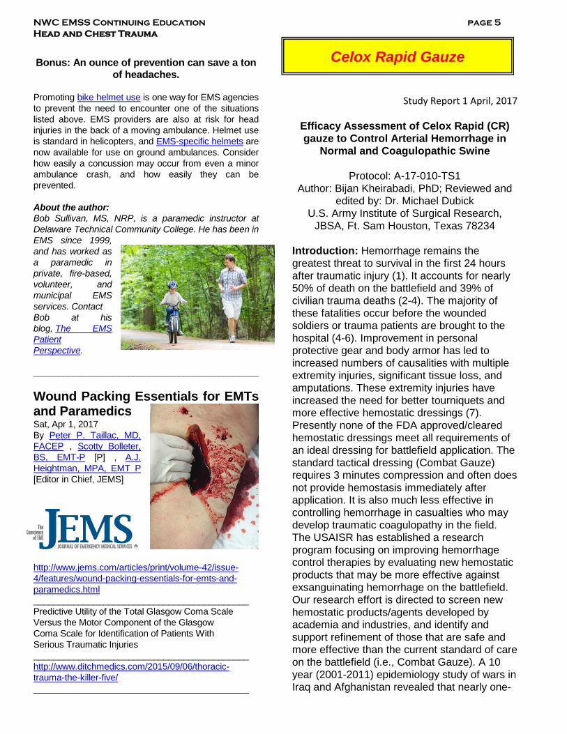
In cases of suspected ICP being so high that portions of the brain may be shifting out of the cranium, the benefit of temporarily reducing cerebral blood with mild hyperventilation may outweigh the harm from less oxygen delivery. Hyperventilation is ONLY indicated for
patients who are posturing or have no motor response, have unequal or dilated and nonreactive pupils, or bradycardia and hypotension. Titrate the respiratory rate to maintain an EtCO2 reading between 30 and 35 (in the absence of multi-system trauma), or 20 breaths per minute.
Outcomes in herniation syndrome Two minute drill: Have two minutes to act after herniation. If medulla brain stem function is altered, the BP will drop to 40/20. HR will increase to 140-150. The brain will not go back to its normal alignment. This is an indication to seek an OLMC order for hyperventilation. 9. You may have a concussion…you may have a concussion… A concussion is a transient change in mentation after blunt head trauma. It can cause retrograde amnesia, and patients often repeatedly ask the same question after being given an answer. It can also cause a
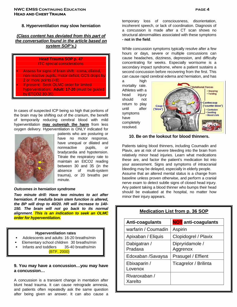
temporary loss of consciousness, disorientation, incoherent speech, or lack of coordination. Diagnosis of a concussion is made after a CT scan shows no structural abnormalities associated with these symptoms - not in the field. While concussion symptoms typically resolve after a few hours or days, severe or multiple concussions can cause headaches, dizziness, depression, and difficulty concentrating for weeks. Especially worrisome is a secondary impact syndrome, where a patient sustains a second concussion before recovering from the first. This can cause rapid cerebral edema and herniation, and has a high mortality rate. Athletes with a head injury should not return to play until after symptoms have completely resolved.
10. Be on the lookout for blood thinners. Patients taking blood thinners, including Coumadin and Plavix, are at risk of severe bleeding into the brain from relatively minor head injuries. Learn what medications these are, and factor the patient’s medication list into your assessment. Signs and symptoms of intracranial bleeding may be delayed, especially in elderly people. Assume that an altered mental status is a change from baseline unless proven otherwise, and perform a cranial nerve exam to detect subtle signs of closed head injury. Any patient taking a blood thinner who bumps their head should be evaluated at the hospital, no matter how minor their injury appears.
Hyperventilation rates Adolescents and adults: 16-20 breaths/min Elementary school children 30 breaths/min Infants and toddlers 35-40 breaths/min
(BTF, 2000)
Head Trauma SOP p. 47 ITC special considerations
Assess for signs of brain shift: coma, dilated, non-reactive pupils, motor deficit; GCS drops by 2 or more points (<8)
If present: Seek OLMC order for limited hyperventilation: Adult: 17-20 (must be guided by ETCO2 30-35)
Medication List from p. 36 SOP
Anti-coagulants NOT anti-coagulants warfarin / Coumadin Aspirin Apixaban / Eliquis Clopidogrel / Plavix Dabigatran / Pradaxa
Dipryridamole / Aggrenox
Edoxaban /Savaysa Prasugel / Effient Ebixaparin / Lovenox
Ticagrelor / Brilinta
Rivaroxaban / Xarelto
NWC EMSS Continuing Education page 5 Head and Chest Trauma
Bonus: An ounce of prevention can save a ton
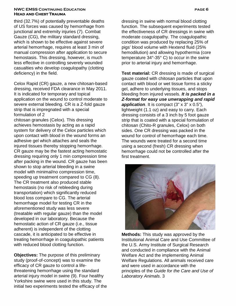
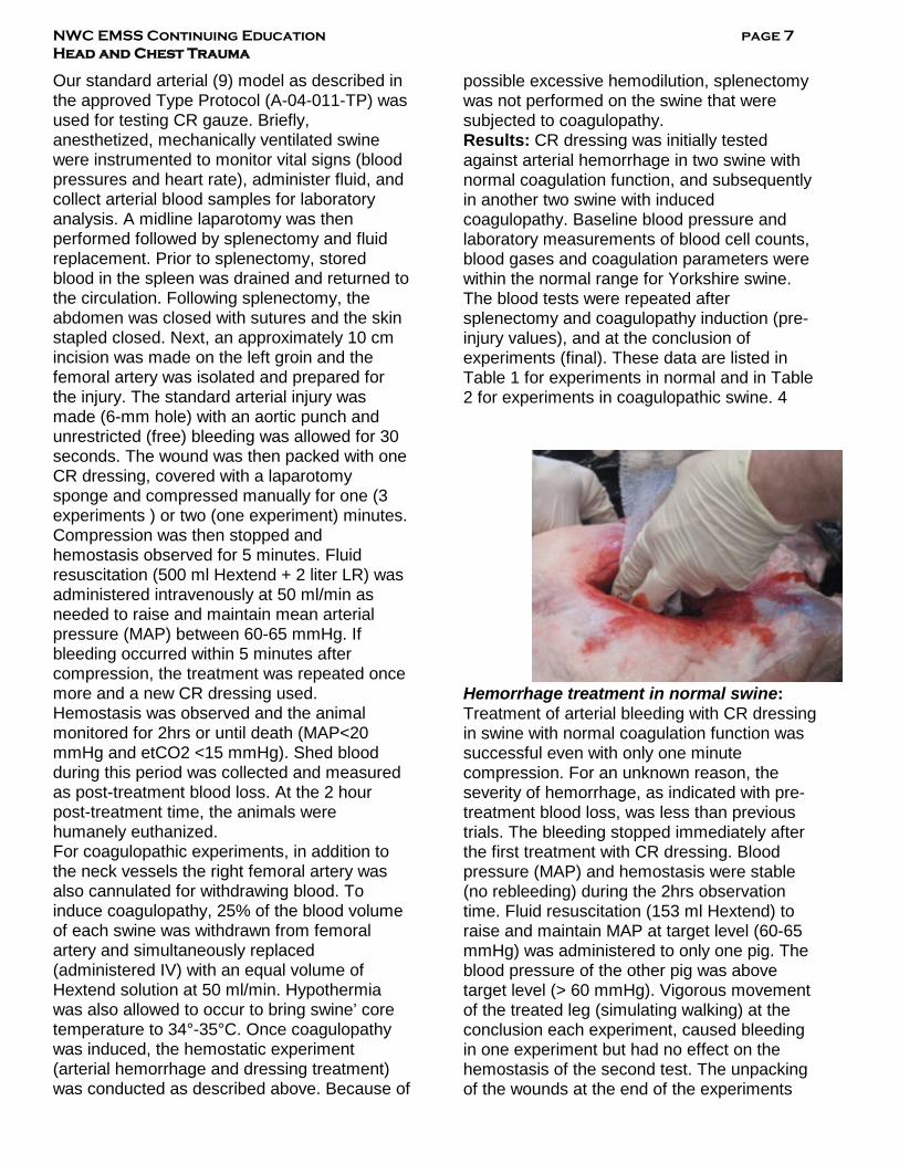
of headaches. Promoting bike helmet use is one way for EMS agencies to prevent the need to encounter one of the situations listed above. EMS providers are also at risk for head injuries in the back of a moving ambulance. Helmet use is standard in helicopters, and EMS-specific helmets are now available for use on ground ambulances. Consider how easily a concussion may occur from even a minor ambulance crash, and how easily they can be prevented. About the author: Bob Sullivan, MS, NRP, is a paramedic instructor at Delaware Technical Community College. He has been in EMS since 1999, and has worked as a paramedic in private, fire-based, volunteer, and municipal EMS services. Contact Bob at his blog, The EMS Patient Perspective. _____________________________________________ Wound Packing Essentials for EMTs and Paramedics Sat, Apr 1, 2017 By Peter P. Taillac, MD, FACEP , Scotty Bolleter, BS, EMT-P [P] , A.J. Heightman, MPA, EMT P [Editor in Chief, JEMS]
http://www.jems.com/articles/print/volume-42/issue-4/features/wound-packing-essentials-for-emts-and-paramedics.html ___________________________________________ Predictive Utility of the Total Glasgow Coma Scale Versus the Motor Component of the Glasgow Coma Scale for Identification of Patients With Serious Traumatic Injuries ___________________________________________ http://www.ditchmedics.com/2015/09/06/thoracic-trauma-the-killer-five/ ___________________________________________
Study Report 1 April, 2017
Efficacy Assessment of Celox Rapid (CR) gauze to Control Arterial Hemorrhage in
Normal and Coagulopathic Swine
Protocol: A-17-010-TS1 Author: Bijan Kheirabadi, PhD; Reviewed and
edited by: Dr. Michael Dubick U.S. Army Institute of Surgical Research,
JBSA, Ft. Sam Houston, Texas 78234 Introduction: Hemorrhage remains the greatest threat to survival in the first 24 hours after traumatic injury (1). It accounts for nearly 50% of death on the battlefield and 39% of civilian trauma deaths (2-4). The majority of these fatalities occur before the wounded soldiers or trauma patients are brought to the hospital (4-6). Improvement in personal protective gear and body armor has led to increased numbers of causalities with multiple extremity injuries, significant tissue loss, and amputations. These extremity injuries have increased the need for better tourniquets and more effective hemostatic dressings (7). Presently none of the FDA approved/cleared hemostatic dressings meet all requirements of an ideal dressing for battlefield application. The standard tactical dressing (Combat Gauze) requires 3 minutes compression and often does not provide hemostasis immediately after application. It is also much less effective in controlling hemorrhage in casualties who may develop traumatic coagulopathy in the field. The USAISR has established a research program focusing on improving hemorrhage control therapies by evaluating new hemostatic products that may be more effective against exsanguinating hemorrhage on the battlefield. Our research effort is directed to screen new hemostatic products/agents developed by academia and industries, and identify and support refinement of those that are safe and more effective than the current standard of care on the battlefield (i.e., Combat Gauze). A 10 year (2001-2011) epidemiology study of wars in Iraq and Afghanistan revealed that nearly one-
Celox Rapid Gauze
NWC EMSS Continuing Education page 6 Head and Chest Trauma
third (32.7%) of potentially preventable deaths of US forces was caused by hemorrhage from junctional and extremity injuries (7). Combat Gauze (CG), the military standard dressing, which is shown to be effective against severe arterial hemorrhage, requires at least 3 min of manual compression after application to secure hemostasis. This dressing, however, is much less effective in controlling severely wounded casualties who develop coagulopathy (clotting deficiency) in the field. Celox Rapid (CR) gauze, a new chitosan-based dressing, received FDA clearance in May 2011. It is indicated for temporary and topical application on the wound to control moderate to severe external bleeding. CR is a Z-fold gauze strip that is impregnated with a special formulation of 2 chitosan granules (Celox). This dressing achieves hemostasis by acting as a rapid system for delivery of the Celox particles which upon contact with blood in the wound forms an adhesive gel which attaches and seals the injured tissues thereby stopping hemorrhage. CR gauze may be the fastest acting hemostatic dressing requiring only 1 min compression time after packing in the wound. CR gauze has been shown to stop arterial bleeding in a swine model with minimal/no compression time, speeding up treatment compared to CG (8). The CR treatment also produced stable hemostasis (no risk of rebleeding during transportation) which significantly reduced blood loss compare to CG. The arterial hemorrhage model for testing CR in the aforementioned study was less severe (treatable with regular gauze) than the model developed in our laboratory. Because the hemostatic action of CR gauze (i.e., tissue adherent) is independent of the clotting cascade, it is anticipated to be effective in treating hemorrhage in coagulopathic patients with reduced blood clotting function. Objectives: The purpose of this preliminary study (proof-of-concept) was to examine the efficacy of CR gauze to control a life-threatening hemorrhage using the standard arterial injury model in swine (9). Four healthy Yorkshire swine were used in this study. The initial two experiments tested the efficacy of the
dressing in swine with normal blood clotting function. The subsequent experiments tested the effectiveness of CR dressings in swine with moderate coagulopathy. The coagulopathic condition was produced by replacing 25% of pigs’ blood volume with Hextend fluid (25% hemodilution) and allowing hypothermia (core temperature 34°-35° C) to occur in the swine prior to arterial injury and hemorrhage. Test material: CR dressing is made of surgical gauze coated with chitosan particles that upon contact with blood or wet tissue forms a sticky gel, adhere to underlying tissues, and stops bleeding from injured vessels. It is packed in a Z-format for easy use unwrapping and rapid application. It is compact (3” x 3” x 0.5”), lightweight (1.1 oz) and easy to carry. Each dressing consists of a 3 inch by 5 foot gauze strip that is coated with a special formulation of chitosan (Chito-R granules, Celox) on both sides. One CR dressing was packed in the wound for control of hemorrhage each time. The wounds were treated for a second time using a second (fresh) CR dressing when hemorrhage could not be controlled after the first treatment.
Methods: This study was approved by the Institutional Animal Care and Use Committee of the U.S. Army Institute of Surgical Research and conducted in compliance with the Animal Welfare Act and the implementing Animal Welfare Regulations. All animals received care and were used in accordance with the principles of the Guide for the Care and Use of Laboratory Animals. 3
NWC EMSS Continuing Education page 7 Head and Chest Trauma
Our standard arterial (9) model as described in the approved Type Protocol (A-04-011-TP) was used for testing CR gauze. Briefly, anesthetized, mechanically ventilated swine were instrumented to monitor vital signs (blood pressures and heart rate), administer fluid, and collect arterial blood samples for laboratory analysis. A midline laparotomy was then performed followed by splenectomy and fluid replacement. Prior to splenectomy, stored blood in the spleen was drained and returned to the circulation. Following splenectomy, the abdomen was closed with sutures and the skin stapled closed. Next, an approximately 10 cm incision was made on the left groin and the femoral artery was isolated and prepared for the injury. The standard arterial injury was made (6-mm hole) with an aortic punch and unrestricted (free) bleeding was allowed for 30 seconds. The wound was then packed with one CR dressing, covered with a laparotomy sponge and compressed manually for one (3 experiments ) or two (one experiment) minutes. Compression was then stopped and hemostasis observed for 5 minutes. Fluid resuscitation (500 ml Hextend + 2 liter LR) was administered intravenously at 50 ml/min as needed to raise and maintain mean arterial pressure (MAP) between 60-65 mmHg. If bleeding occurred within 5 minutes after compression, the treatment was repeated once more and a new CR dressing used. Hemostasis was observed and the animal monitored for 2hrs or until death (MAP<20 mmHg and etCO2 <15 mmHg). Shed blood during this period was collected and measured as post-treatment blood loss. At the 2 hour post-treatment time, the animals were humanely euthanized. For coagulopathic experiments, in addition to the neck vessels the right femoral artery was also cannulated for withdrawing blood. To induce coagulopathy, 25% of the blood volume of each swine was withdrawn from femoral artery and simultaneously replaced (administered IV) with an equal volume of Hextend solution at 50 ml/min. Hypothermia was also allowed to occur to bring swine’ core temperature to 34°-35°C. Once coagulopathy was induced, the hemostatic experiment (arterial hemorrhage and dressing treatment) was conducted as described above. Because of
possible excessive hemodilution, splenectomy was not performed on the swine that were subjected to coagulopathy. Results: CR dressing was initially tested against arterial hemorrhage in two swine with normal coagulation function, and subsequently in another two swine with induced coagulopathy. Baseline blood pressure and laboratory measurements of blood cell counts, blood gases and coagulation parameters were within the normal range for Yorkshire swine. The blood tests were repeated after splenectomy and coagulopathy induction (pre-injury values), and at the conclusion of experiments (final). These data are listed in Table 1 for experiments in normal and in Table 2 for experiments in coagulopathic swine. 4
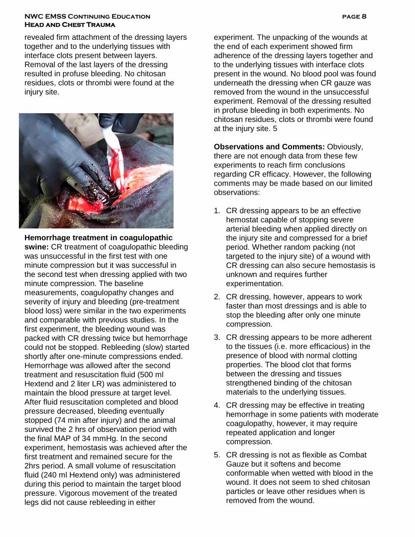
Hemorrhage treatment in normal swine: Treatment of arterial bleeding with CR dressing in swine with normal coagulation function was successful even with only one minute compression. For an unknown reason, the severity of hemorrhage, as indicated with pre-treatment blood loss, was less than previous trials. The bleeding stopped immediately after the first treatment with CR dressing. Blood pressure (MAP) and hemostasis were stable (no rebleeding) during the 2hrs observation time. Fluid resuscitation (153 ml Hextend) to raise and maintain MAP at target level (60-65 mmHg) was administered to only one pig. The blood pressure of the other pig was above target level (> 60 mmHg). Vigorous movement of the treated leg (simulating walking) at the conclusion each experiment, caused bleeding in one experiment but had no effect on the hemostasis of the second test. The unpacking of the wounds at the end of the experiments
NWC EMSS Continuing Education page 8 Head and Chest Trauma
revealed firm attachment of the dressing layers together and to the underlying tissues with interface clots present between layers. Removal of the last layers of the dressing resulted in profuse bleeding. No chitosan residues, clots or thrombi were found at the injury site.
Hemorrhage treatment in coagulopathic swine: CR treatment of coagulopathic bleeding was unsuccessful in the first test with one minute compression but it was successful in the second test when dressing applied with two minute compression. The baseline measurements, coagulopathy changes and severity of injury and bleeding (pre-treatment blood loss) were similar in the two experiments and comparable with previous studies. In the first experiment, the bleeding wound was packed with CR dressing twice but hemorrhage could not be stopped. Rebleeding (slow) started shortly after one-minute compressions ended. Hemorrhage was allowed after the second treatment and resuscitation fluid (500 ml Hextend and 2 liter LR) was administered to maintain the blood pressure at target level. After fluid resuscitation completed and blood pressure decreased, bleeding eventually stopped (74 min after injury) and the animal survived the 2 hrs of observation period with the final MAP of 34 mmHg. In the second experiment, hemostasis was achieved after the first treatment and remained secure for the 2hrs period. A small volume of resuscitation fluid (240 ml Hextend only) was administered during this period to maintain the target blood pressure. Vigorous movement of the treated legs did not cause rebleeding in either
experiment. The unpacking of the wounds at the end of each experiment showed firm adherence of the dressing layers together and to the underlying tissues with interface clots present in the wound. No blood pool was found underneath the dressing when CR gauze was removed from the wound in the unsuccessful experiment. Removal of the dressing resulted in profuse bleeding in both experiments. No chitosan residues, clots or thrombi were found at the injury site. 5 Observations and Comments: Obviously, there are not enough data from these few experiments to reach firm conclusions regarding CR efficacy. However, the following comments may be made based on our limited observations: 1. CR dressing appears to be an effective
hemostat capable of stopping severe arterial bleeding when applied directly on the injury site and compressed for a brief period. Whether random packing (not targeted to the injury site) of a wound with CR dressing can also secure hemostasis is unknown and requires further experimentation.
2. CR dressing, however, appears to work faster than most dressings and is able to stop the bleeding after only one minute compression.
3. CR dressing appears to be more adherent to the tissues (i.e. more efficacious) in the presence of blood with normal clotting properties. The blood clot that forms between the dressing and tissues strengthened binding of the chitosan materials to the underlying tissues.
4. CR dressing may be effective in treating hemorrhage in some patients with moderate coagulopathy, however, it may require repeated application and longer compression.
5. CR dressing is not as flexible as Combat Gauze but it softens and become conformable when wetted with blood in the wound. It does not seem to shed chitosan particles or leave other residues when is removed from the wound.
NWC EMSS Continuing Education page 9 Head and Chest Trauma
In summary, CR gauze is an effective hemostatic dressing that can stop severe arterial bleeding when applied directly on the injured vessels and compressed for one minute. The hemostatic mechanism of this dressing is based on its physical attachment/adhesion to the damaged tissues and appears to be mediated by formation of clots at the interface. Further studies are needed to determine the efficacy of this dressing compared with Combat Gauze and other chitosan-based dressings (Celox gauze and Chitogauze) that are recommend by the committee of Tactical Combat Casualty Care for use on the battlefield. Disclaimer: The opinions or assertions expressed herein are the private views of the authors and are not to be construed as official or as reflecting the views of the US Department of Defense. 6
References
1. Bellamy RF. Causes of death in conventional
warfare: implications for combat casualty care research. Mil Med. 1984;149:55-62.
2. Acosta JA, Yang JC, Winchell RJ, Simons RK, Fortlage DA, Hollingsworth-Fridlund P, and Hoyt DA. Lethal injuries and time to death in a level one trauma center. J Am Coll Surg. 1998;186:528-533.
3. Sauaia A, Moore FA, Moore EE, Moser KS, Brennan R, Read RA, Pons PT. Epidemiology of trauma deaths: a reassessment. J Trauma. 1995;38:185-193.
4. Shackford SR, Mackersie RC, Holbrook TL, Davis JW, Hollingsworth-Fridlund P, Hoyt DB and Wolf PL. The epidemiology of traumatic death: a population-based analysis. Arch Surg. 1993; 128:571-575.
5. Sauaia A, Moore FA, Moore EE, et al. Epidemiology of trauma deaths: reassessment. J Trauma. 1995; 38:185-193.
6. Hoyte D, Bulger E, Knudson M, et al. Deaths in the operating room: an analysis of a multi-center experience. J Trauma. 1994; 37:426-432.
7. Kragh JF, Murphy C, Dubick MA, Baer DG, Johnson J, Blackbourne LH: New tourniquet device concepts for battlefield hemorrhage control. US Army Med Dep J. April-June: 38-48, 2011.
8. Kunio NR, Riha GM, Watson KM, et al., Chitosan based advanced hemostatic dressing is associated with decreased blood loss in a swine uncontrolled hemorrhage model. Am J Surg. 2013; 205:505-510.
9. Kheirabadi BS, et al., Development of a standard swine hemorrhage model for efficacy assessment of topical hemostatic agents. J Trauma, 2011;71(1 Suppl):S139-46.
NWC EMSS Continuing Education page 10 Head and Chest Trauma
Obtaining the GCS: GCS should be obtained through interaction with the patient (i.e., by giving verbal directions or for patients unable to follow commands by application of a painful stimulus).
Ideally, GCS scoring should occur after a clear airway is established, and after hypoxemia and hypotension are corrected, the patient has been resuscitated, and before administration of sedative or paralytic agents or after these drugs have been metabolized.
Components: Unconsciousness can be simplistically defined as failure to respond appropriately to environmental stimuli. Coma is defined as a state of unconsciousness from which the individual cannot be awakened, in which the individual responds minimally or not at all to stimuli, and initiates no voluntary activities (BIAA, 2004). The GCS is the sum of three independent coded values that measure a patient’s eye opening, verbal, and motor responses either spontaneously or in response to verbal or painful stimuli (Healey et al, 2003). Always report the patient’s BEST response even if different on one side of the body from the other.
Best eye opening: Ask the patient, “What happened to you?” If the patient opens his eyes, then ask the questions in the verbal and motor section of the Glasgow Coma Scale to determine the total score. If the patient does not open his or her eyes, apply a central pain stimulus. Pinch the earlobe or apply pressure over the supraorbital ridge. If the patient is spontaneously moving all four extremities, apply blunt pressure to the nailbed or pinch the anterior axillary skin or muscles at the top of shoulder.
Eye opening to command or speech is a higher level of stimulus recognition. Assume that the cerebral cortex is processing information.
Best verbal response: Assesses the eloquent cortex (where we speak) by evaluating the content and context of speech. While this evaluates a high level of cognitive function, its absence does not imply a total loss of function. This is a difficult area to score with consistency, especially between 4 and 5.
Best motor response: This element is the least affected by trauma. It allows evaluation of the interface between sensing a stimulus, interpreting the information and reacting to it (Fischer & Mathieson, 2001). There is currently debate over the sensitivity and specificity of GCS scoring with evolving consensus that the motor component alone can predict neurological outcomes (Gill et al, 2005).
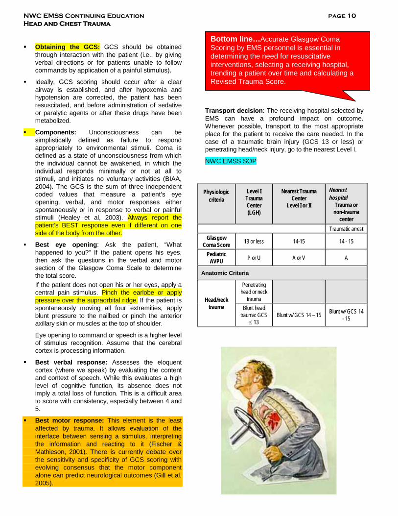
Transport decision: The receiving hospital selected by EMS can have a profound impact on outcome. Whenever possible, transport to the most appropriate place for the patient to receive the care needed. In the case of a traumatic brain injury (GCS 13 or less) or penetrating head/neck injury, go to the nearest Level I.
NWC EMSS SOP
Physiologic criteria
Level I Trauma Center (LGH)
Nearest Trauma Center
Level I or II
Nearest hospital
Trauma or non-trauma
center Traumatic arrest
Glasgow Coma Score 13 or less 14-15 14 - 15
Pediatric AVPU P or U A or V A
Anatomic Criteria
Head/neck trauma
Penetrating head or neck
trauma
Blunt head trauma: GCS
≤ 13 Blunt w/ GCS 14 – 15 Blunt w/ GCS 14
- 15
Bottom line…Accurate Glasgow Coma Scoring by EMS personnel is essential in determining the need for resuscitative interventions, selecting a receiving hospital, trending a patient over time and calculating a Revised Trauma Score.
NWC EMSS Continuing Education page 11 Head and Chest Trauma
Differentiating Immediately Life Threatening Thoracic Injuries
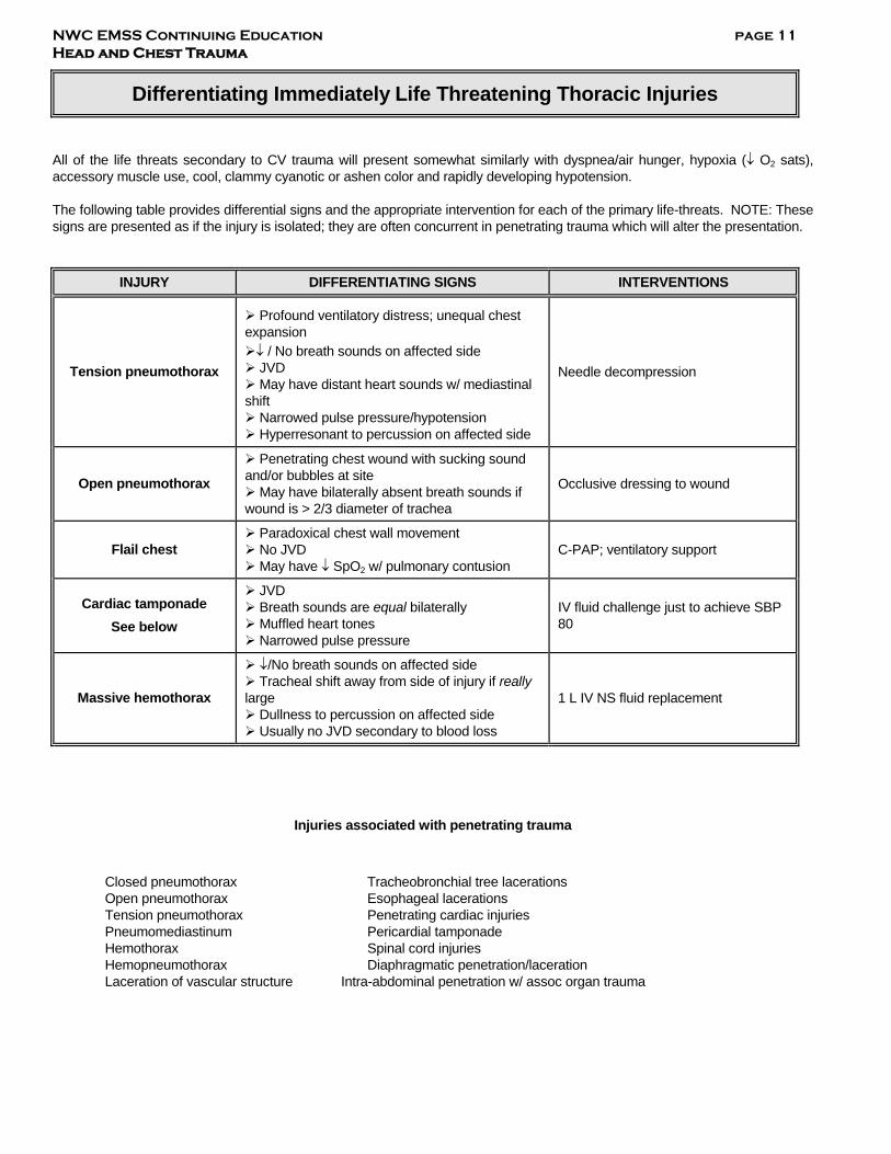
All of the life threats secondary to CV trauma will present somewhat similarly with dyspnea/air hunger, hypoxia (↓ O2 sats), accessory muscle use, cool, clammy cyanotic or ashen color and rapidly developing hypotension. The following table provides differential signs and the appropriate intervention for each of the primary life-threats. NOTE: These signs are presented as if the injury is isolated; they are often concurrent in penetrating trauma which will alter the presentation.
INJURY DIFFERENTIATING SIGNS INTERVENTIONS
Tension pneumothorax
Profound ventilatory distress; unequal chest expansion ↓ / No breath sounds on affected side JVD May have distant heart sounds w/ mediastinal shift Narrowed pulse pressure/hypotension Hyperresonant to percussion on affected side
Needle decompression
Open pneumothorax
Penetrating chest wound with sucking sound and/or bubbles at site May have bilaterally absent breath sounds if wound is > 2/3 diameter of trachea
Occlusive dressing to wound
Flail chest Paradoxical chest wall movement No JVD May have ↓ SpO2 w/ pulmonary contusion
C-PAP; ventilatory support
Cardiac tamponade See below
JVD Breath sounds are equal bilaterally Muffled heart tones Narrowed pulse pressure
IV fluid challenge just to achieve SBP 80
Massive hemothorax
↓/No breath sounds on affected side Tracheal shift away from side of injury if really large Dullness to percussion on affected side Usually no JVD secondary to blood loss
1 L IV NS fluid replacement
Injuries associated with penetrating trauma
Closed pneumothorax Tracheobronchial tree lacerations Open pneumothorax Esophageal lacerations Tension pneumothorax Penetrating cardiac injuries Pneumomediastinum Pericardial tamponade Hemothorax Spinal cord injuries Hemopneumothorax Diaphragmatic penetration/laceration Laceration of vascular structure Intra-abdominal penetration w/ assoc organ trauma
NWC EMSS Continuing Education page 12 Head and Chest Trauma
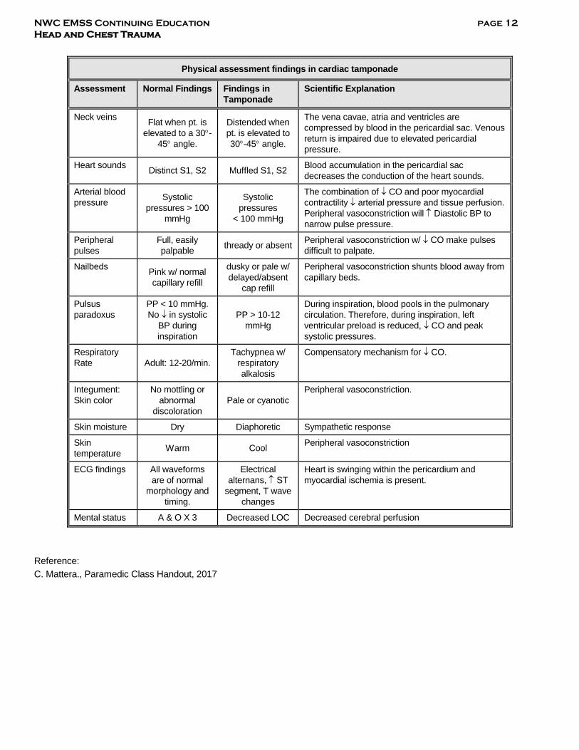
Physical assessment findings in cardiac tamponade
Assessment Normal Findings Findings in Tamponade
Scientific Explanation
Neck veins Flat when pt. is elevated to a 30°-
45° angle.
Distended when pt. is elevated to 30°-45° angle.
The vena cavae, atria and ventricles are compressed by blood in the pericardial sac. Venous return is impaired due to elevated pericardial pressure.
Heart sounds Distinct S1, S2 Muffled S1, S2 Blood accumulation in the pericardial sac decreases the conduction of the heart sounds.
Arterial blood pressure Systolic
pressures > 100 mmHg
Systolic pressures
< 100 mmHg
The combination of ↓ CO and poor myocardial contractility ↓ arterial pressure and tissue perfusion. Peripheral vasoconstriction will ↑ Diastolic BP to narrow pulse pressure.
Peripheral pulses
Full, easily palpable thready or absent Peripheral vasoconstriction w/ ↓ CO make pulses
difficult to palpate.
Nailbeds Pink w/ normal capillary refill
dusky or pale w/ delayed/absent
cap refill
Peripheral vasoconstriction shunts blood away from capillary beds.
Pulsus paradoxus
PP < 10 mmHg. No ↓ in systolic
BP during inspiration
PP > 10-12 mmHg
During inspiration, blood pools in the pulmonary circulation. Therefore, during inspiration, left ventricular preload is reduced, ↓ CO and peak systolic pressures.
Respiratory Rate Adult: 12-20/min.
Tachypnea w/ respiratory alkalosis
Compensatory mechanism for ↓ CO.
Integument: Skin color
No mottling or abnormal
discoloration Pale or cyanotic
Peripheral vasoconstriction.
Skin moisture Dry Diaphoretic Sympathetic response
Skin temperature Warm Cool Peripheral vasoconstriction
ECG findings All waveforms are of normal
morphology and timing.
Electrical alternans, ↑ ST
segment, T wave changes
Heart is swinging within the pericardium and myocardial ischemia is present.
Mental status A & O X 3 Decreased LOC Decreased cerebral perfusion
Reference: C. Mattera., Paramedic Class Handout, 2017