Embed Size (px)
Citation preview

8/30/2016
1
HD OCT Update50470-GO
Michael Cymbor, OD, FAAO Nittany Eye Associates
State College, PA
Adjunct Professor
Pennsylvania College of Optometry
Disclosure Statement• Member of the Speakers Bureau for Alcon, Optovue, and Inspire
• Principal site investigator for Ciba, Vistakon, and Bausch & Lomb
• Has received educational grants from Heidelberg Engineering, Zeiss and EyeIC
8/30/2016
2
In the last 15 years…
• What instrument has changed eye care the most???????
OCT
• O = Optical
• C = Coherence• Coherence comes from a Latin word meaning “to stick together
• T = Tomography• a technique used to obtain an image of a selected plane section of the
human body or some other solid object
How does it work?
• OCT utilizes near-infrared light waves to measure distances of anatomical structures. A beam of light is directed onto the structure and the echo time delay of light is then recorded.
8/30/2016
3
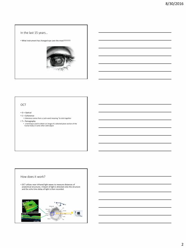
OCT Evolution
OCT
1995
OCT2
2000
OCT3
Stratus OCT
2002
Optovue
HD-OCT
2007
100 A-scans x
500 points
100 A-scans x
500 points
512 A-scans
x1024 points
4096 A-scans x
1024 points
100
100
500
26,000
20
20
10
5
Single line scanScans/
second
Resolution
(microns)
And now…
8/30/2016
4
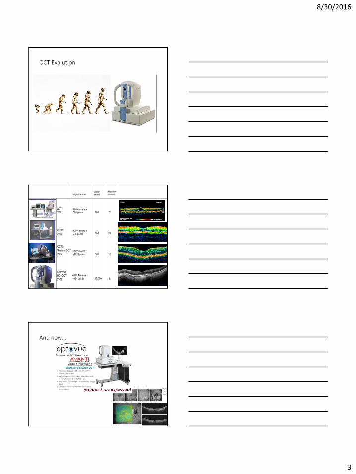
In vivo sub-cellular resolution OCT (A) in a developmental biology animal model (African tadpole).
Resolution down to 1 micron!!!!
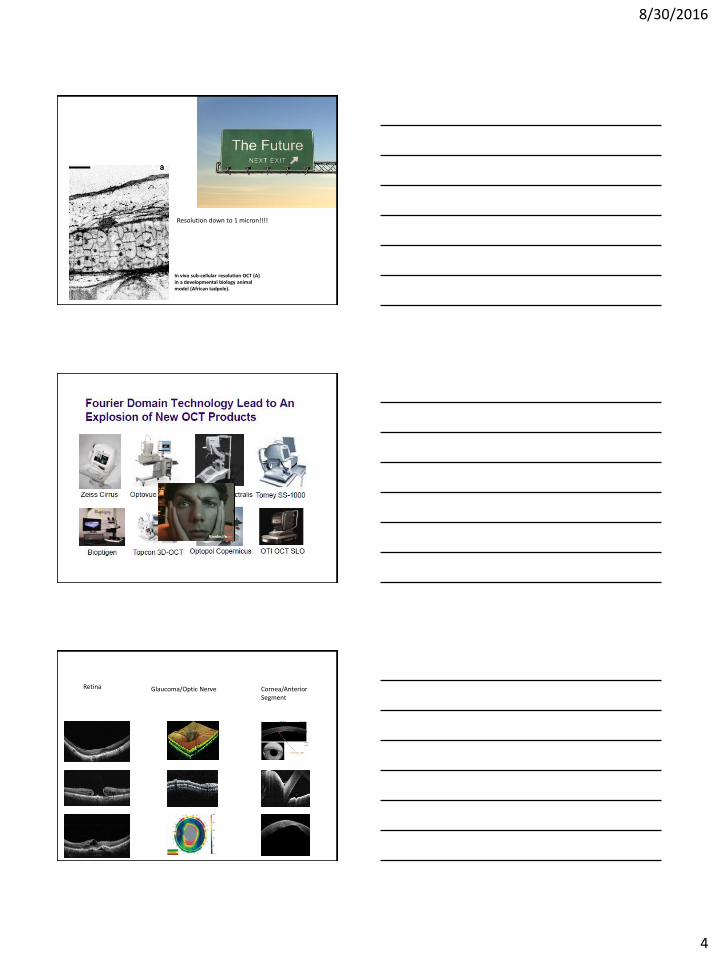
Retina Glaucoma/Optic Nerve Cornea/Anterior Segment
8/30/2016
5
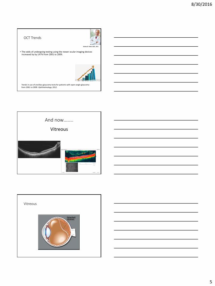
OCT Trends
• The odds of undergoing testing using the newer ocular imaging devices increased by by 147% from 2001 to 2009.
Joshua D. Stein, M.D., M.S.
Trends in use of ancillary glaucoma tests for patients with open-angle glaucoma from 2001 to 2009. Ophthalmology, 2012.
And now……..
Vitreous
Vitreous
8/30/2016
6
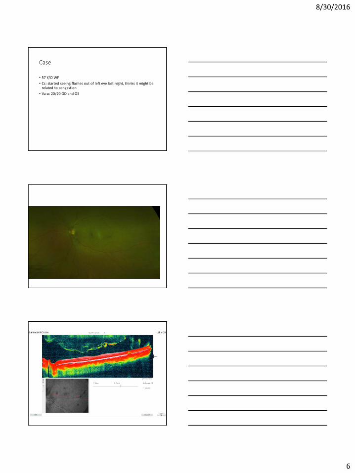
Case
• 57 Y/O WF
• Cc: started seeing flashes out of left eye last night, thinks it might be related to congestion
• Va sc 20/20 OD and OS
8/30/2016
7
Practice Management
• 92134
• Scanning computerized ophthalmic diagnostic imaging, posterior segment, with interpretation and report, unilateral or bilateral, retina
OCT Setup Pearl
• 2 options in our practice• Upload into practice management software
• Networked
8/30/2016
8
Why OCT for Anterior Chamber?
Cornea
8/30/2016
9
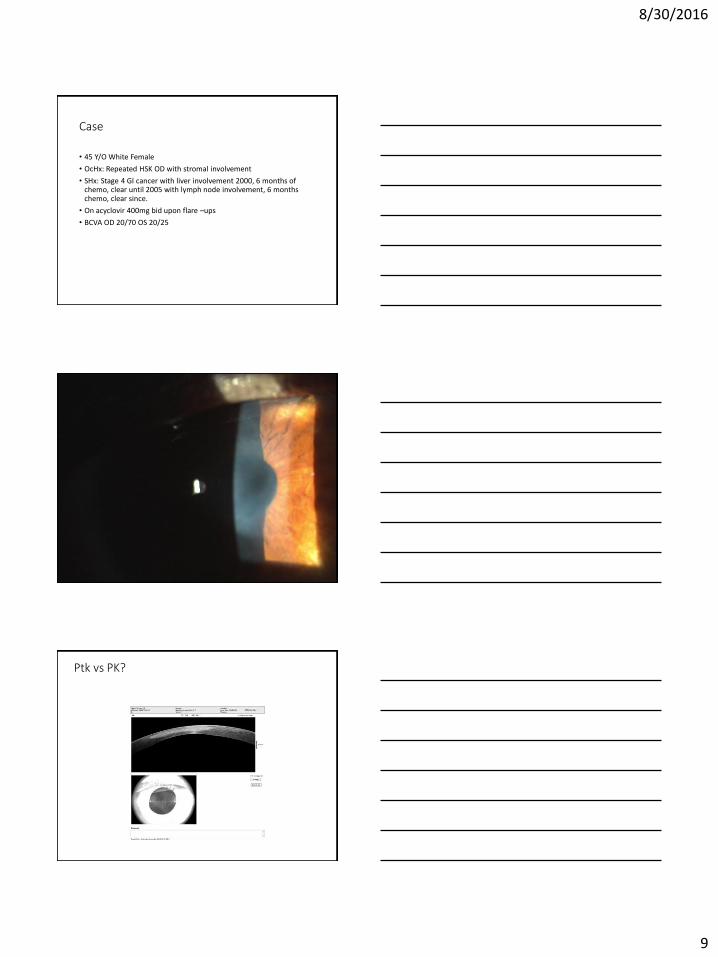
Case
• 45 Y/O White Female
• OcHx: Repeated HSK OD with stromal involvement
• SHx: Stage 4 GI cancer with liver involvement 2000, 6 months of chemo, clear until 2005 with lymph node involvement, 6 months chemo, clear since.
• On acyclovir 400mg bid upon flare –ups
• BCVA OD 20/70 OS 20/25
Ptk vs PK?
8/30/2016
10
Case
• 45 Y/O WM
• Hx of worsening keratoconus OU with occasional hydrops OD
• Attempted to send for corneal crosslinking, pt declines
• OD contact lens uncomfortable with inconsistent vision
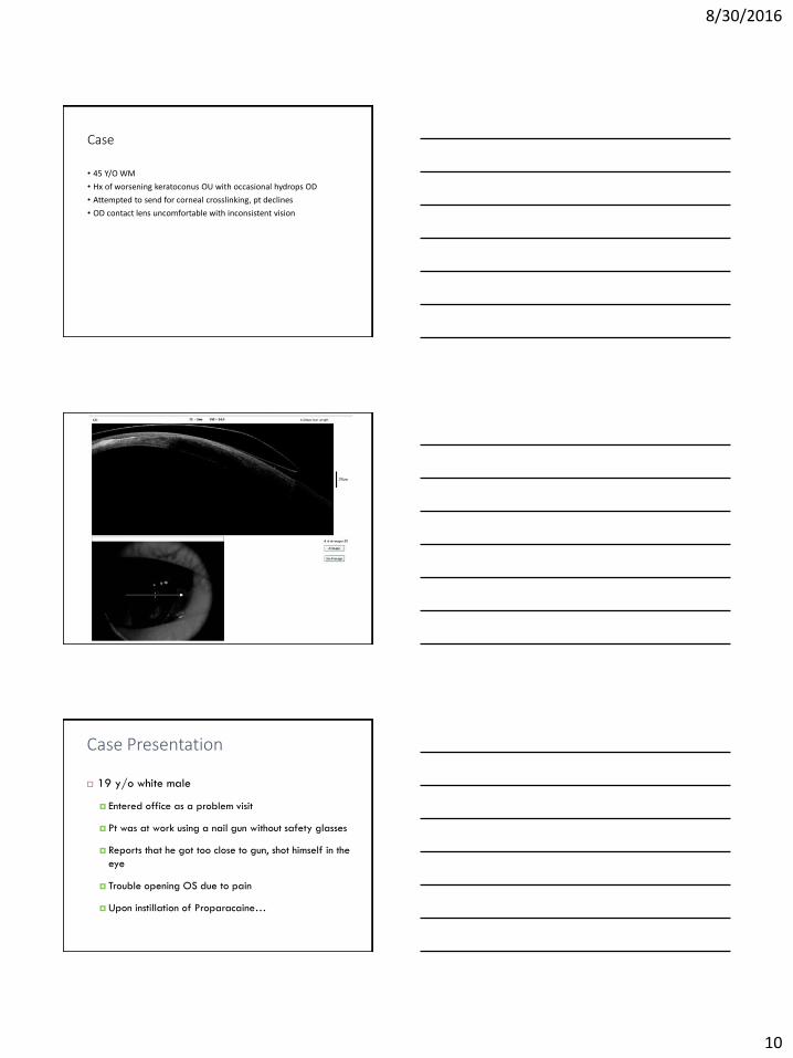
Case Presentation
19 y/o white male
Entered office as a problem visit
Pt was at work using a nail gun without safety glasses
Reports that he got too close to gun, shot himself in the
eye
Trouble opening OS due to pain
Upon instillation of Proparacaine…
8/30/2016
11
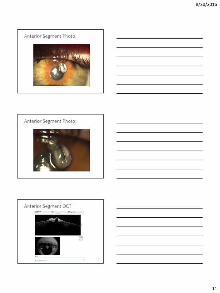
Anterior Segment Photo
Anterior Segment Photo
Anterior Segment OCT
8/30/2016
12
Anterior Segment OCT
Diagnosis/Treatment
Dx: open globe injury due to penetrating
intraocular foreign body
A driver was found and the patient was sent to
tertiary care for immediate surgical repair
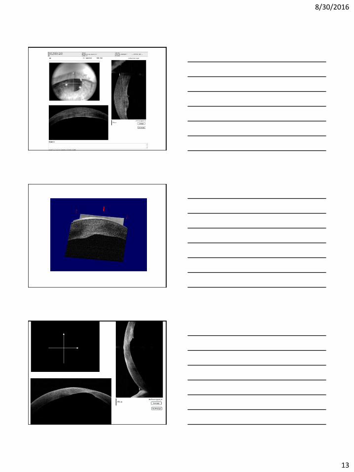
Case
• 60 Y/O WM
• Bilateral Keratoconus
• Cc: Sudden vision loss OD
• BCVA 20/80 OD, 20/40 OS
• Wearing large diameter SoClear Scleral lens
8/30/2016
13
8/30/2016
14
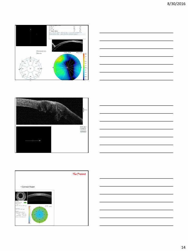
• Corneal Power
The Present
8/30/2016
15
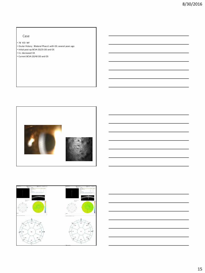
Case
• 78 Y/O WF
• Ocular History: Bilateral Phaco’s with IOL several years ago
• Initial post-op BCVA 20/25 OD and OS
• Cc: decreased VA
• Current BCVA 20/40 OD and OS
8/30/2016
16
Should we be using OCT for determining CCT?
Cornea Rapid Fire
8/30/2016
17
8/30/2016
19
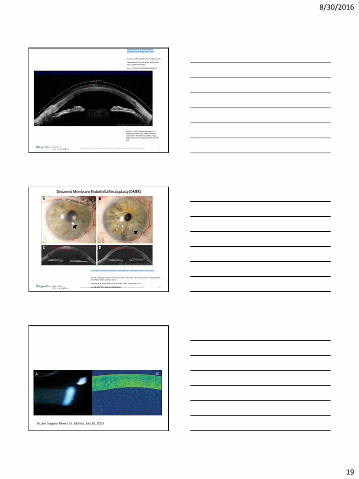
FIGURE 1 This is a cross-sectional OCT image of a scleral lens. Notice that the vault can be directly measured using a caliper tool, in this case, 0.37 mm (or 370 μm).
Copyright © 2013 Optometry & Vision Science. Published by Lippincott Williams & Wilkins. 55
Central Vault in Dry Eye Patients Successfully Wearing Scleral Lens
Sonsino, Jeffrey; Mathe, Dora Sztipanovits
Optometry & Vision Science. 90(9):e248-e251, September 2013.
doi: 10.1097/OPX.0000000000000013
Descemet Membrane Endothelial Keratoplasty (DMEK)
Copyright © 2013 Optometry & Vision Science. Published by Lippincott Williams & Wilkins. 56
Descemet Membrane Endothelial Keratoplasty in Eyes with Glaucoma Implants
Heindl, Ludwig M.; Koch, Konrad R.; Bucher, Franziska; Hos, Deniz; Steven, Philipp; Koch, Hans-Reinhard; Cursiefen, Claus
Optometry & Vision Science. 90(9):e241-e244, September 2013.
doi: 10.1097/OPX.0b013e31829d8e64
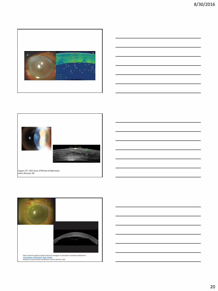
Ocular Surgery News U.S. Edition, July 10, 2013
8/30/2016
20
August 15th, 2013 Issue of Review of OptometryAaron Bronner, OD
Role of anterior segment optical coherence tomogram in Descemet's membrane detachmentSonia Kothari, Kulin Kothari, Rajul S ParikhBombay City Eye Institute and Research Centre, Mumbai, India
8/30/2016
21
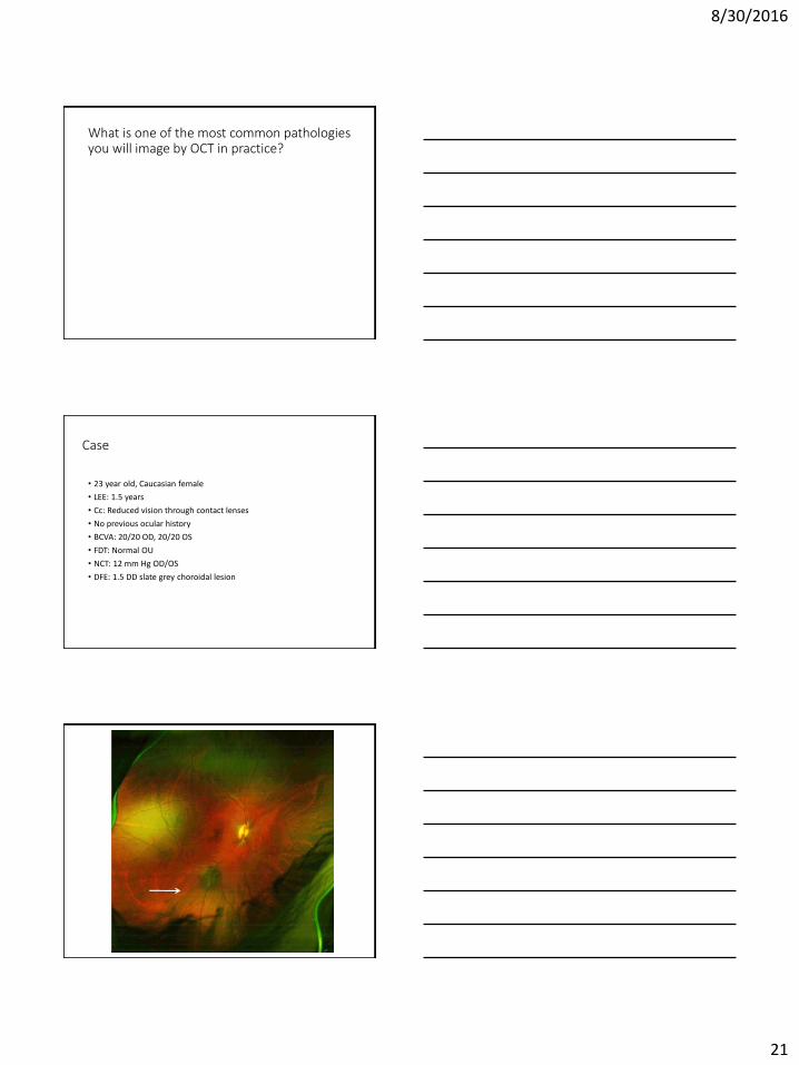
What is one of the most common pathologies you will image by OCT in practice?
Case
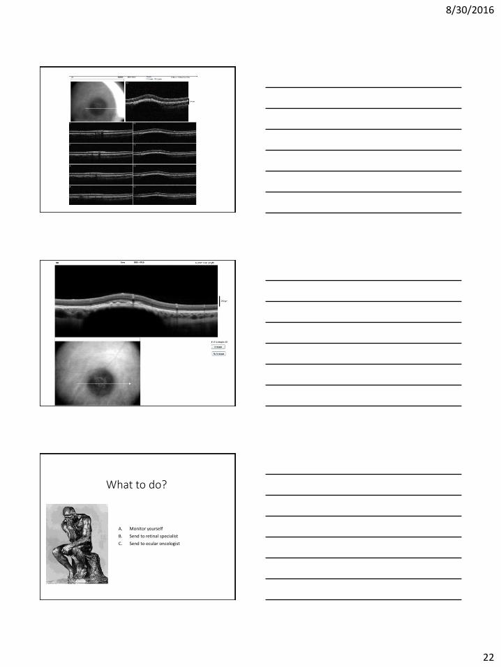
• 23 year old, Caucasian female
• LEE: 1.5 years
• Cc: Reduced vision through contact lenses
• No previous ocular history
• BCVA: 20/20 OD, 20/20 OS
• FDT: Normal OU
• NCT: 12 mm Hg OD/OS
• DFE: 1.5 DD slate grey choroidal lesion
8/30/2016
22
What to do?
A. Monitor yourself
B. Send to retinal specialist
C. Send to ocular oncologist
8/30/2016
23
Ocular Oncology
•Patient referred to Will’s Eye (Drs. Carol and Jerry Shields)
• 1/3/2011
• 2.5 x 2.5 mm
• Thickness: 1.6 mm
• (-) Subretinal fluid, (-) Lipofuscin
• 7/12/2011
• 2.5 x 2.5 mm
• (+) Lipofuscin (auto fluorescence)
• (-) Subretinal fluid
• Return visit to Will’s Eye in one year
• 6/15/2012
• 3mm x 3mm
• Thickness 1.4mm
• Return visit to Wills in one year
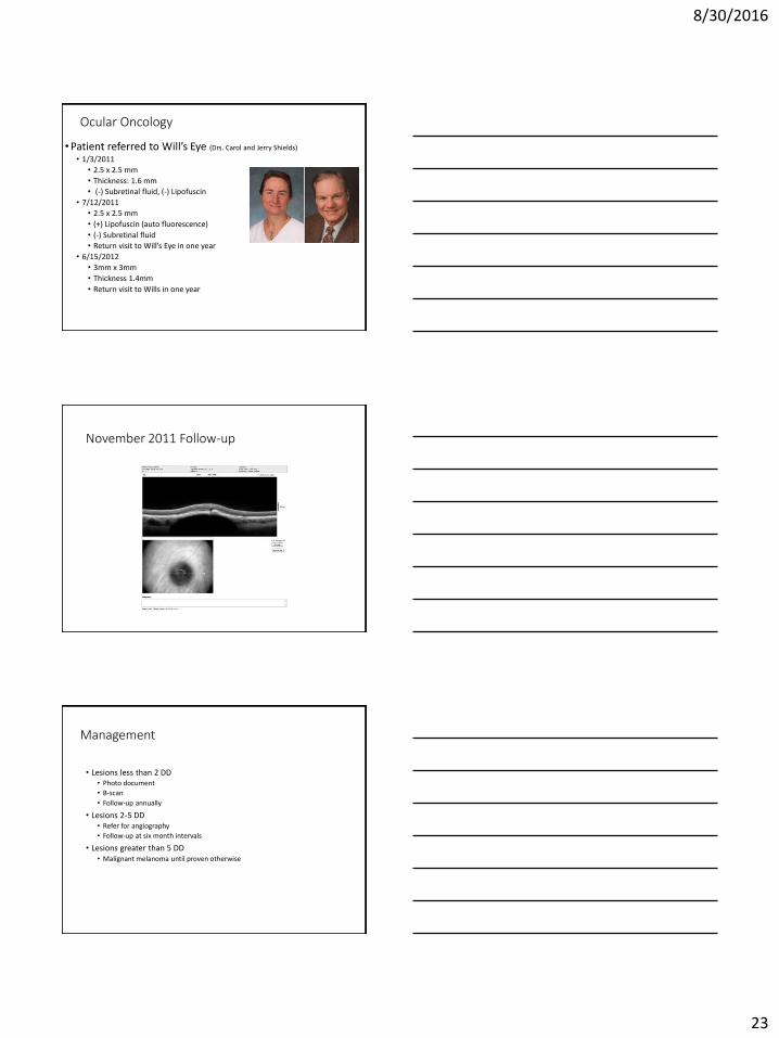
November 2011 Follow-up
Management
• Lesions less than 2 DD• Photo document
• B-scan
• Follow-up annually
• Lesions 2-5 DD• Refer for angiography
• Follow-up at six month intervals
• Lesions greater than 5 DD• Malignant melanoma until proven otherwise
8/30/2016
24
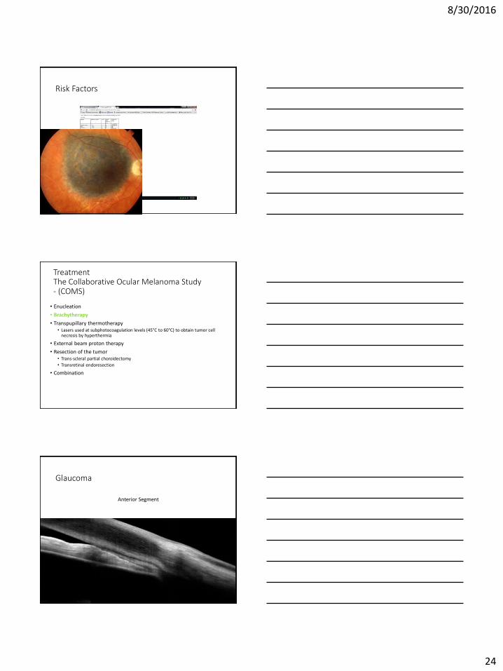
Risk Factors
Treatment The Collaborative Ocular Melanoma Study - (COMS)
• Enucleation
• Brachytherapy
• Transpupillary thermotherapy • Lasers used at subphotocoagulation levels (45°C to 60°C) to obtain tumor cell
necrosis by hyperthermia
• External beam proton therapy
• Resection of the tumor• Trans-scleral partial choroidectomy
• Transretinal endoresection
• Combination
Glaucoma
Anterior Segment
8/30/2016
25
Visante
• Calculates degree of angle
Taken from 2007 Review of Ophthalmology
Case Presentation
• 61 year old white female
• CC: Decreased vision and red eyes
• BCVA: 20/40 OD and 20/20 OS at distance
• IOP’s: 14 OU
• Refractive Status:+1.00-0.50x27
-0.50-1.00x140
• Anterior Segment:
-Cataracts OD>OS
-Blepharitis OU
-Narrow Angles OU (Grade 2 VH, ATM 360 deg. OU by gonio)
• Posterior Segment: Mild RPE mottling OD
Optovue Angle OD

8/30/2016
26
Optovue Angles-OS
Why not just gonio?
Diagnosis
• Narrow anatomical angles OU
• Cataracts OD>OS
• Blepharitis OU
8/30/2016
27
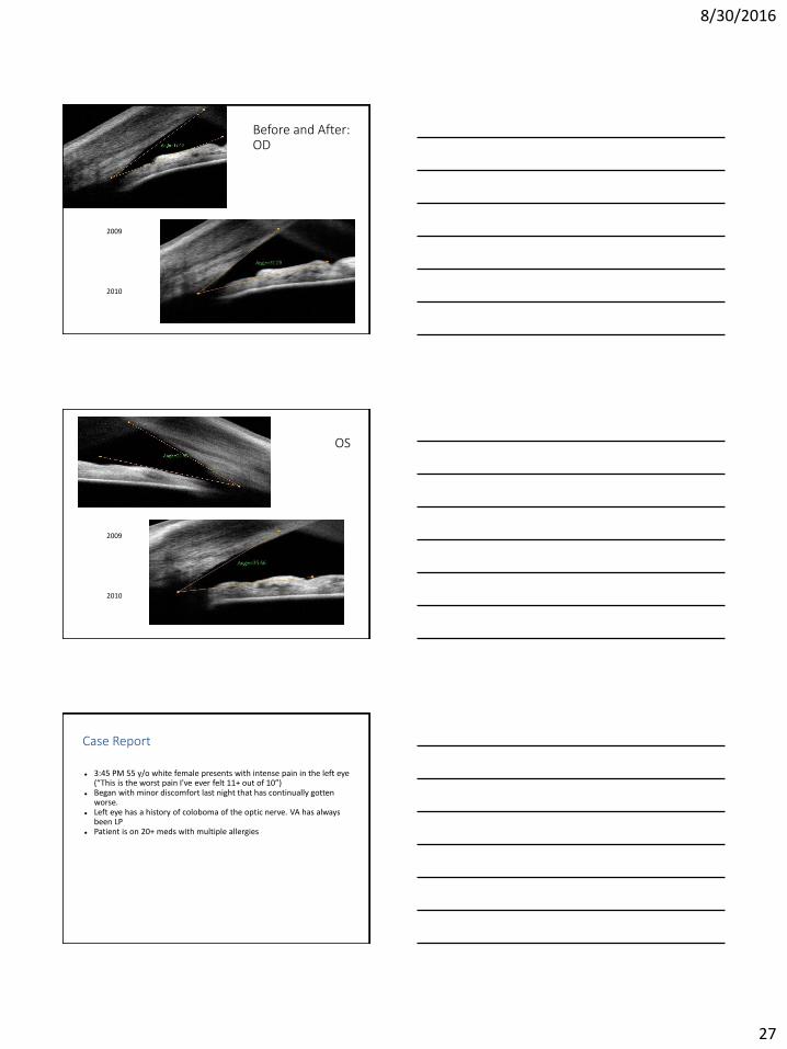
Before and After: OD
2009
2010
OS
2009
2010
Case Report
3:45 PM 55 y/o white female presents with intense pain in the left eye (“This is the worst pain I've ever felt 11+ out of 10”)
Began with minor discomfort last night that has continually gotten worse.
Left eye has a history of coloboma of the optic nerve. VA has always been LP
Patient is on 20+ meds with multiple allergies
8/30/2016
28
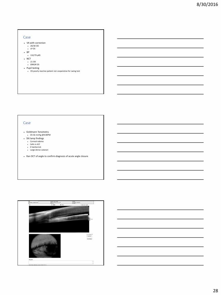
Case
VA with correction 20/30 OD
LP OS
BP 132/79 p85
NCT 21 OD
ERROR OS
Pupil testing OS poorly reactive patient not cooperative for swing test
Case
Goldmann Tonometry OS 56 mmhg @4:00PM
Slit lamp findings Corneal edema
Cells in A/C
0 Vanherrick
Large dense cataract
Ran OCT of angle to confirm diagnosis of acute angle closure
8/30/2016
29
Case
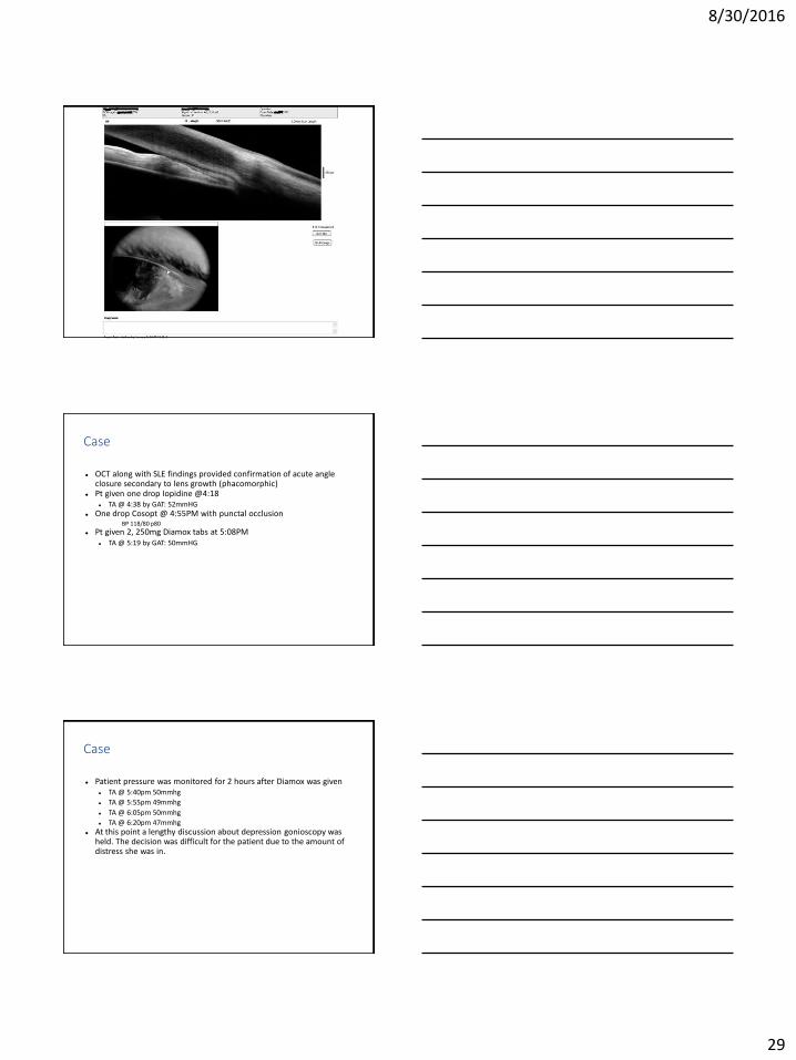
OCT along with SLE findings provided confirmation of acute angle closure secondary to lens growth (phacomorphic)
Pt given one drop Iopidine @4:18 TA @ 4:38 by GAT: 52mmHG
One drop Cosopt @ 4:55PM with punctal occlusion BP 118/80 p80
Pt given 2, 250mg Diamox tabs at 5:08PM TA @ 5:19 by GAT: 50mmHG
Case
Patient pressure was monitored for 2 hours after Diamox was given TA @ 5:40pm 50mmhg
TA @ 5:55pm 49mmhg
TA @ 6:05pm 50mmhg
TA @ 6:20pm 47mmhg At this point a lengthy discussion about depression gonioscopy was
held. The decision was difficult for the patient due to the amount of distress she was in.
8/30/2016
30
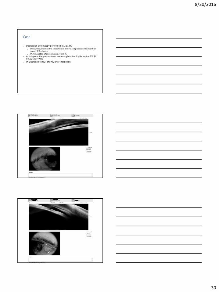
Case
Depression gonioscopy performed at 7:11 PM We saw movement in the apposition on the iris and proceeded to indent for
roughly 1 ½ minutes.
TA immediately after depression 34mmHG At this point the pressure was low enough to instill pilocarpine 2% @
7:14pm???????? Pt was taken to OCT shortly after instillation.
8/30/2016
31
Case
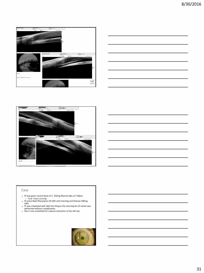
Pt was given second dose of 2- 250mg Diamox tabs at 7:30pm. TA @ 7:52pm 21mmHg
Pt prescribed Pilocarpine 2% QID until morning and Diamox 500mg Q4H
Pt was scheduled with Oph first thing in the morning for LPI which was performed without complication
She is now scheduled for cataract extraction of the left eye.
8/30/2016
32
Case
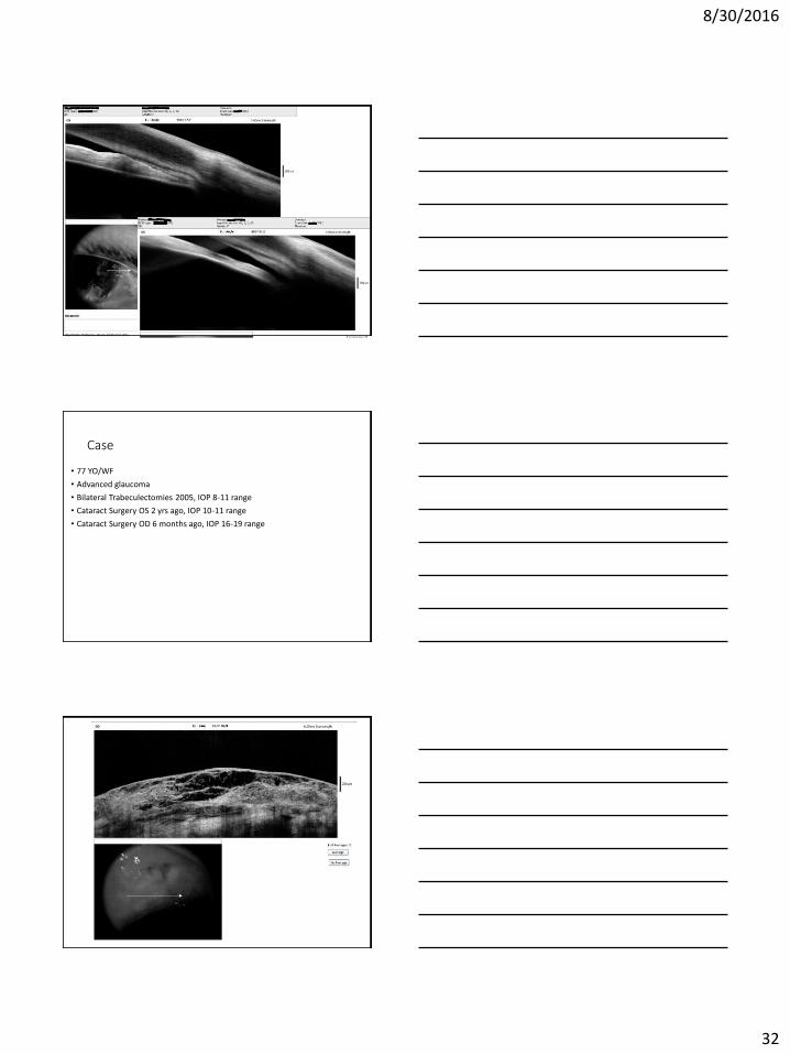
• 77 YO/WF
• Advanced glaucoma
• Bilateral Trabeculectomies 2005, IOP 8-11 range
• Cataract Surgery OS 2 yrs ago, IOP 10-11 range
• Cataract Surgery OD 6 months ago, IOP 16-19 range
8/30/2016
33
8/30/2016
34
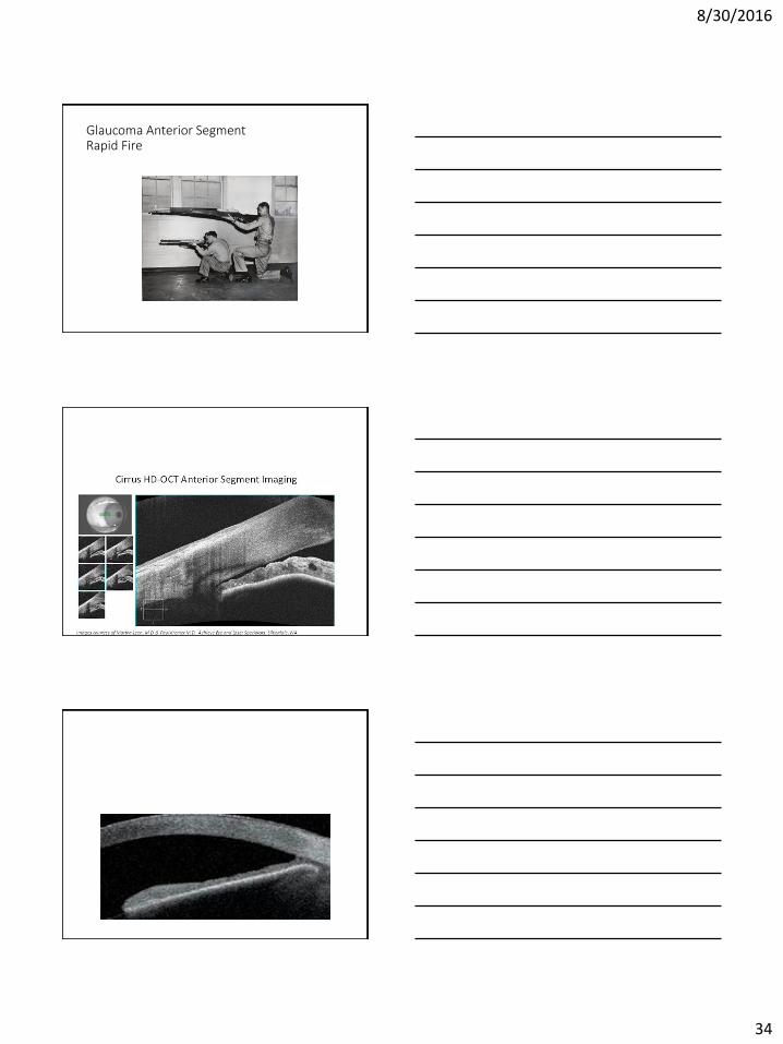
Glaucoma Anterior Segment Rapid Fire
8/30/2016
35
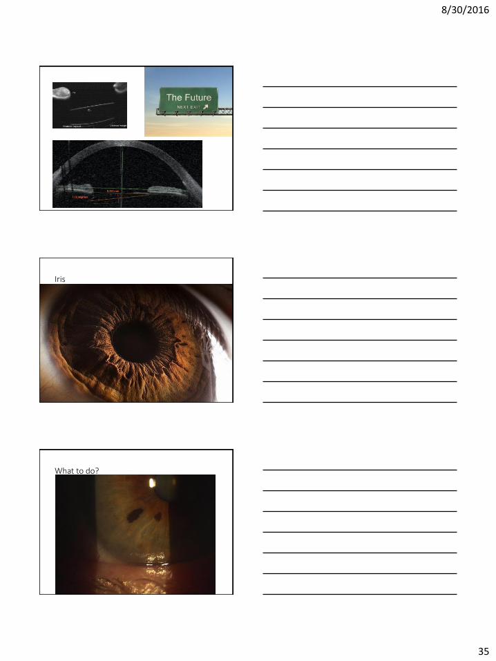
Iris
What to do?
8/30/2016
36
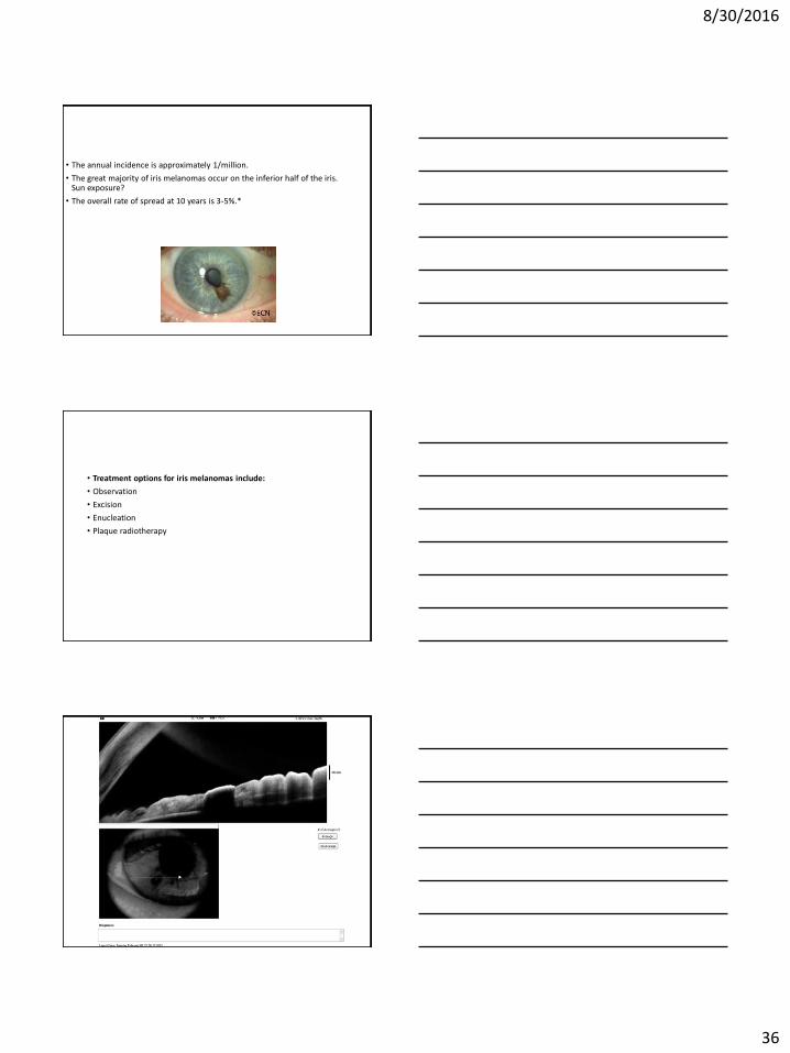
• The annual incidence is approximately 1/million.
• The great majority of iris melanomas occur on the inferior half of the iris. Sun exposure?
• The overall rate of spread at 10 years is 3-5%.*
• Treatment options for iris melanomas include:
• Observation
• Excision
• Enucleation
• Plaque radiotherapy
8/30/2016
37
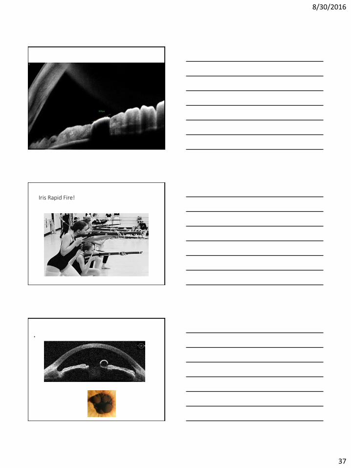
Iris Rapid Fire!
•

8/30/2016
38
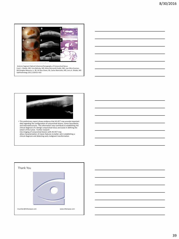
Conjunctiva
8/30/2016
39
Anterior Segment Optical CoherenceTomography of Conjunctival NevusCarol L. Shields, MD, Irina Belinsky, MD, Massi Romanelli-Gobbi, BM, Juan Mica Guzman, MS,Douglas Mazzuca, Jr., BS, W. Ross Green, BS, Carlos Bianciotto, MD, Jerry A. Shields, MD Ophthalmology 2011;118:915–919
• This preliminary report shows evidence that AS-OCT may provide important data regarding the configuration of conjunctival lesions, tumor boundaries, and internalstructures. This information may contribute to establishing the clinical diagnosis of a benign conjunctival nevus and assist in defining the extent of the tumor. Further research into imaging of conjunctival lesions with AS-OCT may allow characterization of classic features to better aid in establishing a clinical diagnosis and detecting early malignant transformation
Thank You
[email protected] www.nittanyeye.com