Embed Size (px)
Citation preview
ApoE-/-hCD1Tg/HJ1Tg/
ApoE-/-hCD1Tg/HJ1Tg/
ApoE+/+
H&E(100x)
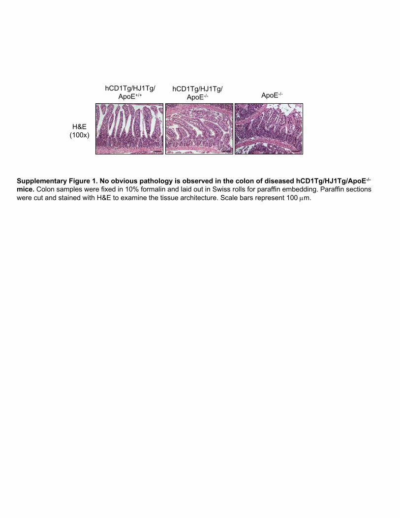
Supplementary Figure 1. No obvious pathology is observed in the colon of diseased hCD1Tg/HJ1Tg/ApoE-/-
mice. Colon samples were fixed in 10% formalin and laid out in Swiss rolls for paraffin embedding. Paraffin sections were cut and stained with H&E to examine the tissue architecture. Scale bars represent 100 µm.
15
10
5
0
2
1
0
Cervical Lymph Node Skin (dermis)
% C
D4+ F
oxp3
+
% C
D4+ F
oxp3
+
hCD1Tg/HJ1Tg/ApoE+/+
hCD1Tg/HJ1Tg/ApoE-/-
ApoE-/-
A B
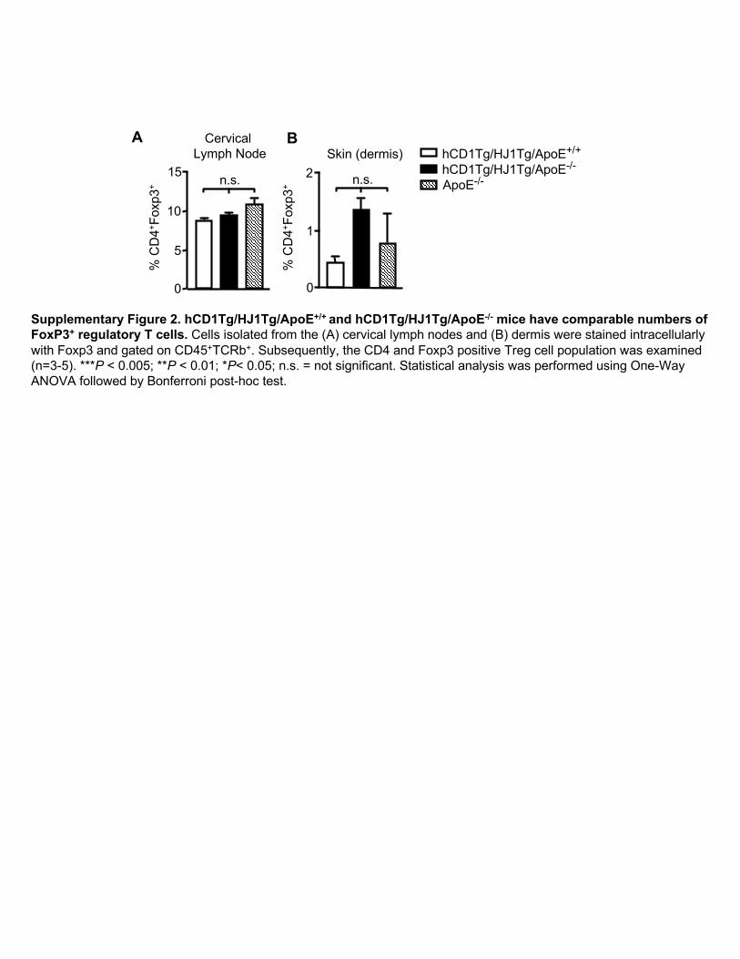
Supplementary Figure 2. hCD1Tg/HJ1Tg/ApoE+/+ and hCD1Tg/HJ1Tg/ApoE-/- mice have comparable numbers of FoxP3+ regulatory T cells. Cells isolated from the (A) cervical lymph nodes and (B) dermis were stained intracellularly with Foxp3 and gated on CD45+TCRb+. Subsequently, the CD4 and Foxp3 positive Treg cell population was examined (n=3-5). ***P < 0.005; **P < 0.01; *P< 0.05; n.s. = not significant. Statistical analysis was performed using One-Way ANOVA followed by Bonferroni post-hoc test.
A B
0.00
6.37
0.15
19.2 5.35
2.49
0.53
6.32
0.00
3.33 4.43
5.92
0.00
0.84
0.00
4.62 3.61
4.43
CD1b
SSC
Gated on CD11b+CD11c+ cells
Gated on CD11b+CD11c- cells
Gated on CD11b-CD11c+ cells
CD
11b
CD11c
ApoE-/-hCD1Tg/HJ1Tg/
ApoE-/-hCD1Tg/HJ1Tg/
ApoE+/+
Dermis
0.890 0.82
CD1b
Gated on CD207+CD11c+ cells
SSC
Epidermis
8.183.38 7.79
CD
207
CD11c
ApoE-/-hCD1Tg/HJ1Tg/
ApoE-/-hCD1Tg/HJ1Tg/
ApoE+/+
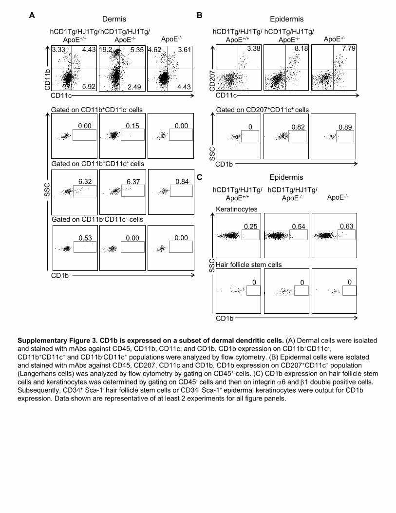
Supplementary Figure 3. CD1b is expressed on a subset of dermal dendritic cells. (A) Dermal cells were isolated and stained with mAbs against CD45, CD11b, CD11c, and CD1b. CD1b expression on CD11b+CD11c-, CD11b+CD11c+ and CD11b-CD11c+ populations were analyzed by flow cytometry. (B) Epidermal cells were isolated and stained with mAbs against CD45, CD207, CD11c and CD1b. CD1b expression on CD207+CD11c+ population (Langerhans cells) was analyzed by flow cytometry by gating on CD45+ cells. (C) CD1b expression on hair follicle stem cells and keratinocytes was determined by gating on CD45- cells and then on integrin a6 and b1 double positive cells. Subsequently, CD34+ Sca-1- hair follicle stem cells or CD34- Sca-1+ epidermal keratinocytes were output for CD1b expression. Data shown are representative of at least 2 experiments for all figure panels.
0.54 0.630.25
00 0
Keratinocytes
Hair follicle stem cells
Epidermis
ApoE-/-hCD1Tg/HJ1Tg/
ApoE-/-hCD1Tg/HJ1Tg/
ApoE+/+
C
CD1b
SSC
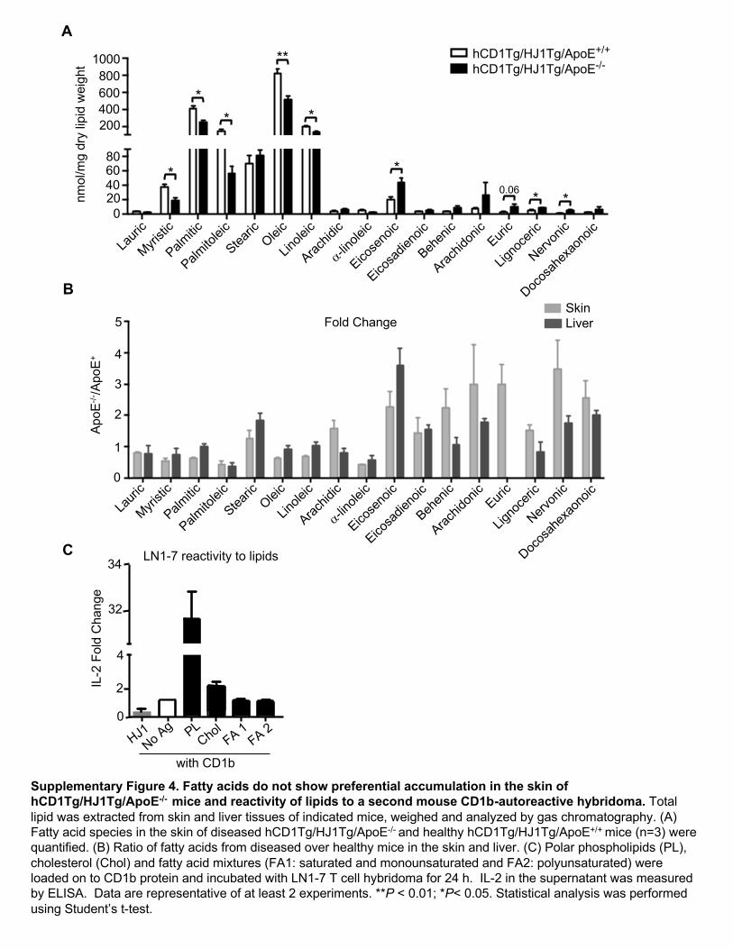
Supplementary Figure 4. Fatty acids do not show preferential accumulation in the skin of hCD1Tg/HJ1Tg/ApoE-/- mice and reactivity of lipids to a second mouse CD1b-autoreactive hybridoma. Total lipid was extracted from skin and liver tissues of indicated mice, weighed and analyzed by gas chromatography. (A) Fatty acid species in the skin of diseased hCD1Tg/HJ1Tg/ApoE-/- and healthy hCD1Tg/HJ1Tg/ApoE+/+ mice (n=3) were quantified. (B) Ratio of fatty acids from diseased over healthy mice in the skin and liver. (C) Polar phospholipids (PL), cholesterol (Chol) and fatty acid mixtures (FA1: saturated and monounsaturated and FA2: polyunsaturated) were loaded on to CD1b protein and incubated with LN1-7 T cell hybridoma for 24 h. IL-2 in the supernatant was measured by ELISA. Data are representative of at least 2 experiments. **P < 0.01; *P< 0.05. Statistical analysis was performed using Student’s t-test.
0
2
4
32
34
with CD1b
LN1-7 reactivity to lipids
IL-2
Fol
d C
hang
e
C Lauric
Acid
Myristi
c Acid
Palmitic
Acid
Palmito
leic A
cid
Stearic
Acid
Oleic A
cid
Linoleic A
cid
Arachidic
Acid
a-Linolen
ic Acid
Eicose
noic Acid
Eicosa
dienoic
Acid
Behen
ic Acid
Arachidonic
Acid
Erucic Acid
Lignoceric
Acid
Nervonic
Acid
Docosa
hexae
noic Acid
0
1
2
3
4
5
Fold
Cha
nge
(Apo
E-/-/A
poE+/
+ )
Fatty acids fold diff
Skin
Liver
SkinLiverFold Change
nmol
/mg
dry
lipid
wei
ght
ApoE
-/-/A
poE+
A
B
Lauri
c
Myristi
c
Palmitic
Palmito
leic
Stearic
Oleic
Linole
ic
Arachid
ic
a-Lino
lenic
Eicose
noic
Eicosa
dieno
ic
Behen
ic
Arachid
onic
Erucic
Ligno
ceric
Nervon
ic
Docos
ahex
aeno
ic0
20406080
200400600800
1000nm
ol/m
g dr
y lip
id w
t.
Fatty acids skin
hCD1Tg+/HJ1Tg+/ApoE+/+
hCD1Tg+/HJ1Tg+/ApoE-/-
1000800600400200
806040200
5
4
3
2
1
0
*
**
**
*
*0.06 * *
hCD1Tg/HJ1Tg/ApoE+/+
hCD1Tg/HJ1Tg/ApoE-/-
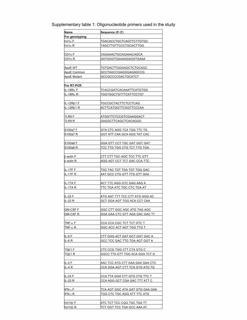
Supplementary table 1: Oligonucleotide primers used in the studyName Sequence (5'-3')For genotypingHJ1a F TGACACCTGCTCAGTTCTTGTGCHJ1a R TAGCTTGTTCCCTGCACTTGG
CD1c F CAGAAACTGCAGAAACAGCACD1c R GATGGGTGAAAAGAGGTGAAA
ApoE WT TGTGACTTGGGAGCTCTGCAGCApoE Common GCCTAGCCGAGGGAGAGCCGApoE Mutant GCCGCCCCGACTGCATCT
For RT-PCRIL-18Ra F TCACCGATCACAAATTCATGTGG IL-18Ra R TGGTGGCTGTTTCATTCCTGT
IL-12Rb1 F TGCCGCTACTTCTCCTCAG IL-12Rb1 R ACTTCATGGTTCGGTTCCCAA
TLR9 F ATGGTTCTCCGTCGAAGGACTTLR9 R GAGGCTTCAGCTCACAGGG
S100a7 F GTA CTC AGG TCA TGG TTC TGS100a7 R GGT ATT CAA GCA AGG TAT CAC
S100a8 F GGA GTT CCT TGC GAT GGT GATS100a8 R TCC TTG TGG CTG TCT TTG TGA
b-actin F CTT CTT TGC AGC TCC TTC GTTb-actin R AGG AGT CCT TCT GAC CCA TTC
IL-17F F TGC TAC TGT TGA TGT TGG GACIL-17F R AAT GCC CTG GTT TTG GTT GAA
IL-17A F ACT TTC AGG GTC GAG AAG AIL-17A R TTC TGA ATC TGC CTC TGA AT
IL-22 F ATG AGT TTT TCC CTT ATG GGG ACIL-22 R GCT GGA AGT TGG ACA CCT CAA
GM-CSF F GGC CTT GGC AGC ATG TAG AGCGM-CSF R GGA GAA CTC GTT AGA GAC GAC TT
TNF-a F CCA CCA CGC TCT TCT GTC TTNF-a R GGC ACC ACT AGT TGG TTG T
IL-6 F CTT GGG ACT GAT GCT GGT GAC AIL-6 R GCC TCC GAC TTG TGA AGT GGT A
TGb1 F CTC CCG TGG CTT CTA GTG CTGb1 R GGCC TTA GTT TGG ACA GGA TCT G
IL-4 F AAC TCC ATG CTT GAA GAA GAA CTCIL-4 R CCA GGA AGT CTT TCA GTG ATG TG
IL-23 F CCA TTA GGA CTT GTG CTG TTC TIL-23 R CCA AGG GCT CGA GAC TTT ATT C
IFN-g F TCA AGT GGC ATA GAT GTG GAA GAAIFN-g R TGG CTC TGC AGG ATT TTC ATG
HJ1Vb F ATC TCT TCC CGG TGC TGA TTHJ1Vb R TCT GGT TCC TGA GCC AAA AT