Embed Size (px)
Citation preview

Handbook of Experimental Pharmacology 240
Harpreet SinghShey-Shing Sheu Editors
Pharmacology of Mitochondria

Handbook of Experimental Pharmacology
Volume 240
Editor-in-Chief
J.E. Barrett, Philadelphia
Editorial Board
V. Flockerzi, Homburg
M.A. Frohman, Stony Brook, NY
P. Geppetti, Florence
F.B. Hofmann, Munchen
M.C. Michel, Mainz
C.P. Page, London
W. Rosenthal, Jena
K. Wang, Beijing

More information about this series at http://www.springer.com/series/164
Harpreet Singh • Shey-Shing SheuEditors
Pharmacologyof Mitochondria

EditorsHarpreet SinghDepartment of Pharmacology and PhysiologyDrexel University College of MedicinePhiladelphia, PennsylvaniaUSA
Shey-Shing SheuDepartment of MedicineThomas Jefferson UniversityPhiladelphia, PennsylvaniaUSA
ISSN 0171-2004 ISSN 1865-0325 (electronic)Handbook of Experimental PharmacologyISBN 978-3-319-57311-3 ISBN 978-3-319-57313-7 (eBook)DOI 10.1007/978-3-319-57313-7
Library of Congress Control Number: 2017940040
# Springer International Publishing AG 2017This work is subject to copyright. All rights are reserved by the Publisher, whether the whole or part ofthe material is concerned, specifically the rights of translation, reprinting, reuse of illustrations,recitation, broadcasting, reproduction on microfilms or in any other physical way, and transmissionor information storage and retrieval, electronic adaptation, computer software, or by similar ordissimilar methodology now known or hereafter developed.The use of general descriptive names, registered names, trademarks, service marks, etc. in thispublication does not imply, even in the absence of a specific statement, that such names are exemptfrom the relevant protective laws and regulations and therefore free for general use.The publisher, the authors and the editors are safe to assume that the advice and information in thisbook are believed to be true and accurate at the date of publication. Neither the publisher nor theauthors or the editors give a warranty, express or implied, with respect to the material containedherein or for any errors or omissions that may have been made. The publisher remains neutral withregard to jurisdictional claims in published maps and institutional affiliations.
Printed on acid-free paper
This Springer imprint is published by Springer NatureThe registered company is Springer International Publishing AGThe registered company address is: Gewerbestrasse 11, 6330 Cham, Switzerland
Preface
In 1870, Eduard Pfluger demonstrated that respiration takes place in cells and
around a full century ago, in 1910, Kingsbury postulated that respiration takes
place in “mitochondria.” However, Carl Benda coined the term “mitochondria”
(thread granules) in 1898 to describe these ubiquitous cellular structures. After the
initial discovery of mitochondria, the respiratory chain was conceptualized by
Keilin, Warburg, Hartree, and others in between the 1910s and 1930s. From the
1920s to 1950s, key aspects of aerobic metabolism were elucidated. The
chemiosmosis hypothesis for ATP generation was then proposed by Peter Mitchell
in 1961. This era was followed by mitochondrial isolation and the publication of
first high-resolution images of mitochondria by George Palade and Fritiof
Sjostrand. In the 1960s and 1970s, much of the focus remained on ATP synthesis,
electron transport chain (ETC), and anion translocators. With the sequence of the
human mitochondrial genome in 1981, a new era of mitochondrial biology was
initiated resulting in the recognition of mitochondrial inheritance. In 1998, the first
mitochondrial proteome was published, and eventually by 2008, the complete
catalog of mitochondrial proteins, “MitoCarta,” was unveiled. The ever growing
interest in mitochondria and pharmacology, as well as the physiology associated
with them, is widely discussed in several scientific meetings and literature.
Classically, mitochondria were defined as the key regulators of cellular energy.
However, now it is universally accepted that they are dynamic, interconnected, and
integrated with other cellular organelles. In addition to their role in the generation
of ATP by oxidative phosphorylation, mitochondria are involved in controlling cell
metabolism and are key players in cellular calcium signaling, free radical homeo-
stasis, lipid transport and biosynthesis, apoptosis, cell cycle and differentiation, and
cellular aging. They also participate in retrograde signaling with nucleus and cross
talk with endoplasmic reticulum either directly or via mitochondria-associated
membranes. Pharmacology of Mitochondria, the focus of this handbook, is a
unique effort by leading experts and researchers to assimilate information on
pharmacology and physiology of mitochondria. Topics covered in the handbook
expand beyond canonical roles assigned to mitochondria. The information
presented will be highly useful for mitochondrial biologists and researchers work-
ing in physiology, medicine, and plant sciences.
v
In the Handbook of Pharmacology, information is delineated from mitochon-
drial genetics to mitochondrial diseases and physiology. Even though mitochondria
possess their own genes, the majority of proteins imported into mitochondria are
encoded by nuclear DNA and a chapter in our book titled “Nuclear Transcription
Factors in the Mitochondria: A New Paradigm in Fine-Tuning Mitochondrial
Metabolism” provides an overview of nuclear-encoded factors. Ionic homeostasis
plays a major role in maintaining the structural and functional integrity of
mitochondria. The majority of the proteins involved in maintaining the mitochon-
drial ionic homeostasis are encoded by the nuclear DNA; however, information on
these proteins responsible for transporting ions is still in its infancy. Several ion
channels and transporters involved in mitochondrial ionic homeostasis have been
shown to present by either pharmacological or genetic approaches. In this hand-
book, chapters titled “The Mitochondrial Permeability Transition Pore and ATP
Synthase”; “The Roles of Mitochondrial Cation Channels Under Physiological
Conditions and in Cancer”; “Anion Channels of Mitochondria”; “Guide to the
Pharmacology of Mitochondrial Potassium Channels”; and “The Mitochondrial
Ca2+ Uniporter: Structure, Function, and Pharmacology” provide an overview of
different classes of ion channels and transporters present in mitochondria, pharma-
cology associated with them, and their roles in diseases.
Mitochondrial structural and functional dynamics are associated with human
physiological and pathological conditions. To maintain the structural and functional
integrity of mitochondria, they continuously undergo fission, fusion, and traffick-
ing, regulate lipid transportations, modulate reactive oxygen species production,
and regulate cellular metabolism. Any deviation or abnormality from mitochondrial
structural and functional integrity can result in human pathological conditions and
diseases. Several chapters titled “Mitochondrial Fission in Human Diseases”;
“Mitochondrial Cholesterol and the Paradox in Cell Death”; “Mitochondrial
Changes in Cancer”; “The Emerging Role of Mitochondrial Targeting in Kidney
Disease”; “Mitochondrial Dynamics as a Therapeutic Target for Treating Cardiac
Diseases”; “Mitochondria in Alzheimer’s Disease and Diabetes-Associated
Neurodegeneration: License to Heal!”; “Leber Hereditary Optic Neuropathy: A
Mitochondrial Disease Unique in Many Ways”; and “Leber Hereditary Optic
Neuropathy: Exemplar of an mtDNA Disease” summarize the association of mito-
chondrial structural and functional integrity with human diseases.
Pharmacologically, mitochondria have undergone a renaissance in the last two
decades. Several novel approaches at pharmacological and genetic levels have been
incorporated to the mitochondrial medicine. The recent first live birth of three-
parent baby carrying mitochondria from donor mother is the spectacular break-
through in treating mitochondrial diseases. However, several new approaches and
treatments are required to treat mitochondrial diseases and disorders. Chapters
discussing new as well as existing therapeutic approaches in this handbook
“Mitochondria-Targeted Agents: Mitochondriotropics, Mitochondriotoxics, and
Mitocans”; “Mitochondrial Flashes: Elemental Signaling Events in Eukaryotic
Cells”; “Role of Mitochondrial Reactive Oxygen Species in the Activation of
Cellular Signals, Molecules, and Function”; “MITO-Porter for Mitochondrial
vi Preface
Delivery and Mitochondrial Functional Analysis”; and “Toxicity of Antiepileptic
Drugs to Mitochondria” provide highly useful overview on mitochondria in phar-
macology. In the current era the amount of data generated by multidisciplinary
approaches on a daily basis, there is an urgent need of integrating the big data to
derive useful information and a chapter focused on understanding the complexity of
mitochondrial phenome titled “Equipping Physiologists with an Informatics Tool
Chest: Toward an Integrated Mitochondrial Phenome” provides an excellent plat-
form for the same.
The authors and editors of the Pharmacology of Mitochondria hope that the
chapters presented herein will provide extremely beneficial and inspiration infor-
mation to mitochondrial enthusiasts, researchers, students, and clinicians. The
chapters contributed by leading mitochondrial researchers in the Handbook ofPharmacology will take us through the novel pharmacological strategies via
mitochondria to understand their physiological and pathological role as well as
present them as therapeutic targets. We hope that the handbook will motivate the
current and new generation of researchers to pursue the unanswered questions and
understand the pharmacological and physiological implications of this fascinating
and complicated organelle, “mitochondrion.”
Philadelphia, PA, USA Harpreet Singh
Shey-Shing Sheu
Preface vii
Contents
Part I Mitochondrial Genetics and Ion Transportation
Nuclear Transcription Factors in the Mitochondria: A New Paradigm
in Fine-Tuning Mitochondrial Metabolism . . . . . . . . . . . . . . . . . . . . . . 3
Naresh Babu V. Sepuri, Prasad Tammineni, Fareed Mohammed,
and Arunkumar Paripati
The Mitochondrial Permeability Transition Pore and ATP Synthase . . . 21
Gisela Beutner, Kambiz N. Alavian, Elizabeth A. Jonas,
and George A. Porter, Jr
The Roles of Mitochondrial Cation Channels Under Physiological
Conditions and in Cancer . . . . . . . . . . . . . . . . . . . . . . . . . . . . . . . . . . . . 47
Ildiko Szabo and Luigi Leanza
Anion Channels of Mitochondria . . . . . . . . . . . . . . . . . . . . . . . . . . . . . . 71
Devasena Ponnalagu and Harpreet Singh
Guide to the Pharmacology of Mitochondrial Potassium Channels . . . . 103
Bartłomiej Augustynek, Wolfram S. Kunz, and Adam Szewczyk
The Mitochondrial Ca2+ Uniporter: Structure, Function,
and Pharmacology . . . . . . . . . . . . . . . . . . . . . . . . . . . . . . . . . . . . . . . . . 129
Jyotsna Mishra, Bong Sook Jhun, Stephen Hurst, Jin O-Uchi,
Gy€orgy Csordas, and Shey-Shing Sheu
Part II Mitochondria in Health and Diseases
Mitochondrial Fission in Human Diseases . . . . . . . . . . . . . . . . . . . . . . . 159
Madhavika N. Serasinghe and Jerry E. Chipuk
Mitochondrial Cholesterol and the Paradox in Cell Death . . . . . . . . . . . 189
Carmen Garcıa-Ruiz, Vicente Ribas, Anna Baulies,
and Jose C. Fernandez-Checa
Mitochondrial Changes in Cancer . . . . . . . . . . . . . . . . . . . . . . . . . . . . . 211
Shubha Gururaja Rao
ix

The Emerging Role of Mitochondrial Targeting in Kidney Disease . . . . 229
Alfonso Eirin, Amir Lerman, and Lilach O. Lerman
Mitochondrial Dynamics as a Therapeutic Target for Treating Cardiac
Diseases . . . . . . . . . . . . . . . . . . . . . . . . . . . . . . . . . . . . . . . . . . . . . . . . . 251
Sang-Bing Ong and Derek J. Hausenloy
Mitochondria in Alzheimer’s Disease and Diabetes-Associated
Neurodegeneration: License to Heal! . . . . . . . . . . . . . . . . . . . . . . . . . . . 281
Susana M. Cardoso, S�onia C. Correia, Cristina Carvalho,and Paula I. Moreira
Leber Hereditary Optic Neuropathy: A Mitochondrial Disease Unique
in Many Ways . . . . . . . . . . . . . . . . . . . . . . . . . . . . . . . . . . . . . . . . . . . . 309
Rui Bi, Ian Logan, and Yong-Gang Yao
Part III Therapeutic Approaches and Mitochondria
Leber Hereditary Optic Neuropathy: Exemplar of an mtDNA Disease . . . 339
Douglas C. Wallace and Marie T. Lott
Equipping Physiologists with an Informatics Tool Chest: Toward
an Integerated Mitochondrial Phenome . . . . . . . . . . . . . . . . . . . . . . . . . 377
Anders Olav Garlid, Jennifer S. Polson, Keith D. Garlid,
Henning Hermjakob, and Peipei Ping
Mitochondrial Flashes: Elemental Signaling Events
in Eukaryotic Cells . . . . . . . . . . . . . . . . . . . . . . . . . . . . . . . . . . . . . . . . . 403
Gaomin Feng, Beibei Liu, Tingting Hou, Xianhua Wang, and Heping Cheng
Mitochondria-Targeted Agents: Mitochondriotropics,
Mitochondriotoxics, and Mitocans . . . . . . . . . . . . . . . . . . . . . . . . . . . . . 423
Diana Guzman-Villanueva and Volkmar Weissig
Role of Mitochondrial Reactive Oxygen Species in the Activation
of Cellular Signals, Molecules, and Function . . . . . . . . . . . . . . . . . . . . . 439
Hiroko P. Indo, Clare L. Hawkins, Ikuo Nakanishi, Ken-ichiro Matsumoto,
Hirofumi Matsui, Shigeaki Suenaga, Michael J. Davies, Daret K. St Clair,
Toshihiko Ozawa, and Hideyuki J. Majima
MITO-Porter for Mitochondrial Delivery and Mitochondrial
Functional Analysis . . . . . . . . . . . . . . . . . . . . . . . . . . . . . . . . . . . . . . . . 457
Yuma Yamada and Hideyoshi Harashima
Toxicity of Antiepileptic Drugs to Mitochondria . . . . . . . . . . . . . . . . . . 473
Josef Finsterer
x Contents

Erratum to: The Mitochondrial Permeability Transition Pore
and ATP Synthase . . . . . . . . . . . . . . . . . . . . . . . . . . . . . . . . . . . . . . . . . 489
Gisela Beutner, Kambiz N. Alavian, Elizabeth A. Jonas,
and George A. Porter, Jr
Index . . . . . . . . . . . . . . . . . . . . . . . . . . . . . . . . . . . . . . . . . . . . . . . . . . . 491
Contents xi
Nuclear Transcription Factorsin the Mitochondria: A New Paradigmin Fine-Tuning Mitochondrial Metabolism
Naresh Babu V. Sepuri, Prasad Tammineni, Fareed Mohammed,and Arunkumar Paripati
Contents
1 Introduction . . . . . . . . . . . . . . . . . . . . . . . . . . . . . . . . . . . . . . . . . . . . . . . . . . . . . . . . . . . . . . . . . . . . . . . . . . . . . . . . . . . 4
2 Nuclear Hormone Receptors Present in the Mitochondria . . . . . . . . . . . . . . . . . . . . . . . . . . . . . . . . . . 5
2.1 Estrogen Receptor . . . . . . . . . . . . . . . . . . . . . . . . . . . . . . . . . . . . . . . . . . . . . . . . . . . . . . . . . . . . . . . . . . . . . . 5
2.2 Glucocorticoid Receptor . . . . . . . . . . . . . . . . . . . . . . . . . . . . . . . . . . . . . . . . . . . . . . . . . . . . . . . . . . . . . . . 6
2.3 Thyroid Hormone Receptor . . . . . . . . . . . . . . . . . . . . . . . . . . . . . . . . . . . . . . . . . . . . . . . . . . . . . . . . . . . . 6
3 Nuclear Transcription Factors Associated with Mitochondria . . . . . . . . . . . . . . . . . . . . . . . . . . . . . . 8
3.1 Interferon Regulatory Factor 3 . . . . . . . . . . . . . . . . . . . . . . . . . . . . . . . . . . . . . . . . . . . . . . . . . . . . . . . . . 8
3.2 p53 . . . . . . . . . . . . . . . . . . . . . . . . . . . . . . . . . . . . . . . . . . . . . . . . . . . . . . . . . . . . . . . . . . . . . . . . . . . . . . . . . . . . . . 8
3.3 cAMP Response Element-Binding Protein . . . . . . . . . . . . . . . . . . . . . . . . . . . . . . . . . . . . . . . . . . . . 10
3.4 NF-kB . . . . . . . . . . . . . . . . . . . . . . . . . . . . . . . . . . . . . . . . . . . . . . . . . . . . . . . . . . . . . . . . . . . . . . . . . . . . . . . . . . . 10
3.5 Signal Transducer and Activator of Transcription . . . . . . . . . . . . . . . . . . . . . . . . . . . . . . . . . . . . 11
3.6 Myocyte-Specific Enhancer Factor-2D . . . . . . . . . . . . . . . . . . . . . . . . . . . . . . . . . . . . . . . . . . . . . . . . 12
4 Mechanistic Insights into the Mitochondrial Transport of Nuclear Receptors
and Transcription Factors . . . . . . . . . . . . . . . . . . . . . . . . . . . . . . . . . . . . . . . . . . . . . . . . . . . . . . . . . . . . . . . . . . . . 12
5 Conclusions and Future Perspectives . . . . . . . . . . . . . . . . . . . . . . . . . . . . . . . . . . . . . . . . . . . . . . . . . . . . . . . . 14
References . . . . . . . . . . . . . . . . . . . . . . . . . . . . . . . . . . . . . . . . . . . . . . . . . . . . . . . . . . . . . . . . . . . . . . . . . . . . . . . . . . . . . . . . 16
Abstract
Noncanonical functions of several nuclear transcription factors in the
mitochondria have been gaining exceptional traction over the years. These
transcription factors include nuclear hormone receptors like estrogen, glucocor-
ticoid, and thyroid hormone receptors: p53, IRF3, STAT3, STAT5, CREB,
NF-kB, and MEF-2D. Mitochondria-localized nuclear transcription factors reg-
ulate mitochondrial processes like apoptosis, respiration and mitochondrial
transcription albeit being nuclear in origin and having nuclear functions.
N.B.V. Sepuri (*) • P. Tammineni • F. Mohammed • A. Paripati
Department of Biochemistry, School of Life Sciences, University of Hyderabad, Telangana
500046, India
e-mail: [email protected]
# Springer International Publishing Switzerland 2016
H. Singh, S.-S. Sheu (eds.), Pharmacology of Mitochondria,Handbook of Experimental Pharmacology 240, DOI 10.1007/164_2016_3
3
Hence, the cell permits these multi-stationed transcription factors to orchestrate
and fine-tune cellular metabolism at various levels of operation. Despite their
ubiquitous distribution in different subcompartments of mitochondria, their
targeting mechanism is poorly understood. Here, we review the current status
of mitochondria-localized transcription factors and discuss the possible targeting
mechanism besides the functional interplay between these factors.
Keywords
Metabolism • Mitochondria • Nuclear receptors and transcription factors •
Protein import • Protein targeting and signaling
1 Introduction
Mitochondria are crucial organelles involved in various cellular processes. Besides
the production of ATP for cellular needs, they are also participating in the metabo-
lism of fatty acids and amino acids. Also, mitochondria play a significant role in
apoptosis by integrating the extracellular cues with intracellular signaling
pathways. Further, the involvement of mitochondria in cell proliferation, motility,
and ion homeostasis is well documented (McBride et al. 2006). Thus, it’s not
surprising that mitochondrial dysfunction is often associated with various disease
conditions like cancer and neurodegenerative disorders. Paradoxically, mitochon-
drial DNA encodes only for a handful of proteins. A majority of mitochondrial
proteins are nuclear encoded. Mitochondria, though, have a handful of proteins
encoded by mitochondrial DNA to execute all these functions; it depends on a
majority of nuclear-encoded proteins for its optimal function. These nuclear-
encoded mitochondrial proteins are synthesized on cytosolic ribosomes and
transported to the mitochondria through mitochondrial protein import machinery
(Mokranjac and Neupert 2009). Therefore, the mitochondria and nucleus act in
concert with each other to mount an appropriate response toward extracellular
stimuli. For example, nuclear transcription factors and coactivators like NRF-1,
NRF-2, and PGC-1 regulate the expression of mitochondrial OXPHOS subunits to
achieve fine-tuning of mitochondrial function toward altered metabolic demands of
the cell (Leigh-Brown et al. 2010; Scarpulla et al. 2012). For this reason, consider-
able interest has been gained toward understating the role of nuclear receptors,
transcription factors, and other signaling proteins in the establishment of harmonic
equilibrium between these two organelles.
Nuclear receptors and transcription factors are activated in response to growth
factors and cytokines to regulate the gene expression in the nucleus. Some of these
transcription factors control mitochondrial function indirectly by regulating the
expression of mitochondria-associated protein factors. On the other hand, increas-
ing evidence suggests that some of the nuclear receptors and transcription factors
are also present in mitochondria to potentially influence various mitochondrial
functions (Szczepanek et al. 2012). For example, glucocorticoids and other tran-
scription factors regulate mitochondrial transcription, while p53 and IRF3 regulate
apoptosis and respiration. Also, p53 and STAT3 are known to influence the opening
4 N.B.V. Sepuri et al.
of mitochondrial permeability transition and also affect ROS generation from the
mitochondrial OXPHOS system (Szczepanek et al. 2012). Sufficiently reasonable
attempts have been made to understand the role of various mitochondrial transcrip-
tion factors. However, information on their mitochondrial targeting is lagging
behind as the nuclear transcription factors entering the mitochondria lack the
canonical mitochondrial targeting sequence.
In this review, we will provide an outline of various transcription factors
associated with mitochondria and discuss how their submitochondrial localization
contributes to their overall biological function. Also, we will summarize the
potential mechanism of their transport to mitochondria and involvement of these
mitochondrial transcription factors in retrograde signaling and functional interplay
between these factors.
2 Nuclear Hormone Receptors Present in the Mitochondria
2.1 Estrogen Receptor
Estrogen receptors (ESRs), ESR1 and ESR2, belong to the nuclear receptor super-
family of transcription factors responsible for estrogen-mediated transcriptional
induction of genes in reproductive and nonreproductive tissues. ESR1 and ESR2
are highly homologous proteins except in the DNA-binding domains that render
ESR2 to be a poor transcription factor when compared to ESR1. Noncanonical
functions of estrogen receptors in the cytosol, endoplasmic reticulum, and plasma
membrane have been well documented. ESR1, ESR2, and its isoform ERβ2 have
been found to be present in various tissues and cell lines (Cammarata et al. 2004;
Chen et al. 2004; Milner et al. 2005; Solakidi et al. 2005). Mitochondria-localized
ESR plays a significant role in apoptosis and reduces ROS by activating manganese
superoxide dismutase (MnSOD) enzyme. When cancer cells are irradiated with
UV, it induces mROS species generation and activation of c-Jun N-terminal kinase
(JNK), and protein kinase C (PKC) δ. This triggers apoptotic cell death. Estradiol(E2) inhibits all these events, by directly activating manganese superoxide
dismutase (Pedram et al. 2006). Mitochondria-localized ESR2 has also been
shown to suppress apoptosis induced by cisplatin in non-small cell lung cancer
cells. ESR interacts with Bad and inhibits translocation of Bax to mitochondria (Xie
et al. 2015). It also has been shown that downregulation of ESR2 enhances Bax
activation and translocation to mitochondria in a ligand-independent manner (Liang
et al. 2015). Interestingly, in ovarian cancer, it was observed that cytoplasmic ERβ2is associated with lower apoptotic rate, whereas mitochondria-localized ERβ2inhibits apoptosis by interacting with Bad protein (Ciucci et al. 2015). Recently,
it has been shown that ESR1 affects beta-oxidation in mitochondria by directly
interacting with beta subunit of HADHB (hydroxyacyl-CoA dehydrogenase/3-
ketoacyl-CoA thiolase/enoyl-CoA hydratase) (Zhou et al. 2012). In addition to
these transcription factors, recent reports also suggest the possible role of G-
protein-coupled estrogen receptor 1 (GPER1) in modulating the opening of
mitochondrial permeability transition pore via phosphorylation of ERK1/ERK2/
Nuclear Transcription Factors in the Mitochondria: A New Paradigm in Fine. . . 5

GSK-3β (Bopassa et al. 2010; Kabir et al. 2015). However, further studies are
needed to understand the mechanistic details of GPER1 action on mitochondria.
2.2 Glucocorticoid Receptor
GRs are expressed in all tissues and cell types and regulate cell metabolism,
immune response, and development in response to glucocorticoids such as cortisol.
GRs are the first nuclear receptors reported to be present in mitochondria. Mobili-
zation of GR from the cytoplasm to mitochondria has been observed when rats were
administered with dexamethasone. However, the precise mechanism of this trans-
location is not clear (Demonacos et al. 1993). GR has been detected in mitochondria
of HeLa cells, cytoplasm, and mitochondria of rat brain synaptosomes (Moutsatsou
et al. 2001; Scheller et al. 2000). GR binds to glucocorticoid response element
(GRE) consensus sequence present on cytochrome c oxidase I and III genes and the
D-loop region of mitochondrial DNA (Demonacos et al. 1995). These studies
indicate that GR present in the mitochondria may be regulating mitochondrial
transcription independent of its nuclear role. Overexpression of mitochondrial GR
increases RNA synthesis, ATP production, and cytochrome c oxidase I protein
(Psarra and Sekeris 2011). Further, it has been shown that GR forms a complex with
Bcl-2 before its translocation into mitochondria in corticosterone-treated brain
cells. This translocation inhibits the release of cytochrome c and calcium ions
from mitochondria, thereby protecting the neuron (Du et al. 2009). In contrast,
mitochondria-localized GR has been shown to induce apoptosis in dexamethasone-
treated thymocytes where the mitochondrial import of GR correlates with the
release of cytochrome c and activation of caspase 3 (Talaber et al. 2009).
2.3 Thyroid Hormone Receptor
Thyroid hormone receptor is a nuclear receptor that regulates cellular metabolism
and heart rate by activating transcription of genes in response to thyroid hormone. It
has been 3 decades since Sterling and his colleagues discovered the presence of
thyroid hormones in mitochondria (Sterling et al. 1984). Thyroid receptors TRα and
TRβ are encoded by two genes c-erbA-1 and c-erbA-2, respectively. p43 and p28
are alternate translational initiation products of TRα, and these are exclusively
localized to mitochondria. Mitochondria-localized TRα induces the expression of
mitochondrial 12S rRNA by binding to the thyroid hormone response element of
mitochondrial DNA (Morrish et al. 2006). p43 depleted mice exhibits decreased
mitochondrial DNA replication and respiratory chain activities. p43 has also been
implicated in maintaining glucose homeostasis and insulin secretion. It would be
interesting to see the effect of p43 depletion in pancreatic cells and on the mito-
chondrial and nuclear cross talk. Depletion of p43 may cause decreased levels of
Glut2 and Kir62 that may ultimately lead to pancreatic cell dysfunction and
6 N.B.V. Sepuri et al.
lowered insulin secretion (Blanchet et al. 2012). Further, overexpression of p43
induces a shift in skeletal muscle fiber types and causes muscle atrophy during
aging through two muscle-specific E3 ubiquitin ligases, atrogin-1/MAFbx and
MuRF1 (Casas et al. 2009; Casas et al. 2008). This data demonstrates the physio-
logical importance of mitochondrial thyroid hormone receptor. Interestingly,
human dermal fibroblasts overexpressing p43 undergo cellular transformation by
upregulating c-Jun and c-Fos and concomitantly decreasing the expression of tumor
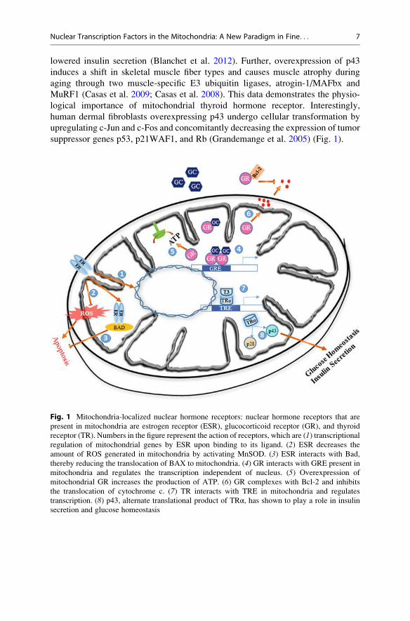
suppressor genes p53, p21WAF1, and Rb (Grandemange et al. 2005) (Fig. 1).
Fig. 1 Mitochondria-localized nuclear hormone receptors: nuclear hormone receptors that are
present in mitochondria are estrogen receptor (ESR), glucocorticoid receptor (GR), and thyroid
receptor (TR). Numbers in the figure represent the action of receptors, which are (1) transcriptionalregulation of mitochondrial genes by ESR upon binding to its ligand. (2) ESR decreases the
amount of ROS generated in mitochondria by activating MnSOD. (3) ESR interacts with Bad,
thereby reducing the translocation of BAX to mitochondria. (4) GR interacts with GRE present in
mitochondria and regulates the transcription independent of nucleus. (5) Overexpression of
mitochondrial GR increases the production of ATP. (6) GR complexes with Bcl-2 and inhibits
the translocation of cytochrome c. (7) TR interacts with TRE in mitochondria and regulates
transcription. (8) p43, alternate translational product of TRα, has shown to play a role in insulin
secretion and glucose homeostasis
Nuclear Transcription Factors in the Mitochondria: A New Paradigm in Fine. . . 7
3 Nuclear Transcription Factors Associatedwith Mitochondria
3.1 Interferon Regulatory Factor 3
IRF3 belongs to an interferon regulatory transcription factor family and is known to
play a major role in innate immune response. Though IRF3 may not be localized to
the mitochondria, it is known to regulate mitochondrial functions in a transcription-
independent manner. In support of this theory, IRF3 mutant’s deficiency in its
transcription activity is still capable of activating apoptosis by releasing cyto-
chrome c from mitochondria (Chattopadhyay et al. 2010). It is well documented
that mitochondria-associated proteins and MAVS are required to orchestrate the
IRF3 nuclear-mediated antiviral signaling and apoptosis. For carrying out its
nuclear and mitochondrial roles, cytosolic IRF3 has to be activated by
mitochondria-associated MAVS through synthetic or viral generated dsRNA-like
vesicular stomatitis viruses. For IRF3 to influence mitochondrial functions, a pool
of cytosolic-activated IRF3 interacts initially with Bax, a pro-apoptotic Bcl2 family
protein through its BH3 domain. This interaction triggers Bax translocation to
mitochondria, and its subsequent oligomerization on the mitochondrial membrane
results in the release of cytochrome c and initiation of apoptosis cascade. Intrigu-
ingly, Bax seems to be also required for phosphorylation and nuclear translocation
of IRF3 (Sharif-Askari et al. 2007). It is not clear whether Bax is directly involved
in the phosphorylation of IRF3. More studies are required to understand if addi-
tional factors are involved in the interaction between phosphorylated IRF3 and Bax
and also the mechanism that prevents the entry of cytosolic IRF3 into nucleus. A
more definitive evidence is required to completely rule out the possibility of IRF3
entry into mitochondria.
3.2 p53
p53 is a tumor suppressor protein that responds to a myriad of stresses that include
oxidative stress, DNA damage, nutrient stress, and ischemia (Zilfou and Lowe
2009). It is believed that p53-induced apoptotic changes were mediated by its
ability to activate transcription of a particular section of genes within the nucleus;
however, recent reports suggest that this ability to induce apoptosis and necrosis is
attributed to its noncanonical role in the mitochondria. Most of the apoptotic
signals, like gamma radiation, oncogenic deregulation, hypoxia, and genotoxic
and oxidative stress, stimulate the translocation of p53 from cytosol to the outer
mitochondrial membranes and to interact with multi-domain Bcl-2 family of
proteins so that it can promote membrane permeabilization and apoptosis (March-
enko et al. 2007). p53 interacts with Bcl-2 family of proteins such as pro-apoptotic
BAX and BAK and anti-apoptotic proteins Bcl-xL and Bcl-2. During stress, p53
competes with anti-apoptotic Mcl1 protein for its interaction with BAK in order to
induce apoptosis (Leu et al. 2004). Similarly, p53 interaction with BAX disrupts the
8 N.B.V. Sepuri et al.
sequestration of BAX by Bcl-xL. This disruption allows for the oligomerization of
BAX on the outer mitochondrial membranes and opening of the permeability
transition pore (PTP) (Chipuk et al. 2004). Unlike other transcription factors that
reside in the mitochondria, the DNA-binding domain of p53 is critical for its
mitochondrial function. In addition to its influence on outer mitochondrial Bcl2
proteins, p53 is also present in the subcompartments of mitochondria. A fraction of
p53 resides in the mitochondrial matrix and interacts with cyclophilin D (CypD).
This interaction facilitates the ROS-mediated opening of PTP and induction of
necrosis. Thus, mitochondria-localized p53 is required for induction of
mitochondria-mediated apoptosis and necrosis. In addition, another fraction of
p53 that is also present in the mitochondrial matrix interacts with mtHSP70,
mtHSP60, mitochondrial DNA polymerase γ, and mtTFA. However, the precise
function of matrix-localized p53 in mitochondrial transcription, DNA binding, and
protein folding is yet to be understood. Matrix-localized p53 has also been shown to
sequester MnSOD to initiate apoptosis (Zhao et al. 2005). It appears that necrosis is
operated when apoptotic process is stalled and necrosis seems to be operated only
under oxidative stress conditions. However, it is not clear whether there is any cross
talk between these two pathways to initiate the death signaling. Elucidation of the
upstream signaling mechanism that determines the translocation of p53 to different
subcompartments of mitochondria will provide valuable insights into p53 function
vis-a-vis its mitochondrial positioning.
Though the mitochondrial translocation mechanism of p53 is not clear, mito-
chondrial p53 is known to regulate the OPA1 processing, mitochondrial dynamics
in cisplatin (cis-diamminedichloroplatinum (II)), and CDDP-induced mitochon-
drial fragmentation (Kong et al. 2014). In response to oxidative stress, p53 interacts
with Drp1, a mitochondrial fission protein before translocating to mitochondria.
Although the mechanism of p53 translocation is yet to be unraveled, there are some
indications of the possible path that p53 might be taking. It has been shown that
Mdm2-dependent mono-ubiquitination of cytosolic p53 triggers p53 translocation
to mitochondria (Marchenko et al. 2007). Drp1 may act as a facilitator for the
Mdm2-dependent mono-ubiquitination of p53 (Guo et al. 2014). The translocated
p53 probably undergoes deubiquitination in a HAUSP-dependent manner to revert
back to a fully functional form on mitochondrial membrane. Curiously, serine
phosphorylated p53 appears to be accumulated in the mitochondrial matrix of
mouse cortical neurons (Pei et al. 2014). This suggests that posttranslational
modifications of p53 may be employed as a signature for the accurate targeting of
p53 into the mitochondrial subcompartments. This possibility is all the more
appealing as p53 is known to undergo different kinds of posttranslational
modifications including methylation and acetylation besides phosphorylation. Phar-
macological drugs and stress conditions can directly modulate mitochondrial trans-
location of p53 without affecting its nuclear translocation (Strom et al. 2006).
Hence, understanding the mechanistic details of p53 translocation to mitochondria
might be useful for novel therapeutic interventions as p53 regulates tumor devel-
opment and ischemic reperfusion injury.
Nuclear Transcription Factors in the Mitochondria: A New Paradigm in Fine. . . 9

3.3 cAMP Response Element-Binding Protein
The cAMP response element-binding (CREB) protein is a transcriptional factor
known to be involved in synaptic transmission and neuron survival. CREB is
activated by a set of kinases such as cyclic AMP-dependent protein kinase A
(PKA), extracellular regulated kinases (ERKs), and calcium-activated calmodulin
kinases (CaMKs) (Altarejos and Montminy 2011). CREB activates transcription
upon binding to cAMP-responsive elements present in the upstream region of target
genes. Although CREB does not contain any mitochondrial localization signals, it
has been shown that CREB gets imported into mitochondria through the TOM
complex in a membrane potential-dependent manner with the aid of mitochondrial
matrix-residing heat shock protein, mtHSP70. mtHSP70 is known to be involved in
the unfolding and translocation of proteins across the mitochondrial inner mem-
brane (Lee et al. 2005). In support of CREB’s movement to mitochondria, CREB-
binding sites have been detected on the D-loop of mitochondrial genome by
chromatin immunoprecipitation (ChIP) assay (Marinov et al. 2014). Mitochondrial
depletion of CREB has shown to decrease the expression of several mitochondria-
encoded RNAs of complex I with a concomitant reduction in the complex I activity
(Lee et al. 2005).
Intriguingly, recent reports have suggested the presence of phosphorylated
CREB in mitochondria similar to phosphorylated p53. It is possible that
mitochondria choose a similar mechanism for import of both CREB and p53.
CREB has also been implicated in the regulation of neuronal survival by regulating
mitochondrial gene expression in response to various stimuli in a cell (De Rasmo
et al. 2009). Besides the aforementioned functions of CREB in the mitochondria, it
also appears to be responsible for pathophysiology of Huntington disease
(HD) (Bogdanov et al. 1998; Lee et al. 2005). In addition, CREB is also known
to be involved in the regulation of cell death. CREB binds to the CRE element
present in the promoter of Bcl-2 gene to induce Bcl-2 overexpression that causes
inhibition of apoptosis (Wilson et al. 1996). Hence, CREB multitasks to contribute
not only to the activation of a subset of nuclear genes but also in the regulation of
apoptosis and neuronal survival.
3.4 NF-kB
NF-kB family of transcription factors responds toward diverse stimuli which result
in the expression of genes involved in inflammation, metabolism, cancer, and
development. It is well documented that NF-kB transcription factors regulate
mitochondrial metabolism through a canonical transcriptional activation pathway.
However, there are reports suggesting that they might be playing additional roles in
a transcription-independent manner. Along with its inhibitor, Ik-B, several
members of the NF-kB family are present in mitochondria. NF-kB was shown to
interact with ATP-ADP translocator-1 (Bottero et al. 2001). This interaction
promotes the mitochondrial recruitment of NF-kB with a concomitant decrease in
10 N.B.V. Sepuri et al.
its nuclear activity. This result is further corroborated with decreased expression of
known nuclear anti-apoptotic NF-kB target genes, Bcl-xL and c-IAP-2 (Zamora
et al. 2004).
Coincidentally, RelA, a NF-kB family member, is also present in the
mitochondria, binds to mitochondrial DNA, and inhibits the expression of cyto-
chrome c oxidase III (CoxIII) (Cogswell et al. 2003). It is striking to note that
mitochondrial p53 levels negatively correlate with mitochondrial RelA levels. This
potential interplay between mitochondrial p53 and RelA levels is further supported
by the fact that overexpression of p53 mitigates the inhibitory effect of RelA on
mitochondrial gene expression (Johnson et al. 2011). It has been proposed that the
actions of p53 and RelA on mitochondrial respiration influence the metabolic
switch from OXPHOS to glycolysis (Johnson et al. 2011).
3.5 Signal Transducer and Activator of Transcription
In response to a variety of cytokine stimuli, signal transducer and activator of
transcription (STAT) family proteins translocate to the nucleus from cytoplasm to
regulate target gene expression (Bromberg et al. 1999). These transcription factors
often work together or in opposite manner to regulate various cellular processes. In
fact, they regulate energy metabolism and mitochondrial function by modulating
the expression of nuclear-encoded mitochondrial genes (Avalle et al. 2012; Walker
et al. 2009). Despite their well-established nuclear functions, it has been reported
that a pool of STATs are also present in mitochondria and regulate diverse
mitochondrial functions (Bourke et al. 2013; Chueh et al. 2010; Wegrzyn
et al. 2009). To date, only three of the STAT family members have been reported
to be present in mitochondria. These are STAT3, STAT5, and STAT1.
STAT3 was the first STAT family member to be found in the mitochondria. It
was shown to regulate the activities of complexes I and II (Wegrzyn et al. 2009).
Mitochondrial STAT3 was also shown to mediate the Ras-induced cellular trans-
formation (Gough et al. 2009). Though Tyr705 phosphorylation is required for
nuclear functions of STAT3, mitochondrial functions of STAT3 also require
phosphorylation, however, at Ser727 (Gough et al. 2009; Wegrzyn et al. 2009).
Mitochondrial STAT3 was also shown to protect against ischemic injury by
preventing the leakage of electrons from complex I of ETC (Szczepanek
et al. 2011). Studies indicate that mitochondrial STAT3 interacts with CypD,
thereby suggesting a possible role of this transcription factor in permeability
transition (Boengler et al. 2010). A recent study also suggested the involvement
of STAT3 in gene expression. SIRT1 is a major NAD-dependent deacetylase and an
important marker for cardiovascular, neurological, and aging disorders. SIRT1 is
known to be involved in mitochondrial metabolism through deacetylation of
PGC-1α and LKB1 (an upstream kinase of AMPK) besides regulating STAT3-
mediated mitochondrial respiration (Nemoto et al. 2005). SIRT1 knockdown in
MEFs enhances the mitochondrial respiration rate and enzyme activities due to the
accumulation of phosphorylated STAT3 in the mitochondria (Bernier et al. 2011).
Nuclear Transcription Factors in the Mitochondria: A New Paradigm in Fine. . . 11
Taken together, the above studies underscore the importance of the intricate
interplay between STAT3 and SIRT1 for executing their mitochondrial functions.
STAT5 is a transcription factor majorly involved in the growth and development
of blood cells. However, recently it was also shown to be present in mitochondria.
The IL-2 treatment increases mitochondrial recruitment of STAT5 to mitochondria.
It also binds to D-loop of mitochondrial DNA and interacts with an E2 subunit of
mitochondrial pyruvate dehydrogenase complex (Chueh et al. 2010).
STAT1, a key regulator of antiviral immune response, is also localized to
mitochondria. Though there is no function ascribed to this transcription factor, it
may be repressing the mitochondrial gene expression as IFN-β inhibition activates
STAT1 (Bourke et al. 2013), mitochondrial RNAs, as well as nuclear-encoded
mRNAs of ETC.
3.6 Myocyte-Specific Enhancer Factor-2D
The myocyte-specific enhancer factor-2 (MEF-2) family of transcription factors
play a major role in immune response, muscle differentiation, and carbohydrate
metabolism. Though the involvement of MEF-2 family protein in mitochondrial
biogenesis has been known for a long time, only recently the localization of
MEF-2D in mitochondria was demonstrated (She et al. 2011). MEF-2D binds to
the consensus sequence present in the light strand of the mitochondrial DNA that
encodes a complex I subunit called ND6. Disruption of MEF-2D resulted in
decreased complex I activity, increased ROS production, and decreased ATP
levels. Rotenone treatment decreases the binding of MEF-2D to the ND6 promoter
(She et al. 2011). In addition, MMP+ treatment resulted in declined levels of
MEF-2D and ND6, which is associated with decreased neuronal viability in brains
from MMP+-treated mice. Intriguingly, reduced levels of ND6 and mitochondrial
MEF-2D have been documented in postmortem brains of PD patients. These results
suggest the crucial role of MEF-2D in regulating the mitochondrial metabolism
through modulation of ND6 (Fig. 2).
4 Mechanistic Insights into the Mitochondrial Transportof Nuclear Receptors and Transcription Factors
Mitochondria being a semiautonomous organelle, a majority of its proteins are
nuclear DNA encoded. These proteins are synthesized on the cytosolic ribosomes
and imported into mitochondria posttranslationally. Nuclear-encoded mitochon-
drial matrix-targeted proteins, in general, possess cleavable, N-terminal
pre-sequence which forms an amphipathic alpha helical structure on the mitochon-
drial surface. Import receptors that are present on the outer and inner mitochondrial
membranes known as TOM and TIM complex, respectively, drive the precursor
protein across the inner membrane into the matrix with the help of matrix-localized
Hsp70 molecular motor (Schulke et al. 1999; Schulke et al. 1997). Most of the inner
12 N.B.V. Sepuri et al.

membrane, outer membrane, and intermembrane space mitochondrial proteins do
not contain any cleavable N-terminal targeting sequence; instead, the targeting
sequences are embedded within the mature protein sequence (Chacinska
et al. 2009). Precursor proteins that contain internal targeting sequence are
recognized and made to traverse through the outer membrane receptors Tom70
followed by Tom20 and Tom40/Tom22 of the TOM complex
(Anandatheerthavarada et al. 2008; Sepuri et al. 2007). However, pre-sequence-
containing proteins are first recognized by Tom20 followed by Tom40/Tom22 of
TOM complex. Besides, the mitochondrial recruitment of these proteins requires a
cytosolic chaperone system. Both Hsp90 and Hsp70 chaperones guide proteins that
harbor an internal targeting sequence, while Hsp70 alone is sufficient to accompany
proteins with pre-sequence to the outer membrane receptors.
In contrast nuclear transcription factors despite lacking either of the canonical or
non-canonical mitochondrial targeting sequences can translocate to the mitochondria.
These proteins make a significant contribution to the myriad functions of
mitochondria (Szczepanek et al. 2012). The investigation into their mitochondrial
Fig. 2 Mitochondria-localized nuclear transcription factors and their role. (a) During stress
conditions, mono-ubiquitylated p53 translocates into mitochondria. Matrix-localized p53
sequesters MnSOD and initiates apoptosis. In matrix p53 interacts with CypD and mediates
ROS-mediated permeability transition pore opening. p53 interacts with Bcl2 family proteins and
anti-apoptotic proteins Bcl-xL and Bcl2. (b) During viral infections, RIGI binds to viral dsRNA
and interacts with mitochondrial MAVS protein, which activates IRF3. This activated IRF3
interacts with Bax and induces the release of cytochrome c for apoptosis initiation. (c, d ) CREBand MEF-2D translocate to mitochondria by mtHSP70 and induce mitochondrial gene expression.
(e) GRIM-19 acts as a chaperon to import STAT3 into mitochondria
Nuclear Transcription Factors in the Mitochondria: A New Paradigm in Fine. . . 13
recruitment has become an active area of research in cell biology.Attemptsweremade
to study the role of Hsp70, Hsp90, TOMcomplex, and alternative translation initiation
in the mitochondrial recruitment of few transcription factors like CREB, RelA, and
p53. The exact mechanism adopted bymitochondria to import these nuclear transcrip-
tion factors is yet to be unraveled despite studies that tried to understand the role of
chaperones and mitochondrial import receptors in their import. Studies on alternative
translation initiation have also been futile.
One of our studies had focused on the mitochondrial pool of STAT3 and its role
in cellular respiration. Our studies showed that mitochondrial STAT3 plays a very
critical role in a vast array of cellular processes. Using in vitro and in vivo studies,
we have shown that the gene associated with retinoid interferon-induced cell
mortality 19 (GRIM-19), a complex I subunit, involved in the recruitment of
STAT3 into mitochondria. GRIM-19 acts like a chaperone to enhance the import
and integration of STAT3 into mitochondrial complex I. GRIM-19 mediated import
of STAT3 requires a Ser727 phosphorylation as a phospho-mutant fails to integrate
into the membrane (Tammineni et al. 2013; Zhang et al. 2013).
5 Conclusions and Future Perspectives
In general, transcription factors translocate to the nucleus in response to extracellu-
lar cues and regulate gene expression. Recently, novel functions for these transcrip-
tion factors were also being reported in mitochondria. These mitochondria-
localized transcription factors regulate myriad cellular functions like apoptosis,
cell survival, and mitochondrial gene expression. In fact, the list of transcription
factors associated with mitochondria and their functions is increasing exponen-
tially. These findings suggest a new paradigm that mitochondrial function of
transcription factors serves as the key determinant of cell fate over their nuclear
function (Szczepanek et al. 2012). Despite the significant role of nuclear transcrip-
tion factors in mitochondria, our understanding of their mitochondrial functions and
targeting is limited at this moment.
Mitochondrial proteins utilize either cleavable canonical mitochondrial
targeting sequence or internal bipartite sequence for their transport to mitochondria
(Chacinska et al. 2009). Since most of the transcription factors do not have either of
the mitochondrial targeting sequences, understanding their mechanism of mito-
chondrial transport would be an active area of research. Initial studies in this
direction identify the importance of some of the chaperones and mitochondria-
associated proteins in this process. However, it would be of more interest to see
whether these transcription factors compete or cooperate for their mitochondrial
targeting or utilize unique or shared pathways for their transport to mitochondria.
Since most of the transcription factors identified so far are associated with various
disease conditions like cancer and cardiac injury, understanding the mechanism of
mitochondrial recruitment may also provide novel insights into precise therapeutic
interventions sparing their nuclear functions intact.
14 N.B.V. Sepuri et al.
Moreover, each subcompartment of mitochondria is discrete in structure and
function. Precise submitochondrial localization of transcription factors rather than
mitochondrial localization per se would provide novel insights into our current
understanding of various cellular functions.
Another important aspect would be to investigate the cross talk between various
transcription factors in regulating the mitochondrial functions. The role of tran-
scription factors in controlling each other’s function in the nucleus is well
established. However, it remains largely unknown whether such regulation also
exists in the mitochondria to regulate either their targeting or mitochondrial func-
tion. For instance, STAT3 promotes cell proliferation and motility to promote
tumorigenesis. On the other hand, STAT1, in general, triggers antiproliferative
and pro-apoptotic responses in tumor cells (Avalle et al. 2012). As these two
transcription factors are present in mitochondria and mitochondria play a significant
role in tumorigenesis, it would be interesting to see how these transcription factors
regulate mitochondrial function and whether loss of this balance sufficiently
triggers tumorigenesis. Likewise, IRF3 and p53, though, respond to similar kinds
of stresses and translocate to mitochondria. Do they have opposing effects on
mitochondrial RNA expression? Hence, investigating the cross talk among tran-
scription factors would probably provide novel insights into our current understand-
ing of these processes.
Steady-state levels of most of the nuclear transcription factors, under normal
physiological conditions, in the mitochondria are very minimal. These transcription
factors are subject to change in response to various stimuli. Very little progress is
being made in understanding the role of the posttranslational modification in
directing the transcription factors to mitochondria. For instance, phosphorylation
of STAT3 at Ser727 is shown to be essential for its mitochondrial functions (Gough
et al. 2009; Wegrzyn et al. 2009), whereas upstream kinase or signal that is
responsible for STAT3 phosphorylation in the context of mitochondria remains to
be understood. Similarly, it remains to be elucidated the significance of mono-
ubiquitination in mitochondrial targeting of p53 (Marchenko et al. 2007). Hence,
further studies are needed to understand whether posttranslational modifications are
sufficed to target them to mitochondria or increase their interaction with chaperons
or other mitochondria-associated proteins to share their ride to mitochondria. It is
also possible that phosphorylation may expose cryptic mitochondrial targeting
sequence present (Robin et al. 2002) or induce conformational changes such that
noncontiguous sequences brought together to generate the mitochondria-targeting
sequence. However, further studies may shed light on the mechanistic details
employed by mitochondria to import these nuclear transcription factors. Neverthe-
less, mounting evidence suggests that nuclear transcription factors in their novel
milieu help in fine-tuning mitochondrial metabolism to evoke response toward
environmental cues.
Acknowledgments We would like to thank all the members of Dr. Sepuri lab for critical
comments on the manuscript. We are grateful to Dr. Thanuja Krishnamoorthy for critically
evaluating the manuscript. We thank the funding agency SERB to Dr. Sepuri lab and DST-FIST
Nuclear Transcription Factors in the Mitochondria: A New Paradigm in Fine. . . 15

and UGC-SAP to the department. Mr. Arun Kumar and Fareed Mohammed are supported by CSIR
and UGC Junior Research Fellowship, respectively.
References
Altarejos JY, Montminy M (2011) CREB and the CRTC co-activators: sensors for hormonal and
metabolic signals. Nat Rev Mol Cell Biol 12:141–151. doi:10.1038/nrm3072
Anandatheerthavarada HK, Sepuri NB, Biswas G, Avadhani NG (2008) An unusual TOM20/
TOM22 bypass mechanism for the mitochondrial targeting of cytochrome P450 proteins
containing N-terminal chimeric signals. J Biol Chem 283:36060
Avalle L, Pensa S, Regis G, Novelli F, Poli V (2012) STAT1 and STAT3 in tumorigenesis: a
matter of balance. JAKSTAT 1:65–72. doi:10.4161/jkst.20045
Bernier M, Paul RK, Martin-Montalvo A, Scheibye-Knudsen M, Song S, He HJ, Armour SM,
Hubbard BP, Bohr VA, Wang L, Zong Y, Sinclair DA, de Cabo R (2011) Negative regulation
of STAT3 protein-mediated cellular respiration by SIRT1 protein. J Biol Chem
286:19270–19279. doi:10.1074/jbc.M110.200311
Blanchet E, Bertrand C, Annicotte JS, Schlernitzauer A, Pessemesse L, Levin J, Fouret G, Feillet-
Coudray C, Bonafos B, Fajas L, Cabello G, Wrutniak-Cabello C, Casas F (2012) Mitochondrial
T3 receptor p43 regulates insulin secretion and glucose homeostasis. FASEB J 26:40–50.
doi:10.1096/fj.11-186841
Boengler K, Hilfiker-Kleiner D, Heusch G, Schulz R (2010) Inhibition of permeability transition
pore opening by mitochondrial STAT3 and its role in myocardial ischemia/reperfusion. Basic
Res Cardiol 105:771–785. doi:10.1007/s00395-010-0124-1
Bogdanov MB, Ferrante RJ, Kuemmerle S, Klivenyi P, Beal MF (1998) Increased vulnerability to
3-nitropropionic acid in an animal model of Huntington’s disease. J Neurochem 71:2642–2644
Bopassa JC, Eghbali M, Toro L, Stefani E (2010) A novel estrogen receptor GPER inhibits
mitochondria permeability transition pore opening and protects the heart against ischemia-
reperfusion injury. Am J Physiol Heart Circ Physiol 298:H16–H23. doi:10.1152/ajpheart.
00588.2009
Bottero V, Rossi F, Samson M, Mari M, Hofman P, Peyron JF (2001) Ikappa b-alpha, the
NF-kappa B inhibitory subunit, interacts with ANT, the mitochondrial ATP/ADP translocator.
J Biol Chem 276:21317–21324. doi:10.1074/jbc.M005850200M005850200
Bourke LT, Knight RA, Latchman DS, Stephanou A, McCormick J (2013) Signal transducer and
activator of transcription-1 localizes to the mitochondria and modulates mitophagy. JAKSTAT
2, e25666. doi:10.4161/jkst.256662013JAKS0144R1
Bromberg JF, Wrzeszczynska MH, Devgan G, Zhao Y, Pestell RG, Albanese C, Darnell JE Jr
(1999) Stat3 as an oncogene. Cell 98:295–303, doi: S0092-8674(00)81959-5 [pii]
Cammarata PR, Chu S, Moor A, Wang Z, Yang SH, Simpkins JW (2004) Subcellular distribution
of native estrogen receptor alpha and beta subtypes in cultured human lens epithelial cells. Exp
Eye Res 78:861–871. doi:10.1016/j.exer.2003.09.027S0014483503003907
Casas F, Pessemesse L, Grandemange S, Seyer P, Gueguen N, Baris O, Lepourry L, Cabello G,
Wrutniak-Cabello C (2008) Overexpression of the mitochondrial T3 receptor p43 induces a
shift in skeletal muscle fiber types. PLoS One 3, e2501. doi:10.1371/journal.pone.0002501
Casas F, Pessemesse L, Grandemange S, Seyer P, Baris O, Gueguen N, Ramonatxo C, Perrin F,
Fouret G, Lepourry L, Cabello G, Wrutniak-Cabello C (2009) Overexpression of the mito-
chondrial T3 receptor induces skeletal muscle atrophy during aging. PLoS One 4, e5631.
doi:10.1371/journal.pone.0005631
Chacinska A, Koehler CM, Milenkovic D, Lithgow T, Pfanner N (2009) Importing mitochondrial
proteins: machineries and mechanisms. Cell 138:628–644. doi:10.1016/j.cell.2009.08.005
Chattopadhyay S, Marques JT, Yamashita M, Peters KL, Smith K, Desai A, Williams BR, Sen GC
(2010) Viral apoptosis is induced by IRF-3-mediated activation of Bax. EMBO J
29:1762–1773. doi:10.1038/emboj.2010.50
16 N.B.V. Sepuri et al.
Chen JQ, Delannoy M, Cooke C, Yager JD (2004) Mitochondrial localization of ERalpha and
ERbeta in human MCF7 cells. Am J Physiol Endocrinol Metab 286:E1011–E1022. doi:10.
1152/ajpendo.00508.200300508.2003
Chipuk JE, Kuwana T, Bouchier-Hayes L, Droin NM, Newmeyer DD, Schuler M, Green DR
(2004) Direct activation of Bax by p53 mediates mitochondrial membrane permeabilization
and apoptosis. Science 303:1010–1014. doi:10.1126/science.1092734303/5660/1010
Chueh FY, Leong KF, Yu CL (2010) Mitochondrial translocation of signal transducer and
activator of transcription 5 (STAT5) in leukemic T cells and cytokine-stimulated cells.
Biochem Biophys Res Commun 402:778–783. doi:10.1016/j.bbrc.2010.10.112
Ciucci A, Zannoni GF, Travaglia D, Scambia G, Gallo D (2015) Mitochondrial estrogen receptor
beta2 drives antiapoptotic pathways in advanced serous ovarian cancer. Hum Pathol
46:1138–1146. doi:10.1016/j.humpath.2015.03.016
Cogswell PC, Kashatus DF, Keifer JA, Guttridge DC, Reuther JY, Bristow C, Roy S, Nicholson
DW, Baldwin AS Jr (2003) NF-kappa B and I kappa B alpha are found in the mitochondria.
Evidence for regulation of mitochondrial gene expression by NF-kappa B. J Biol Chem
278:2963–2968. doi:10.1074/jbc.M209995200M209995200
De Rasmo D, Signorile A, Roca E, Papa S (2009) cAMP response element-binding protein
(CREB) is imported into mitochondria and promotes protein synthesis. FEBS J
276:4325–4333. doi:10.1111/j.1742-4658.2009.07133.x
Demonacos C, Tsawdaroglou NC, Djordjevic-Markovic R, Papalopoulou M, Galanopoulos V,
Papadogeorgaki S, Sekeris CE (1993) Import of the glucocorticoid receptor into rat liver
mitochondria in vivo and in vitro. J Steroid Biochem Mol Biol 46:401–413
Demonacos C, Djordjevic-Markovic R, Tsawdaroglou N, Sekeris CE (1995) The mitochondrion
as a primary site of action of glucocorticoids: the interaction of the glucocorticoid receptor with
mitochondrial DNA sequences showing partial similarity to the nuclear glucocorticoid respon-
sive elements. J Steroid Biochem Mol Biol 55:43–55, doi: 096007609500159W [pii]
Du J, McEwen B, Manji HK (2009) Glucocorticoid receptors modulate mitochondrial function: a
novel mechanism for neuroprotection. Commun Integr Biol 2:350–352
Gough DJ, Corlett A, Schlessinger K, Wegrzyn J, Larner AC, Levy DE (2009) Mitochondrial
STAT3 supports Ras-dependent oncogenic transformation. Science 324:1713–1716. doi:10.
1126/science.1171721
Grandemange S, Seyer P, Carazo A, Becuwe P, Pessemesse L, Busson M, Marsac C, Roger P,
Casas F, Cabello G, Wrutniak-Cabello C (2005) Stimulation of mitochondrial activity by p43
overexpression induces human dermal fibroblast transformation. Cancer Res 65:4282–4291.
doi:10.1158/0008-5472.CAN-04-3652
Guo X, Sesaki H, Qi X (2014) Drp1 stabilizes p53 on the mitochondria to trigger necrosis under
oxidative stress conditions in vitro and in vivo. Biochem J 461:137–146. doi:10.1042/
BJ20131438
Johnson RF, Witzel II, Perkins ND (2011) p53-dependent regulation of mitochondrial energy
production by the RelA subunit of NF-kappaB. Cancer Res 71:5588–5597. doi:10.1158/0008-
5472.CAN-10-4252
Kabir ME, Singh H, Lu R, Olde B, Leeb-Lundberg LM, Bopassa JC (2015) G protein-coupled
estrogen receptor 1 mediates acute estrogen-induced cardioprotection via MEK/ERK/GSK-
3beta pathway after ischemia/reperfusion. PLoS One 10, e0135988. doi:10.1371/journal.pone.
0135988
Kong B, Wang Q, Fung E, Xue K, Tsang BK (2014) p53 is required for cisplatin-induced
processing of the mitochondrial fusion protein L-Opa1 that is mediated by the mitochondrial
metallopeptidase Oma1 in gynecologic cancers. J Biol Chem 289:27134–27145. doi:10.1074/
jbc.M114.594812
Lee J, Kim CH, Simon DK, Aminova LR, Andreyev AY, Kushnareva YE, Murphy AN, Lonze BE,
Kim KS, Ginty DD, Ferrante RJ, Ryu H, Ratan RR (2005) Mitochondrial cyclic AMP response
element-binding protein (CREB) mediates mitochondrial gene expression and neuronal sur-
vival. J Biol Chem 280:40398–40401. doi:10.1074/jbc.C500140200
Nuclear Transcription Factors in the Mitochondria: A New Paradigm in Fine. . . 17

Leigh-Brown S, Enriquez JA, Odom DT (2010) Nuclear transcription factors in mammalian
mitochondria. Genome Biol 11:215. doi:10.1186/gb-2010-11-7-215
Leu JI, Dumont P, Hafey M, Murphy ME, George DL (2004) Mitochondrial p53 activates Bak and
causes disruption of a Bak-Mcl1 complex. Nat Cell Biol 6:443–450. doi:10.1038/
ncb1123ncb1123
Liang J, Xie Q, Li P, Zhong X, Chen Y (2015) Mitochondrial estrogen receptor beta inhibits cell
apoptosis via interaction with Bad in a ligand-independent manner. Mol Cell Biochem
401:71–86. doi:10.1007/s11010-014-2293-y
Marchenko ND, Wolff S, Erster S, Becker K, Moll UM (2007) Monoubiquitylation promotes
mitochondrial p53 translocation. EMBO J 26:923–934. doi:10.1038/sj.emboj.7601560
Marinov GK, Wang YE, Chan D, Wold BJ (2014) Evidence for site-specific occupancy of the
mitochondrial genome by nuclear transcription factors. PLoS One 9, e84713. doi:10.1371/
journal.pone.0084713PONE-D-13-34454
McBride HM, Neuspiel M, Wasiak S (2006) Mitochondria: more than just a powerhouse. Curr
Biol 16:R551–R560. doi:10.1016/j.cub.2006.06.054
Milner TA, Ayoola K, Drake CT, Herrick SP, Tabori NE, McEwen BS, Warrier S, Alves SE
(2005) Ultrastructural localization of estrogen receptor beta immunoreactivity in the rat
hippocampal formation. J Comp Neurol 491:81–95. doi:10.1002/cne.20724
Mokranjac D, Neupert W (2009) Thirty years of protein translocation into mitochondria: unex-
pectedly complex and still puzzling. Biochim Biophys Acta 1793:33–41. doi:10.1016/j.
bbamcr.2008.06.021
Morrish F, Buroker NE, Ge M, Ning XH, Lopez-Guisa J, Hockenbery D, Portman MA (2006)
Thyroid hormone receptor isoforms localize to cardiac mitochondrial matrix with potential for
binding to receptor elements on mtDNA. Mitochondrion 6:143–148. doi:10.1016/j.mito.2006.
04.002
Moutsatsou P, Psarra AM, Tsiapara A, Paraskevakou H, Davaris P, Sekeris CE (2001) Localiza-
tion of the glucocorticoid receptor in rat brain mitochondria. Arch Biochem Biophys
386:69–78. doi:10.1006/abbi.2000.2162
Nemoto S, Fergusson MM, Finkel T (2005) SIRT1 functionally interacts with the metabolic
regulator and transcriptional coactivator PGC-1{alpha}. J Biol Chem 280:16456–16460.
doi:10.1074/jbc.M501485200
Pedram A, Razandi M, Wallace DC, Levin ER (2006) Functional estrogen receptors in the
mitochondria of breast cancer cells. Mol Biol Cell 17:2125–2137. doi:10.1091/mbc.E05-11-
1013
Pei L, Shang Y, Jin H, Wang S, Wei N, Yan H, Wu Y, Yao C, Wang X, Zhu LQ, Lu Y (2014)
DAPK1-p53 interaction converges necrotic and apoptotic pathways of ischemic neuronal
death. J Neurosci 34:6546–6556. doi:10.1523/JNEUROSCI.5119-13.2014
Psarra AM, Sekeris CE (2011) Glucocorticoids induce mitochondrial gene transcription in HepG2
cells: role of the mitochondrial glucocorticoid receptor. Biochim Biophys Acta
1813:1814–1821. doi:10.1016/j.bbamcr.2011.05.014
Robin MA, Anandatheerthavarada HK, Biswas G, Sepuri NB, Gordon DM, Pain D, Avadhani NG
(2002) Bimodal targeting of microsomal CYP2E1 to mitochondria through activation of an
N-terminal chimeric signal by cAMP-mediated phosphorylation. J Biol Chem
277:40583–40593. doi:10.1074/jbc.M203292200M203292200
Scarpulla RC, Vega RB, Kelly DP (2012) Transcriptional integration of mitochondrial biogenesis.
Trends Endocrinol Metab 23:459–466. doi:10.1016/j.tem.2012.06.006
Scheller K, Sekeris CE, Krohne G, Hock R, Hansen IA, Scheer U (2000) Localization of
glucocorticoid hormone receptors in mitochondria of human cells. Eur J Cell Biol
79:299–307. doi:10.1078/S0171-9335(04)70033-3
Schulke N, Sepuri NB, Pain D (1997) In vivo zippering of inner and outer mitochondrial
membranes by a stable translocation intermediate. Proc Natl Acad Sci U S A 94:7314–7319
18 N.B.V. Sepuri et al.
Schulke N, Sepuri NB, Gordon DM, Saxena S, Dancis A, Pain D (1999) A multisubunit complex
of outer and inner mitochondrial membrane protein translocases stabilized in vivo by translo-
cation intermediates. J Biol Chem 274:22847–22854
Sepuri NB, Yadav S, Anandatheerthavarada HK, Avadhani NG (2007) Mitochondrial targeting of
intact CYP2B1 and CYP2E1 and N-terminal truncated CYP1A1 proteins in Saccharomyces
cerevisiae--role of protein kinase A in the mitochondrial targeting of CYP2E1. FEBS J
274:4615–4630. doi:10.1111/j.1742-4658.2007.05990.x
Sharif-Askari E, Nakhaei P, Oliere S, Tumilasci V, Hernandez E, Wilkinson P, Lin R, Bell J,
Hiscott J (2007) Bax-dependent mitochondrial membrane permeabilization enhances IRF3-
mediated innate immune response during VSV infection. Virology 365:20–33. doi:10.1016/j.
virol.2007.03.011
She H, Yang Q, Shepherd K, Smith Y, Miller G, Testa C, Mao Z (2011) Direct regulation of
complex I by mitochondrial MEF2D is disrupted in a mouse model of Parkinson disease and in
human patients. J Clin Invest 121:930–940. doi:10.1172/JCI43871
Solakidi S, Psarra AM, Sekeris CE (2005) Differential subcellular distribution of estrogen receptor
isoforms: localization of ERalpha in the nucleoli and ERbeta in the mitochondria of human
osteosarcoma SaOS-2 and hepatocarcinoma HepG2 cell lines. Biochim Biophys Acta
1745:382–392. doi:10.1016/j.bbamcr.2005.05.010
Sterling K, Campbell GA, Brenner MA (1984) Purification of the mitochondrial triiodothyronine
(T3) receptor from rat liver. Acta Endocrinol (Copenh) 105:391–397
Strom E, Sathe S, Komarov PG, Chernova OB, Pavlovska I, Shyshynova I, Bosykh DA, Burdelya
LG, Macklis RM, Skaliter R, Komarova EA, Gudkov AV (2006) Small-molecule inhibitor of
p53 binding to mitochondria protects mice from gamma radiation. Nat Chem Biol 2:474–479.
doi:10.1038/nchembio809
Szczepanek K, Chen Q, Derecka M, Salloum FN, Zhang Q, Szelag M, Cichy J, Kukreja RC,
Dulak J, Lesnefsky EJ, Larner AC (2011) Mitochondrial-targeted Signal transducer and
activator of transcription 3 (STAT3) protects against ischemia-induced changes in the electron
transport chain and the generation of reactive oxygen species. J Biol Chem 286:29610–29620.
doi:10.1074/jbc.M111.226209
Szczepanek K, Lesnefsky EJ, Larner AC (2012) Multi-tasking: nuclear transcription factors with
novel roles in the mitochondria. Trends Cell Biol 22:429–437. doi:10.1016/j.tcb.2012.05.001
Talaber G, Boldizsar F, Bartis D, Palinkas L, Szabo M, Berta G, Setalo G Jr, Nemeth P, Berki T
(2009) Mitochondrial translocation of the glucocorticoid receptor in double-positive
thymocytes correlates with their sensitivity to glucocorticoid-induced apoptosis. Int Immunol
21:1269–1276. doi:10.1093/intimm/dxp093
Tammineni P, Anugula C, Mohammed F, Anjaneyulu M, Larner AC, Sepuri NB (2013) The
import of the transcription factor STAT3 into mitochondria depends on GRIM-19, a compo-
nent of the electron transport chain. J Biol Chem 288:4723–4732. doi:10.1074/jbc.M112.
378984
Walker SR, Nelson EA, Zou L, Chaudhury M, Signoretti S, Richardson A, Frank DA (2009)
Reciprocal effects of STAT5 and STAT3 in breast cancer. Mol Cancer Res 7:966–976. doi:10.
1158/1541-7786.MCR-08-0238
Wegrzyn J, Potla R, Chwae YJ, Sepuri NB, Zhang Q, Koeck T, Derecka M, Szczepanek K,
Szelag M, Gornicka A, Moh A, Moghaddas S, Chen Q, Bobbili S, Cichy J, Dulak J, Baker DP,
Wolfman A, Stuehr D, Hassan MO, Fu XY, Avadhani N, Drake JI, Fawcett P, Lesnefsky EJ,
Larner AC (2009) Function of mitochondrial Stat3 in cellular respiration. Science
323:793–797. doi:10.1126/science.1164551
Wilson BE, Mochon E, Boxer LM (1996) Induction of bcl-2 expression by phosphorylated CREB
proteins during B-cell activation and rescue from apoptosis. Mol Cell Biol 16:5546–5556
Xie Q, Huang Z, Liu Y, Liu X, Huang L (2015) Mitochondrial estrogen receptor beta inhibits
non-small cell lung cancer cell apoptosis via interaction with Bad. Nan Fang Yi Ke Da Xue
Xue Bao 35:98–102
Nuclear Transcription Factors in the Mitochondria: A New Paradigm in Fine. . . 19

Zamora M, Merono C, Vinas O, Mampel T (2004) Recruitment of NF-kappaB into mitochondria is
involved in adenine nucleotide translocase 1 (ANT1)-induced apoptosis. J Biol Chem
279:38415–38423. doi:10.1074/jbc.M404928200M404928200
Zhang Q, Raje V, Yakovlev VA, Yacoub A, Szczepanek K, Meier J, Derecka M, Chen Q, Hu Y,
Sisler J, Hamed H, Lesnefsky EJ, Valerie K, Dent P, Larner AC (2013) Mitochondrial localized
Stat3 promotes breast cancer growth via phosphorylation of serine 727. J Biol Chem
288:31280–31288. doi:10.1074/jbc.M113.505057
Zhao Y, Chaiswing L, Velez JM, Batinic-Haberle I, Colburn NH, Oberley TD, St Clair DK (2005)
p53 translocation to mitochondria precedes its nuclear translocation and targets mitochondrial
oxidative defense protein-manganese superoxide dismutase. Cancer Res 65:3745–3750.
doi:10.1158/0008-5472.CAN-04-3835
Zhou Z, Zhou J, Du Y (2012) Estrogen receptor alpha interacts with mitochondrial protein
HADHB and affects beta-oxidation activity. Mol Cell Proteomics 11(M111):011056. doi:10.
1074/mcp.M111.011056
Zilfou JT, Lowe SW (2009) Tumor suppressive functions of p53. Cold Spring Harb Perspect Biol
1:a001883. doi:10.1101/cshperspect.a001883
20 N.B.V. Sepuri et al.

The Mitochondrial Permeability TransitionPore and ATP Synthase
Gisela Beutner, Kambiz N. Alavian, Elizabeth A. Jonas,and George A. Porter, Jr
Contents
1 Mitochondrial Energy Production . . . . . . . . . . . . . . . . . . . . . . . . . . . . . . . . . . . . . . . . . . . . . . . . . . . . . . . . . . . . 22
1.1 Metabolic Substrates and Energy Supply . . . . . . . . . . . . . . . . . . . . . . . . . . . . . . . . . . . . . . . . . . . . . . 22
1.2 The Electron Transport Chain . . . . . . . . . . . . . . . . . . . . . . . . . . . . . . . . . . . . . . . . . . . . . . . . . . . . . . . . . . 22
1.3 Electron Transport Chain Assembly and Respirasomes . . . . . . . . . . . . . . . . . . . . . . . . . . . . . . 24
1.4 ATP Synthase . . . . . . . . . . . . . . . . . . . . . . . . . . . . . . . . . . . . . . . . . . . . . . . . . . . . . . . . . . . . . . . . . . . . . . . . . . . 25
2 The Permeability Transition Pore . . . . . . . . . . . . . . . . . . . . . . . . . . . . . . . . . . . . . . . . . . . . . . . . . . . . . . . . . . . . 26
2.1 Physiologic Consequences of the Permeability Transition . . . . . . . . . . . . . . . . . . . . . . . . . . . 26
2.2 Defining the Permeability Transition Pore . . . . . . . . . . . . . . . . . . . . . . . . . . . . . . . . . . . . . . . . . . . . 28
2.3 The Search for the Identity of the Permeability Transition Pore . . . . . . . . . . . . . . . . . . . . . 30
3 ATP Synthase and the Permeability Transition Pore . . . . . . . . . . . . . . . . . . . . . . . . . . . . . . . . . . . . . . . . 33
3.1 ATP Synthase and the Permeability Transition Pore Interact . . . . . . . . . . . . . . . . . . . . . . . . 33
3.2 The Permeability Transition Pore Lies Within or Around ATP Synthase . . . . . . . . . . . 34
3.3 The C-Ring of ATP Synthase Creates the Permeability Transition Pore . . . . . . . . . . . . 36
4 A Current Model of the Permeability Transition Pore . . . . . . . . . . . . . . . . . . . . . . . . . . . . . . . . . . . . . . 38
5 Summary . . . . . . . . . . . . . . . . . . . . . . . . . . . . . . . . . . . . . . . . . . . . . . . . . . . . . . . . . . . . . . . . . . . . . . . . . . . . . . . . . . . . . . 40
References . . . . . . . . . . . . . . . . . . . . . . . . . . . . . . . . . . . . . . . . . . . . . . . . . . . . . . . . . . . . . . . . . . . . . . . . . . . . . . . . . . . . . . . . 40
The original version of this chapter was revised. An erratum to this chapter can be found at
DOI 10.1007/164_2016_87
G. Beutner • G.A. Porter, Jr (*)
Division of Cardiology, Department of Pediatrics, University of Rochester Medical Center, 601
Elmwood Ave., Box 631, Rochester 14642, NY, USA
e-mail: [email protected]
K.N. Alavian
Division of Brain Sciences, Department of Medicine, Imperial College London, London, UK
E.A. Jonas
Department of Internal Medicine, Section of Endocrinology, Yale University, New Haven, CT,
USA
# Springer International Publishing Switzerland 2016
H. Singh, S.-S. Sheu (eds.), Pharmacology of Mitochondria,Handbook of Experimental Pharmacology 240, DOI 10.1007/164_2016_5
21