Embed Size (px)
Citation preview
REVIEW ARTICLEpublished: 27 June 2014
doi: 10.3389/fnhum.2014.00377
Harnessing the power of neuroplasticity for interventionBryan Kolb* and Arif Muhammad
Canadian Centre for Behavioural Neuroscience, University of Lethbridge, Lethbridge, AB, Canada
Edited by:
Edward Taub, University of Alabamaat Birmingham, USA
Reviewed by:
Antoni Rodriguez-Fornells,University of Barcelona, SpainEdward Taub, University of Alabamaat Birmingham, USA
*Correspondence:
Bryan Kolb, Canadian Centre forBehavioural Neuroscience,University of Lethbridge, 4401University Drive, Lethbridge,AB T1K 3M4, Canadae-mail: [email protected]
A fundamental property of the brain is its capacity to change with a wide varietyof experiences, including injury. Although there are spontaneous reparative changesfollowing injury, these changes are rarely sufficient to support significant functionalrecovery. Research on the basic principles of brain plasticity is leading to new approachesto treating the injured brain. We review factors that affect synaptic organization in thenormal brain, evidence of spontaneous neuroplasticity after injury, and the evidence thatfactors including postinjury experience, pharmacotherapy, and cell-based therapies, canform the basis of rehabilitation strategies after brain injuries early in life and in adulthood.
Keywords: brain plasticity, neurorehabilitation, recovery of function
Over the past 20 years, our understanding of how the brain ischanged by experience, usually referred to as neural plasticity,has exploded. Once believed to be fairly constant in its organiza-tion and function, it has become clear that the brain is inherentlycapable of changing after injury to enable at least some behav-ioral restitution. What is less clear, however, is what factors mightpotentiate (or attenuate) the endogenous response to injury andwhat rules might guide the reparative changes.
Although the idea of brain plasticity is well recognized inneuroscience and rehabilitation, it is not easily defined andcan refer to many changes ranging from behavior to molecularevents such as gene expression (e.g., Shaw and McEachern, 2001).Nonetheless, it is possible to identify some general principles thatcan guide us in harnessing the power of neural plasticity to designnew rehabilitation strategies.
CHALLENGES IN STUDYING NEURAL PLASTICITY ANDBEHAVIORCORRELATION IS NOT CAUSATIONBy its nature, behavioral neuroscience is correlational. Consideran example. If a laboratory animal is trained in some type ofmotor task such as reaching for food with fine forelimb move-ments, we can look for accompanying changes in the structure ofcells in the putative motor circuit (e.g., Withers and Greenough,1989). Now if we administer a drug such as nicotine while animalslearn the task we may find enhanced learning and a correspond-ingly larger change in the neural circuit (e.g., Gonzalez et al.,2006). But what caused what? The drug may have acted directlyon the motor circuit making it easier to change, or the drugcould have had some less direct effect such as increasing activ-ity, which in turn enhanced the behavior and synaptic changes.Furthermore, just because neurons change with the learning doesnot mean that those changes have anything to do with the mem-ory of the task per se, but could simply reflect the motor activityinherent in performing the task. As a result, behavioral studiesseeking to link behavioral and synaptic changes are often criti-cized for “simply showing correlates.” The problem is even larger
when we are studying the effects of treatments on motor recoveryafter brain injury. Stroke may produce motor deficits and thesedeficits may be reduced with a treatment such as nicotine and thismay be correlated with synaptic changes. There is little doubt thatthe drug changed behavior but we do not know what caused thesynaptic changes.
One solution is to try to break the correlations and see if thebehavioral change persists. For example, if we can prevent thestructural changes but the behavior still improves, then they arenot related. But even if we cannot break the correlation, we stilldo not have proof of causation. The challenge is to prove how thesynaptic changes arose, such as by showing that a change in geneexpression is caused by a treatment and that the epigenetic changeleads to the behavioral changes. For the majority of studies, thisis impractical. Proving causation in behavioral neuroscience is anextremely difficult process but for the purposes of designing treat-ments to facilitate improvement from brain injury, it probablydoes not matter. As a beginning it is sufficient to demonstrate thatbehavior is improved and that the improvement is associated withchanges in the brain.
WHAT IS RECOVERY?It is common to refer to functional improvement after braininjury as “recovery.” But this term is ambiguous. It might implya complete return of function, a marked improvement in func-tion, or just some improvement. It is rare indeed for true returnof the pre-injury state to occur. We like to refer to this as the“three-legged cat problem.” If a hindlimb of a cat is seriouslyinjured, it is not uncommon for a veterinarian to amputate theleg. Postoperatively the cat has great difficulty in moving aroundbut over a period of months eventually adapts and can be sur-prisingly agile. Has the cat recovered? No. True recovery wouldrequire the regrowth of the missing limb. Rather, the cat has com-pensated for the lost limb. It would be simple to design a taskto demonstrate things that the cat would have difficulty doing.Nonetheless, for the most part the cat adapts well. A similarphenomenon occurs after brain injury. Functional improvement
Frontiers in Human Neuroscience www.frontiersin.org June 2014 | Volume 8 | Article 377 | 1
HUMAN NEUROSCIENCE
Kolb and Muhammad Harnessing the power of neuroplasticity for intervention
may occur but there is still dead or dysfunctional tissue. Theperson has learned to cope with the disability, either physically orcognitively. As we search for mechanisms underlying functionalimprovement we need to wary of thinking that there is true recov-ery because this may mislead our investigations (see review byAlaverdashvili and Whishaw, 2013).
WHAT IS THE BEST LEVEL OF ANALYSIS?Research on the neural basis of rehabilitation can be conductedat many levels of analysis beginning with behavior, and movingincreasingly more reductionistic to neural imaging and electro-physiological recording from the scalp, cerebral mapping, invasivephysiological recordings, neuronal morphology, genetics and epi-genetics, and finally to proteins and other molecules. The choiceof level will depend upon the question being asked. For example,if the goal is to develop pharmacological therapies, the appro-priate level will likely be more molecular in order to examinesynaptic mechanisms or structure. On the other hand, to under-stand how compensatory neural circuits are organized it makessense to examine neural circuitry using neural imaging or to iden-tify changes to sensory or motor maps. The key point here is thatthere is no “correct” or “best” level. Studies need to be at all levelsif we are to understand how to design the best therapies.
WHAT ARE APPROPRIATE ANIMAL MODELS?Although many studies can be undertaken in humans withoutprior preclinical studies in laboratory animals, our understandingof the mechanisms underlying the neural basis of rehabilitationmust usually begin in the laboratory using animal models. Thechoice of the best animal model is problematic. One signifi-cant issue is that larger brains, such as humans, have a relativelylarger amount of white matter compared to gray matter thansmaller-brained mammals that are normally used in laboratorystudies (Zhang and Sejnowski, 2000). One consequence of thisis that humans are more likely to have white matter injury thanlab animals with similar gray matter injury. Approximately 25%of human strokes involve subcortical white matter injury andalthough there are several animal models that attempt to mimichuman white matter injury, to date none are ideal (Hainsworthand Marjus, 2008; Sozmen et al., 2012). The failure to ade-quately mimic human white matter injury is problematic in tryingto model the neural basis of rehabilitation. Specifically, a realquestion is whether treatments that are effective in stimulatingenhanced compensation in laboratory animals with gray matterinjury generalize to people with white matter injury.
A second problem is related to how results are generalizedfrom lab animals to human patients. Animal studies generallyhave well-defined injuries with far less variance than in humanconditions. In addition, animal studies rarely include very largeinjuries because smaller injuries generally show a much betterresponse to therapies than larger injuries. Human clinical trialstypically choose patients with larger injuries, presumably becausethey are perceived to have greater need of help, even though theproof of principle in a translation to humans might be easier todemonstrate in people with less severe symptoms.
A third problem relates to models outside primary sensoryor motor regions. It is common to make unilateral injurieswhen studying treatments for motor cortex injuries but bilateral
injuries when studying cognitive functions such as those per-formed by the prefrontal cortex, posterior parietal cortex, ormedial temporal regions. Bilateral injuries are needed in suchstudies in rats because unilateral lesions produce only very smalldeficits. In contrast, humans normally have only unilateral injuryto prefrontal, posterior parietal or temporal regions but they canhave quite substantial behavioral symptoms. Given that studiesof rats with unilateral motor cortex injury have shown that theintact contralateral hemisphere plays a significant role in func-tional improvement (e.g., Jones and Schallert, 1992; Gonzalez andKolb, 2003), we might predict a similar mechanism with uni-lateral prefrontal injuries. Indeed, this may be the reason thatthe unilateral lesions in rodents show such minimal functionaleffects. One possible solution here is to focus the animal studieson the neural compensation rather than the behavior, althoughthis seems counterintuitive.
BRAIN INJURY MAY HAVE EFFECTS ON SOMATIC ORGANSOne assumption of most studies of rehabilitation and neural com-pensation is that changes in the nervous system are closely relatedto functional outcomes. There are a few clinical reports, however,that suggest some relationship between stroke and somatic organfunction, and especially kidney function (e.g., Losito et al., 2012).In most such studies the implications are either that cardio-vascular problems and renal problems either share predisposingconditions (e.g., high blood pressure) or that renal problemsincrease the risk of stroke. Another possibility, however, is thatbrain injury induces functional changes both in brain and somaticorgans. A recent study looked at molecular epigenetic changesin kidney, heart, and liver of rats who had suffered ischemicstroke (Kovalchuk et al., 2012). They found changes in all threeorgans but the effects were largest in the kidney where theyfound changes in methylation, acetylation, gene expression, andmicroRNA expression that were still present 4 months poststroke.These changes may significantly alter kidney function, which inturn may influence the course of functional improvement. Thisstudy serves as a warning that rehabilitation programs may needto be wary of the effects of stroke on distal tissues and organs.
GENERAL PRINCIPLES OF PLASTICITY IN NORMAL BRAINBefore we address treatments that enhance functional outcomesand neural plasticity after brain injury we will briefly review keyprinciples of plasticity in the normal brain.
NEURAL PLASTICITY IS FOUND IN ALL NERVOUS SYSTEMS AND THEPRINCIPLES ARE CONSERVEDAll animals, including very simple ones like C. elegans, can showvarious forms of learning, which is correlated with neuronal plas-ticity (e.g., Ardiel and Rankin, 2010). This plasticity includes bothpre- and postsynaptic changes and are remarkably similar to thoseobserved in animals with much more complex nervous systems.It is believed, for example, that there are NMDA-like changes inlearning in both invertebrates and mammals (e.g., Roberts andGlanzman, 2003). There are likely differences in the details, suchas the nature of gene expression changes and second messengersbut the general principles appear to be conserved across diversephyla. This is important because it allows researchers to use a widerange of models to search for the neural mechanisms of plasticity.
Frontiers in Human Neuroscience www.frontiersin.org June 2014 | Volume 8 | Article 377 | 2
Kolb and Muhammad Harnessing the power of neuroplasticity for intervention
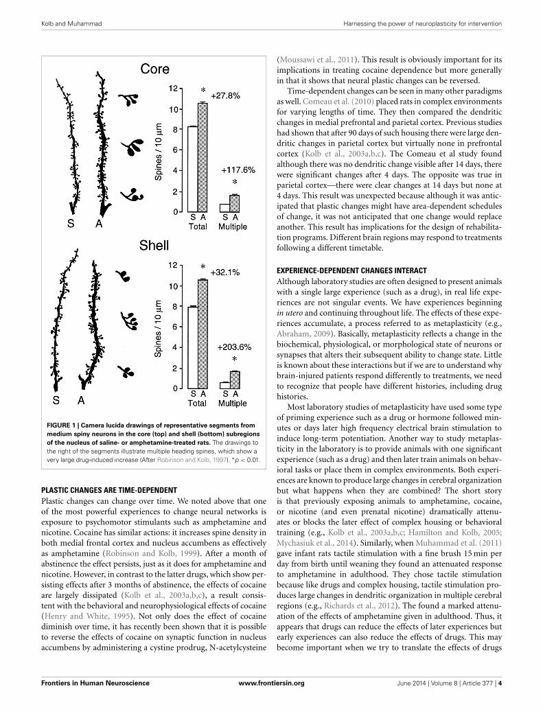
A WIDE VARIETY OF EXPERIENCES ALTER THE BRAIN THROUGHOUTTHE LIFESPANVirtually any experience can change the brain, especially if there isan associated behavioral change (Table 1). We learn and remem-ber, create new thoughts, and behavior changes throughoutour lifetime. Changing behavior requires changes in the neuralcircuits that underlie it. For example, there are many stud-ies showing that learning neuropsychological tasks is associatedwith synaptic changes in regions believed to be requisite for thelearning (e.g., Greenough and Chang, 1989; Kolb et al., 2008).Similarly, repeated exposure to psychoactive drugs can result inlong-lasting changes in behavior, which is correlated with largealterations in morphology of neurons in many brain structuresincluding medial and orbital prefrontal cortex, nucleus accum-bens, caudate-putamen, and hippocampus (see Figure 1) (e.g.,Robinson and Kolb, 2004). Indeed, it is the capacity of drugssuch as amphetamine and nicotine that have led to their use astreatments for brain injury (see below). Indeed, the role of expe-riences in general in changing the brain is especially important aswe search for treatments for brain injuries.
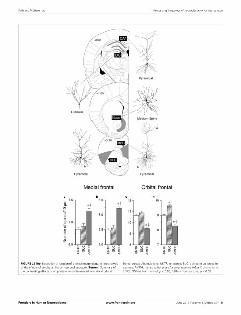
PLASTIC CHANGES ARE AREA DEPENDENTIt is generally assumed that powerful experiences such as com-plex housing would produce similar neuronal changes. In fact,it is not uncommon for investigators to choose one structure(often the hippocampus) as a surrogate for how experiences arealtering the brain. It has been therefore surprising to us thatthis is wrong. For example, when one compares the effects ofamphetamine on the medial prefrontal and orbital prefrontal cor-tex, parietal cortex, nucleus accumbens, CA1 of the hippocampus,and the dentate gyrus the effects are wildly different (Crombaget al., 2005). Thus, whereas neurons in the medial prefrontalcortex, nucleus accumbens, and CA1 show increased spine den-sity, neurons in the orbital frontal cortex show a decrease inspine density and the other structures no change at all (seeFigure 2). Similarly, when Mychasiuk et al. (2012) examined theeffects of prenatal stress on gene expression in hippocampus andmedial prefrontal cortex they found over 100 genes changed ineach region but there was virtually no overlap in which geneschanged. Using one structure or the other (or blood) as a surro-gate marker for epigenetic change throughout the brain is clearlymisleading.
The area-dependent nature of plastic changes in the brain hasobvious implications for developing therapies for brain injury.The therapies need to be informed by where the changes arehoped to occur.
THE DEGREE OF PLASTIC CHANGE IS RELATED TO BOTH THERELEVANCE OF AN EXPERIENCE AND THE INTENSITY OR FREQUENCYOF THE EVENTSExperiences that highly relevant to an animal are likely to producemuch more rapid neuronal changes than less relevant experi-ences. Fowl become imprinted on a moving object appearingshortly after their hatching, which in the natural world wouldnormally be a parent. There are immediate changes in thechick’s hyperstriatum after visual imprinting including decreasedspine density, increased NMDA receptor density, and increased
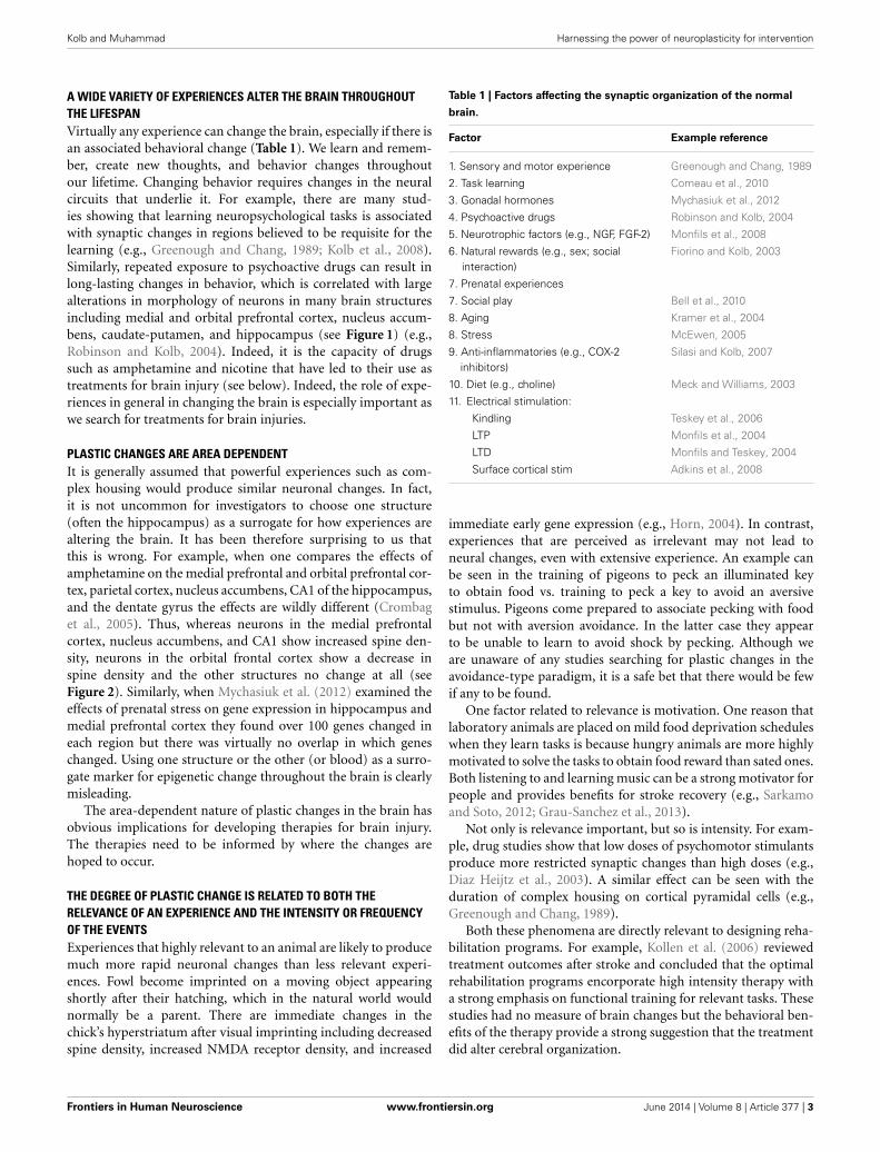
Table 1 | Factors affecting the synaptic organization of the normal
brain.
Factor Example reference
1. Sensory and motor experience Greenough and Chang, 1989
2. Task learning Comeau et al., 2010
3. Gonadal hormones Mychasiuk et al., 2012
4. Psychoactive drugs Robinson and Kolb, 2004
5. Neurotrophic factors (e.g., NGF, FGF-2) Monfils et al., 2008
6. Natural rewards (e.g., sex; socialinteraction)
Fiorino and Kolb, 2003
7. Prenatal experiences
7. Social play Bell et al., 2010
8. Aging Kramer et al., 2004
8. Stress McEwen, 2005
9. Anti-inflammatories (e.g., COX-2inhibitors)
Silasi and Kolb, 2007
10. Diet (e.g., choline) Meck and Williams, 2003
11. Electrical stimulation:
Kindling Teskey et al., 2006
LTP Monfils et al., 2004
LTD Monfils and Teskey, 2004
Surface cortical stim Adkins et al., 2008
immediate early gene expression (e.g., Horn, 2004). In contrast,experiences that are perceived as irrelevant may not lead toneural changes, even with extensive experience. An example canbe seen in the training of pigeons to peck an illuminated keyto obtain food vs. training to peck a key to avoid an aversivestimulus. Pigeons come prepared to associate pecking with foodbut not with aversion avoidance. In the latter case they appearto be unable to learn to avoid shock by pecking. Although weare unaware of any studies searching for plastic changes in theavoidance-type paradigm, it is a safe bet that there would be fewif any to be found.
One factor related to relevance is motivation. One reason thatlaboratory animals are placed on mild food deprivation scheduleswhen they learn tasks is because hungry animals are more highlymotivated to solve the tasks to obtain food reward than sated ones.Both listening to and learning music can be a strong motivator forpeople and provides benefits for stroke recovery (e.g., Sarkamoand Soto, 2012; Grau-Sanchez et al., 2013).
Not only is relevance important, but so is intensity. For exam-ple, drug studies show that low doses of psychomotor stimulantsproduce more restricted synaptic changes than high doses (e.g.,Diaz Heijtz et al., 2003). A similar effect can be seen with theduration of complex housing on cortical pyramidal cells (e.g.,Greenough and Chang, 1989).
Both these phenomena are directly relevant to designing reha-bilitation programs. For example, Kollen et al. (2006) reviewedtreatment outcomes after stroke and concluded that the optimalrehabilitation programs encorporate high intensity therapy witha strong emphasis on functional training for relevant tasks. Thesestudies had no measure of brain changes but the behavioral ben-efits of the therapy provide a strong suggestion that the treatmentdid alter cerebral organization.
Frontiers in Human Neuroscience www.frontiersin.org June 2014 | Volume 8 | Article 377 | 3
Kolb and Muhammad Harnessing the power of neuroplasticity for intervention
FIGURE 1 | Camera lucida drawings of representative segments from
medium spiny neurons in the core (top) and shell (bottom) subregions
of the nucleus of saline- or amphetamine-treated rats. The drawings tothe right of the segments illustrate multiple heading spines, which show avery large drug-induced increase (After Robinson and Kolb, 1997). ∗p < 0.01.
PLASTIC CHANGES ARE TIME-DEPENDENTPlastic changes can change over time. We noted above that oneof the most powerful experiences to change neural networks isexposure to psychomotor stimulants such as amphetamine andnicotine. Cocaine has similar actions: it increases spine density inboth medial frontal cortex and nucleus accumbens as effectivelyas amphetamine (Robinson and Kolb, 1999). After a month ofabstinence the effect persists, just as it does for amphetamine andnicotine. However, in contrast to the latter drugs, which show per-sisting effects after 3 months of abstinence, the effects of cocaineare largely dissipated (Kolb et al., 2003a,b,c), a result consis-tent with the behavioral and neurophysiological effects of cocaine(Henry and White, 1995). Not only does the effect of cocainediminish over time, it has recently been shown that it is possibleto reverse the effects of cocaine on synaptic function in nucleusaccumbens by administering a cystine prodrug, N-acetylcysteine
(Moussawi et al., 2011). This result is obviously important for itsimplications in treating cocaine dependence but more generallyin that it shows that neural plastic changes can be reversed.
Time-dependent changes can be seen in many other paradigmsas well. Comeau et al. (2010) placed rats in complex environmentsfor varying lengths of time. They then compared the dendriticchanges in medial prefrontal and parietal cortex. Previous studieshad shown that after 90 days of such housing there were large den-dritic changes in parietal cortex but virtually none in prefrontalcortex (Kolb et al., 2003a,b,c). The Comeau et al study foundalthough there was no dendritic change visible after 14 days, therewere significant changes after 4 days. The opposite was true inparietal cortex—there were clear changes at 14 days but none at4 days. This result was unexpected because although it was antic-ipated that plastic changes might have area-dependent schedulesof change, it was not anticipated that one change would replaceanother. This result has implications for the design of rehabilita-tion programs. Different brain regions may respond to treatmentsfollowing a different timetable.
EXPERIENCE-DEPENDENT CHANGES INTERACTAlthough laboratory studies are often designed to present animalswith a single large experience (such as a drug), in real life expe-riences are not singular events. We have experiences beginningin utero and continuing throughout life. The effects of these expe-riences accumulate, a process referred to as metaplasticity (e.g.,Abraham, 2009). Basically, metaplasticity reflects a change in thebiochemical, physiological, or morphological state of neurons orsynapses that alters their subsequent ability to change state. Littleis known about these interactions but if we are to understand whybrain-injured patients respond differently to treatments, we needto recognize that people have different histories, including drughistories.
Most laboratory studies of metaplasticity have used some typeof priming experience such as a drug or hormone followed min-utes or days later high frequency electrical brain stimulation toinduce long-term potentiation. Another way to study metaplas-ticity in the laboratory is to provide animals with one significantexperience (such as a drug) and then later train animals on behav-ioral tasks or place them in complex environments. Both experi-ences are known to produce large changes in cerebral organizationbut what happens when they are combined? The short storyis that previously exposing animals to amphetamine, cocaine,or nicotine (and even prenatal nicotine) dramatically attenu-ates or blocks the later effect of complex housing or behavioraltraining (e.g., Kolb et al., 2003a,b,c; Hamilton and Kolb, 2005;Mychasiuk et al., 2014). Similarly, when Muhammad et al. (2011)gave infant rats tactile stimulation with a fine brush 15 min perday from birth until weaning they found an attenuated responseto amphetamine in adulthood. They chose tactile stimulationbecause like drugs and complex housing, tactile stimulation pro-duces large changes in dendritic organization in multiple cerebralregions (e.g., Richards et al., 2012). The found a marked attenu-ation of the effects of amphetamine given in adulthood. Thus, itappears that drugs can reduce the effects of later experiences butearly experiences can also reduce the effects of drugs. This maybecome important when we try to translate the effects of drugs
Frontiers in Human Neuroscience www.frontiersin.org June 2014 | Volume 8 | Article 377 | 4
Kolb and Muhammad Harnessing the power of neuroplasticity for intervention
FIGURE 2 | Top: Illustration of location of, and cell morphology, for the analysisof the effects of amphetamine on neuronal structure. Bottom: Summary ofthe contrasting effects of amphetamine on the medial frontal and orbital
frontal cortex. Abbreviations: UNTR, untrained; SUC, trained to bar press forsucrose; AMPH, trained to bar press for amphetamine (After Crombag et al.,2005). ∗Differs from control, p < 0.05; †differs from sucrose, p < 0.05.
Frontiers in Human Neuroscience www.frontiersin.org June 2014 | Volume 8 | Article 377 | 5
Kolb and Muhammad Harnessing the power of neuroplasticity for intervention
on recovery in animal studies to humans with a rich lifetime ofexperiences that may modulate the action of the drugs.
The idea of metaplasticity is likely related to the concept of cog-nitive reserve, which refers to the differences in cognitive capacityin older people related to a lifetime of intellectual activities (e.g.,Barulli and Stern, 2013). For example, Verghese et al. (2003)showed that extensive participation in leisure activities in olderpeople (>75 year) is associated with a reduced risk of dementia.The hypothesis is that cognitive reserves stemming from previouslearning experiences play a protective role in coping with neu-rodegenerative diseases. The idea can be extended to hypothesizethat cognitive reserve might also have the benefit of enhancing theefficacy of rehabilitative strategies after brain injury, which wouldbe an example of metaplasticity.
PLASTIC CHANGES ARE AGE-DEPENDENTThe developing brain is more responsive to experiences than theadult brain but there are qualitative differences in response tosimilar experience at different ages. It is known, for example,that stress in adulthood decreases spine density in medial pre-frontal cortex but increases it in orbital frontal cortex (Listonet al., 2006). In contrast, however, prenatal stress produces justthe opposite pattern in the adult brain, namely an increase inmedial prefrontal cortex and a decrease in orbital frontal cor-tex (Muhammad et al., 2012). Similar age-dependent qualitativechanges can be seen in the effects of complex housing (Kolb et al.,2003c). When adult animals are placed in such environmentsthere is a general increase in spine density in cortical pyramidalcells whereas similar experience at weaning leads to a decreasein spine density. The age-dependent nature of synaptic changeis clearly significant when we consider most effective treatmentsneurological disorders at different ages such as in pediatric vs.adult disorders.
BOTH PRENATAL AND PRECONCEPTUAL EXPERIENCE CAN ALTER THEADULT BRAINAlthough the brain continues to develop long after birth, it hasbecome clear in the last decade that prenatal and preconcep-tual experiences can profoundly alter the adult brain. Exposureto psychoactive drugs, including prescription drugs, stress, andcomplex housing all change synaptic organization of the pre-frontal cortex (e.g., Kolb et al., 2012a,b; Muhammad et al.,2012; Mychasiuk et al., 2013). In addition, mild prenatal stressincreases global methylation in prefrontal cortex and hippocam-pus (Mychasiuk et al., 2011a,b) and alters gene expression ina sexually-dimorphic and and region-specific manner in brainsexamined at weaning (Mychasiuk et al., 2011a,b). The frontalcortex changes were largely related to neurotransmitter function,whereas hippocampal changes were more prominent in femalesand concentrated around growth factors. It seems likely thatthe preconceptual experiences could influence the brain’s laterresponse to injury.
Although there have been many reports that parental experi-ences could influence epigenetics and subsequent health in theoffspring (e.g., Barker, 1998; Kaati et al., 2002; Pembrey, 2004), itis only recently that experiences of the parents have been shownto affect epigenetics in the brain of offspring (Mychasiuk et al.,
2011a,b, 2013). For example, Mychasiuk et al. (2013) found thatpaternal stress alters offspring behavior and DNA methylationpatterns in a sexually-dimorphic manner, presumably because ofexperience-dependent effects on spermatogenesis. The authorssuggest that brain development is influenced not only by post-natal experiences but is also influenced by earlier maternal andpaternal experiences, which combine to produce the various phe-notypes and individual differences that we perceive in closelyrelated individuals. Once again, such differences are likely toinfluence how the brain responds to injury and rehabilitationmuch later in life.
NOT ALL PLASTICITY IS GOODMost studies of brain plasticity and behavior emphasize the ideathat plastic changes can support improved cognitive and motorfunction. But plastic changes are not all good and can interferewith behavior. For example, it is likely that drug-related alter-ations in prefrontal cortex and other brain regions could underliesome of the maladaptive behavior observed in drug addicts.Another example can be seen in focal hand dystonia in musicians.
Focal hand dystonia refers to abnormal finger and hand posi-tions, cramps, and difficulty in coordinating hand and fingermovements. Dystonia can be so disabling that some musiciansmust give up their occupation. Dystonia is most common ininstruments, such as stringed instruments, that require maximalfine finger movements. Although the precise cause of dystonia isstill debated, one hypothesis is that it results from extensive prac-tice of the more intensively used hand, which likely results from adistortion of the somatosensory and/or motor maps in the cortex(Candia et al., 2003). Another hypothesis is that the practice leadsto abnormal activation patterns in the premotor cortex (Kadotaet al., 2010), which in turn could be related to abnormal corti-cal maps. In either event, the dystonia symptoms appear to resultfrom pathological plasticity.
There are many other examples of pathological plasticityincluding dementia (Mattson et al., 2001) epilepsy (Teskey, 2001),and pathological pain (Baranauskas, 2001). It is not known ifthere is pathological plasticity after brain injury but one sugges-tive example comes from Nudo et al. (1996) who showed thatwithout rehabilitation after injury to the motor cortex, the arearegulating hand movements becomes smaller—a phenomenonsometimes referred to as a disuse syndrome. With rehabilitationthe hand area retains its cortical representation. The reversal ofthis type of pathological plasticity is presumably important inunderstanding the success of restraint-induced therapies.
HARNASSING PLASTICITY FOR NEUROREHABILITATIONThe studies of neural plasticity in normal animals provide alaunch pad to develop new strategies for designing rehabilitationprograms (for an extensive review, see Cramer et al., 2011). Ourbasic assumption is that experiences that change the normal brainwill likely produce similar, and hopefully even larger, changes inthe injured brain. This is often the case but not always. For exam-ple we have shown that tactile stimulation in early developmenthas a profound effect on neural organization and behavior (e.g.,Richards et al., 2012) so we anticipated that tactile stimulationmight be a good treatment for brain injury, which it is (see
Frontiers in Human Neuroscience www.frontiersin.org June 2014 | Volume 8 | Article 377 | 6
Kolb and Muhammad Harnessing the power of neuroplasticity for intervention
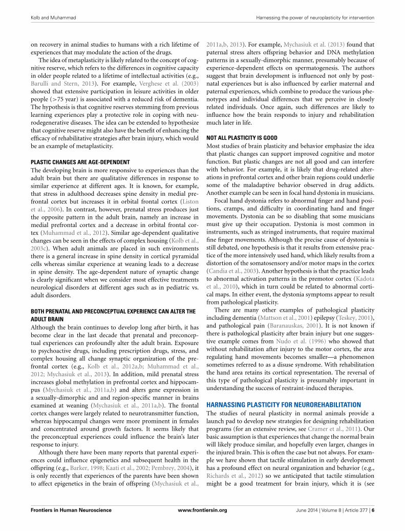
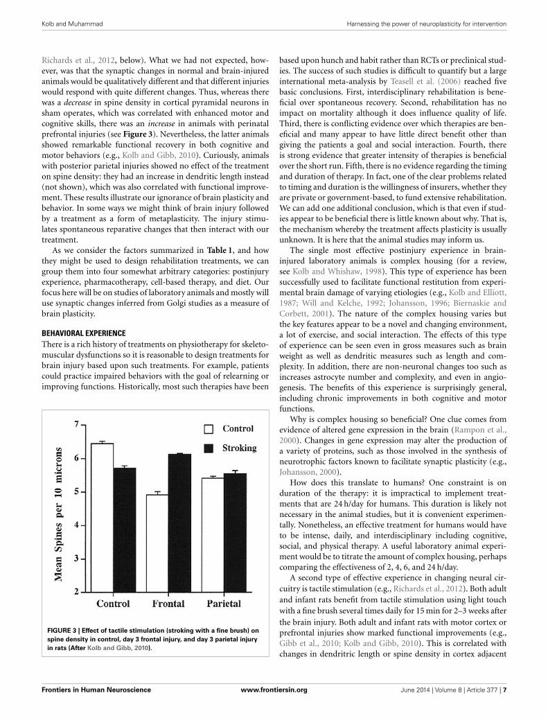
Richards et al., 2012, below). What we had not expected, how-ever, was that the synaptic changes in normal and brain-injuredanimals would be qualitatively different and that different injurieswould respond with quite different changes. Thus, whereas therewas a decrease in spine density in cortical pyramidal neurons insham operates, which was correlated with enhanced motor andcognitive skills, there was an increase in animals with perinatalprefrontal injuries (see Figure 3). Nevertheless, the latter animalsshowed remarkable functional recovery in both cognitive andmotor behaviors (e.g., Kolb and Gibb, 2010). Curiously, animalswith posterior parietal injuries showed no effect of the treatmenton spine density: they had an increase in dendritic length instead(not shown), which was also correlated with functional improve-ment. These results illustrate our ignorance of brain plasticity andbehavior. In some ways we might think of brain injury followedby a treatment as a form of metaplasticity. The injury stimu-lates spontaneous reparative changes that then interact with ourtreatment.
As we consider the factors summarized in Table 1, and howthey might be used to design rehabilitation treatments, we cangroup them into four somewhat arbitrary categories: postinjuryexperience, pharmacotherapy, cell-based therapy, and diet. Ourfocus here will be on studies of laboratory animals and mostly willuse synaptic changes inferred from Golgi studies as a measure ofbrain plasticity.
BEHAVIORAL EXPERIENCEThere is a rich history of treatments on physiotherapy for skeleto-muscular dysfunctions so it is reasonable to design treatments forbrain injury based upon such treatments. For example, patientscould practice impaired behaviors with the goal of relearning orimproving functions. Historically, most such therapies have been
FIGURE 3 | Effect of tactile stimulation (stroking with a fine brush) on
spine density in control, day 3 frontal injury, and day 3 parietal injury
in rats (After Kolb and Gibb, 2010).
based upon hunch and habit rather than RCTs or preclinical stud-ies. The success of such studies is difficult to quantify but a largeinternational meta-analysis by Teasell et al. (2006) reached fivebasic conclusions. First, interdisciplinary rehabilitation is bene-ficial over spontaneous recovery. Second, rehabilitation has noimpact on mortality although it does influence quality of life.Third, there is conflicting evidence over which therapies are ben-eficial and many appear to have little direct benefit other thangiving the patients a goal and social interaction. Fourth, thereis strong evidence that greater intensity of therapies is beneficialover the short run. Fifth, there is no evidence regarding the timingand duration of therapy. In fact, one of the clear problems relatedto timing and duration is the willingness of insurers, whether theyare private or government-based, to fund extensive rehabilitation.We can add one additional conclusion, which is that even if stud-ies appear to be beneficial there is little known about why. That is,the mechanism whereby the treatment affects plasticity is usuallyunknown. It is here that the animal studies may inform us.
The single most effective postinjury experience in brain-injured laboratory animals is complex housing (for a review,see Kolb and Whishaw, 1998). This type of experience has beensuccessfully used to facilitate functional restitution from experi-mental brain damage of varying etiologies (e.g., Kolb and Elliott,1987; Will and Kelche, 1992; Johansson, 1996; Biernaskie andCorbett, 2001). The nature of the complex housing varies butthe key features appear to be a novel and changing environment,a lot of exercise, and social interaction. The effects of this typeof experience can be seen even in gross measures such as brainweight as well as dendritic measures such as length and com-plexity. In addition, there are non-neuronal changes too such asincreases astrocyte number and complexity, and even in angio-genesis. The benefits of this experience is surprisingly general,including chronic improvements in both cognitive and motorfunctions.
Why is complex housing so beneficial? One clue comes fromevidence of altered gene expression in the brain (Rampon et al.,2000). Changes in gene expression may alter the production ofa variety of proteins, such as those involved in the synthesis ofneurotrophic factors known to facilitate synaptic plasticity (e.g.,Johansson, 2000).
How does this translate to humans? One constraint is onduration of the therapy: it is impractical to implement treat-ments that are 24 h/day for humans. This duration is likely notnecessary in the animal studies, but it is convenient experimen-tally. Nonetheless, an effective treatment for humans would haveto be intense, daily, and interdisciplinary including cognitive,social, and physical therapy. A useful laboratory animal experi-ment would be to titrate the amount of complex housing, perhapscomparing the effectiveness of 2, 4, 6, and 24 h/day.
A second type of effective experience in changing neural cir-cuitry is tactile stimulation (e.g., Richards et al., 2012). Both adultand infant rats benefit from tactile stimulation using light touchwith a fine brush several times daily for 15 min for 2–3 weeks afterthe brain injury. Both adult and infant rats with motor cortex orprefrontal injuries show marked functional improvements (e.g.,Gibb et al., 2010; Kolb and Gibb, 2010). This is correlated withchanges in dendritric length or spine density in cortex adjacent
Frontiers in Human Neuroscience www.frontiersin.org June 2014 | Volume 8 | Article 377 | 7
Kolb and Muhammad Harnessing the power of neuroplasticity for intervention
to the injuries. As shown in Figure 3, the rats with infant lesionsshowed a reversal of the decrease in spine density related to theinjury. In contrast, rats with adult lesions, who showed exten-sive atrophy of adjacent cortical pyramidal neurons, showed anincrease in dendritic length, not shown by shams, related to thetactile stimulation.
Recently, Livingston-Thomas et al. (2013, 2014) devised anovel behavioral approach, which they called voluntary forced usemovement therapy. Rats were placed in plastic pet activity ballsfor 30 min per day for 21 days beginning 5 days after ischemicstroke. The therapy resulted in small but consistent acceleration offorelimb performance in several behavioral tests. The functionalimprovement was associated with an increase in migrating neu-ral precursor cells originating in the subventricular zone as wellas increased expression of BDNF (brain-derived neurotrophicfactor) that were presumed to be in microglia.
Few laboratory studies have examined the beneficial effectsof physical therapy on recovery. One emerging strategy is toimplement specialized training protocols, with the best knownbeing constraint-induced movement therapy for the arm andhand (Wolf et al., 2002). More recently robotic devices (e.g.,Hidler et al., 2009) behavioral shaping, bilateral arm training (Linet al., 2010), body weight-supported treadmill training (Dobkinet al., 2006; Duncan et al., 2007),task oriented physical therapy(Jonsdottir et al., 2010), and music therapy (Schneider et al.,2007) have also proven effective. The reasons for the effective-ness of these treatments is unknown but they presumably leadto synaptic changes that may be identified in mapping studiesusing noninvasive imaging or intracortical transcortical mag-netic stimulation (TMS). Indeed, a recent study by Amengualet al. (2013) used music-supported therapy followed by TMSand found improved motor functions correlated with plasticchanges in the form of increased cortical excitability following thetraining.
PHARMACOTHERAPYWe showed above that many drugs are extremely effective in stim-ulating neuroplastic changes in cerebral neurons, and especiallypsychomotor stimulants. Early studies by Feeney and colleagues(e.g., Feeney and Sutton, 1987) used amphetamine as a postin-jury treatment following unilateral motor cortex injury and foundstriking benefits provided that the animals received physical ther-apy while under the influence of the drug (see also Goldstein,2003). One complication of the amphetamine treatment is thatlesion size, location, and route of administration appear to influ-ence efficacy. Moroz and Kolb (2005) compared the effect ofamphetamine on focal and extensive unilateral ischemic injuriesfinding that whereas amphetamine was useful in the former con-dition, it was not helpful with the larger injuries. This may explainwhy clinical studies with amphetamine have had mixed success.Clinical studies tend to select patients most in need of ther-apy, which are those with larger injuries. (Alaverdashvili et al.,2007) made small lesions similar to those of Moroz and Kolbbut administered the same dose (1 mg/kg) orally rather thansubcutaneously. They found no beneficial effect of the drug.
In unpublished studies we used amphetamine after bilateralmedial frontal lesions, finding no benefit on the performance of
cognitive tests. Given that all of the positive preclinical studiesusing amphetamine have been with animals with motor cortexinjuries, the jury is still out on whether it will be effective for cog-nitive improvement. As mentioned above, it is usually necessaryto make bilateral injuries in rodents to obtain robust cognitivedeficits. Thus, it may not be the motor/cognitive distinction thatis important but rather the unilateral/bilateral difference.
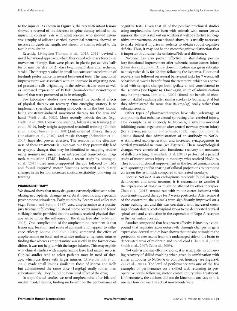
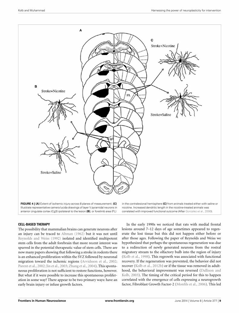
Nicotine has also proven effective in stimulating postin-jury functional improvement after ischemic motor cortex injury(Gonzalez et al., 2006). A low dose of nicotine was given subcuta-neously twice daily for 12 days following the ischemia. Functionalrecovery was followed on several behavioral tasks for 7 weeks. Allbehaviors showed a benefit from the treatment, which was corre-lated with synaptic changes both ipsilateral and contralateral tothe ischemia (see Figure 4). Once again, route of administrationmay be important. Lim et al. (2009) found no benefit of nico-tine on skilled reaching after similar strokes to Gonzalez et al butthey administered the same dose (0.3 mg/kg) orally rather thansubcutaneously.
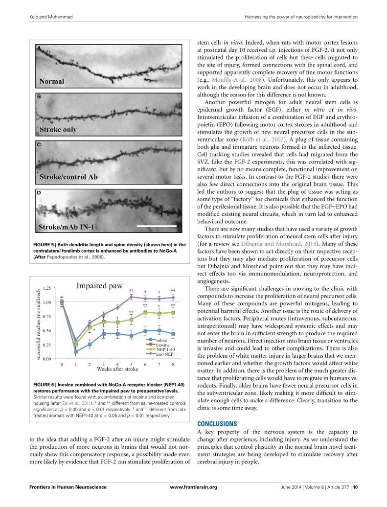
Other types of pharmacotherapies have taken advantage ofcompounds that enhance axonal sprouting after cerebral injury.One example is an antibody to NoGo-A, a myelin-associatedinhibiting axonal regeneration after central nervous system injury(for a review, see Kempf and Schwab, 2013). Papadopoulos et al.(2006) showed that administration of an antibody to NoGo-A stimulated axon generation and increased synaptogenesis incortical pyramidal neurons (see Figure 5). These morphologicalchanges were correlated with functional recovery on measuresof skilled reaching. Hamadjida et al. (2012) performed a parallelstudy of motor cortex injury in monkeys who received NoGo-A.They found functional improvement in the treated animals alongwith sprouting and/or sparing of callosal projections to premotorcortex on the lesion side compared to untreated monkeys.
Because NoGo-A is an endogenous molecule found in oligo-dendrocytes and some neurons, it is reasonable to wonder ifthe expression of NoGo-A might be affected by other therapies.Zhao et al. (2013) treated rats with motor cortex ischemia withconstraint-induced therapy for 3 weeks poststroke. After removalof the constraint, the animals were significantly improved on abeam-walking test and this was correlated with increased cross-ings of contralateral corticospinal axons to the denervated cervicalspinal cord and a reduction in the expression of Nogo-A receptorin the peri-infarct cortex.
Another compound that has proven effective is inosine, a com-pound that regulates axon outgrowth through changes in geneexpression. Several studies have shown that inosine stimulates theprojection of new axons from the undamaged side of the brain todenervated areas of midbrain and spinal cord (Chen et al., 2002;Smith et al., 2007; Zai et al., 2009).
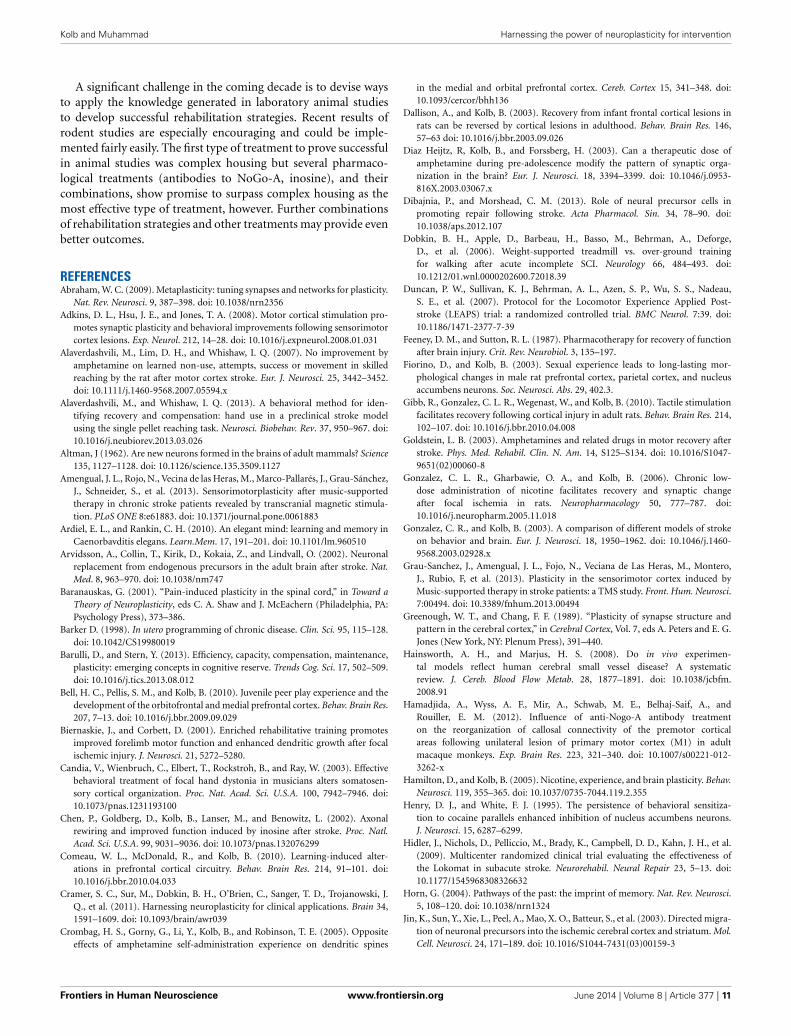
Not only is inosine effective alone, it is synergistic in enhanc-ing recovery of skilled reaching when given in combination witheither antibodies to NoGo-A or complex housing (see Figure 6;Zai et al., 2011). The level of performance was one of the fewexamples of performance on a skilled task returning to pre-operative levels following motor cortex injury plus treatment.Unfortunately, the authors did not do kinematic analysis so it isunclear how normal the actual movements were.
Frontiers in Human Neuroscience www.frontiersin.org June 2014 | Volume 8 | Article 377 | 8
Kolb and Muhammad Harnessing the power of neuroplasticity for intervention
FIGURE 4 | (A) Extent of ischemic injury across 8 planes of measurement. (C)
Illustrate representative camera lucida drawings of layer V pyramidal neurons inanterior cingulate cortex (Cg3) ipsilateral to the lesion (B), or forelimb area (FL)
in the contralesional hemisphere (C) from animals treated either with saline ornicotine. Increased dendritic length in the nicotine-treated animals wascorrelated with improved functional outcome (After Gonzalez et al., 2006).
CELL-BASED THERAPYThe possibility that mammalian brains can generate neurons afteran injury can be traced to Altman (1962) but it was not untilReynolds and Weiss (1992) isolated and identified multipotentstem cells from the adult forebrain that more recent interest wasspurred in the potential therapeutic value of stem cells. There arenow many papers showing that following a stroke in rodents thereis an enhanced proliferation within the SVZ followed by neuronalmigration toward the ischemic regions (Arvidsson et al., 2002;Parent et al., 2002; Jin et al., 2003; Zhang et al., 2004). This sponta-neous proliferation is not sufficient to restore functions, however.But what if it were possible to increase this spontaneous prolifer-ation in some way? There appear to be two primary ways: have anearly brain injury or infuse growth factors.
In the early 1990s we noticed that rats with medial frontallesions around 7–12 days of age sometimes appeared to regen-erate the lost tissue but this did not happen either before orafter those ages. Following the paper of Reynolds and Weiss wehypothesized that perhaps the spontaneous regeneration was dueto a redirection of newly generated neurons from the rostralmigratory stream to the olfactory bulb into the region of injury(Kolb et al., 1998). This regrowth was associated with functionalrecovery. If the regeneration was prevented, the behavior did notrecover (Kolb et al., 2012b) or if the tissue was removed in adult-hood, the behavioral improvement was reversed (Dallison andKolb, 2003). The timing of the critical period for this to happencorrelated with the emergence of cells expressing a neurogrowthfactor, Fibroblast Growth Factor-2 (Monfils et al., 2006). This led
Frontiers in Human Neuroscience www.frontiersin.org June 2014 | Volume 8 | Article 377 | 9
Kolb and Muhammad Harnessing the power of neuroplasticity for intervention
FIGURE 5 | Both dendritic length and spine density (shown here) in the
contralateral forelimb cortex is enhanced by antibodies to NoGo-A
(After Papadopoulos et al., 2006).
FIGURE 6 | Inosine combined with NoGo-A receptor blocker (NEP1-40)
restores performance with the impaired paw to preoperative levels.
Similar results were found with a combination of inosine and complexhousing (after Zai et al., 2011). ∗ and ∗∗ different from saline-treated controlssignificant at p < 0.05 and p < 0.01 respectively. † and †† different from ratstreated animals with NEP1-40 at p < 0.05 and p < 0.01 respectively.
to the idea that adding a FGF-2 after an injury might stimulatethe production of more neurons in brains that would not nor-mally show this compensatory response, a possibility made evenmore likely by evidence that FGF-2 can stimulate proliferation of
stem cells in vitro. Indeed, when rats with motor cortex lesionsat postnatal day 10 received i.p. injections of FGF-2, it not onlystimulated the proliferation of cells but these cells migrated tothe site of injury, formed connections with the spinal cord, andsupported apparently complete recovery of fine motor functions(e.g., Monfils et al., 2008). Unfortunately, this only appears towork in the developing brain and does not occur in adulthood,although the reason for this difference is not known.
Another powerful mitogen for adult neural stem cells isepidermal growth factor (EGF), either in vitro or in vivo.Intraventricular infusion of a combination of EGF and erythro-poietin (EPO) following motor cortex strokes in adulthood andstimulates the growth of new neural precursor cells in the sub-ventricular zone (Kolb et al., 2007). A plug of tissue containingboth glia and immature neurons formed in the infarcted tissue.Cell tracking studies revealed that cells had migrated from theSVZ. Like the FGF-2 experiments, this was correlated with sig-nificant, but by no means complete, functional improvement onseveral motor tasks. In contrast to the FGF-2 studies there werealso few direct connections into the original brain tissue. Thisled the authors to suggest that the plug of tissue was acting assome type of “factory” for chemicals that enhanced the functionof the perilesional tissue. It is also possible that the EGF+EPO hadmodified existing neural circuits, which in turn led to enhancedbehavioral outcome.
There are now many studies that have used a variety of growthfactors to stimulate proliferation of neural stem cells after injury(for a review see Dibajnia and Morshead, 2013). Many of thesefactors have been shown to act directly on their respective recep-tors but they may also mediate proliferation of precursor cellsbut Dibajnia and Morshead point out that they may have indi-rect effects too via immunomodulation, neuroprotection, andangiogenesis.
There are significant challenges in moving to the clinic withcompounds to increase the proliferation of neural precursor cells.Many of these compounds are powerful mitogens, leading topotential harmful effects. Another issue is the route of delivery ofactivation factors. Peripheral routes (intravenous, subcutaneous,intraperitoneal) may have widespread systemic effects and maynot enter the brain in sufficient strength to produce the requirednumber of neurons. Direct injection into brain tissue or ventriclesis invasive and could lead to other complications. There is alsothe problem of white matter injury in larger brains that we men-tioned earlier and whether the growth factors would affect whitematter. In addition, there is the problem of the much greater dis-tance that proliferating cells would have to migrate in humans vs.rodents. Finally, older brains have fewer neural precursor cells inthe subventricular zone, likely making it more difficult to stim-ulate enough cells to make a difference. Clearly, transition to theclinic is some time away.
CONCLUSIONSA key property of the nervous system is the capacity tochange after experience, including injury. As we understand theprinciples that control plasticity in the normal brain novel treat-ment strategies are being developed to stimulate recovery aftercerebral injury in people.
Frontiers in Human Neuroscience www.frontiersin.org June 2014 | Volume 8 | Article 377 | 10
Kolb and Muhammad Harnessing the power of neuroplasticity for intervention
A significant challenge in the coming decade is to devise waysto apply the knowledge generated in laboratory animal studiesto develop successful rehabilitation strategies. Recent results ofrodent studies are especially encouraging and could be imple-mented fairly easily. The first type of treatment to prove successfulin animal studies was complex housing but several pharmaco-logical treatments (antibodies to NoGo-A, inosine), and theircombinations, show promise to surpass complex housing as themost effective type of treatment, however. Further combinationsof rehabilitation strategies and other treatments may provide evenbetter outcomes.
REFERENCESAbraham, W. C. (2009). Metaplasticity: tuning synapses and networks for plasticity.
Nat. Rev. Neurosci. 9, 387–398. doi: 10.1038/nrn2356Adkins, D. L., Hsu, J. E., and Jones, T. A. (2008). Motor cortical stimulation pro-
motes synaptic plasticity and behavioral improvements following sensorimotorcortex lesions. Exp. Neurol. 212, 14–28. doi: 10.1016/j.expneurol.2008.01.031
Alaverdashvili, M., Lim, D. H., and Whishaw, I. Q. (2007). No improvement byamphetamine on learned non-use, attempts, success or movement in skilledreaching by the rat after motor cortex stroke. Eur. J. Neurosci. 25, 3442–3452.doi: 10.1111/j.1460-9568.2007.05594.x
Alaverdashvili, M., and Whishaw, I. Q. (2013). A behavioral method for iden-tifying recovery and compensation: hand use in a preclinical stroke modelusing the single pellet reaching task. Neurosci. Biobehav. Rev. 37, 950–967. doi:10.1016/j.neubiorev.2013.03.026
Altman, J (1962). Are new neurons formed in the brains of adult mammals? Science135, 1127–1128. doi: 10.1126/science.135.3509.1127
Amengual, J. L., Rojo, N., Vecina de las Heras, M., Marco-Pallarés, J., Grau-Sánchez,J., Schneider, S., et al. (2013). Sensorimotorplasticity after music-supportedtherapy in chronic stroke patients revealed by transcranial magnetic stimula-tion. PLoS ONE 8:e61883. doi: 10.1371/journal.pone.0061883
Ardiel, E. L., and Rankin, C. H. (2010). An elegant mind: learning and memory inCaenorbavditis elegans. Learn.Mem. 17, 191–201. doi: 10.1101/lm.960510
Arvidsson, A., Collin, T., Kirik, D., Kokaia, Z., and Lindvall, O. (2002). Neuronalreplacement from endogenous precursors in the adult brain after stroke. Nat.Med. 8, 963–970. doi: 10.1038/nm747
Baranauskas, G. (2001). “Pain-induced plasticity in the spinal cord,” in Toward aTheory of Neuroplasticity, eds C. A. Shaw and J. McEachern (Philadelphia, PA:Psychology Press), 373–386.
Barker D. (1998). In utero programming of chronic disease. Clin. Sci. 95, 115–128.doi: 10.1042/CS19980019
Barulli, D., and Stern, Y. (2013). Efficiency, capacity, compensation, maintenance,plasticity: emerging concepts in cognitive reserve. Trends Cog. Sci. 17, 502–509.doi: 10.1016/j.tics.2013.08.012
Bell, H. C., Pellis, S. M., and Kolb, B. (2010). Juvenile peer play experience and thedevelopment of the orbitofrontal and medial prefrontal cortex. Behav. Brain Res.207, 7–13. doi: 10.1016/j.bbr.2009.09.029
Biernaskie, J., and Corbett, D. (2001). Enriched rehabilitative training promotesimproved forelimb motor function and enhanced dendritic growth after focalischemic injury. J. Neurosci. 21, 5272–5280.
Candia, V., Wienbruch, C., Elbert, T., Rockstroh, B., and Ray, W. (2003). Effectivebehavioral treatment of focal hand dystonia in musicians alters somatosen-sory cortical organization. Proc. Nat. Acad. Sci. U.S.A. 100, 7942–7946. doi:10.1073/pnas.1231193100
Chen, P., Goldberg, D., Kolb, B., Lanser, M., and Benowitz, L. (2002). Axonalrewiring and improved function induced by inosine after stroke. Proc. Natl.Acad. Sci. U.S.A. 99, 9031–9036. doi: 10.1073/pnas.132076299
Comeau, W. L., McDonald, R., and Kolb, B. (2010). Learning-induced alter-ations in prefrontal cortical circuitry. Behav. Brain Res. 214, 91–101. doi:10.1016/j.bbr.2010.04.033
Cramer, S. C., Sur, M., Dobkin, B. H., O’Brien, C., Sanger, T. D., Trojanowski, J.Q., et al. (2011). Harnessing neuroplasticity for clinical applications. Brain 34,1591–1609. doi: 10.1093/brain/awr039
Crombag, H. S., Gorny, G., Li, Y., Kolb, B., and Robinson, T. E. (2005). Oppositeeffects of amphetamine self-administration experience on dendritic spines
in the medial and orbital prefrontal cortex. Cereb. Cortex 15, 341–348. doi:10.1093/cercor/bhh136
Dallison, A., and Kolb, B. (2003). Recovery from infant frontal cortical lesions inrats can be reversed by cortical lesions in adulthood. Behav. Brain Res. 146,57–63 doi: 10.1016/j.bbr.2003.09.026
Diaz Heijtz, R, Kolb, B., and Forssberg, H. (2003). Can a therapeutic dose ofamphetamine during pre-adolescence modify the pattern of synaptic orga-nization in the brain? Eur. J. Neurosci. 18, 3394–3399. doi: 10.1046/j.0953-816X.2003.03067.x
Dibajnia, P., and Morshead, C. M. (2013). Role of neural precursor cells inpromoting repair following stroke. Acta Pharmacol. Sin. 34, 78–90. doi:10.1038/aps.2012.107
Dobkin, B. H., Apple, D., Barbeau, H., Basso, M., Behrman, A., Deforge,D., et al. (2006). Weight-supported treadmill vs. over-ground trainingfor walking after acute incomplete SCI. Neurology 66, 484–493. doi:10.1212/01.wnl.0000202600.72018.39
Duncan, P. W., Sullivan, K. J., Behrman, A. L., Azen, S. P., Wu, S. S., Nadeau,S. E., et al. (2007). Protocol for the Locomotor Experience Applied Post-stroke (LEAPS) trial: a randomized controlled trial. BMC Neurol. 7:39. doi:10.1186/1471-2377-7-39
Feeney, D. M., and Sutton, R. L. (1987). Pharmacotherapy for recovery of functionafter brain injury. Crit. Rev. Neurobiol. 3, 135–197.
Fiorino, D., and Kolb, B. (2003). Sexual experience leads to long-lasting mor-phological changes in male rat prefrontal cortex, parietal cortex, and nucleusaccumbens neurons. Soc. Neurosci. Abs. 29, 402.3.
Gibb, R., Gonzalez, C. L. R., Wegenast, W., and Kolb, B. (2010). Tactile stimulationfacilitates recovery following cortical injury in adult rats. Behav. Brain Res. 214,102–107. doi: 10.1016/j.bbr.2010.04.008
Goldstein, L. B. (2003). Amphetamines and related drugs in motor recovery afterstroke. Phys. Med. Rehabil. Clin. N. Am. 14, S125–S134. doi: 10.1016/S1047-9651(02)00060-8
Gonzalez, C. L. R., Gharbawie, O. A., and Kolb, B. (2006). Chronic low-dose administration of nicotine facilitates recovery and synaptic changeafter focal ischemia in rats. Neuropharmacology 50, 777–787. doi:10.1016/j.neuropharm.2005.11.018
Gonzalez, C. R., and Kolb, B. (2003). A comparison of different models of strokeon behavior and brain. Eur. J. Neurosci. 18, 1950–1962. doi: 10.1046/j.1460-9568.2003.02928.x
Grau-Sanchez, J., Amengual, J. L., Fojo, N., Veciana de Las Heras, M., Montero,J., Rubio, F, et al. (2013). Plasticity in the sensorimotor cortex induced byMusic-supported therapy in stroke patients: a TMS study. Front. Hum. Neurosci.7:00494. doi: 10.3389/fnhum.2013.00494
Greenough, W. T., and Chang, F. F. (1989). “Plasticity of synapse structure andpattern in the cerebral cortex,” in Cerebral Cortex, Vol. 7, eds A. Peters and E. G.Jones (New York, NY: Plenum Press), 391–440.
Hainsworth, A. H., and Marjus, H. S. (2008). Do in vivo experimen-tal models reflect human cerebral small vessel disease? A systematicreview. J. Cereb. Blood Flow Metab. 28, 1877–1891. doi: 10.1038/jcbfm.2008.91
Hamadjida, A., Wyss, A. F., Mir, A., Schwab, M. E., Belhaj-Saif, A., andRouiller, E. M. (2012). Influence of anti-Nogo-A antibody treatmenton the reorganization of callosal connectivity of the premotor corticalareas following unilateral lesion of primary motor cortex (M1) in adultmacaque monkeys. Exp. Brain Res. 223, 321–340. doi: 10.1007/s00221-012-3262-x
Hamilton, D., and Kolb, B. (2005). Nicotine, experience, and brain plasticity. Behav.Neurosci. 119, 355–365. doi: 10.1037/0735-7044.119.2.355
Henry, D. J., and White, F. J. (1995). The persistence of behavioral sensitiza-tion to cocaine parallels enhanced inhibition of nucleus accumbens neurons.J. Neurosci. 15, 6287–6299.
Hidler, J., Nichols, D., Pelliccio, M., Brady, K., Campbell, D. D., Kahn, J. H., et al.(2009). Multicenter randomized clinical trial evaluating the effectiveness ofthe Lokomat in subacute stroke. Neurorehabil. Neural Repair 23, 5–13. doi:10.1177/1545968308326632
Horn, G. (2004). Pathways of the past: the imprint of memory. Nat. Rev. Neurosci.5, 108–120. doi: 10.1038/nrn1324
Jin, K., Sun, Y., Xie, L., Peel, A., Mao, X. O., Batteur, S., et al. (2003). Directed migra-tion of neuronal precursors into the ischemic cerebral cortex and striatum. Mol.Cell. Neurosci. 24, 171–189. doi: 10.1016/S1044-7431(03)00159-3
Frontiers in Human Neuroscience www.frontiersin.org June 2014 | Volume 8 | Article 377 | 11
Kolb and Muhammad Harnessing the power of neuroplasticity for intervention
Johansson, B. B. (1996). Functional outcome in rats transferred to an enrichedenvironment 15 days after focal brain ischemia. Stroke 27, 324–326. doi:10.1161/01.STR.27.2.324
Johansson, B. B. (2000). Brain plasticity and stroke rehabilitation. The Willislecture. Stroke 31, 223–230. doi: 10.1161/01.STR.31.1.223
Jones, T. A., and Schallert, T. (1992). Overgrowth and pruning of dendrites inadult rats recovering from neocortical damage. Brain Res. 581, 156–160. doi:10.1016/0006-8993(92)90356-E
Jonsdottir, J., Cattaneo, D., Recalcati, M., Regola, A., Rabuffetti, M., Ferrarin,M., et al. (2010). Task-oriented biofeedback to improve gait in individualswith chronic stroke: motor learning approach. Neurorehabil. Neural Repair 24,478–485. doi: 10.1177/1545968309355986
Kaati, G, Bygren, L. O, and Edvinsson, S. (2002). Cardiovascular and dia-betes mortality determined by nutrition during parents’ and grandparents’slow growth period. Eur. J. Hum. Genet. 10, 682–688. doi: 10.1038/sj.ejhg.5200859
Kadota, H., Nakajima, Y., Miyazaki, M., Sekiguchi, H., Kohno, Y., Amako, M., et al.(2010). An fMRI study of musicians with focal dystonia during tapping tasks.J. Neurol. 257, 1092–1098. doi: 10.1007/s00415-010-5468-9
Kempf, A., and Schwab, M. E. (2013). Nogo-a represses anatomical and synap-tic plasticity in the central nervous system. Physiology 28, 151–163. doi:10.1152/physiol.00052.2012
Kolb, B., Cioe, J., and Comeau, W. (2008). Contrasting effects of motor and visuallearning tasks on dendritic arborization and spine density in rats. Neurobiol.Learn. Mem. 90, 295–300. doi: 10.1016/j.nlm.2008.04.012
Kolb, B., and Elliott, W. (1987). Effects of experience on anatomy and behaviorfollowing frontal lesions at 1 or 5 days of age. Behav. Brain Res. 26, 47–56. doi:10.1016/0166-4328(87)90015-5
Kolb, B., and Gibb, R. (2010). Tactile stimulation facilitates functional recov-ery and dendritic change after neonatal medial frontal or posterior pari-etal lesions in rats. Behav. Brain Res. 214, 115–120. doi: 10.1016/j.bbr.2010.04.024
Kolb, B., Gibb, R., and Gorny, G. (2003c). Experience-dependent changes in den-dritic arbor and spine density in neocortex vary with age and sex. Neurobiol.Learn. Mem. 79, 1–10. doi: 10.1016/S1074-7427(02)00021-7
Kolb, B., Gibb, R., Gorny, G., and Whishaw, I. Q. (1998). Possible brain regrowthafter cortical lesions in rats. Behav. Brain Res. 91, 127–141. doi: 10.1016/S0166-4328(97)00112-5
Kolb, B., Gorny, G., Li, Y., Samaha, A. N., and Robinson, T. E. (2003a).Amphetamine or cocaine limits the ability of later experience to promote struc-tural plasticity in the neocortex and nucleus accumbens. Proc. Natl. Acad. Sci.U.S.A. 100, 10523–10528. doi: 10.1073/pnas.1834271100
Kolb, B., Gorny, G., Sonderpalm, A., and Robinson, T. E. (2003b). Environmentalcomplexity has different effects on the structure of neurons in the prefrontalcortex versus the parietal cortex or nucleus accumbens. Synapse 48, 149–153.doi: 10.1002/syn.10196
Kolb, B., Morshead, C, Gonzalez, C., Kim, N., Shingo, T., and Weiss, S. (2007).Growth factor-stimulated generation of new cortical tissue and functionalrecovery after stroke damage to the motor cortex of rats. J. Cereb. Blood FlowMetab. 27, 983–997. doi: 10.1038/sj.jcbfm.9600402
Kolb, B., Mychasiuk, R., Muhammad, A., Li, Y., Frost, D. O., and Gibb, R. (2012a).Experience and the developing prefrontal cortex. Proc. Natl. Acad Sci. U.S.A.109(Suppl. 2), 17186–17193. doi: 10.1073/pnas.1121251109
Kolb, B., Pedersen, B., and Gibb, R. (2012b). Embryonic pretreatment withbromodeoxyuridine blocks neurogenesis and functional recovery from peri-natal frontal lesions in rats. Dev. Neurosci. 34, 228–239. doi: 10.1159/000336645
Kolb, B., and Whishaw, I. Q. (1998). Brain plasticity and behavior. Annu. Rev.Psychol. 49, 43–64. doi: 10.1146/annurev.psych.49.1.43
Kollen, B., Kwakkel, G., and Lindeman, E. (2006). Functional recovery after stroke:a review of current developments in stroke rehabilitation research. Rev. RecentClin. Trials 1, 75–80. doi: 10.2174/157488706775246111
Kovalchuk, A., Lowings, M., Rodriguez-Juarez, R., Muhammad, A., Ilnytskyy, S.,Kolb, B., et al. (2012). Distal epigenetic bystander-like effects of stroke insomatic organs of rats. Aging 4, 224–234.
Kramer, A. F., Bherer, L., Colcombe, S. J., Dong, W., and Greenough,W. T. (2004). Environmental influences on cognitive and brain plastic-ity during aging. J. Gerontol. A Biol. Sci. Med. Sci. 59, M940–M957. doi:10.1093/gerona/59.9.M940
Lim, D. H., Alaverdashvili, M., and Whishaw, I. Q. (2009). Nicotine does notimprove recovery from learned nonuse nor enhance constraint-induced ther-apy after motor cortex stroke in the rat. Behav. Brain Res. 1978, 411–419. doi:10.1016/j.bbr.2008.11.038
Lin, K. C., Chen, Y. A., Chen, C. L., Wu, C. Y., and Chang, Y. F. (2010). The effects ofbilateral arm training on motor control and functional performance in chronicstroke: a randomized controlled study. Neurorehabil. Neural Repair 24, 42–51.doi: 10.1177/1545968309345268
Liston, C., Miller, M. M., Goldwater, D. S., Radley, J. J., Rocher, A. B., Hof, P. R., et al.(2006). Stress-induced alterations in prefrontal cortical dendritic morphologypredict selective impairments in perceptual attentional set-shifting. J. Neurosci.26, 7870–7874. doi: 10.1523/JNEUROSCI.1184-06.2006
Livingston-Thomas, J., Hume, A. H., Doucette, T. A., and Tasker, R. A. (2013). Anovel approach to induction and rehabilitation of deficits in forelimb func-tion a rat model of ischemic stroke. Acta. Pharmacol. Sin. 34, 104–112. doi:10.1038/aps.2012.106
Livingston-Thomas, J. M., McGuire, E. P., Doucette, T. A., and Tasker, R. A. (2014).Voluntary forced use of the impaired limb following stroke facilitates functionalrecovery in the rat. Behav. Brain Res. 261, 210–219. doi: 10.1016/j.bbr.2013.12.032
Losito, A., Pittavini, L., Ferri, C., and De Angelis, L. (2012). Reduced kid-ney function and outcome in acute ischaemic stroke: relationship to arterialhypertension and diabetes. Nephrol. Dial. Transplant. 27, 1054–1058. doi:10.1093/ndt/gfr378
Mattson, M. P., Duan, W., Chan, S. L., and Guo, Z. (2001). “Modification of brainaging and neurodegenerative disorders by genes, diet, and behavior,” in Towarda Theory of Neuroplasticity, eds C. A. Shaw and J. McEachern (Philadelphia, PA:Psychology Press), 402–426.
McEwen, B. S. (2005). Glucocorticoids, depression, and mood disorders: struc-tural remodeling in the brain. Metabolism 54 (Suppl. 1), 20–23. doi:10.1016/j.metabol.2005.01.008
Meck, W. H., and Williams, C. L. (2003). Metabolic imprinting of choline byits availability during gestation: implications for memory and attentionalprocessing across the lifespan. Neurosci. Biobehav. Rev. 27, 385–399. doi:10.1016/S0149-7634(03)00069-1
Monfils, H-H., Driscoll, I., Melvin, N., and Kolb, B. (2006). Differential expres-sion of basic fibroblast growth factor in developing rat brain. Neuroscience 141,213–221. doi: 10.1016/j.neuroscience.2006.03.047
Monfils, M.-H., Driscoll, I., Vavrek, R., Kolb, B., and Fouad, K. (2008). FGF-2 induced functional improvement from neonatal motor cortex injury viacorticospinal projections. Exp. Brain Res. 185, 453–460. doi: 10.1007/s00221-007-1172-0
Monfils, M. H., and Teskey, G. C. (2004). Induction of long-term depressionis associated with decreased dendritic length and spine density in layers IIIand V of sensorimotor neocortex. Synapse 53, 114–121. doi: 10.1002/syn.20039
Monfils, M. H., VandenBerg, P. M., Kleim, J. A., and Teskey, G. C. (2004). Longtermpotentiation induces expanded movement representations and dendritic hyper-trophy in layer V of rat sensorimotor neocortex. Cereb. Cortex 14, 586–593. doi:10.1093/cercor/bhh020
Moroz, I. A., and Kolb, B. (2005). Amphetamine facilitates recovery of skilledreaching following cortical devascularization in the rat. Soc. Neurosci. Abst.669.6.
Moussawi, K., Zhou, W., Shen, H., Reichel, C. M., See, R. E., Carr, D. B.,et al. (2011). Reversing cocaine-induced synaptic potentiation provides endur-ing protection from relapse. Proc. Nat. Acad. Sc. U.S.A. 108, 385–390. doi:10.1073/pnas.1011265108
Muhammad, A., Carroll, C., and Kolb, B. (2012). Stress during develop-ment alters dendritic morphology in the nucleus accumbens and pre-frontal cortex. Neuroscience 216, 103–109. doi: 10.1016/j.neuroscience.2012.04.041
Muhammad, A., Hossain, S., Pellis, S. M., and Kolb, B. (2011). Tactile stimula-tion during development attenuates amphetamine sensitization and structurallyreorganizes prefrontal cortex and striatum in a sex-dependent manner. Behav.Neurosci. 125, 161–174. doi: 10.1037/a0022628
Mychasiuk, R., Gibb, R., and Kolb, B. (2012). Prenatal stress produces sexuallydimorphic and regionally-specific changes in gene expression in hippocampusand frontal cortex of developing rat offspring. Dev. Neurosci. 33, 531–538. doi:10.1159/000335524
Frontiers in Human Neuroscience www.frontiersin.org June 2014 | Volume 8 | Article 377 | 12
Kolb and Muhammad Harnessing the power of neuroplasticity for intervention
Mychasiuk, R., Ilnytskyy, S., Kovalchuk, O., Kolb, B. and Gibb, R. (2011a).Intensity matters: brain, behaviour and the epigenome of prenatally stressedrats. Neuroscience 180, 105–110. doi: 10.1016/j.neuroscience.2011.02.026
Mychasiuk, R., Muhammad, A., Carroll, C., and Kolb, B. (2013). Does prena-tal nicotine exposure alter the brain’s response to nicotine in adolescence? ANeuroanatomical Analysis. Eur. J. Neurosci. 38, 2491–2503. doi: 10.1111/ejn12245
Mychasiuk, R., Muhammad, A., and Kolb, B. (2014). Environmental enrichmentalters structural plasticity of the adolescent brain but does not remediate theeffects of prenatal nicotine exposure. Synapse 68, 293–305. doi: 10.1002/syn.21737
Mychasiuk, R., Schmold, N., Ilnytskyy, S., Kovalchuk, O., Kolb, B., and Gibb, R.(2011b). Prenatal bystander stress alters brain, behavior, recovery from injury,and the epigenome of developing rat offspring. Dev. Neurosci. 33, 159–169. doi:10.1159/000330034
Nudo, R. J., Wise, B. M., SiFuentes, F., and Milliken, G. W. (1996). Neural substratesfor the effects of rehabilitative training on motor recovery after ischemic infarct.Science 272, 1791–1794. doi: 10.1126/science.272.5269.1791
Papadopoulos, C., Tsai, S.-Y., Cheatwood, J. L., Bollnow, M. R., Kolb, B., Schwab,M., et al. (2006). Dendritic plasticity in the adult rat following middle cere-bral artery occlusion and Nogo-A neutralization. Cereb. Cortex 16, 529–536. doi:10.1093/cercor/bhi132
Parent, J. M., Vexler, Z. S., Gong, C., Derugin, N., and Ferriero, D. M. (2002). Ratforebrain neurogenesis and striatal neuron replacement after focal stroke. Ann.Neurol. 52, 802–811. doi: 10.1002/ana.10393
Pembrey, M. (2004). The avon longitudinal study of parents and children(ALSPAC): a resource for genetic epidemiology. Eur. J. Endocrinol. 151(Suppl 3),U125–U129.
Rampon, C., Jiang, C. H., Dong, H., Tang, Y. P., Lockart, D. J., Schultz, P. G., et al.(2000). Effects of environmental enrichment on gene expression in the brain.Proc. Natl. Acad. Sci. U.S.A. 97, 12880–12884. doi: 10.1073/pnas.97.23.12880
Reynolds, B. A., and Weiss, S. (1992). Generation of neurons and astrocytes fromisolated cells of the adult mammalian central nervous system. Science 255,1613–1808. doi: 10.1126/science.1553558
Richards, S., Mychasiuk, R., Kolb, B., and Gibb, R. (2012). Tactile stimulation dur-ing development alters behaviour and neuroanatomical organization of normalrats. Behav. Brain Res. 231, 86–91. doi: 10.1016/j.bbr.2012.02.043
Roberts, A. C., and Glanzman, D. L. (2003). Learning in Aplysia: lookingat synaptic plasticity from both sides. Trends Neurosci. 26, 662–670. doi:10.1016/j.tins.2003.09.014
Robinson, T. E., and Kolb, B. (1997). Persistent structural adaptations in nucleusaccumbens and prefrontal cortex neurons produced by prior experience withamphetamine. J. Neurosci. 17, 8491–8498.
Robinson, T. E., and Kolb, B. (1999). Alterations in the morphology of dendritesand dendritic spines in the nucleus accumbens and prefrontal cortex follow-ing repeated treatment with amphetamine or cocaine. Eur. J. Neurosci. 11,1598–1604. doi: 10.1046/j.1460-9568.1999.00576.x
Robinson, T. E., and Kolb, B. (2004). Structural plasticity associated with drugsof abuse. Neuropharmacology 47(Suppl. 1), 33–46. doi: 10.1016/j.neuropharm.2004.06.025
Sarkamo, T., and Soto, D. (2012). Musci listening after stroke: beneficial effectsand potential neural mechanisms. Ann. N.Y. Acad. Sci. 1252, 266–281. doi:10.1111/j.1749-6632.2011.06405.x
Schneider, S., Schonle, P., Altenmuller, E., and Munte, T. (2007). Using musicalinstruments to improve motor skill recovery following a stroke. J. Neurol. 254,1339–1346. doi: 10.1007/s00415-006-0523-2
Shaw, C. A., and McEachern, J. C. (eds.). (2001). Toward a Theory of Neuroplasticity.New York, NY: Elsevier.
Silasi, G., and Kolb, B. (2007). Chronic inhibition of cyclooxygenase-2 induces den-dritic hypertrophy and limited functional improvement following motor cortexstroke. Neuroscience 144, 1160–1168. doi: 10.1016/j.neuroscience.2006.10.030
Smith, J. M., Lunga, P., Story, D., Harris, N., Le Belle, J., James, M. F., et al. (2007).Inosine promotes recovery of skilled motor function in a model of focal braininjury. Brain 130, 915–935. doi: 10.1093/brain/awl393
Sozmen, E. G., Hinman, J. D., and Carmichael, S. T. (2012). Models that matter:white matter stroke models. Neurotherapeutics 9, 349–358. doi: 10.1007/s13311-012-0106-0
Teasell, R., Bayona, N., Salter, K., Hellings, C., and Bitensky, J. (2006). Progress inclinical neurosciences: stroke recovery and rehabilitation. Can. J. Neurol. Sci. 33,357–364.
Teskey, G. C. (2001). “Using kindling to model the neuroplastic changes associatedwith learning and memory, neuropsychiatric disorders, and epilepsy,” in Towarda Theory of Neuroplasticity, eds C. A. Shaw and J., McEachern (Philadelphia, PA:Psychology Press), 347–358.
Teskey, G. C., Monfils, M. H., Silasi, G., and Kolb, B. (2006). Neocortical kindlingis associated with opposing alterations in dendritic morphology in neocor-tical layer V and striatum from neocortical layer III. Synapse 59, 1–9. doi:10.1002/syn.20215
Verghese, J., Lipton, R. B., Katz, M. J., Hall, C. B., Derby, C. A., Kuslansky, G., et al.(2003). Leisure activities and the risk of dementia in the elderly. N. Engl. J. Med.348, 2508–2516. doi: 10.1056/NEJMoa022252
Will, B., and Kelche, C. (1992). “Environmental approaches to recovery of functionfrom brain damage: a review of animal studies (1981 to 1991),” in Recovery fromBrain Damage: Reflections and Directions, eds F. D. Rose and D. A. Johnson (NewYork, NY: Plenum), 79–104.
Withers, G. G., and Greenough, W. T. (1989). Reach training selectively altersdendritic branching in subpopulations of layer II-III pyramids in rat motor-somatosensory forelimb cortex. Neuropsychologia 27, 61–69. doi: 10.1016/0028-3932(89)90090-0
Wolf, S., Blanton, S., Baer, H., Breshears, J., and Butler, A. (2002). Repetitivetask practice: a critical review of constraint-induced movement ther-apy in stroke. Neurologist 8, 325–338. doi: 10.1097/00127893-200211000-00001
Zai, L., Ferrari, C., Dice, C., Subbaiah, S., Havton, L. A., Coppola, G., et al.(2011). Inosine augments the effects of a nogo receptor blocker and of envi-ronmental enrichment to restore skilled forelimb use after stroke. J. Neurosci.31, 5977–5988. doi: 10.1523/JNEUROSCI.4498-10.2011
Zai, L., Ferrari, C., Subbaiah, S., Havton, L. A., Coppola, G., Strittmater, S.,et al. (2009). Inosine alters gene expression and axonal projections in neu-rons contralateral to a cortical infarct and improves skilled use of theimpaired limb. J. Neurosci. 29, 8187–8197. doi: 10.1523/JNEUROSCI.0414-09.2009
Zhang, K., and Sejnowski, T. J. (2000). A universal scaling law between gray matterand white matter of cerebral cortex. Proc. Natl. Acad. Sci. U.S.A. 97, 5621–5626.doi: 10.1073/pnas.090504197
Zhang, R., Zhang, Z., Wang, L., Wang, Y., Gousev, A., Zhang, L., et al. (2004).Activated neural stem cells contribute to stroke-induced neurogenesis and neu-roblast migration toward the infarct boundary in adult rats. J. Cereb. Blood FlowMetab. 24, 441–448. doi: 10.1097/00004647-200404000-00009
Zhao, S., Zhao, M., Xiao, T., Jolkkonen, J., and Zhao, C. (2013). Constraint-induced movement therapy overcomes the intrinsic axonal growth-inhibitorysignals in stroke rats. Stroke 44, 1698–1705. doi: 10.1161/STROKEAHA111.000361
Conflict of Interest Statement: The authors declare that the research was con-ducted in the absence of any commercial or financial relationships that could beconstrued as a potential conflict of interest.
Received: 05 June 2013; accepted: 14 May 2014; published online: 27 June 2014.Citation: Kolb B and Muhammad A (2014) Harnessing the power of neuroplasticityfor intervention. Front. Hum. Neurosci. 8:377. doi: 10.3389/fnhum.2014.00377This article was submitted to the journal Frontiers in Human Neuroscience.Copyright © 2014 Kolb and Muhammad. This is an open-access article distributedunder the terms of the Creative Commons Attribution License (CC BY). The use, dis-tribution or reproduction in other forums is permitted, provided the original author(s)or licensor are credited and that the original publication in this journal is cited, inaccordance with accepted academic practice. No use, distribution or reproduction ispermitted which does not comply with these terms.
Frontiers in Human Neuroscience www.frontiersin.org June 2014 | Volume 8 | Article 377 | 13