Embed Size (px)
Citation preview
Identifying Hard to Detect Pressure Ulcers in Individuals
with Dark Skin Tones
Sheila Carter, MSN, RN FNP‐BC, CWON, CFCNPam Damron, MSN, RN, CWONPatricia Moore, RN, ASN, CWCN
Jennifer Vandiver, RN, BSN, CWONHope Voegeli, RN, CWON
Free Template from www.brainybetty.com 1
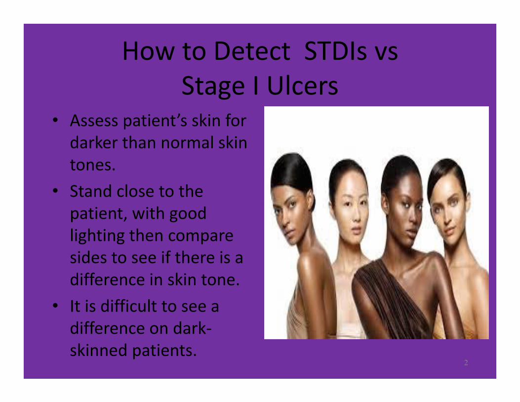
How to Detect STDIs vsStage I Ulcers
• Assess patient’s skin for darker than normal skin tones.
• Stand close to the patient, with good lighting then compare sides to see if there is a difference in skin tone.
• It is difficult to see a difference on dark‐skinned patients.
2
Free Template from www.brainybetty.com 3
The Wound Ostomy Continence (WOC) nurses were asked by one of our hospitals nursing units directors to educate the nursing staff on how to identify suspected deep tissue injury (SDTI) pressure ulcers, particularly in dark skin tone individuals. The director recognized this as a problem on her unit, but one that is system‐wide at Jackson‐Madison County General Hospital (JMCGH), and an issue in most acute and long‐term care facilities.
The WOC nurses wanted to educate the nursing staff on recognition of SDTI pressure ulcers in dark skin patients but light skin tone individuals as well, and to take it a step further with education on early identification of Stage I pressure ulcers.
Consent was obtained from the patient and/or family members and physicians for photographs taken. Photos were taken of the suspected deep tissue injuries and stage I ulcers. Other photos of SDTI and Stage I pressure ulcers were taken from web sites of WOCN and Google.
Free Template from www.brainybetty.com 4
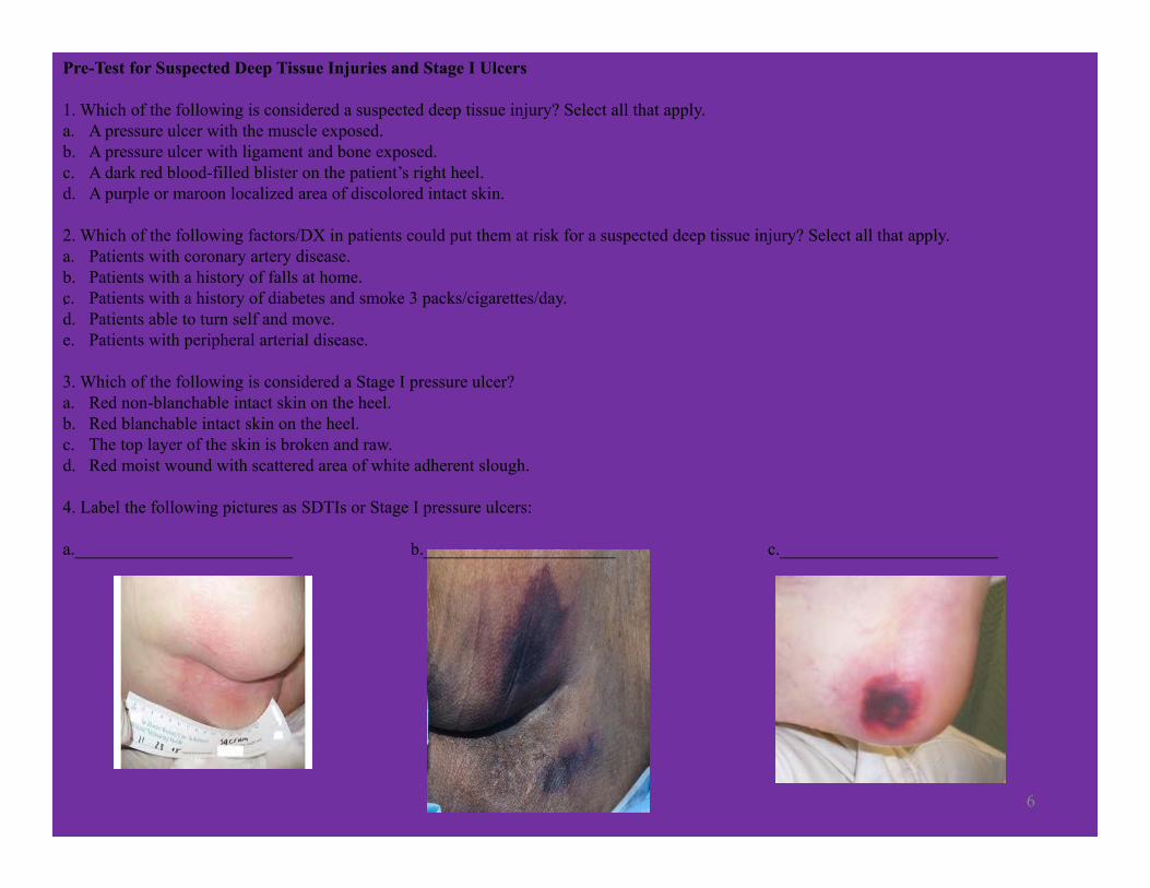
A pre‐test which included pictures to identify, was given to the staff nurses on their current knowledge of suspected deep tissue injuries and stage I ulcers.
Free Template from www.brainybetty.com 5
6
Pre-Test for Suspected Deep Tissue Injuries and Stage I Ulcers
1. Which of the following is considered a suspected deep tissue injury? Select all that apply.a. A pressure ulcer with the muscle exposed.b. A pressure ulcer with ligament and bone exposed.c. A dark red blood-filled blister on the patient’s right heel.d. A purple or maroon localized area of discolored intact skin.
2. Which of the following factors/DX in patients could put them at risk for a suspected deep tissue injury? Select all that apply.a. Patients with coronary artery disease.b. Patients with a history of falls at home.c. Patients with a history of diabetes and smoke 3 packs/cigarettes/day.d. Patients able to turn self and move.e. Patients with peripheral arterial disease.
3. Which of the following is considered a Stage I pressure ulcer?a. Red non-blanchable intact skin on the heel.b. Red blanchable intact skin on the heel.c. The top layer of the skin is broken and raw.d. Red moist wound with scattered area of white adherent slough.
4. Label the following pictures as SDTIs or Stage I pressure ulcers:
a._________________________ b.______________________ c._________________________
.
The staff nurses were then shown a PowerPoint presentation with detailed pictures of suspected deep tissue injuries, stage I ulcers; methods, such as looking, listening, and feeling (touch) when performing a thorough skin assessment; risk assessment tools, such as the Braden Scale; and recognizing key diagnoses when completing the patient’s health history (such as history of diabetes mellitus, coronary artery disease, peripheral vascular disease, etc.) that places the patient at higher risk of developing suspected deep tissue injuries, stage I pressure ulcers, and skin breakdown. The presentation concluded with treatment options for suspected deep tissue injuries, stage I pressure ulcers, and prevention of skin breakdown.
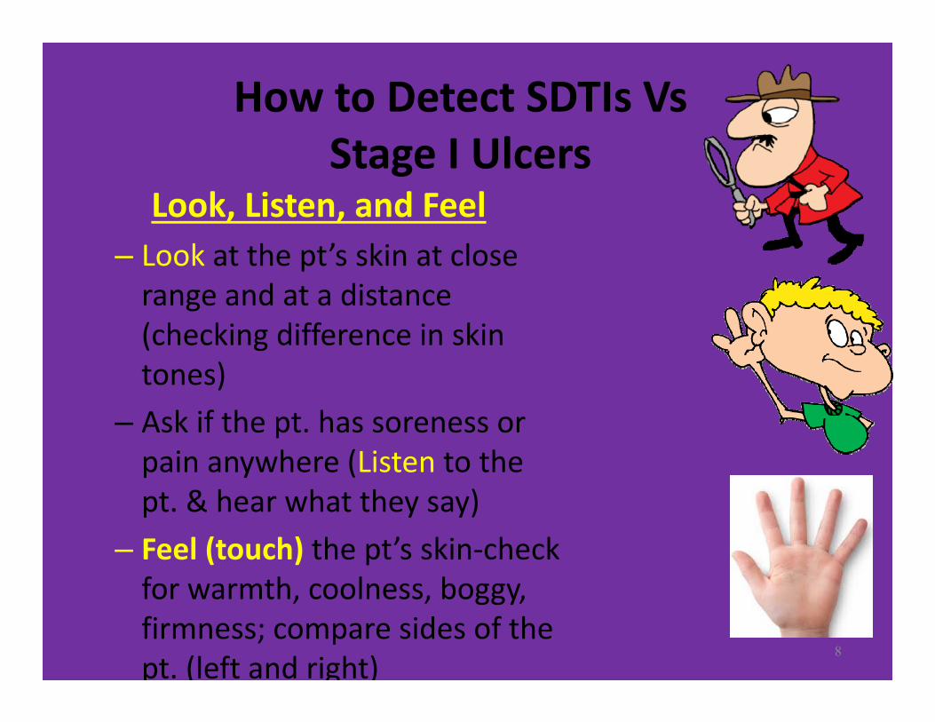
How to Detect SDTIs Vs Stage I Ulcers
Look, Listen, and Feel– Look at the pt’s skin at close range and at a distance (checking difference in skin tones)
– Ask if the pt. has soreness or pain anywhere (Listen to the pt. & hear what they say)
– Feel (touch) the pt’s skin‐check for warmth, coolness, boggy, firmness; compare sides of the pt. (left and right)
8

How to Detect a Stage I Pressure Ulcer?
• Red, nonblanchable intact skin
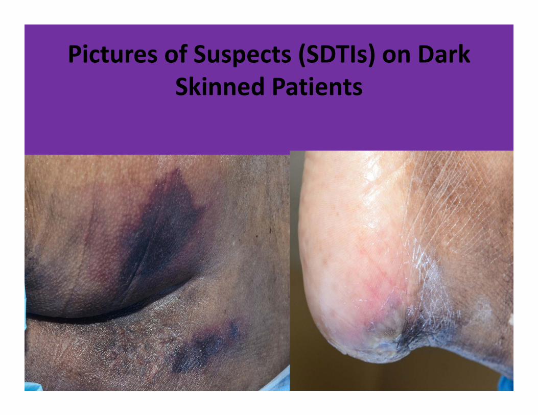
Pictures of Suspects (SDTIs) on Dark Skinned Patients
10
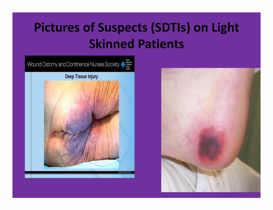
Pictures of Suspects (SDTIs) on Light Skinned Patients
11

Free Template from www.brainybetty.com 12
After the presentation, the staff nurses were given a post‐test to determine their knowledge of STDIs and stage I pressure ulcers.
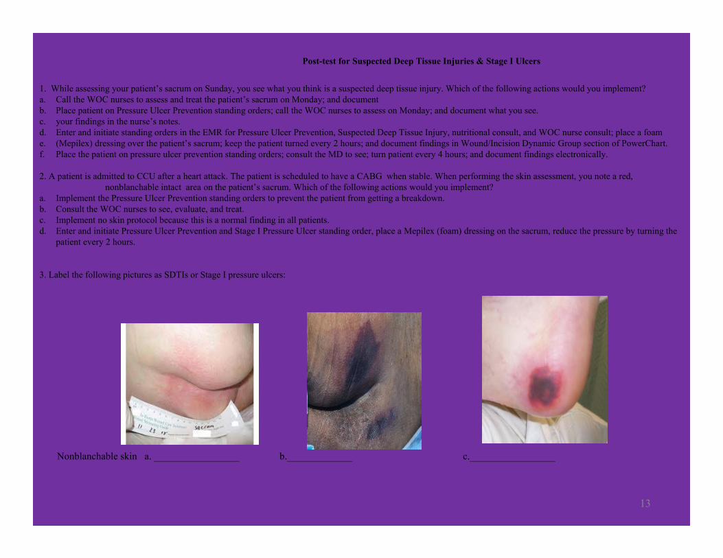
13
Post-test for Suspected Deep Tissue Injuries & Stage I Ulcers
1. While assessing your patient’s sacrum on Sunday, you see what you think is a suspected deep tissue injury. Which of the following actions would you implement?a. Call the WOC nurses to assess and treat the patient’s sacrum on Monday; and documentb. Place patient on Pressure Ulcer Prevention standing orders; call the WOC nurses to assess on Monday; and document what you see.c. your findings in the nurse’s notes.d. Enter and initiate standing orders in the EMR for Pressure Ulcer Prevention, Suspected Deep Tissue Injury, nutritional consult, and WOC nurse consult; place a foame. (Mepilex) dressing over the patient’s sacrum; keep the patient turned every 2 hours; and document findings in Wound/Incision Dynamic Group section of PowerChart.f. Place the patient on pressure ulcer prevention standing orders; consult the MD to see; turn patient every 4 hours; and document findings electronically.
2. A patient is admitted to CCU after a heart attack. The patient is scheduled to have a CABG when stable. When performing the skin assessment, you note a red, nonblanchable intact area on the patient’s sacrum. Which of the following actions would you implement?
a. Implement the Pressure Ulcer Prevention standing orders to prevent the patient from getting a breakdown.b. Consult the WOC nurses to see, evaluate, and treat.c. Implement no skin protocol because this is a normal finding in all patients.d. Enter and initiate Pressure Ulcer Prevention and Stage I Pressure Ulcer standing order, place a Mepilex (foam) dressing on the sacrum, reduce the pressure by turning the
patient every 2 hours.
3. Label the following pictures as SDTIs or Stage I pressure ulcers:
Nonblanchable skin a. _________________ b._____________ c._________________
Visual aids and educational preparation improved success on the post‐test and enhanced the staff nurses ability to identify SDTI and stage I pressure ulcers in individuals with dark skin tones. This form of education will be used on other units throughout the hospital as well as shorter versions without the PowerPoint presentation, using posters with pictures and bullet points still including the pre and post‐test. Ongoing education is necessary to keep current and new staff knowledgeable about early assessment and identification of SDTIs and stage I pressure ulcers.
Free Template from www.brainybetty.com 14
RESULTS

References
Brindle, T. (2010, July 7). Deep Tissue Injury and Differential Diagnosis. Retrieved on June 3, 2014 from www.browardhealth.org/upload/docs/conferences/woundostomy13/2Brindle_Differential‐Diagnosis.pdf
Google images: https://google.com/imghp?hl=en&tab=wi&ei=dhjRU5i6oo7ZigKT‐ICADw&ved=0CAQQqi4oAgHettric, H. (2014, June). Skin & Wound Considerations in Non‐Caucasian Skin. Presented at WOCN 46th Annual Conference, Nashville, TN.Lyder, C. (2009). Closing the Skin Assessment Disparity Gap Between Patients With Light and Darkly Pigmented Skin. Journal of Wound
Ostomy Continence Nursing, 36(3):285.McCurren, C., Constable, K., Carroll, M., & Bean, N. (2004, May/june). Darkly Pigmented Skin: Assessment Strategies and Pressure Ulcer
Detection. Journal of Wound, Ostomy & Continence Nursing, 31(S), S23‐S24.The National Pressure Ulcer Advisory Panel. (2014). NPUAP Pressure Ulcer Stages/Categories. Retrieved on June 6, 2014 from
http://www.npuap.org/resources/educational‐and‐clinical‐resources/npuap‐pressure‐ulcer‐stagescategories/
Free Template from www.brainybetty.com 15