Embed Size (px)
Citation preview
8/21/2019 HAP-CAH DiagImag Prepub July2015release 20150105
http://slidepdf.com/reader/full/hap-cah-diagimag-prepub-july2015release-20150105 1/7
1Copyright 2015 The Joint Commission
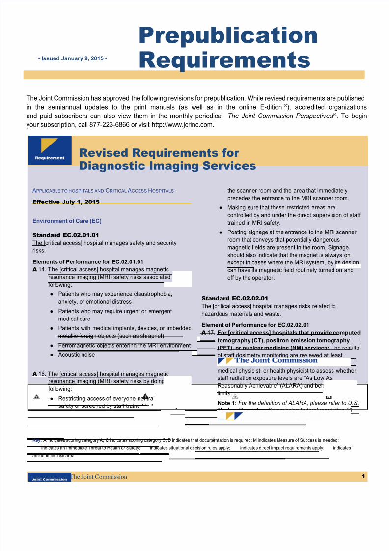
PrepublicationRequirements• Issued January 9, 2015 •
The Joint Commission has approved the following revisions for prepublication. While revised requirements are publishein the semiannual updates to the print manuals (as well as in the online E-dition®), accredited organization
and paid subscribers can also view them in the monthly periodical The Joint Commission Perspectives®. To begi
your subscription, call 877-223-6866 or visit http://www.jcrinc.com.
Revised Requirements forDiagnostic Imaging Services
APPLICABLE TO HOSPITALS AND
CRITICAL
ACCESS
HOSPITALS
Effective July 1, 2015
Environment of Care (EC)
Standard EC.02.01.01
The [critical access] hospital manages safety and security
risks.
Elements of Performance for EC.02.01.01
A 14. The [critical access] hospital manages magnetic
resonance imaging (MRI) safety risks associated with the
following:
● Patients who may experience claustrophobia,
anxiety, or emotional distress
● Patients who may require urgent or emergent
medical care
● Patients with medical implants, devices, or imbedded
metallic foreign objects (such as shrapnel)
● Ferromagnetic objects entering the MRI environment
● Acoustic noise
A 16. The [critical access] hospital manages magnetic
resonance imaging (MRI) safety risks by doing the
following:
● Restricting access of everyone not trained in MRI
safety or screened by staff trained in MRI safety from
the scanner room and the area that immediatelyprecedes the entrance to the MRI scanner room.
● Making sure that these restricted areas are
controlled by and under the direct supervision of staff
trained in MRI safety.
● Posting signage at the entrance to the MRI scanner
room that conveys that potentially dangerous
magnetic fields are present in the room. Signage
should also indicate that the magnet is always on
except in cases where the MRI system, by its design,
can have its magnetic field routinely turned on and
off by the operator.
Standard EC.02.02.01
The [critical access] hospital manages risks related to
hazardous materials and waste.
Element of Performance for EC.02.02.01
A 17. For [critical access] hospitals that provide computed
tomography (CT), positron emission tomography
(PET), or nuclear medicine (NM) services: The results
of staff dosimetry monitoring are reviewed at least
quarterly by the radiation safety officer, diagnostic
medical physicist, or health physicist to assess whether
staff radiation exposure levels are “As Low As
Reasonably Achievable” (ALARA) and below regulatorylimits.
Note 1: For the definition of ALARA, please refer to U.S.
Nuclear Regulatory Commission federal regulation 10
CFR 20.1003.
Key: A indicates scoring category A; C indicates scoring category C; D indicates that documentation is required; M indicates Measure of Success is needed;
indicates an Immediate Threat to Health or Safety; indicates situational decision rules apply; indicates direct impact requirements apply; indicates
an identified risk area

8/21/2019 HAP-CAH DiagImag Prepub July2015release 20150105
http://slidepdf.com/reader/full/hap-cah-diagimag-prepub-july2015release-20150105 2/7
Prepublication Requirements continued January 9, 2015
2Copyright 2015 The Joint Commission
abdomen, pediatric brain, and pediatric abdomen. If
one or more of these protocols is not used by the
[critical access] hospital, other commonly used CT
protocols may be substituted.
● Verifies that the radiation dose (in the form ofCTDIvol) produced and measured for each protocol
tested is within 20 percent of the CTDIvol displayed
on the CT console. The dates, results, and
verifications of these measurements are
documented.
Note 1: This element of performance is only applicable
for systems capable of calculating and displaying
radiation doses.
Note 2: This element of performance does not apply to
dental cone beam CT radiographic imaging studies
performed for diagnosis of conditions affecting the
maxillofacial region or to obtain guidance for thetreatment of such conditions.
Note 3: Medical physicists are accountable for these
activities. They may be assisted with the testing and
evaluation of equipment performance by individuals who
have the required training and skills, as determined by
the physicist. (See also HR.01.02.01, EP 1;
HR.01.02.05, EP 20; HR.01.02.07, EPs 1 and 2;
HR.01.06.01, EP 1; and LD.03.06.01, EP 4)
A 19. For diagnostic computed tomography (CT)
services: At least annually, a diagnostic medical
physicist conducts a performance evaluation of all CTimaging equipment. The evaluation results, along with
recommendations for correcting any problems identified,
are documented. The evaluation includes the use of
phantoms to assess the following imaging metrics:
● Image uniformity
● Slice thickness accuracy
● Slice position accuracy (when prescribed from a
scout image)
● Alignment light accuracy
● Table travel accuracy
● Radiation beam width
● High-contrast resolution
● Low-contrast resolution
● Geometric or distance accuracy
● CT number accuracy and uniformity
● Artifact evaluation
Note 1: This element of performance does not apply to
dental cone beam CT radiographic imaging studies
performed for diagnosis of conditions affecting the
Note 2: This element of performance does not apply to
dental cone beam CT radiographic imaging studies
performed for diagnosis of conditions affecting the
maxillofacial region or to obtain guidance for the
treatment of such conditions.
Standard EC.02.04.01
The [critical access] hospital manages medical equipment
risks.
Element of Performance for EC.02.04.01
A 10. The [critical access] hospital identifies quality control and
maintenance activities to maintain the quality of the
diagnostic computed tomography (CT), positron
emission tomography (PET), magnetic resonance
imaging (MRI), and nuclear medicine (NM) images
produced. The [critical access] hospital identifies how
often these activities should be conducted. (See alsoEC.02.04.03, EP 15)
Standard EC.02.04.03
The [critical access] hospital inspects, tests, and maintains
medical equipment.
Elements of Performance for EC.02.04.03
C 15. The [critical access] hospital maintains the quality of the
diagnostic computed tomography (CT), positron
emission tomography (PET), magnetic resonance
imaging (MRI), and nuclear medicine (NM) images
produced. (See also EC.02.04.01, EP 7)
A 17. For [critical access] hospitals in California that
provide computed tomography (CT) services: A
qualified medical physicist measures the actual radiation
dose * produced by each diagnostic CT imaging system
at least annually and verifies that the radiation dose
displayed on the system for standard adult brain, adult
abdomen, and pediatric brain protocols is within 20
percent of the actual amount of radiation dose delivered.
The dates of these verifications are documented.
Note: This element of performance is applicable only for
systems capable of calculating and displaying radiationdoses.
* For the definition of “radiation dose” refer to section 115111(f) of theCalifornia Health and Safety Code.
For diagnostic computed tomography (CT) services: At
least annually, a diagnostic medical physicist does the
following:
● Measures the radiation dose (in the form of volume
computed tomography dose index [CTDIvol])
produced by each diagnostic CT imaging system for
the following four CT protocols: adult brain, adult

8/21/2019 HAP-CAH DiagImag Prepub July2015release 20150105
http://slidepdf.com/reader/full/hap-cah-diagimag-prepub-july2015release-20150105 3/7
Prepublication Requirements continued January 9, 2015
3Copyright 2015 The Joint Commission
maxillofacial region or to obtain guidance for the
treatment of such conditions.
Note 2: Medical physicists are accountable for these
activities. They may be assisted w ith the testing and
evaluation of equipment performance by individuals who
have the required training and skills, as determined by
the physicist. (See also HR.01.02.01, EP 1;
HR.01.02.05, EP 20; HR.01.02.07, EPs 1 and 2;
HR.01.06.01, EP 1; and LD.03.06.01, EP 4)
A 20. At least annually, a diagnostic medical physicist or
magnetic resonance imaging (MRI) scientist conducts a
performance evaluation of all MRI imaging equipment.
The evaluation results, along with recommendations for
correcting any problems identified, are documented. The
evaluation includes the use of phantoms to assess the
following imaging metrics:
● Image uniformity for all radiofrequency (RF) coils
used clinically
● Signal-to-noise ratio (SNR) for all coils used clinically
● Slice thickness accuracy
● Slice position accuracy
● Alignment light accuracy
● High-contrast resolution
● Low-contrast resolution (or contrast-to-noise ratio)
● Geometric or distance accuracy
● Magnetic field homogeneity
● Artifact evaluation
Note: Medical physicists or MRI scientists are
accountable for these activities. They may be assisted
with the testing and evaluation of equipment
performance by individuals who have the required
training and skills, as determined by the medical
physicist or MRI scientist. (See also HR.01.02.01, EP 1;
HR.01.02.05, EP 20; HR.01.02.07, EPs 1 and 2;
HR.01.06.01, EP 1; and LD.03.06.01, EP 4)
A 21. At least annually, a diagnostic medical physicist or
nuclear medicine physicist conducts a performance
evaluation of all nuclear medicine imaging equipment.The evaluation results, along with recommendations for
correcting any problems identified, are documented. The
evaluations are conducted for all of the image types
produced clinically by each NM scanner (for example,
planar and/or tomographic) and include the use of
phantoms to assess the following imaging metrics:
● Image uniformity/system uniformity
● High-contrast resolution/system spatial resolution
● Sensitivity
● Energy resolution
● Count-rate performance
● Artifact evaluation
Note 1: The following test is recommended, but not
required: Low-contrast resolution or detectability for non-
planar acquisitions.
Note 2: The medical physicist or nuclear medicine
physicist is accountable for these activities. He or she
may be assisted with the testing and evaluation of
equipment performance by individuals who have the
required training and skills, as determined by the medical
physicist or nuclear medicine physicist. (See also
HR.01.02.01, EP 1; HR.01.02.05, EP 20; HR.01.02.07,
EPs 1 and 2; HR.01.06.01, EP 1; and LD.03.06.01, EP
4)
A 22. At least annually, a diagnostic medical physicist
conducts a performance evaluation of all positron
emission tomography (PET) imaging equipment. The
evaluation results, along with recommendations for
correcting any problems identified, are documented. The
evaluations are conducted for all of the image types
produced clinically by each PET scanner (for example,
planar and/or tomographic) and include the use of
phantoms to assess the following imaging metrics:
● Image uniformity/system uniformity
● High-contrast resolution/system spatial resolution
● Low-contrast resolution or detectability (not
applicable for planar acquisitions)
● Artifact evaluation
Note 1: The following tests are recommended, but not
required, for PET scanner testing: sensitivity, energy
resolution, and count-rate performance.
Note 2: Medical physicists are accountable for these
activities. They may be assisted with the testing and
evaluation of equipment performance by individuals who
have the required training and skills, as determined by
the medical physicist. (See also HR.01.02.01, EP 1;
HR.01.02.05, EP 20; HR.01.02.07, EPs 1 and 2;
HR.01.06.01, EP 1; and LD.03.06.01, EP 4)
A 23. For computed tomography (CT), positron emission
tomography (PET), nuclear medicine (NM), or
magnetic resonance imaging (MRI) services: The
annual performance evaluation conducted by the
diagnostic medical physicist or MRI scientist (for MRI
only) includes testing of image acquisition display

8/21/2019 HAP-CAH DiagImag Prepub July2015release 20150105
http://slidepdf.com/reader/full/hap-cah-diagimag-prepub-july2015release-20150105 4/7
Prepublication Requirements continued January 9, 2015
4Copyright 2015 The Joint Commission
monitors for maximum and minimum luminance,
luminance uniformity, resolution, and spatial accuracy.
Note 1: This element of performance does not apply to
dental cone beam CT radiographic imaging studies
performed for diagnosis of conditions affect ing the
maxillofacial region or to obtain guidance for the
treatment of such conditions.
Note 2: Medical physicists or MRI scientists are
accountable for these activities. They may be assisted
with the testing and evaluation of equipment
performance by individuals who have the required
training and skills, as determined by the physicist or MRI
scientist. (See also HR.01.02.01, EP 1; HR.01.02.05, EP
20; HR.01.02.07, EPs 1 and 2; HR.01.06.01, EP 1; and
LD.03.06.01, EP 4)
Standard EC.02.06.05The [critical access] hospital manages its environment during
demolition, renovation, or new construction to reduce risk to
those in the organization.
Elements of Performance for EC.02.06.05
A 4. For computed tomography (CT), positron emission
tomography (PET), or nuclear medicine (NM)
services: Prior to installation of new imaging equipment,
replacement of existing imaging equipment, or
modification to rooms where ionizing radiation will be
emitted or radioactive materials will be stored (such as
scan rooms or hot labs), a medical physicist or health
physicist conducts a structural shielding design *
assessment to specify required radiation shielding.
Note: This element of performance does not apply to
dental cone beam CT radiographic imaging studies
performed for diagnosis of conditions affecting the
maxillofacial region or to obtain guidance for the
treatment of such conditions.
* For additional guidance on shielding designs and radiation protectionsurveys, see National Council on Radiation Protection andMeasurements Report No. 147 (NCRP-147).
A 6. For computed tomography (CT), positron emission
tomography (PET), or nuclear medicine (NM)
services: After installation of imaging equipment orconstruction in rooms where ionizing radiation will be
emitted or radioactive materials will be stored, a medical
physicist or health physicist conducts a radiation
protection survey to verify the adequacy of installed
shielding. * This survey is conducted prior to clinical use
of the room.
Note: This element of performance does not apply to
dental cone beam CT radiographic imaging studies
performed for diagnosis of conditions affecting the
maxillofacial region or to obtain guidance for the
treatment of such conditions.
* For additional guidance on shielding designs and radiation protectionsurveys, see National Council on Radiation Protection andMeasurements Report No. 147 (NCRP-147).
Human Resources (HR)
Standard HR.01.02.05
The [critical access] hospital verifies staff qualifications.
Element of Performance for HR.01.02.05
C 20. The [critical access] hospital verifies and documents
that diagnostic medical physicists who support computed
tomography (CT) services have board certification in
diagnostic radiologic physics or radiologic physics by the
American Board of Radiology, or in Diagnostic Imaging
Physics by the American Board of Medical Physics, or in
Diagnostic Radiological Physics by the CanadianCollege of Physicists in Medicine, or meet all of the
following requirements:
● A graduate degree in physics, medical physics,
biophysics, radiologic physics, medical health
physics, or a closely related science or engineering
discipline from an accredited college or university
● College coursework in the biological sciences with at
least one course in biology or radiation biology and
one course in anatomy, physiology, or a similar topic
related to the practice of medical physics
● Documented experience in a clinical CT environment
conducting at least 10 CT performance evaluationsunder the direct supervision of a board-certified
medical physicist
Note: This element of performance does not apply to
dental cone beam CT radiographic imaging studies
performed for diagnosis of conditions affecting the
maxillofacial region or to obtain guidance for the
treatment of such conditions.
Standard HR.01.05.03
Staff participate in ongoing education and training.
Elements of Performance for HR.01.05.03
C 14. The [critical access] hospital verifies and documents
that technologists who perform diagnostic computed
tomography (CT) examinations participate in ongoing
education that includes annual training on the following:
● Radiation dose optimization techniques and tools for
pediatric and adult patients addressed in the Image
Gently® and Image Wisely® campaigns

8/21/2019 HAP-CAH DiagImag Prepub July2015release 20150105
http://slidepdf.com/reader/full/hap-cah-diagimag-prepub-july2015release-20150105 5/7
Prepublication Requirements continued January 9, 2015
5Copyright 2015 The Joint Commission
● Safe procedures for operation of the types of CT
equipment they will use
Note 1: Information on the Image Gently and Image
Wisely initiatives can be found online at
http://www.imagegently.org and
http://www.imagewisely.org, respectively.
Note 2: This element of performance does not apply to
CT systems used for therapeutic radiation treatment
planning or delivery, or for calculat ing attenuation
coefficients for nuclear medicine studies.
Note 3: This element of performance does not apply to
dental cone beam CT radiographic imaging studies
performed for diagnosis of conditions affecting the
maxillofacial region or to obtain guidance for the
treatment of such conditions.
C 25. The [critical access] hospital verifies and documentsthat technologists who perform magnetic resonance
imaging (MRI) examinations participate in ongoing
education that includes annual training on safe MRI
practices in the MRI environment, including the following:
● Patient screening criteria that address ferromagnetic
items, electrically conductive items, medical implants
and devices, and risk for Nephrogenic Systemic
Fibrosis (NSF)
● Proper patient and equipment positioning activities to
avoid thermal injuries
●
Equipment and supplies that have been determinedto be acceptable for use in the MRI environment (MR
safe or MR conditional) *
● MRI safety response procedures for patients who
require urgent or emergent medical care
● MRI system emergency shutdown procedures, such
as MRI system quench and cryogen safety
procedures
● Patient hearing protection
● Management of patients with claustrophobia,
anxiety, or emotional distress
* Terminology for defining the safety of items in the magnetic resonance
environment is provided in ASTM F2503 Standard Practice for MarkingMedical Devices and Other Items for Safety in the Magnetic ResonanceEnvironment (http://www.astm.org).
Medication Management (MM)
Standard MM.06.01.01
The [critical access] hospital safely administers medications.
Element of Performance for MM.06.01.01
C 13. Before administering a radioactive pharmaceutical for
diagnostic purposes, staff verify that the dose to be
administered is within 20% of the prescribed dose, or, if
the dose is prescribed as a range, staff verify that thedose to be administered is within the prescribed range.
Provision of Care, Treatment, and Services (PC)
Standard PC.01.02.15
The [critical access] hospital provides for diagnostic testing.
Elements of Performance for PC.01.02.15
C 5. For [critical access] hospitals in California that provide
computed tomography (CT) services: The [critical
access] hospital documents in the patient’s record the
radiation dose index (CTDIvol, DLP, or size-specific doseestimate [SSDE]) on every study produced during a
diagnostic computed tomography (CT) examination. The
radiation dose index must be exam specific, summarized
by series or anatomic area, and documented in a
retrievable format.
Note 1: This element of performance is only applicable
only for systems capable of calculating and displaying
radiation doses indices.
Note 2: This element of performance does not apply to
systems used for therapeutic radiation treatment
planning or del ivery, or for calculating attenuation
coefficients for nuclear medicine studies.Note 3: This element of performance does not apply to
dental cone beam CT radiographic imaging studies
performed for diagnosis of conditions affecting the
maxillofacial region or to obtain guidance for the
treatment of such conditions.
Note 4: While the CTDIvol, DLP, and SSDE are useful
indicators for monitoring radiation dose indices from the
CT machine, they do not represent the patient’s radiation
dose.
* For the definition of “radiation dose” refer to section 115111(f) of theCalifornia Health and Safety Code.
C 6. For [critical access] hospitals that provide
diagnostic computed tomography (CT) services: The
interpretive report of a diagnostic CT study includes the
volume computed tomography dose index (CTDIvol) or
dose-length product (DLP) radiation dose. The dose is
either recorded in the patient's interpretive report or
included on the protocol page.

8/21/2019 HAP-CAH DiagImag Prepub July2015release 20150105
http://slidepdf.com/reader/full/hap-cah-diagimag-prepub-july2015release-20150105 6/7
Prepublication Requirements continued January 9, 2015
6Copyright 2015 The Joint Commission
C 7. For [critical access] hospitals in California that
provide computed tomography (CT) services: The
[critical access] hospital electronically sends each CT
study and protocol page that lists the radiation dose *
and related technical factors to the [critical access]
hospital’s electronic picture archiving andcommunications system.
Note: This element of performance is applicable only forsystems capable of calculating and displaying radiationdoses.
* For the definition of "radiation dose" refer to section 115111(f) of theCalifornia Health and Safety Code.
A 10. For [critical access] hospitals that provide
diagnostic computed tomography (CT), magnetic
resonance imaging (MRI), positron emission
tomography (PET), or nuclear medicine (NM)
services: Prior to conducting a diagnostic imaging
study, the [critical access] hospital verifies the following:
● Correct patient
● Correct imaging site
● Correct patient positioning
● For CT only: Correct imaging protocol
● For CT only: Correct scanner parameters
Note: This element of performance does not apply to
dental cone beam CT radiographic imaging studies
performed for diagnosis of conditions affecting the
maxillofacial region or to obtain guidance for the
treatment of such conditions.
A 12. For [critical access] hospitals that provide
diagnostic computed tomography (CT), magnetic
resonance imaging (MRI), positron emission
tomography (PET), or nuclear medicine (NM)
services: The [critical access] hospital considers the
patient’s age and recent imaging exams when deciding
on the most appropriate type of imaging exam.
Note 1: Knowledge of a patient’s recent imaging exams
can help to prevent unnecessary duplication of these
examinations.
Note 2: This element of performance does not apply to
dental cone beam CT radiographic imaging studies
performed for diagnosis of conditions affecting the
maxillofacial region or to obtain guidance for the
treatment of such conditions.
Standard PC.01.03.01
The [critical access] hospital plans the patient’s care.
Elements of Performance for PC.01.03.01
25. The [critical access] hospital establishes or adopts
diagnostic computed tomography (CT) imaging protocols
based on current standards of practice, which address
key criteria including clinical indication, contrast
administration, age (to indicate whether the patient is
pediatric or an adult), patient size and body habitus, and
the expected radiation dose index range.
Note: This element of performance does not apply to
dental cone beam CT radiographic imaging studies
performed for diagnosis of conditions affecting the
maxillofacial region or to obtain guidance for the
treatment of such conditions.
26. Diagnostic computed tomography (CT) imaging
protocols are reviewed and kept current with input from
an interpreting radiologist, medical physicist, and lead
imaging technologist to make certain that they adhere to
current standards of practice and account for changes in
CT imaging equipment. These reviews are conducted at
time frames identified by the [critical access] hospital.
Note: This element of performance does not apply to
dental cone beam CT radiographic imaging studies
performed for diagnosis of conditions affecting the
maxillofacial region or to obtain guidance for the
treatment of such conditions.
Performance Improvement (PI)
Standard PI.01.01.01
The [critical access] hospital collects data to monitor its
performance.
Elements of Performance for PI.01.01.01
46. The [critical access] hospital collects data on patient
thermal injuries that occur during magnetic resonance
imaging exams.
47. The [critical access] hospital collects data on the
following:
● Incidents where ferromagnetic objects unintentionally
entered the magnetic resonance imaging (MRI)scanner room
● Injuries resulting from the presence of ferromagnetic
objects in the MRI scanner room
Standard PI.02.01.01
The [critical access] hospital compiles and analyzes data.

8/21/2019 HAP-CAH DiagImag Prepub July2015release 20150105
http://slidepdf.com/reader/full/hap-cah-diagimag-prepub-july2015release-20150105 7/7
Prepublication Requirements continued January 9, 2015
7Copyright 2015 The Joint Commission
Element of Performance for PI.02.01.01
A 6. The [critical access] hospital reviews and analyzes
incidents where the radiation dose index (CTDIvol, DLP,
or size-specific dose estimate [SSDE]) from diagnostic
CT examinations exceeded expected dose index rangesidentified in imaging protocols. These incidents are then
compared to external benchmarks.
Note 1: While the CTDIvol, DLP, and SSDE are useful
indicators for monitoring radiation dose indices from the
CT machine, they do not represent the patient’s radiation
dose.
Note 2: This element of performance does not apply to
dental cone beam CT radiographic imaging studies
performed for diagnosis of conditions affecting the
maxillofacial region or to obtain guidance for the
treatment of such conditions.