Embed Size (px)
Citation preview

This article appeared in a journal published by Elsevier. The attachedcopy is furnished to the author for internal non-commercial researchand education use, including for instruction at the authors institution
and sharing with colleagues.
Other uses, including reproduction and distribution, or selling orlicensing copies, or posting to personal, institutional or third party
websites are prohibited.
In most cases authors are permitted to post their version of thearticle (e.g. in Word or Tex form) to their personal website orinstitutional repository. Authors requiring further information
regarding Elsevier’s archiving and manuscript policies areencouraged to visit:
http://www.elsevier.com/copyright
Author's personal copy
Complications ofEquineWoundManagement andDermatologic Surgery
R. Reid Hanson, DVM
Complications of wounds and cosmetic surgery although frequent can be accuratelymanaged with a combination of timely surgical and medical intervention to ensure thebest possible outcome. The lack of soft tissue protection and a large quantity of sus-ceptible synovial, tendon, ligament, and neurovascular structures make early and me-ticulous evaluation of limb wounds critical. Chronic wounds should be consideredinfected and may become afflicted with sarcoid, Pythium spp, Habronema spp, orDraschia spp, and rarely neoplasia. Methicillin-resistant Staphylococcus aureus(MRSA) infections can consequently be difficult to treat and are associated withincreased morbidity, mortality, and treatment costs. Skin grafting is usually usedfollowing a period of open wound management and after healthy granulation tissueformation. Penetrating wounds of the abdomen or thorax have a guarded prognosisresulting from the ensuing potential for infection and pneumothorax. Gunshot woundslimited to the skeletal muscles have a good prognosis, whereas injuries that involvevital organs decrease survivability.
COMPLICATIONS OF REPAIROsseous Sequestration
The distal limbs of horses are extremely susceptible to damage of the periosteum andunderlying bone because of the lack of soft tissue protection. Because the periosteumprovides the blood supply to the outer one third of the cortical bone, disruption of theperiosteum leads to ischemia of the bone with eventual bone death secondary tothese alterations in blood flow. This ischemic locale is very susceptible to bacterial col-onization and proliferation originating from the inciting trauma. Blunt trauma with noexternal entry wound may result in sequestrum formation indicating that bacterial
Department of Clinical Sciences, College of Veterinary Medicine, Auburn University, JTVaughan Hall, 1500 Wire Road, Auburn, AL 36849, USAE-mail address: [email protected]
KEYWORDS
� Equine � Wounds � Complications� Management � Dermatologic surgery
Vet Clin Equine 24 (2009) 663–696doi:10.1016/j.cveq.2008.10.005 vetequine.theclinics.com0749-0739/08/$ – see front matter ª 2009 Elsevier Inc. All rights reserved.
Author's personal copy
inoculation may also occur by way of the bloodstream. This type of injury is morecommonly present with trauma to the metacarpal or metatarsal bones.1
The body may be able to resorb the sequestrum or expel it from the draining tractdepending on the size of the devitalized bone fragment. Larger fragments usually per-sist and lead to more extensive clinical disease. The sequestrum can becomea chronic nidus thereby delaying the healing process. Because of the lack of vascular-ization, the immune response at the site is inadequate, leading to a chronic infectionthat persists in the face of a normal immune system.2,3 The lack of a relative reliableblood flow impedes the migration of osteoclasts to the site hindering the natural re-sorption of bone. These two events collectively result in a chronic inflammatory focusthat the body cannot or only slowly resolves.1
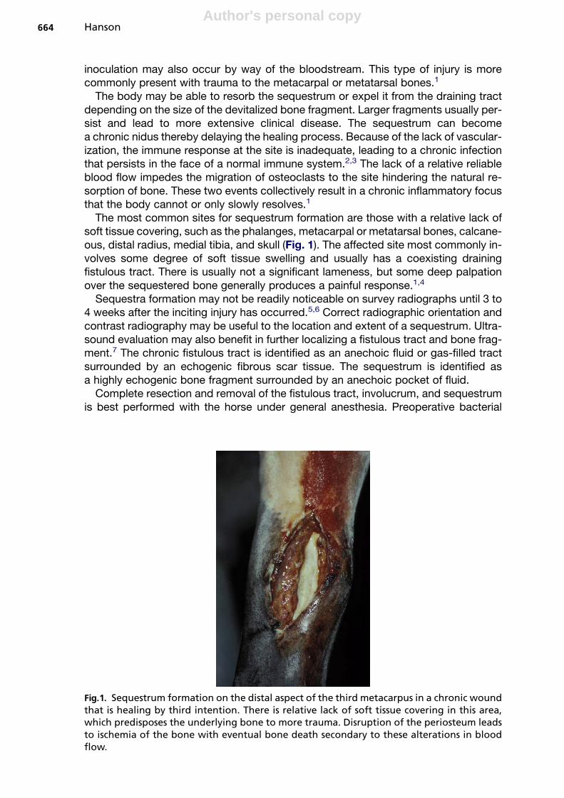
The most common sites for sequestrum formation are those with a relative lack ofsoft tissue covering, such as the phalanges, metacarpal or metatarsal bones, calcane-ous, distal radius, medial tibia, and skull (Fig. 1). The affected site most commonly in-volves some degree of soft tissue swelling and usually has a coexisting drainingfistulous tract. There is usually not a significant lameness, but some deep palpationover the sequestered bone generally produces a painful response.1,4
Sequestra formation may not be readily noticeable on survey radiographs until 3 to4 weeks after the inciting injury has occurred.5,6 Correct radiographic orientation andcontrast radiography may be useful to the location and extent of a sequestrum. Ultra-sound evaluation may also benefit in further localizing a fistulous tract and bone frag-ment.7 The chronic fistulous tract is identified as an anechoic fluid or gas-filled tractsurrounded by an echogenic fibrous scar tissue. The sequestrum is identified asa highly echogenic bone fragment surrounded by an anechoic pocket of fluid.
Complete resection and removal of the fistulous tract, involucrum, and sequestrumis best performed with the horse under general anesthesia. Preoperative bacterial
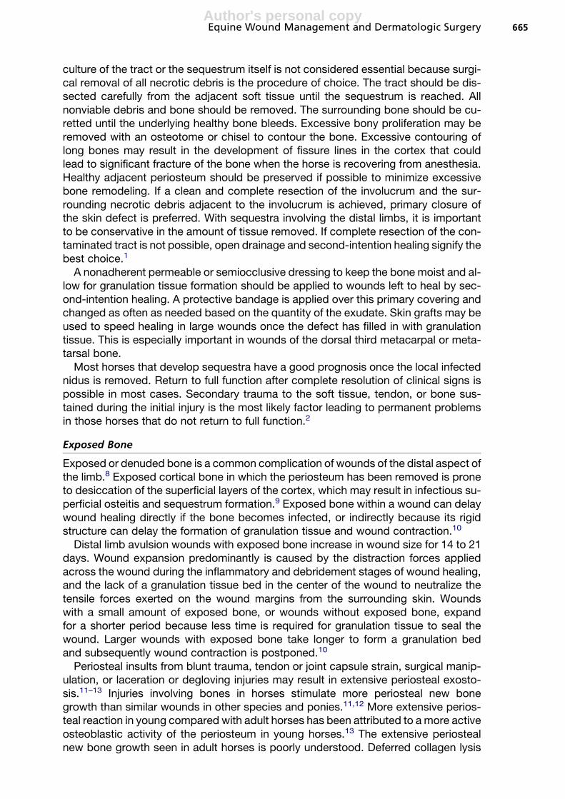
Fig.1. Sequestrum formation on the distal aspect of the third metacarpus in a chronic woundthat is healing by third intention. There is relative lack of soft tissue covering in this area,which predisposes the underlying bone to more trauma. Disruption of the periosteum leadsto ischemia of the bone with eventual bone death secondary to these alterations in bloodflow.
Hanson664
Author's personal copy
culture of the tract or the sequestrum itself is not considered essential because surgi-cal removal of all necrotic debris is the procedure of choice. The tract should be dis-sected carefully from the adjacent soft tissue until the sequestrum is reached. Allnonviable debris and bone should be removed. The surrounding bone should be cu-retted until the underlying healthy bone bleeds. Excessive bony proliferation may beremoved with an osteotome or chisel to contour the bone. Excessive contouring oflong bones may result in the development of fissure lines in the cortex that couldlead to significant fracture of the bone when the horse is recovering from anesthesia.Healthy adjacent periosteum should be preserved if possible to minimize excessivebone remodeling. If a clean and complete resection of the involucrum and the sur-rounding necrotic debris adjacent to the involucrum is achieved, primary closure ofthe skin defect is preferred. With sequestra involving the distal limbs, it is importantto be conservative in the amount of tissue removed. If complete resection of the con-taminated tract is not possible, open drainage and second-intention healing signify thebest choice.1
A nonadherent permeable or semiocclusive dressing to keep the bone moist and al-low for granulation tissue formation should be applied to wounds left to heal by sec-ond-intention healing. A protective bandage is applied over this primary covering andchanged as often as needed based on the quantity of the exudate. Skin grafts may beused to speed healing in large wounds once the defect has filled in with granulationtissue. This is especially important in wounds of the dorsal third metacarpal or meta-tarsal bone.
Most horses that develop sequestra have a good prognosis once the local infectednidus is removed. Return to full function after complete resolution of clinical signs ispossible in most cases. Secondary trauma to the soft tissue, tendon, or bone sus-tained during the initial injury is the most likely factor leading to permanent problemsin those horses that do not return to full function.2
Exposed Bone
Exposed or denuded bone is a common complication of wounds of the distal aspect ofthe limb.8 Exposed cortical bone in which the periosteum has been removed is proneto desiccation of the superficial layers of the cortex, which may result in infectious su-perficial osteitis and sequestrum formation.9 Exposed bone within a wound can delaywound healing directly if the bone becomes infected, or indirectly because its rigidstructure can delay the formation of granulation tissue and wound contraction.10
Distal limb avulsion wounds with exposed bone increase in wound size for 14 to 21days. Wound expansion predominantly is caused by the distraction forces appliedacross the wound during the inflammatory and debridement stages of wound healing,and the lack of a granulation tissue bed in the center of the wound to neutralize thetensile forces exerted on the wound margins from the surrounding skin. Woundswith a small amount of exposed bone, or wounds without exposed bone, expandfor a shorter period because less time is required for granulation tissue to seal thewound. Larger wounds with exposed bone take longer to form a granulation bedand subsequently wound contraction is postponed.10
Periosteal insults from blunt trauma, tendon or joint capsule strain, surgical manip-ulation, or laceration or degloving injuries may result in extensive periosteal exosto-sis.11–13 Injuries involving bones in horses stimulate more periosteal new bonegrowth than similar wounds in other species and ponies.11,12 More extensive perios-teal reaction in young compared with adult horses has been attributed to a more activeosteoblastic activity of the periosteum in young horses.13 The extensive periostealnew bone growth seen in adult horses is poorly understood. Deferred collagen lysis
Equine Wound Management and Dermatologic Surgery 665
Author's personal copy
compared with other species may be a contributing factor.11 The more extensive peri-osteal new bone formation in horses compared with ponies is alleged to be the resultof a slower onset and longer duration of the periosteal response and prolonged exten-sive limb swelling in horses, as compared with ponies.12
Despite the common occurrence of exposed bone associated with trauma to thedistal aspect of the limb, there has been little investigation into methods of stimulatingcoverage of granulation tissue over exposed bone in horses. Granulation tissue devel-opment is a very important role in second-intention healing because it provides a bar-rier to infection and mechanical trauma for the underlying tissues. Healthy granulationtissue is resistant to infection and provides a moist surface for epithelialization. Thedelay in wound healing caused by exposed bone has prompted the search for differentmethods to promote granulation tissue coverage of bone in other species.
Head trauma, thermal injury, and surgical oncology often results in exposed bone ofthe cranium in humans.9,14 In these cases the outer cortex of the uncovered portion ofthe cranium is fenestrated with drill holes, burrs, or lasers to expose the medullary cav-ity from which granulation tissue grows to cover the exposed bone.9,15–17 Similarly, ex-posed cortices of long bones in humans have been fenestrated with drill holes topromote granulation tissue formation.12 It has been suggested that the drill holes pro-mote healing by allowing osteogenic factors from the medullary cavity access to thewound, or by the enhancement of healing of bone and soft tissue by a nonspecific re-sponse known as ‘‘the regional acceleratory phenomenon.’’18 Cortical fenestrationcombined with drugs that promote topical granulation tissue may accelerate granula-tion tissue coverage compared with control wounds, but further investigation isneeded.
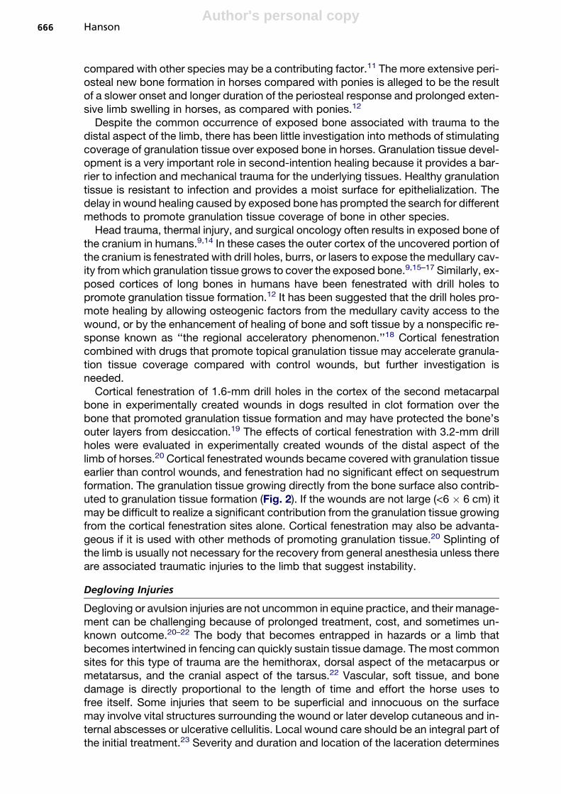
Cortical fenestration of 1.6-mm drill holes in the cortex of the second metacarpalbone in experimentally created wounds in dogs resulted in clot formation over thebone that promoted granulation tissue formation and may have protected the bone’souter layers from desiccation.19 The effects of cortical fenestration with 3.2-mm drillholes were evaluated in experimentally created wounds of the distal aspect of thelimb of horses.20 Cortical fenestrated wounds became covered with granulation tissueearlier than control wounds, and fenestration had no significant effect on sequestrumformation. The granulation tissue growing directly from the bone surface also contrib-uted to granulation tissue formation (Fig. 2). If the wounds are not large (<6 � 6 cm) itmay be difficult to realize a significant contribution from the granulation tissue growingfrom the cortical fenestration sites alone. Cortical fenestration may also be advanta-geous if it is used with other methods of promoting granulation tissue.20 Splinting ofthe limb is usually not necessary for the recovery from general anesthesia unless thereare associated traumatic injuries to the limb that suggest instability.
Degloving Injuries
Degloving or avulsion injuries are not uncommon in equine practice, and their manage-ment can be challenging because of prolonged treatment, cost, and sometimes un-known outcome.20–22 The body that becomes entrapped in hazards or a limb thatbecomes intertwined in fencing can quickly sustain tissue damage. The most commonsites for this type of trauma are the hemithorax, dorsal aspect of the metacarpus ormetatarsus, and the cranial aspect of the tarsus.22 Vascular, soft tissue, and bonedamage is directly proportional to the length of time and effort the horse uses tofree itself. Some injuries that seem to be superficial and innocuous on the surfacemay involve vital structures surrounding the wound or later develop cutaneous and in-ternal abscesses or ulcerative cellulitis. Local wound care should be an integral part ofthe initial treatment.23 Severity and duration and location of the laceration determines
Hanson666
Author's personal copy
the best approach to the treatment of degloving injuries because healing of woundsinvolving the distal limb is often delayed when compared with other areas of thebody, further complicating the healing process.
Primary repair of the wound is the preferred treatment for wounds that involvedetachment of skin with maintenance of an intact blood supply (Fig. 3). Complications,such as sequestrum formation, are lessened and healing is improved when theexposed bone and tendons are covered with skin and soft tissue in the immediateposttrauma period. Closing as much of the wound as possible improves the cosmeticand functional outcome and lessens the amount of healing having to occur by secondintention.
Delayed closure of a degloving injury is preferred when there is significant contam-ination, swelling, and trauma of the wound without loss of skin. Initial treatment for thefirst 2 to 3 days after injury includes debridement and lavage of the wound followed bywet to dry bandages to facilitate further debridement. Pressure bandaging is indicatedto remove edema associated with the injury. Debridement of the wound edges andappropriately applied tension sutures facilitate closure of the wound because skinretraction is a complication of delayed closure.
Second-intention healing is indicated for degloving injuries in which there is a con-siderable loss of skin immediately at the time of injury or in which a closed degloving
Fig. 2. (A) Degloving injury (9 � 5 cm) of the dorsal metatarsus with exposed bone proximalto the fetlock. Exposed bone within a wound can delay wound healing directly if the bonebecomes infected, or indirectly because its rigid structure can delay the formation of gran-ulation tissue and wound contraction. (B) Cortical fenestration of the dorsal metacarpuswith a 4-mm drill penetrating into the medullary canal. (C) Cortical fenestrated area afterdrilling. The granulation tissue growing directly from the cortical fenestration sites to serveas an extra source for granulation tissue production. (D) Granulation tissue production andcomplete coverage of the wound 12 days after cortical fenestration. A significant contribu-tion of the granulation tissue from the cortical fenestration sites is especially noticeable inwounds larger than 6 � 6 cm.
Equine Wound Management and Dermatologic Surgery 667

Author's personal copy
injury has developed avascular necrosis of the skin with subsequent sloughage. Thewound is sharply debrided until only healthy tissue remains. A hydrogel Carradressdressing (Carrington, Irving, Texas) is applied to the region of the wound that remainsopen. These dressings are able to contribute moisture to dehydrated tissue, augmentautolytic debridement, and absorb some moisture from an exudating wound. Thedressing is applied to the wound bed followed by application of a conformable ab-sorptive dressing (Kerlix, Kendall, Mansfield, Massachusetts). A firm cotton bandageis used to provide warmth and support and to minimize excessive movement of the
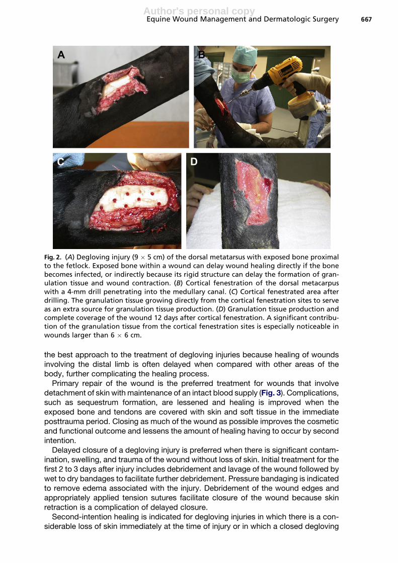
Fig. 3. (A) Degloving injury to the right hemithorax secondary to a laceration with a metalpost. There is extensive undermining of the skin along the dorsal and ventral caudal aspectsof the wound. (B) Primary repair of laceration. Debridement of the wound edges, walkingsutures, and appropriately applied tension sutures facilitate closure. (C) The laceration re-pair dehisced 6 days after primary repair. The wound is debrided, cleansed, and honey is ap-plied topically to facilitate third-intention wound healing. (D) A secondary topical pad andreusable body meshing is used to facilitate wound cleansing. (E) Laceration 25 days after ini-tial injury. Contraction, epithelialization, and granulation tissue formation is progressingwell.
Hanson668
Author's personal copy
limb and associated wound area. Depending on the size and location of the wound,skin grafting may be indicated to facilitate complete healing. Grafting should bedelayed to permit maximum wound contraction, which depending on the locationand size of the wound may be 4 to 8 weeks after injury.
Dorsal knuckling of the fetlock and an inability to extend the digit is a common com-plication of distal limb wounds that is usually associated with the loss of the extensortendon of the distal limb.24 Supporting the dorsal aspect of the limb to counteract thepull of the flexor tendons on the palmar or plantar aspect of the limb is the premise formanagement of extensor tendon disruption. The wound and extensor tendon lacera-tion is managed by second-intention healing without suturing the extensor ten-don.22,25 A rigid polyvinyl chloride splint is applied to the dorsal or palmar or plantaraspect of the distal limb after wound bandaging. The bandage and splint, which main-tains the limb in extension and prevents dorsal knuckling of the fetlock, are retaineduntil normal limb function returns, which may vary from 7 days to 6 weeks.1
Wound Infection and Dehiscence
Wound infection and dehiscence occur in both surgically or trauma-induced wounds.Tissue integrity and perfusion, wound repair processes, and bacterial challenge andhost responses heavily influence infection. A very important determinant of woundinfection is the bacterial inosculation dose. An inoculum size of 105 organisms pergram of tissue is a bacterial challenge below which soft tissue wounds may heal with-out infection. Samples from infected wounds should be taken on a sterile swab fromdeep within the infected site or tract after cleansing of the wound with dilute povidone-iodine or chlorhexidine scrub, followed by a thorough lavage with sterile isotonic fluidor by harvesting fresh exudate on a sterile swab. Debridement of wounds should beperformed on cleaned wounds before the administration of systemic antimicrobials.Necrotic, devitalized, or macerated tissue and organic debris should be removed. Co-pious lavage should be performed with dilute solutions, such as 0.1% povidone-iodineor 0.05% chlorhexidine solution, which maintain antiseptic properties and minimizetissue toxicity. Pressures of greater than 8 psi and up to 70 psi dislodge adherent bac-teria without forcing them deeper into tissue. A 60-mL syringe with a 14-gauge needlecan generate 8 psi and a Water Pik (Water Pik, Fort Collins, Colorado) can generate upto 70 psi.
There is no replacement for a representative culture and sensitivity of a wound. It ishelpful, however, to have an idea of the organism to expect when faced with a need fortherapy in the absence of culture results. Common isolates from equine woundsinclude Streptococcus spp most predominantly followed by coagulase-positive andcoagulase- negative Staphylococcus spp in addition to Enterobacteriaceae, Pseudo-monas spp, and anaerobes. Gentamicin and penicillin or cephalothin is a good treat-ment choice. Aminoglycosides have a concentration-dependent bactericidal actionand a good concentration-dependent postantibiotic affect that remains for severalhours after the dose is administered and bacteria continue to take up the drug througha combination of passive and oxygen-dependent facilitated processes. Antimicrobialsusceptibility of Pseudomonas spp is unpredictable and therapy should be based onculture and sensitivity. Local wound care with silver sulfadiazine is effective in mostconfirmed pseudomonas skin infections. Systemic antibiotics are generally adminis-tered for 7 to 10 days in combination with local wound debridement and care.21
Regional Limb Perfusion
Regional intravenous infusion achieves high concentrations of antibiotic by diffusionfrom the vascular space into the traumatized and infected synovial membranes.
Equine Wound Management and Dermatologic Surgery 669
Author's personal copy
Survival rates of horses treated with systemically injected antibiotics in conjunctionwith regional intravenous antibiotic infusion is greatly increased. Higher concentra-tions of antibiotic are detected sooner in joints after regional intravenous comparedwith regional intraosseous antibiotic infusion.
To perform standing regional intravenous perfusion, the horse is sedated. A highfour-point block is performed using mepivacaine when the synovial structure to betreated is located at or below the fetlock. Anesthesia of the ulnar and median nerves,or tibial and peroneal nerves, is performed when the area to be treated is located at thelevel of the carpal or tarsal joints, respectively.
An Esmarch’s bandage or a pneumatic tourniquet is applied proximal to the affectedsynovial structure to occlude the venous system. Usually, the bandage or tourniquet isapplied to the mid-metacarpus or metatarsus, but in cases of infection of the carpal ortarsal structures, the bandage or the tourniquet is applied to the distal aspect of theradius or the mid tibial region. The Esmarch’s bandage is maintained in place afterantibiotic infusion for a period of 30 to 40 minutes and then released.
The antibiotic, generally an aminoglycoside or cephalosporin (amikacin, 1 g dilutedin 20–30 mL of saline solution; gentamicin, 1 g diluted in 20–30 mL of saline solution;cephotaxim [Claforan], 1–2 g diluted in 20–30 mL of sterile water) is injected into one ofthe local superficial veins when the Esmarch’s bandage is in place. The palmar-plantarmedial and lateral veins are used when the infusion is performed at the level of the fet-lock. The cephalic and saphenous veins are used when the tissues intended to be per-fused are localized at the levels of the carpal and the tarsal joints. The skin over thevein is surgically prepared using an antiseptic technique. The vein is catheterized us-ing a 22-gauge, 2.5-cm catheter with an infusion plug. A 23-gauge butterfly catheterwith an incorporated extension set also works well for this purpose.
The volume of infusion varies between 20 and 30 mL, but smaller volumes can beused for treating foals. Different recommendations for the rate of infusion are reported.The rate of infusion can be as fast as 60 mL/min or 2 mL/min for a total delivery time of30 minutes for a 30-mL volume. For standing treatment, a fast rate of administration ismore convenient and seems to offer satisfactory results. Once the catheter is with-drawn several gauze sponges are applied over the venipuncture site and the site iswrapped with an elastic bandage to avoid a hematoma formation.
Foreign Bodies
Most horses with foreign bodies present with a nonhealing persistent draining tract.This wound is recognized only after prolonged medical treatments have been unsuc-cessful in resolving a local infection.22 Most foreign bodies are not evident radiograph-ically unless, however, a gas line is evident secondary to a bacterial infection. Plasticand wood have the same radiodensity as soft tissue and are not visible radiographi-cally. Ultrasound, contrast radiographs, or probing the wound with a surgicalinstrument may aid in the identification of these foreign bodies.1,7,22
Thermal Injuries
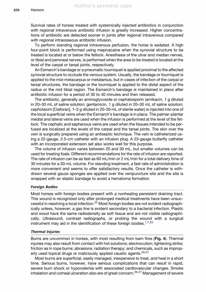
Burns are uncommon in horses, with most resulting from barn fires (Fig. 4). Thermalinjuries may also result from contact with hot solutions; electrocution; lightening strike;friction as in rope burns; abrasions; radiation therapy; and chemicals, such as improp-erly used topical drugs or maliciously applied caustic agents.26,27
Most burns are superficial, easily managed, inexpensive to treat, and heal in a shorttime. Serious burns, however, have serious complications that can result in rapid,severe burn shock or hypovolemia with associated cardiovascular changes. Smokeinhalation and corneal ulceration also are of great concern.26,27 Management of severe
Hanson670
Author's personal copy
and extensive burns is difficult, expensive, and time consuming. The large surfacearea of the burn dramatically increases the potential for loss of fluids, electrolytes,and calories. Burns covering up to 50% or more of the body are usually fatal, althoughthe depth of the burn also influences mortality. Massive wound infection is almostimpossible to prevent because of the difficulty of maintaining a sterile wound environ-ment. Long-term care is required to prevent continued trauma, because burn woundsare often pruritic and self-mutilation is common. Burned horses are frequently disfig-ured, preventing them from returning to full function. Before treatment, the patientmust be carefully examined, with particular attention paid to cardiovascular function,pulmonary status, ocular lesions, and the extent and severity of the burns.26,28–30
Although specific guidelines do not exist for burns of horses, euthanasia should berecommended for deep partial-thickness to full-thickness burns involving 30% to 50%of the total body surface area.29,30 The availability of adequate treatment facilities,cost of treatment, and pain experienced by the horse during long-term care shouldbe considered when deciding treatment. Euthanasia is often an acceptable alternativebecause convalescence may take up to 2 years.31 Cost of treatment and prognosisshould be thoroughly discussed with the owner.26,27,32,33
Excessive Skin Tension
Skin sutured with excessive tension is likely to have complications of healing becauseof local ischemia with pressure necrosis of the surrounding skin and the pull through ofsutures at the skin edge with subsequent wound disruption. Undermining the sur-rounding skin, relief incisions, and appropriately applied tension sutures are themost common methods that can be used to lessen tension along the skin margins.
The surrounding skin can be undermined up to 4 cm from the wound edge withoutassociated complications.1,34 Relief incisions can be closed after the primary incisionis closed or left to heal by second intention.
Fig. 4. (A) Superficial to deep second-degree burn of the muzzle, periocular region, neck,and chest. These wounds may heal spontaneously in approximately 30 days if further dermalischemia does not develop, which may lead to full-thickness necrosis. The eye needs to beevaluated daily for evidence of trauma or inflammation. (B) Third-degree burn of the distalhind limbs 3 weeks after initial injury. Third-degree burns are characterized by loss of epi-dermal and dermal components including the adnexa. Healing is by means of contractionand epithelialization from the wound margin or acceptance of an autograft.
Equine Wound Management and Dermatologic Surgery 671
Author's personal copy
To not interrupt the blood supply to the primary suture line, tension sutures arepositioned well away from the wound margin. Once the tension suture is in place,the primary incision line is sutured to close the wound edges. Tension suture patternsinclude vertical mattress, horizontal mattress, far-far-near-near, and far-near-near-farpatterns. Vertical mattress sutures with or without skin support to prevent laceration ofthe wound edges, such as polyethylene or rubber tubing, are useful in reducing ten-sion on the primary suture line. This tension suture support method is used in areasthat cannot be bandaged well, such as the upper limb, body, and neck region. It iscontraindicated to use tension suture supports under a limb cast or heavy bandagebecause these supports may cause tissue necrosis and suture line failure. Tension su-tures are not effective after 7 to 10 days and should be removed in a staggered fashionwith one half removed initially followed by the remaining sutures later.1
Nerve Damage
Nerve damage or transection of a nerve in the limb or trunk is not readily recognized atthe initial time of the injury. Many lower limb lacerations in the pastern, fetlock, andheel bulb areas with significant injury almost certainly have concurrent transectionof the palmar or plantar digital nerve that is not recognized during the examinationof the injury. Unilateral transection of the palmar or plantar nerves associated witha traumatic laceration seems to regrow after transection to reinnervate the affectedarea and cause minimal clinical problems in horses.4
Neuroma formation after nerve transection, although rare, can occur and may causelameness, together with focal pain directly over the wound and nerve site. The lame-ness improves after local anesthetic is placed at the site of the neuroma. Surgicalremoval of the neuroma and associated nerve is the treatment of choice to whichmost horses respond favorably.
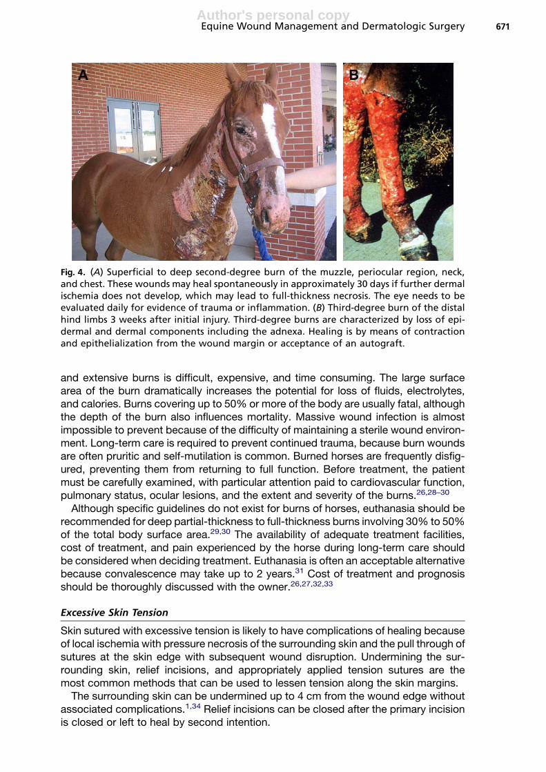
Potential sites for wound-associated nerve transection other than the limb includethe lateral aspect of the proximal radius and elbow where the radial nerve lies fairly su-perficial and the shoulder region in which lacerations and blunt trauma may contributeto suprascapular nerve injury (Fig. 5). Nerve injury to either location, however, isuncommon.
Injuries to smaller nerve branches most likely occur with all types of traumaticwounds but seem to have minimal impact on wound healing. Occasionally, focal areasof wounds may be hypersensitive to touch and other stimuli, which may indicateprevious damage to nerve branches and potential small neuroma formation. Thishypersensitivity tends to resolve as the wound is covered with healthy granulationtissue.1
Major Blood Vessels
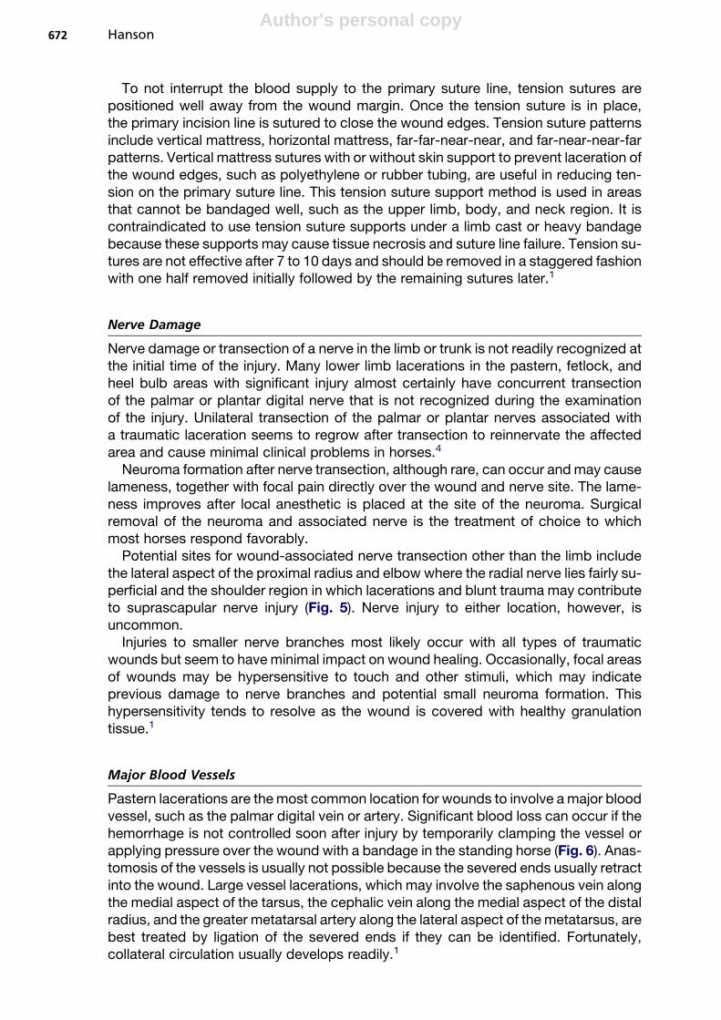
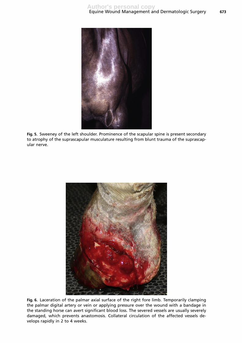
Pastern lacerations are the most common location for wounds to involve a major bloodvessel, such as the palmar digital vein or artery. Significant blood loss can occur if thehemorrhage is not controlled soon after injury by temporarily clamping the vessel orapplying pressure over the wound with a bandage in the standing horse (Fig. 6). Anas-tomosis of the vessels is usually not possible because the severed ends usually retractinto the wound. Large vessel lacerations, which may involve the saphenous vein alongthe medial aspect of the tarsus, the cephalic vein along the medial aspect of the distalradius, and the greater metatarsal artery along the lateral aspect of the metatarsus, arebest treated by ligation of the severed ends if they can be identified. Fortunately,collateral circulation usually develops readily.1
Hanson672
Author's personal copy
Fig. 5. Sweeney of the left shoulder. Prominence of the scapular spine is present secondaryto atrophy of the suprascapular musculature resulting from blunt trauma of the suprascap-ular nerve.
Fig. 6. Laceration of the palmar axial surface of the right fore limb. Temporarily clampingthe palmar digital artery or vein or applying pressure over the wound with a bandage inthe standing horse can avert significant blood loss. The severed vessels are usually severelydamaged, which prevents anastomosis. Collateral circulation of the affected vessels de-velops rapidly in 2 to 4 weeks.
Equine Wound Management and Dermatologic Surgery 673
Author's personal copy
Movement
The extent of movement of the skin relative to the underlying bed of granulation tissueis usually much higher in the limb regions than in the trunk. This is possibly exacer-bated by the relative lack of skin elasticity and the obvious proximity of the limbskin to structures with a high degree of motion, such as joints and tendons. Trunkwounds have a better available reparative blood supply than those of the distal limb.
An injury to the distal limb metacarpal or metatarsal region of a horse that involvesthe flexor tendons or their sheaths requires healing by the ingress of blood vesselsfrom adjacent structures. As healing attempts to progress, however, repeated tendoncontraction and limb movement moves the injury away from the site of the skin woundleaving the damaged tissues with no effective mechanism for healing.
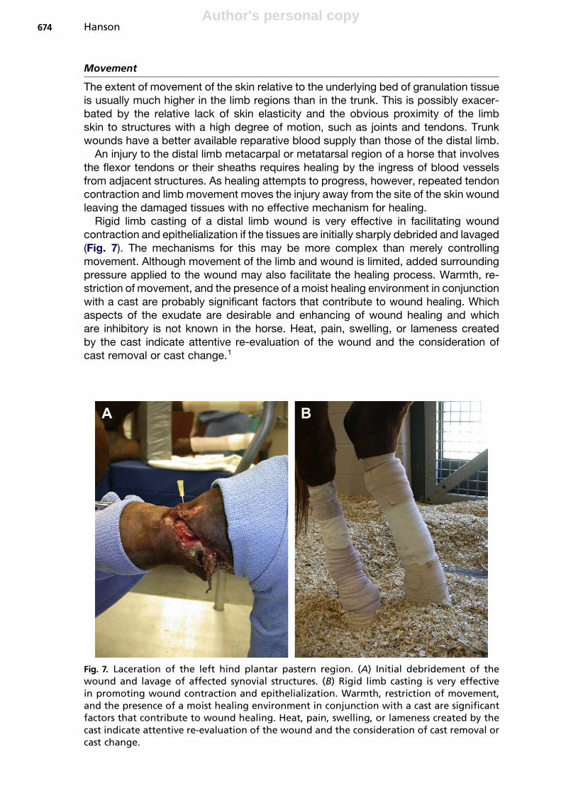
Rigid limb casting of a distal limb wound is very effective in facilitating woundcontraction and epithelialization if the tissues are initially sharply debrided and lavaged(Fig. 7). The mechanisms for this may be more complex than merely controllingmovement. Although movement of the limb and wound is limited, added surroundingpressure applied to the wound may also facilitate the healing process. Warmth, re-striction of movement, and the presence of a moist healing environment in conjunctionwith a cast are probably significant factors that contribute to wound healing. Whichaspects of the exudate are desirable and enhancing of wound healing and whichare inhibitory is not known in the horse. Heat, pain, swelling, or lameness createdby the cast indicate attentive re-evaluation of the wound and the consideration ofcast removal or cast change.1
Fig. 7. Laceration of the left hind plantar pastern region. (A) Initial debridement of thewound and lavage of affected synovial structures. (B) Rigid limb casting is very effectivein promoting wound contraction and epithelialization. Warmth, restriction of movement,and the presence of a moist healing environment in conjunction with a cast are significantfactors that contribute to wound healing. Heat, pain, swelling, or lameness created by thecast indicate attentive re-evaluation of the wound and the consideration of cast removal orcast change.
Hanson674
Author's personal copy
Self-Mutilation
Significant self-mutilation of wounds through rubbing, biting, and pawing can occur ifthe horse is not adequately restrained or medicated. Usually, the most intense pruriticepisodes occur in the first weeks of wound healing during the inflammatory phase ofrepair and during eschar sloughing but can be a later complication associated withburn wounds.27,35 To prevent extreme self-mutilation, the horse should be crosstied or sedated at this time and use of a neck collar may be considered. Delayed heal-ing, poor epithelialization, and complications of second-intention healing may limit re-turn of the animal to their previous use.
Exuberant Granulation Tissue
Surgical resection is a simple and effective method to control exuberant granulationtissue. The procedure is performed with the horse standing, because granulation tis-sue is not innervated. Strips of granulation tissue can be shaved from the wound bed ina distal to proximal direction to produce a flat surface level with or slightly (approxi-mately 2 mm) below the surrounding wound edges. The epithelial margin should bepreserved to allow continued healing. A pressure bandage is usually necessary to con-trol hemorrhage after excision. In lower limb wounds of horses this technique has beensuccessful in enhancing second-intention healing that was delayed because of pro-truding granulation tissue. This technique is preferred for the removal of exuberantgranulation tissue over other methods, such as application of caustic drugs, becauseit is easy to perform; provides tissue for histologic evaluation, if needed; and preservesthe epithelial margin for continued healing. As with any alternate technique, healing bycontraction and epithelialization must subsequently be supported by maintaining thelimb in a firm support bandage and limiting excessive motion of the wound or exces-sive granulation recurs. Corticosteroids may be applied topically to curb the early for-mation of exuberant granulation tissue, hence facilitating epithelialization and woundrepair. The ability of some corticosteroids to suppress the formation of exuberantgranulation tissue in the early phases of healing may be related to their ability selec-tively to decrease the release of profibrotic transforming growth factor-b from mono-cytes and macrophages, inhibiting lysosomal activity and fibroblastic proliferation.Corticosteroids are generally applied at the earliest signs of formation of exuberantgranulation tissue with one or two applications being all that is needed to achievethe desired effect. Continued applications are not recommended, because this mayexert negative effects on wound contraction, epithelialization, and angiogenesis. Cor-ticosteroids should not be applied to an infected wound because they inhibit the in-flammatory response required to eliminate microorganisms.
Application of a cast to a lower limb wound is indicated in cases in which it is difficultto control exuberant granulation tissue. Wounds over joints or tendons may require im-mobilization because continued movement disrupts healing. Frequently, the hock orcarpus is involved in these types of compound injuries. When the limb is mechanicallystable, the wound should be bandaged for a few days before applying a cast, to allowsuperior wound debridement and permit dissipation of edema, which ensures a better-fitting cast. Casts minimize the formation of exuberant granulation tissue by reducingmotion. Casts should be maintained no longer than necessary over lower limb woundsfor reasons similar to those mentioned for bandages and to minimize the developmentof cast sores. Generally, casts over wounds should be changed every 3 to 10 days, butthis depends on the nature and location of the wound and the temperament of thehorse. Skin grafts can be used after cast removal to facilitate wound coverage. A splintbandage is continued during this period.
Equine Wound Management and Dermatologic Surgery 675
Author's personal copy
COMPLICATIONS OF CONTAMINATEDWOUNDSMethicillin-Resistant Staphylococcus Aureus Infection
There have been increasing reports of MRSA infection and colonization in horses andother domestic animals.36–39 MRSA is resistant to all b-lactam antimicrobials and fre-quently to a wide range of additional antimicrobial classes because of the presence ofan altered penicillin-binding protein. Infections can consequently be difficult to treatand are associated with increased morbidity, mortality, and treatment costs, com-pared with infections caused by methicillin-susceptible S aureus strains.40–42
Identification of MRSA infection and colonization in horses in veterinary hospitalsand in the community, and reports of transmission of MRSA between humans and an-imals, have raised concern about the role of animals in MRSA infection in humans andthe potential for animals to become a reservoir of MRSA.36,38,39,43 In one recent vet-erinary hospital study the incidence rate of nosocomial MRSA infection was at therate of 1.8 per 1000 admissions, with an incidence density of 0.88 per 1000 patientdays. Administrations of ceftiofur or aminoglycosides during hospitalization were thetwo risk factors associated with nosocomial MRSA colonization. In another veterinaryhospital study horses that had received at least 72 hours of penicillin treatment hada 5.8-times higher chance (odds) of harboring penicillin-resistant staphylococci thanhorses that were not admitted to the clinic and did not receive penicillin treatment.Control horses with a 72-hour stay at the clinic but no penicillin treatment still hada 2.4-times higher chance (odds) of harboring penicillin-resistant strains. This workdemonstrated that horses entering the hospital harbor staphylococci-containing anti-biotic-resistance genes. Shortly after hospitalization, horses acquired a specific mul-tidrug-resistant skin flora that was presumably selected for and maintained in thehospital by the use of penicillin. These authors proposed that antibiotics should be lim-ited to the treatment of infection and not used for infection prevention and that suchprudent use could help prevent selection for multidrug-resistant strains, such asMRSA strains in animals.44 MRSA screening of horses admitted to a veterinary hospi-tal was useful for identification of community-associated and nosocomial colonizationand infection, and for monitoring of infection control practices.45,46
MRSA infections are generally classified as hospital or community acquired and assuperficial colonization of a wound without signs of infection, superficial soft tissue in-fection or cellulitis, complex skin and skin-structure infection, or osteomyelitis. Super-ficial wounds may be treated without the use of oral or IV antibiotics. Regular cleansingof the wound with 4% chlorhexidine gluconate and soft tissue debridement is effectiveat reducing the colony-forming bacteria load. Topical application of honey or silver-coated dressings has been shown to be effective. Careful monitoring of the woundis imperative to ensure adequate response to treatment.47–49
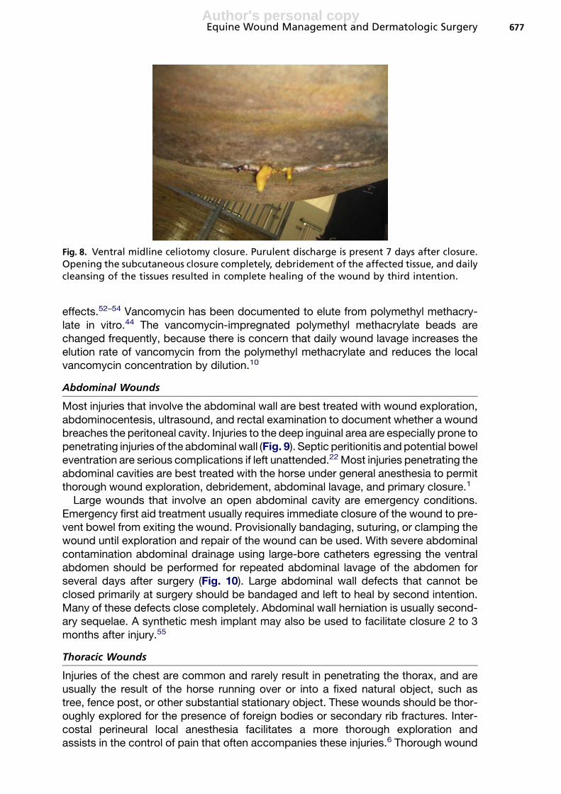
Ventral midline celiotomy closures are prone to MRSA infection (Fig. 8). Clinicalsigns usually present 5 to 10 days after surgery with a purulent exudate escapingfrom the subcutaneous tissue. Opening of the skin closure accompanied by localsuperficial debridement with the application of 4% chlorhexidine, topical honey, orsilver-coated dressings has been an effective treatment. Although intranasal mupiro-cin can prevent endogenous acquired MRSA infections in an ICU and is effective inoverall decolonizing of nasal carriers, mupirocin is less effective in decolonizing extra-nasal sites, such as wounds.50,51 Doxycycline in combination with rifampin has beenshown to have a synergistic activity against MRSA activity in people.
Local antimicrobial treatment, consisting of implantation of vancomycin-impreg-nated polymethyl methacrylate beads, has also been performed to increase the con-centration of antimicrobials in the local environment and decrease adverse systemic
Hanson676
Author's personal copy
effects.52–54 Vancomycin has been documented to elute from polymethyl methacry-late in vitro.44 The vancomycin-impregnated polymethyl methacrylate beads arechanged frequently, because there is concern that daily wound lavage increases theelution rate of vancomycin from the polymethyl methacrylate and reduces the localvancomycin concentration by dilution.10
Abdominal Wounds
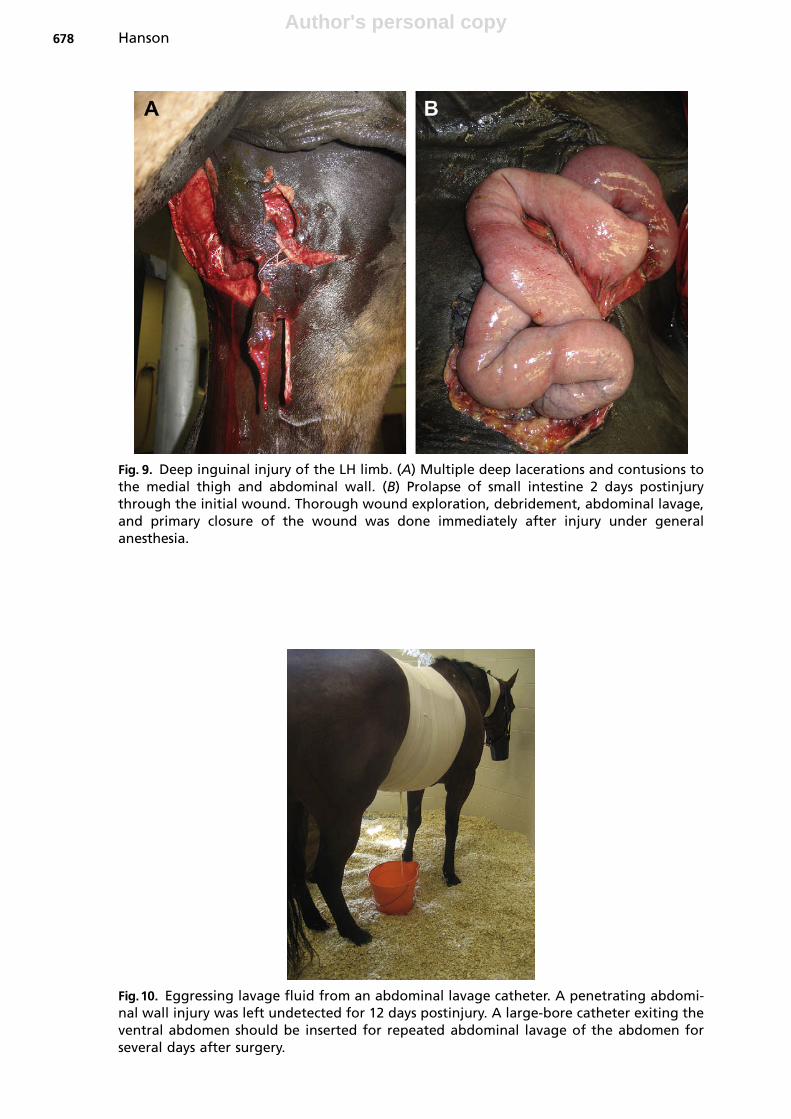
Most injuries that involve the abdominal wall are best treated with wound exploration,abdominocentesis, ultrasound, and rectal examination to document whether a woundbreaches the peritoneal cavity. Injuries to the deep inguinal area are especially prone topenetrating injuries of the abdominal wall (Fig. 9). Septic peritionitis and potential boweleventration are serious complications if left unattended.22 Most injuries penetrating theabdominal cavities are best treated with the horse under general anesthesia to permitthorough wound exploration, debridement, abdominal lavage, and primary closure.1
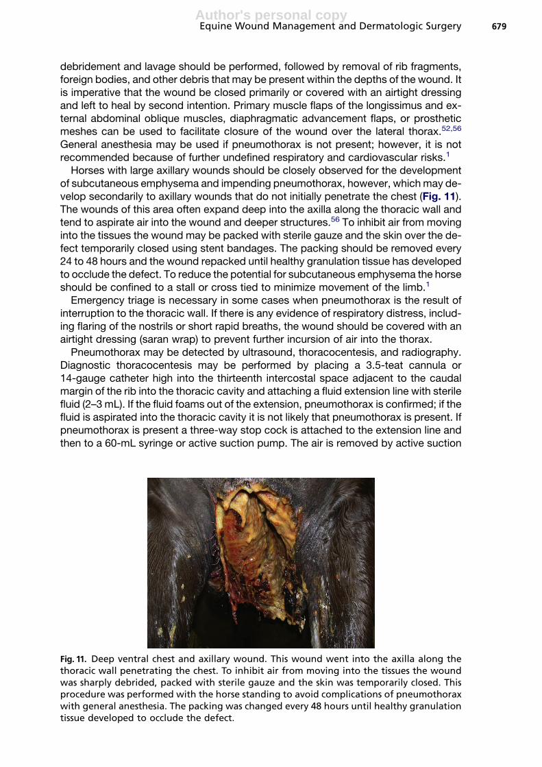
Large wounds that involve an open abdominal cavity are emergency conditions.Emergency first aid treatment usually requires immediate closure of the wound to pre-vent bowel from exiting the wound. Provisionally bandaging, suturing, or clamping thewound until exploration and repair of the wound can be used. With severe abdominalcontamination abdominal drainage using large-bore catheters egressing the ventralabdomen should be performed for repeated abdominal lavage of the abdomen forseveral days after surgery (Fig. 10). Large abdominal wall defects that cannot beclosed primarily at surgery should be bandaged and left to heal by second intention.Many of these defects close completely. Abdominal wall herniation is usually second-ary sequelae. A synthetic mesh implant may also be used to facilitate closure 2 to 3months after injury.55
Thoracic Wounds
Injuries of the chest are common and rarely result in penetrating the thorax, and areusually the result of the horse running over or into a fixed natural object, such astree, fence post, or other substantial stationary object. These wounds should be thor-oughly explored for the presence of foreign bodies or secondary rib fractures. Inter-costal perineural local anesthesia facilitates a more thorough exploration andassists in the control of pain that often accompanies these injuries.6 Thorough wound
Fig. 8. Ventral midline celiotomy closure. Purulent discharge is present 7 days after closure.Opening the subcutaneous closure completely, debridement of the affected tissue, and dailycleansing of the tissues resulted in complete healing of the wound by third intention.
Equine Wound Management and Dermatologic Surgery 677
Author's personal copy
Fig. 9. Deep inguinal injury of the LH limb. (A) Multiple deep lacerations and contusions tothe medial thigh and abdominal wall. (B) Prolapse of small intestine 2 days postinjurythrough the initial wound. Thorough wound exploration, debridement, abdominal lavage,and primary closure of the wound was done immediately after injury under generalanesthesia.
Fig.10. Eggressing lavage fluid from an abdominal lavage catheter. A penetrating abdomi-nal wall injury was left undetected for 12 days postinjury. A large-bore catheter exiting theventral abdomen should be inserted for repeated abdominal lavage of the abdomen forseveral days after surgery.
Hanson678
Author's personal copy
debridement and lavage should be performed, followed by removal of rib fragments,foreign bodies, and other debris that may be present within the depths of the wound. Itis imperative that the wound be closed primarily or covered with an airtight dressingand left to heal by second intention. Primary muscle flaps of the longissimus and ex-ternal abdominal oblique muscles, diaphragmatic advancement flaps, or prostheticmeshes can be used to facilitate closure of the wound over the lateral thorax.52,56
General anesthesia may be used if pneumothorax is not present; however, it is notrecommended because of further undefined respiratory and cardiovascular risks.1
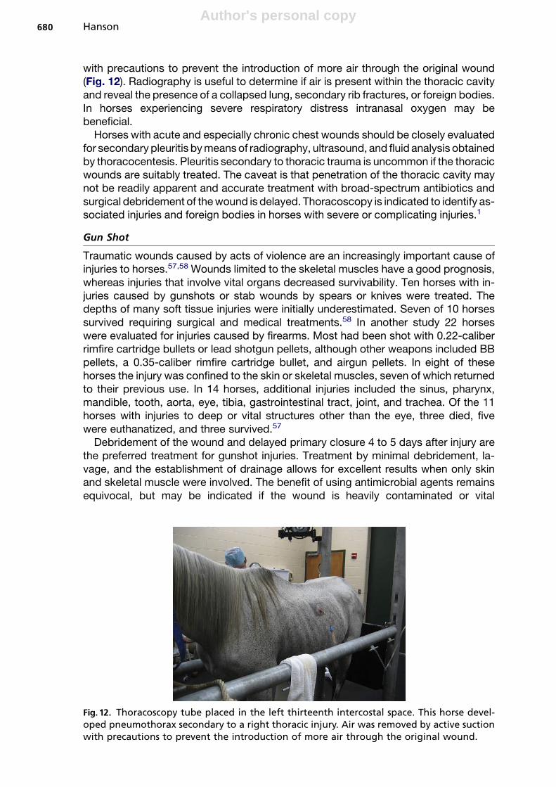
Horses with large axillary wounds should be closely observed for the developmentof subcutaneous emphysema and impending pneumothorax, however, which may de-velop secondarily to axillary wounds that do not initially penetrate the chest (Fig. 11).The wounds of this area often expand deep into the axilla along the thoracic wall andtend to aspirate air into the wound and deeper structures.56 To inhibit air from movinginto the tissues the wound may be packed with sterile gauze and the skin over the de-fect temporarily closed using stent bandages. The packing should be removed every24 to 48 hours and the wound repacked until healthy granulation tissue has developedto occlude the defect. To reduce the potential for subcutaneous emphysema the horseshould be confined to a stall or cross tied to minimize movement of the limb.1
Emergency triage is necessary in some cases when pneumothorax is the result ofinterruption to the thoracic wall. If there is any evidence of respiratory distress, includ-ing flaring of the nostrils or short rapid breaths, the wound should be covered with anairtight dressing (saran wrap) to prevent further incursion of air into the thorax.
Pneumothorax may be detected by ultrasound, thoracocentesis, and radiography.Diagnostic thoracocentesis may be performed by placing a 3.5-teat cannula or14-gauge catheter high into the thirteenth intercostal space adjacent to the caudalmargin of the rib into the thoracic cavity and attaching a fluid extension line with sterilefluid (2–3 mL). If the fluid foams out of the extension, pneumothorax is confirmed; if thefluid is aspirated into the thoracic cavity it is not likely that pneumothorax is present. Ifpneumothorax is present a three-way stop cock is attached to the extension line andthen to a 60-mL syringe or active suction pump. The air is removed by active suction
Fig. 11. Deep ventral chest and axillary wound. This wound went into the axilla along thethoracic wall penetrating the chest. To inhibit air from moving into the tissues the woundwas sharply debrided, packed with sterile gauze and the skin was temporarily closed. Thisprocedure was performed with the horse standing to avoid complications of pneumothoraxwith general anesthesia. The packing was changed every 48 hours until healthy granulationtissue developed to occlude the defect.
Equine Wound Management and Dermatologic Surgery 679
Author's personal copy
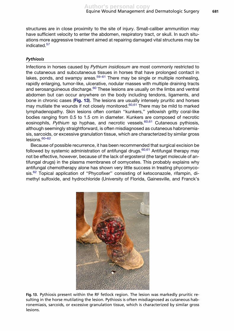
with precautions to prevent the introduction of more air through the original wound(Fig. 12). Radiography is useful to determine if air is present within the thoracic cavityand reveal the presence of a collapsed lung, secondary rib fractures, or foreign bodies.In horses experiencing severe respiratory distress intranasal oxygen may bebeneficial.
Horses with acute and especially chronic chest wounds should be closely evaluatedfor secondary pleuritis by means of radiography, ultrasound, and fluid analysis obtainedby thoracocentesis. Pleuritis secondary to thoracic trauma is uncommon if the thoracicwounds are suitably treated. The caveat is that penetration of the thoracic cavity maynot be readily apparent and accurate treatment with broad-spectrum antibiotics andsurgical debridement of the wound is delayed. Thoracoscopy is indicated to identify as-sociated injuries and foreign bodies in horses with severe or complicating injuries.1
Gun Shot
Traumatic wounds caused by acts of violence are an increasingly important cause ofinjuries to horses.57,58 Wounds limited to the skeletal muscles have a good prognosis,whereas injuries that involve vital organs decreased survivability. Ten horses with in-juries caused by gunshots or stab wounds by spears or knives were treated. Thedepths of many soft tissue injuries were initially underestimated. Seven of 10 horsessurvived requiring surgical and medical treatments.58 In another study 22 horseswere evaluated for injuries caused by firearms. Most had been shot with 0.22-caliberrimfire cartridge bullets or lead shotgun pellets, although other weapons included BBpellets, a 0.35-caliber rimfire cartridge bullet, and airgun pellets. In eight of thesehorses the injury was confined to the skin or skeletal muscles, seven of which returnedto their previous use. In 14 horses, additional injuries included the sinus, pharynx,mandible, tooth, aorta, eye, tibia, gastrointestinal tract, joint, and trachea. Of the 11horses with injuries to deep or vital structures other than the eye, three died, fivewere euthanatized, and three survived.57
Debridement of the wound and delayed primary closure 4 to 5 days after injury arethe preferred treatment for gunshot injuries. Treatment by minimal debridement, la-vage, and the establishment of drainage allows for excellent results when only skinand skeletal muscle were involved. The benefit of using antimicrobial agents remainsequivocal, but may be indicated if the wound is heavily contaminated or vital
Fig. 12. Thoracoscopy tube placed in the left thirteenth intercostal space. This horse devel-oped pneumothorax secondary to a right thoracic injury. Air was removed by active suctionwith precautions to prevent the introduction of more air through the original wound.
Hanson680
Author's personal copy
structures are in close proximity to the site of injury. Small-caliber ammunition mayhave sufficient velocity to enter the abdomen, respiratory tract, or skull. In such situ-ations more aggressive treatment aimed at repairing damaged vital structures may beindicated.57
Pythiosis
Infections in horses caused by Pythium insidiosum are most commonly restricted tothe cutaneous and subcutaneous tissues in horses that have prolonged contact inlakes, ponds, and swampy areas.59–61 There may be single or multiple nonhealing,rapidly enlarging, tumor-like, ulcerative, nodular masses with multiple draining tractsand serosanguineous discharge.60 These lesions are usually on the limbs and ventralabdomen but can occur anywhere on the body including tendons, ligaments, andbone in chronic cases (Fig. 13). The lesions are usually intensely pruritic and horsesmay mutilate the wounds if not closely monitored.60,61 There may be mild to markedlymphadenopathy. Skin lesions often contain ‘‘kunkers,’’ yellowish gritty coral-likebodies ranging from 0.5 to 1.5 cm in diameter. Kunkers are composed of necroticeosinophils, Pythium sp hyphae, and necrotic vessels.60,61 Cutaneous pythiosis,although seemingly straightforward, is often misdiagnosed as cutaneous habronemia-sis, sarcoids, or excessive granulation tissue, which are characterized by similar grosslesions.60–62
Because of possible recurrence, it has been recommended that surgical excision befollowed by systemic administration of antifungal drugs.60,61 Antifungal therapy maynot be effective, however, because of the lack of ergosterol (the target molecule of an-tifungal drugs) in the plasma membranes of oomycetes. This probably explains whyantifungal chemotherapy alone has shown very little success in treating phycomyco-sis.62 Topical application of ‘‘Phycofixer’’ consisting of ketoconazole, rifampin, di-methyl sulfoxide, and hydrochloride (University of Florida, Gainesville, and Franck’s
Fig. 13. Pythiosis present within the RF fetlock region. The lesion was markedly pruritic re-sulting in the horse mutilating the lesion. Pythiosis is often misdiagnosed as cutaneous hab-ronemiasis, sarcoids, or excessive granulation tissue, which is characterized by similar grosslesions.
Equine Wound Management and Dermatologic Surgery 681
Author's personal copy
Compounding Pharmacy, Ocala, Florida) beneath an absorbent bandage has met withgood success in wounds that cannot be surgically debrided.
Recently, immunotherapy using a newly formulated vaccine has been successful intreating cutaneous pythiosis in horses and dogs.62 This vaccine has been shown to beeffective for both acute and chronic cutaneous pythiosis in the horse.61,62
For most phycomycosis infections the prognosis is guarded to poor regardless ofthe advances in treatment. The reason is that many horses have multiple bone lesionsat the time of initial presentation.60,61 The rate of recurrence after attempted surgicalexcision has also been a major factor in the failure of treatment.
Myiasis
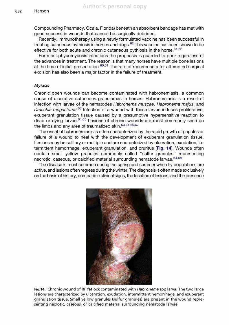
Chronic open wounds can become contaminated with habronemiasis, a commoncause of ulcerative cutaneous granulomas in horses. Habronemiasis is a result ofinfection with larvae of the nematodes Habronema muscae, Habronema majus, andDraschia megastoma.63 Infection of a wound with these larvae induces proliferative,exuberant granulation tissue caused by a presumptive hypersensitive reaction todead or dying larvae.64,65 Lesions of chronic wounds are most commonly seen onthe limbs and any area of traumatized skin.63,64,66,67
The onset of habronemiasis is often characterized by the rapid growth of papules orfailure of a wound to heal with the development of exuberant granulation tissue.Lesions may be solitary or multiple and are characterized by ulceration, exudation, in-termittent hemorrhage, exuberant granulation, and pruritus (Fig. 14). Wounds oftencontain small yellow granules commonly called ‘‘sulfur granules’’ representingnecrotic, caseous, or calcified material surrounding nematode larvae.64,68
The disease is most common during the spring and summer when fly populations areactive, and lesionsoften regressduring thewinter. The diagnosis isoftenmadeexclusivelyon the basis of history, compatible clinical signs, the location of lesions, and the presence
Fig.14. Chronic wound of RF fetlock contaminated with Habronema spp larva. The two largelesions are characterized by ulceration, exudation, intermittent hemorrhage, and exuberantgranulation tissue. Small yellow granules (sulfur granules) are present in the wound repre-senting necrotic, caseous, or calcified material surrounding nematode larvae.
Hanson682

Author's personal copy
of yellowish granules. Biopsy is the method of choice for confirming the diagnosis, whichreveals nodular to diffuse eosinophilic dermatitis. Multiple fociof coagulative necrosiswithor without degenerating nematode larvae in the center are characteristic of this disease.64
Habronemiasis must be differentiated from proliferative granulation tissue, sarcoids, py-thiosis, squamous cell carcinoma, and other neoplasms.63–67
A combination of local and systemic treatment aimed at reducing the size of thelesions, reducing associated inflammation, and preventing reinfestation is most effec-tive.63–67 Massive lesions and wounds refractory to medical treatment may be re-moved surgically or at least debulked before topical and systemic treatment.Corticosteroids are used to reduce inflammatory hypersensitivity reactions, and forsmaller lesions, topical or intralesional application is favored. Topical preparationsthat contain anti-inflammatory, larvicidal, antimicrobial, penetrating, and protective in-gredients are commonly used.63–67 Ivermectin has been reported to be effective in thetreatment of habronemiasis in horses, because it kills infective larvae and adult wormsin the stomach.69
COMPLICATIONS OF NEOPLASIASquamous Cell Carcinoma
A variety of tumors have been reported in association with chronic wounds. Neoplasiasecondary to trauma or thermal injury is not uncommon in humans, and can developacutely but more often latently, several years after the injury.70 Squamous cell carci-noma has a prevalence rate of almost 2% in human burn wounds.70 Although scar ma-lignancy in animals is less frequently diagnosed than in humans, various causes,including chronic irritation, as in the case of the chemicals applied to a wound, maypotentially lead to development of squamous cell carcinoma.70 Squamous cell carci-noma and fibrosarcoma have been identified in association with scars resulting fromburns in the horse (Fig. 15). Squamous cell carcinoma has been associated with cu-taneous scars in a llama and a horse.71,72 Other complications include habronemiasis,keloid-like fibroblastic proliferations, sarcoids, and other burn-induced neoplasia.31
The clinical management of squamous cell carcinoma can include surgical excision,cryosurgery, electrosurgery, intralesional chemotherapy with such agents as cisplatinor 5-fluorouracil cream, immunotherapy, hyperthermia, and radiotherapy.72,73 Cryo-therapy controls squamous cell carcinoma of the skin but is not optimal for recurrent
Fig.15. Squamous cell carcinoma present along the dorsal gluteal region of a horse, which de-veloped secondarily after deep second-degree burn to the area. (Courtesy of J. Schumacher,DVM, MS, DACVS, Knoxville, TN.)
Equine Wound Management and Dermatologic Surgery 683
Author's personal copy
squamous cell carcinoma or larger tumors.74 Intralesional injection with cisplatin in oilor topical or intralesional administration of 5-fluorouracil is effective. Hypertrophicscars, which commonly develop following deep second-degree burns, generallyremodel in a cosmetic manner without surgery within 1 to 2 years. Because scarredskin is hairless and often depigmented, solar exposure should be limited. Chronic non-healing areas should be excised and autografted to prevent neoplastic transformation.Return of squamous cell carcinoma associated with incomplete removal of the lesionis not uncommon, which subsequently requires chronic retreatment.
Interstitial brachytherapy with iridium-192 sources has several advantages com-pared with other possible treatments.74 Although management is intensive andmore costly than other treatments, it is very effective in eliminating the lesion andcauses little tissue defect. Delayed healing, poor epithelialization, and complicationsof second-intention healing may limit return of the animal to previous uses.
Sarcoid
Sarcoid is a benign tumor of fibrous tissue of horses, donkeys, and mules. Sarcoids arethe most frequently diagnosed tumor in horses, with sarcoid transformation anincreasingly important cause of wound healing failure. Surveys have estimated theprevalence of sarcoid at 20% of all equine neoplasms and 36% of all skin tumors.75–79
Sarcoids are presumably caused by bovine papilloma virus type 1 and 2 infection,although retrovirus etiologies have been suggested.75–79 The mode of virus transmis-sion and presence of latent infections are not clearly understood. Peripheral bloodmononuclear cells may serve as host cells for bovine papilloma virus type 1 and 2DNA and contribute to virus latency.80 Sarcoids are recognized as having six differentclinical manifestations (occult, verrucose, nodular, fibroblastic, mixed, and malignant-malevolent) and can occur at any cutaneous site.34,81–83 These lesions are character-ized by proliferation of neoplastic fibroblasts and thickening or ulceration of the skin.
Horses with sarcoids at other sites seem to be particularly prone to sarcoid trans-formation, as are those that have close contact with other horses having sarcoids.34,79
Transformation is also more likely in uncovered wounds in summer months when fliesare abundant.81 Body, trunk, or facial wounds that contain sarcoid cells usually de-velop verrucose sarcoids, whereas limb wounds develop fibroblastic sarcoids thatare easily confused with exuberant granulation tissue.34,81,82 Some wounds partiallyheal, whereas others fail to heal at all even if the overall extent of sarcoid involvementis small. Wounds on horses with sarcoids at other sites should be treated particularlycarefully, with attention not to cross-contaminate the wounds, no matter how smalland insignificant the wound.
Fibroblastic sarcoids strongly resemble exuberant granulation tissue or staphylo-coccal pyogranuloma, especially when it progresses at the site of a wound, and espe-cially in limb wounds.34,81,82 Traumatic skin injuries that fail to heal may containconsiderable sarcoid components in the wound margins, and the sarcoid tissuescan be interdispersed with granulation tissue. Sporadic infiltration of sarcoid tissueat a granulation wound site is very difficult to identify and can be easily missed onbiopsy. Careful deep biopsy and a skilled pathologist are essential. The presence ofsarcoid transformation in a wound site adds critically to the therapeutic challenges.82
Early diagnosis and treatment is the most effective course for resolution because mostsarcoids involving wounds are prone to getting worse with time and develop into largermore invasive lesions.
Surgically debulking a wound with sarcoid tissue is the most effective treatment forlesions involving wounds. Surgical removal is complicated by the regrowth of the sar-coid at the surgical site and complications of complete wound healing because of lack
Hanson684
Author's personal copy
of a suitable means for wound closure or the subsequent interference with normalfunction. The rate of regrowth of the equine sarcoid following surgical excision isclosely dependent on the extent of the tumor and the degree to which the surgeoncan define its limits. Small, well-defined tumors carry the best prognosis for surgicalremoval, whereas extensive areas of poorly defined verrucose and mixed sarcoidmay result in rapid regrowth of a more aggressive sarcoid type. The earliest regrowthof sarcoids occurs within days of incomplete excision and is usually accompanied byrapid wound dehiscence and subsequent failure to heal.83 Viral latency may be oneexplanation for the high rate of recurrence following surgical excision of sarcoids. Inone study bovine papillomaviral DNA was detected in essentially all sarcoids exam-ined. There seems to be a regional variation in the prevalence of viral types with sar-coid tumors. Viral DNA in normal skin samples from horses with sarcoids suggests thepossibility of a latent vial phase.84
Cryotherapy and immune-mediated therapy have mixed results when used to elim-inate sarcoid tissue in wounds. Cryotherapy is generally used as an adjunct therapyafter surgical debulking of the wound and in areas where complete excision of the sar-coid is not possible. Repeat treatments are often needed and complicate the healingof the wound. Poor cosmesis related to regrowth of white hair and scarring is one ofthe most common ‘‘cosmetic’’ complications associated with cryotherapy.
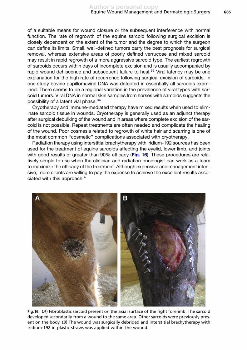
Radiation therapy using interstitial brachytherapy with iridium-192 sources has beenused for the treatment of equine sarcoids affecting the eyelid, lower limb, and jointswith good results of greater than 90% efficacy (Fig. 16). These procedures are rela-tively simple to use when the clinician and radiation oncologist can work as a teamto maximize the efficacy of the treatment. Although expensive and management inten-sive, more clients are willing to pay the expense to achieve the excellent results asso-ciated with this approach.4
Fig.16. (A) Fibroblastic sarcoid present on the axial surface of the right forelimb. The sarcoiddeveloped secondarily from a wound to the same area. Other sarcoids were previously pres-ent on the body. (B) The wound was surgically debrided and interstitial brachytherapy withiridium-192 in plastic straws was applied within the wound.
Equine Wound Management and Dermatologic Surgery 685
Author's personal copy
COMPLICATIONS OF SKIN GRAFTS
Skin grafting decreases healing time and is one of the best techniques for coveringa wound that has been chronically affected by exuberant granulation tissue. Skingrafting of lower limb wounds should be considered to cover the granulating woundbed if contraction has ceased and the wound bed is large.8,85 Frequently, however,wounds in horses are treated for several weeks before skin grafting is initiated. Atthis point granulation tissue is mature, fibrous, and has less of a blood supply thannewly formed granulation tissue. Other complications of graft acceptance and healingare wound infection and sequestra formation.
Chronic inflammation, inherently present during second-intention healing of woundson the distal portion of limbs of horses, may be at least as important as infection be-cause it reduces the quality of the granulation bed and results in the production ofa moderate amount of purulent exudate, both of which negatively influence accep-tance of grafts.12 As a result, the ability of a wound bed to accept a graft is lessened.It is imperative that chronic granulating wounds be debrided to a level below the skinsurface down to a level of healthy granulation tissue before graft application.8,86–88
To increase the success of graft acceptance wound bacteria must be minimized.b-Hemolytic Streptococcus spp, Proteus spp, and Pseudomonas spp are capableof producing destructive proteolytic enzymes and excessive purulent discharge thatbreakdown fibrinous attachments between the graft and recipient bed.89,90 Topicalantiseptics have better efficacy than antibiotics in reducing bacterial wound load be-cause the latter increase the risk of patient sensitization and the development of resis-tant organisms especially when used routinely over prolonged periods in uninfectedwounds.88,90 Infected wounds, however, should be treated with broad-spectrumantibiotics while awaiting culture results. The bone underlying the wound should beradiographed for evidence of sequestra and excessive pericortical dystrophic miner-alization. Large wounds often develop healthy granulating tissue around the perimeterbefore a sequestrum completely defines itself.
Donor site is influenced by the method of grafting, color and texture of the donorhair, cosmesis of the donor site, and ease of obtaining skin. Common sites for obtain-ing donor skin include pectoral, dorsal neck region, perineum, ventral midline, ventrallateral abdomen, and sternal region caudal to the girth area.8,91
Pinch Grafts
Pinch grafts are distinct pieces of skin (3 mm in diameter) produced by excising an el-evated cone of skin. Graft acceptance is as high as 75% using pinch grafts partiallybecause of the fact that the pockets of granulation tissue hold the graft in contactwith the wound. Complications include necrosis of the graft, slower wound healing,improper orientation of hair, and thin skin coverage of the wound.
Necrotic spots along the top of the granulation pockets normally occur during heal-ing, after which the graft epithelializes circumferentially. Because pinch grafts aresmall, complete epithelialization of the wound often takes more than 70 days.91 Im-proper orientation of hair growth is a complication of pinch graft application despiterepeated efforts properly to align the hair to match that of the recipient area. A cobble-stone appearance with thin subcutaneous tissue is sequelae of pinch graft applica-tions that may not be cosmetically acceptable for show horses.
Punch Grafts
Punch grafts are circular pieces of skin that are directly removed from the locally anes-thetized donor site or by obtaining biopsies from an excised piece of donor skin.
Hanson686
Author's personal copy
Common complications of punch graft failure are incomplete removal of the underly-ing subcutaneous tissue from the graft, recipient site hemorrhage, and motion.
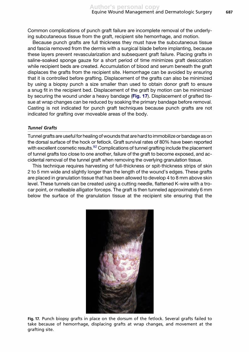
Because punch grafts are full thickness they must have the subcutaneous tissueand fascia removed from the dermis with a surgical blade before implanting, becausethese layers prevent revascularization and subsequent graft failure. Placing grafts insaline-soaked sponge gauze for a short period of time minimizes graft desiccationwhile recipient beds are created. Accumulation of blood and serum beneath the graftdisplaces the grafts from the recipient site. Hemorrhage can be avoided by ensuringthat it is controlled before grafting. Displacement of the grafts can also be minimizedby using a biopsy punch a size smaller than used to obtain donor graft to ensurea snug fit in the recipient bed. Displacement of the graft by motion can be minimizedby securing the wound under a heavy bandage (Fig. 17). Displacement of grafted tis-sue at wrap changes can be reduced by soaking the primary bandage before removal.Casting is not indicated for punch graft techniques because punch grafts are notindicated for grafting over moveable areas of the body.
Tunnel Grafts
Tunnel grafts are useful forhealing of wounds that arehard to immobilize or bandage asonthe dorsal surface of the hock or fetlock. Graft survival rates of 80% have been reportedwith excellent cosmetic results.92 Complications of tunnel grafting include the placementof tunnel grafts too close to one another, failure of the graft to become exposed, and ac-cidental removal of the tunnel graft when removing the overlying granulation tissue.
This technique requires harvesting of full-thickness or spit-thickness strips of skin2 to 5 mm wide and slightly longer than the length of the wound’s edges. These graftsare placed in granulation tissue that has been allowed to develop 4 to 8 mm above skinlevel. These tunnels can be created using a cutting needle, flattened K-wire with a tro-car point, or malleable alligator forceps. The graft is then tunneled approximately 6 mmbelow the surface of the granulation tissue at the recipient site ensuring that the
Fig. 17. Punch biopsy grafts in place on the dorsum of the fetlock. Several grafts failed totake because of hemorrhage, displacing grafts at wrap changes, and movement at thegrafting site.
Equine Wound Management and Dermatologic Surgery 687
Author's personal copy
epidermal side of the graft faces the surface of the wound. Tunnel grafts should not beplaced closer than 2 cm apart to prevent excessive necrosis of granulation tissue.92
The cut ends of the skin strips are sutured to the skin on either side of the granulationbed. A tourniquet may be useful to control hemorrhage and improve visualization ofthe strips for procedures that involve grafting on a limb. If placed the correct depth,the granulation tissue overlying the graft should slough in 7 to 10 days.93 If thisdoes not occur, it should be excised at this time. Adjacent granulation tissue that israised should be excised at this time. Most tunnel graft failures are attributable toaccidental removal of the graft during removal of the overlying granulation tissue orfailure of the graft to become exposed. Exposure of the graft if necessary may befacilitated by placing malleable probes or wires through the tunnels to cut throughthe overlying granulation tissue.92
FULL-THICKNESS SHEET GRAFT
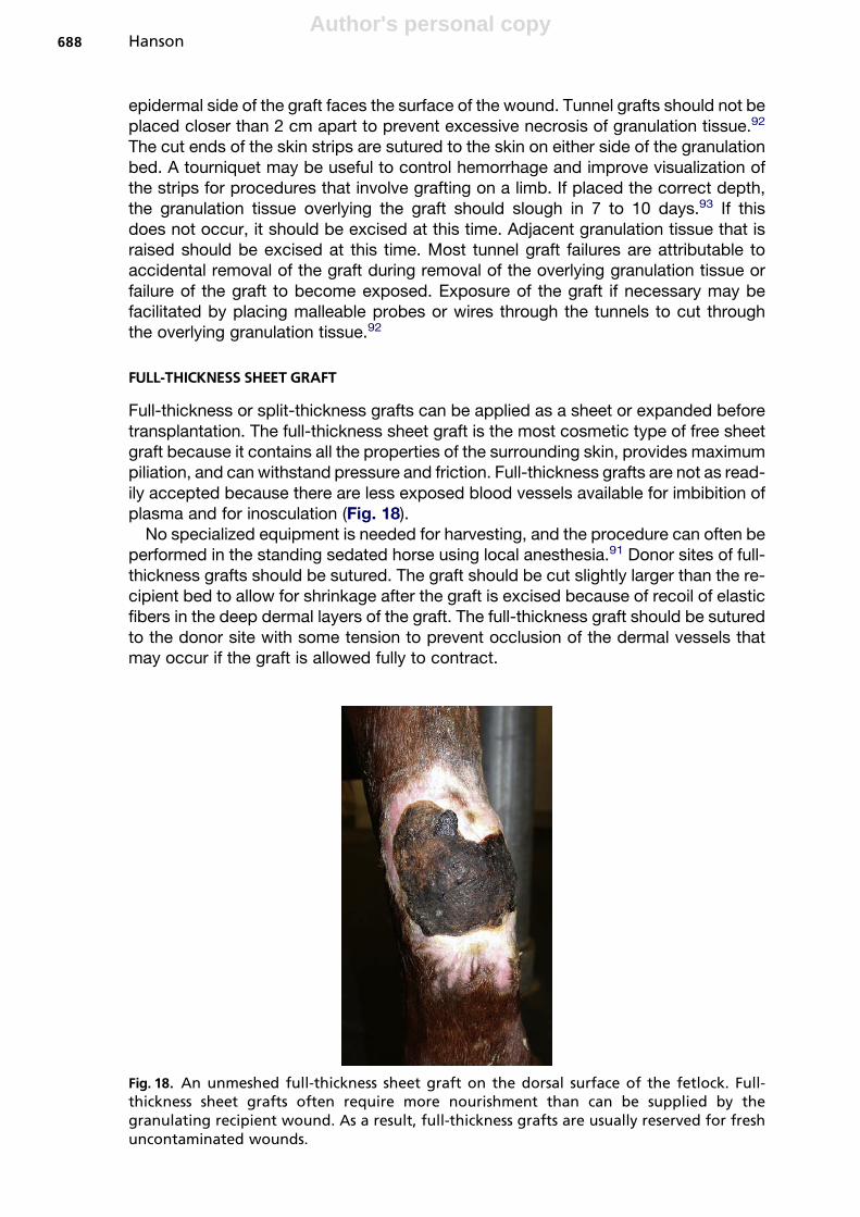
Full-thickness or split-thickness grafts can be applied as a sheet or expanded beforetransplantation. The full-thickness sheet graft is the most cosmetic type of free sheetgraft because it contains all the properties of the surrounding skin, provides maximumpiliation, and can withstand pressure and friction. Full-thickness grafts are not as read-ily accepted because there are less exposed blood vessels available for imbibition ofplasma and for inosculation (Fig. 18).
No specialized equipment is needed for harvesting, and the procedure can often beperformed in the standing sedated horse using local anesthesia.91 Donor sites of full-thickness grafts should be sutured. The graft should be cut slightly larger than the re-cipient bed to allow for shrinkage after the graft is excised because of recoil of elasticfibers in the deep dermal layers of the graft. The full-thickness graft should be suturedto the donor site with some tension to prevent occlusion of the dermal vessels thatmay occur if the graft is allowed fully to contract.
Fig. 18. An unmeshed full-thickness sheet graft on the dorsal surface of the fetlock. Full-thickness sheet grafts often require more nourishment than can be supplied by thegranulating recipient wound. As a result, full-thickness grafts are usually reserved for freshuncontaminated wounds.
Hanson688
Author's personal copy
A high oxygen gradient between the wound and the graft is essential for neovascu-larization of the graft and graft acceptance. Full-thickness grafts treated withhyperbaric oxygen therapy developed less granulation tissue, edema, and neovascu-larization, but more inflammation. The superficial portion of these full-thickness graftswas also less viable than the superficial portion of those not treated with hyperbaricoxygen therapy.94
Full-thickness sheet grafts are often considered compromised because they oftenrequire more nourishment than can be supplied by the granulating recipient wound.As a result, full-thickness grafts are usually reserved for fresh uncontaminated wounds.The upper layers of a full-thickness graft are more likely to slough because full-thicknessgrafts require more nourishment and have fewer exposed vessels for this purpose. Be-cause of the lack of abundant donor skin in the horse, the graft often must be meshedand expanded to achieve coverage of the wound larger than the donor area.
Split-Thickness Grafts
Split-thickness grafts are more readily accepted than full-thickness grafts, and may beused to cover granulation beds that are less than ideal.95 Because blood vesselsbranch as they become more superficial in the dermis, more vessels are cut andexposed with split-thickness grafts. The greater the number of exposed vessels thebetter the absorption of nutrients is from the granulation bed. A split-thickness sheetgraft is more cosmetic than a pinch or punch graft because the thickness of the graftand orientation of the hair are uniform and coverage by the graft is more complete.
A mechanical dermatome or a free-hand knife (Watson Skin Graft Knife, Down’sSurgical, Sheffield, England) is used to split the dermis. The latter is preferred becauseit is easy and economical to use. General anesthesia is necessary to obtain the graft;split-thickness donor sites are very painful to the horse, because many nerve endingsare exposed. Grafts less than 0.5-mm thickness in the horse lack strength, durability,and have sparse or no hair follicles or exocrine glands, which results in less sebaceoussecretion. Grafts harvested between 0.63 and 0.75 mm have good coverage of hairand greater durability than do thinner grafts.96,97 Unlike full-thickness grafts, suturingof the donor site is not required and primary graft contraction is minimal because a por-tion of the dermis remains intact and heals with a scarred appearance.97
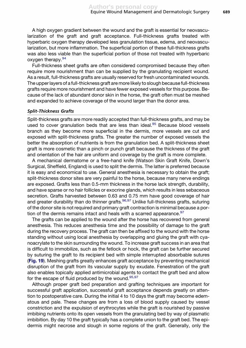
The grafts can be applied to the wound after the horse has recovered from generalanesthesia. This reduces anesthesia time and the possibility of damage to the graftduring the recovery process. The graft can then be affixed to the wound with the horsestanding without using local anesthesia by overlapping and gluing the graft with cya-noacrylate to the skin surrounding the wound. To increase graft success in an area thatis difficult to immobilize, such as the fetlock or hock, the graft can be further securedby suturing the graft to its recipient bed with simple interrupted absorbable sutures(Fig. 19). Meshing grafts greatly enhances graft acceptance by preventing mechanicaldisruption of the graft from its vascular supply by exudate. Fenestration of the graftalso enables topically applied antimicrobial agents to contact the graft bed and allowfor the escape of fluid produced by the wound.95,97
Although proper graft bed preparation and grafting techniques are important forsuccessful graft application, successful graft acceptance depends greatly on atten-tion to postoperative care. During the initial 4 to 10 days the graft may become edem-atous and pale. These changes are from a loss of blood supply caused by vesselconstriction and the expulsion of erythrocytes while the graft is nourished by passiveimbibing nutrients onto its open vessels from the granulating bed by way of plasmaticimbibition. By day 10 the graft typically has a complete union to the graft bed. The epi-dermis might necrose and slough in some regions of the graft. Generally, only the
Equine Wound Management and Dermatologic Surgery 689
Author's personal copy
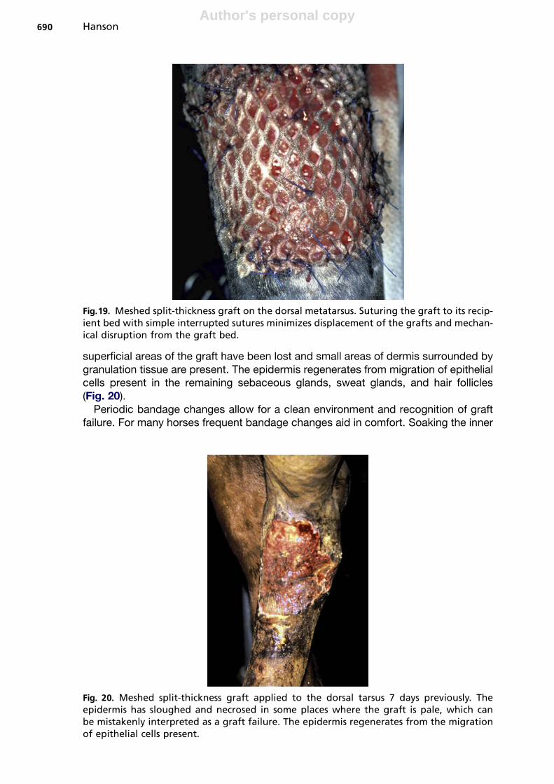
superficial areas of the graft have been lost and small areas of dermis surrounded bygranulation tissue are present. The epidermis regenerates from migration of epithelialcells present in the remaining sebaceous glands, sweat glands, and hair follicles(Fig. 20).
Periodic bandage changes allow for a clean environment and recognition of graftfailure. For many horses frequent bandage changes aid in comfort. Soaking the inner
Fig.19. Meshed split-thickness graft on the dorsal metatarsus. Suturing the graft to its recip-ient bed with simple interrupted sutures minimizes displacement of the grafts and mechan-ical disruption from the graft bed.
Fig. 20. Meshed split-thickness graft applied to the dorsal tarsus 7 days previously. Theepidermis has sloughed and necrosed in some places where the graft is pale, which canbe mistakenly interpreted as a graft failure. The epidermis regenerates from the migrationof epithelial cells present.
Hanson690
Author's personal copy
bandage with sterile saline for 5 minutes and then carefully removing the bandage pre-vents destruction of many grafts. The presence of purulent material on the initial ban-dage change does not have a detrimental effect on acceptance of individual grafts.Silver sulfadiazine in a 1% water-miscible cream is effective against most gram-positive and gram-negative organisms and may enhance wound epithelialization.Additional immobilization gained with a cast is usually unnecessary to facilitate accep-tance of grafts after 10 to 14 days. Immobilization may, however, lessen edema anddecrease the possibility of self-mutilation.8,96,97 Persistence in regrafting on horsesthat self-mutilate wounds has resulted in satisfactory wound healing in most cases.
SUMMARY
Complications of wounds and cosmetic surgery although frequent can be accuratelymanaged with a combination of timely surgical and medical intervention to ensure thebest possible outcome. The lack of soft tissue protection and a large quantity ofsusceptible synovial, tendon, ligament, and neurovascular structures make earlyand meticulous evaluation of limb wounds critical. Radiographs of traumatized areasshould be performed in all cases of distal limb wounds. Devitalized bone subject tosequestrum formation may not be evident initially. Ultrasonography provides informa-tion about the details of soft tissue structures and radiolucent foreign material.
Chronic wounds should be considered infected and may become afflicted withsarcoid, Pythium spp, Habronema spp, or Draschia spp and rarely squamous cellcarcinoma. Identification of pathologic tissue from a biopsy sample of the woundconfirms the diagnosis. MRSA infections can consequently be difficult to treat andare associated with increased morbidity, mortality, and treatment costs, comparedwith infections caused by methicillin-susceptible S aureus strains. Penetrating woundsof the abdomen or thorax have a guarded prognosis resulting from the ensuing poten-tial for infection and pneumothorax. Gunshot wounds limited to the skeletal muscleshave a good prognosis, whereas injuries that involve vital organs decreasesurvivability.
Skin grafting is usually used following a period of open wound management and af-ter healthy granulation tissue formation. Skin graft may be applied to fresh woundsthat are vascular enough rapidly to produce granulation tissue. Failure to adhere tothese basic principles in wound care results in poor graft survival.
REFERENCES
1. Hendrix SM, Baxter GM. Management of complicated wounds. Vet Clin North AmEquine Pract 2005;21(1):217–30.
2. Clem MF, Debowes RM, Yovich JV, et al. Osseous sequestration in horses, a re-view of 68 cases. Vet Surg 1988;11:2–5.
3. Clem MF, Debowes RM, Yovich JV, et al. Osseous sequestration in horses.Compend Contin Educ Pract Vet 1987;9:1219–24.
4. Moens Y, Vershooten F, DeMoor A, et al. Bone sequestration as a consequence oflimb wounds in the horse. Vet Radiol 1980;21:40–4.
5. Gift LJ, DeBowes RM. Wounds associated with osseous sequestration andpenetrating foreign bodies. Vet Clin North Am Equine Pract 1989;5(3):659–708.
6. Booth LC, Feeney DA. Superficial osteitis and sequestrum formation as a result ofskin avulsion in the horse. Vet Surg 1982;11:2–8.
7. Cartee RE, Rumpg PF. Ultrasonic detection of fistulous tracts and foreign objectsin muscles of horse. J Am Vet Med Assoc 1984;184:1127–32.
Equine Wound Management and Dermatologic Surgery 691
Author's personal copy
8. Stashak TS. Wound management and reconstructive surgery of problems associ-ated with the distal limbs. In: Equine wound management. Philadelphia: Lea &Febiger; 1991. p. 163–217.
9. Latenser J, Snow SN, Mohs FE, et al. Power drills to fenestrate exposed bone tostimulate wound healing. J Dermatol Surg Oncol 1991;17:265–70.
10. Hurwitz DJ. Osseous interference of soft tissue healing. Surg Clin North Am 1984;64:699–704.
11. Silver IA. Basic physiology of wound healing in the horse. Equine Vet J 1982;14:7–15.
12. Wilmink JA, Stolk PW, VanWeeren PR, et al. Differences in second-intentionwound healing between horses and ponies: macroscopic aspects. Equine VetJ 1999;31:53–60.
13. Caron JP, Barber SM, Doige CE, et al. The radiographic and histologic appear-ance of controlled surgical manipulation of the equine periosteum. Vet Surg1987;16:13–20.
14. Bradley DM, Swaim SF, Stuart SW. An animal model for research on woundhealing over exposed bone. Vet Comp Orthop Traumatol 1998;11:131–5.
15. Bailin PL, Wheeland RG. Carbon dioxide (CO2) laser perforation of exposed cra-nial bone to stimulate granulation tissue. Plast Reconstr Surg 1985;75:898–902.
16. Cabbage EB, Korock SW, Malik PA. Skin grafting denuded skull. Ann Plast Surg1982;8:318–21.
17. Brown PW. The fate of exposed bone. Am J Surg 1979;137:464–9.18. Specht TE, Colahan PT. Osteostixis for incomplete cortical fracture of the third
metacarpal bone: results in 11 horses. Vet Surg 1990;19:34–40.19. Lee AH, Swaim SF, Newton JC, et al. Wound healing over denuded bone. J Am
Anim Hosp Assoc 1987;23:75–84.20. Johnson RJ. The effects of cortical fenestration on second intention healing of
wounds over exposed bone of the distal aspect of the limb of horses. MastersThesis, Auburn University July 11, 2000.
21. Adam EN, Southwood LL. Surgical and traumatic wound infections, cellulitis, andmyositis in horses. Vet Clin North Am Equine Pract 2006;12:335–61.
22. Baxter GM. Wound management. In: White NA, Moore JN, editors. Current tech-niques in equine surgery and lameness. 2nd edition. Philadelphia: WB Saunders;1998. p. 72–80.
23. Farstvedt EG, Hendrickson DA, Dickenson CE, et al. Treatment of suppurativefacial cellulitis and panniculitis caused by Corynebacterium pseudotuberculosisin two horses. J Am Vet Med Assoc 2004;224:1139–42.
24. Belknap JK, Baxter GM, Nickels FA. Extensor tendon laceration in horses:50 cases (1982–1988). J Am Vet Med Assoc 1993;203(3):428–31.
25. Bertone AL. Tendon lacerations. Vet Clin North Am Equine Pract 1995;11(2):293–314.
26. Fubini SL. Burns. In: Robinson NE, editor. Current therapy in equine medicine 2.Philadelphia: WB Saunders; 1987. p. 639–41.
27. Fox SM. Management of a large thermal burn in a horse. Comp Cont Educ PractVet 1988;10:88–95.
28. Hanson RR. Management of burn injury in the horse. Vet Clin North Am EquinePract 2005;21:105–23.
29. Johnston DE. Burns: electrical, chemical and cold injuries. In: Slatter D, editor.Textbook of small animal surgery. Philadelphia: WB Saunders; 1985. p. 516–33.
30. Fox SM, Goring RI, Probst CW. Management of thermal burn injuries, Part II.Compend Cont Educ Pract Vet 1986;8:439–44.
Hanson692
Author's personal copy
31. Schumacher J, Watkins JP, Wilson JP, et al. Burn induced neoplasia in twohorses. Equine Vet J 1986;18:410–3.
32. Baxter GM. Management of burns. In: Colahan PT, Mayhew IG, Merritt AM, ed-itors. Equine medicine and surgery. 5th edition. St. Louis (MO): Mosby; 1999.p. 1843–7.
33. Warden GD. Outpatient care of thermal injuries. Surg Clin North Am 1987;67:147–57.
34. Bailey JV, Jacobs KA. The mesh expansion method of suturing wounds on thelegs of horses. Vet Surg 1983;12(2):78–82.
35. Geiser D, Walker RD. Management of large animal thermal injuries. CompendCont Educ Pract Vet 1985;7:S69–78.
36. Cefai C, Ashurst S, Owens C. Human carriage of methicillin-resistant Staphylo-coccus aureus linked with pet dog. Lancet 1994;344:539–40.
37. Loeffler AK, Boag J, Sung JA, et al. Prevalence of methicillin-resistant Staphylo-coccus aureus among staff and pets in a small animal referral hospital in the UK.J Antimicrob Chemother 2005;56:692–7.
38. Weese JS, Archambault M, Willey BM, et al. Methicillin-resistant Staphylococcusaureus in horses and horse personnel, 2000–2002. Emerg Infect Dis 2005;11:430–5.
39. Weese JS, Rousseau J, Traub-Dargatz JL, et al. Community-associated methicil-lin-resistant Staphylococcus aureus in horses and humans who work with horses.J Am Vet Med Assoc 2005;226:580–3.
40. Blot SI, Vandewoude KH, Hoste EA, et al. Outcome and attributable mortalityin critically ill patients with bacteremia involving methicillin-susceptible andmethicillin-resistant Staphylococcus aureus. Arch Intern Med 2002;162:2229–35.
41. Engemann JJ, Carmeli Y, Cosgrove SE, et al. Adverse clinical and economic out-comes attributable to methicillin resistance among patients with Staphylococcusaureus surgical site infection. Clin Infect Dis 2003;36:592–8.
42. Melzer M, Eykyn SJ, Graunsden WR, et al. Is methicillin-resistant Staphylococcusaureus more virulent than methicillin-susceptible S. aureus? A comparativecohort study of British patients with nosocomial infection and bacteremia. ClinInfect Dis 2003;37:1453–60.
43. Manian FA. Asymptomatic nasal carriage of mupirocin-resistant, methicillin-resis-tant Staphylococcus aureus (MRSA) in a pet dog associated with MRSA infectionin household contacts. Clin Infect Dis 2003;36:e26–8.
44. Greene N, Holtom PD, Warren CA, et al. In vitro elution of tobramycin and vanco-mycin polymethyl-methacrylate beads and spaces from simplex and palacos.Am J Orthop 1998;27:201–5.
45. Weese JS, Lefebvre SL. Risk factors for methicillin-resistant Staphylococcus au-reus colonization in horses admitted to a veterinary teaching hospital. Can Vet J2007;48(9):921–6.
46. Trostle SS, Peavy CL, King DS, et al. Treatment of methicillin-resistant Staphylo-coccus epidermidis infection following repair of an ulnar fracture and humerora-dial joint luxation in a horse. J Am Vet Med Assoc 2001;218:554–9.
47. Schnellmann C, Gerber V, Rossano A, et al. Presence of new mec(A) and mph(C)variants conferring antibiotic resistance in Staphylococcus spp isolated from theskin of horses before and after clinic admission. J Clin Microbiol 2006;44(12):4444–54.
48. Visavada BG, Honneysett J, Danford MH. Manuka honey dressing: an effectivetreatment for chronic wound infections. Br J Oral Maxillofac Surg 2008;46:55–6.
Equine Wound Management and Dermatologic Surgery 693
Author's personal copy
49. Wright JB, Lam K, Burrell RE. Wound management in an era of increasing bac-terial antibiotic resistance: a role for topical silver treatment. Am J Infect Control1998;26(6):572–7.
50. Wertheim HFL, Verveer J, Boelens HAM, et al. Effect of mupirocin treatment onnasal, pharyngeal, and perineal carriage of Staphylococcus aureus in healthyadults. Antimicrob Agents Chemother 2005;49:1465–7.
51. Muller A, Talon D, Potier A, et al. Use of intranasal mupirocin to prevent methicil-lin-resistant Staphylococcus aureus infection in intensive care units. Crit Care2005;9(3):R246–50.
52. Santschi LM. Diagnosis and management of surgical site infection and antimicro-bial prophylaxis. In: Auer JA, Stick JA, editors. Equine surgery. 2nd edition. Phil-adelphia: WB Saunders; 1999. p. 55–60.
53. Holcombe SJ, Schneider RK, Bramlage LR, et al. Use of antibiotic-impregnatedpolymethyl methacrylate in horses with open or infected fractures or joints: 19cases (1987–1995). J Am Vet Med Assoc 1997;211:889–93.
54. Swalec Tobias KM, Schneider RK, Besser TE. The use of anitmicrobial-impreg-nated polymethyl methacrylate. J Am Vet Med Assoc 1996;208:841–5.
55. Kawcak CE, Stashak TS. Predisposing factors, diagnosis and management oflarge abdominal wall defects in horses and cattle. J Am Vet Med Assoc 1995;206(5):607–11.
56. Holcombe SJ, Laverty S. Thoracic trauma. In: Auer JA, Stick JA, editors. Equinesurgery. 2nd edition. Philadelphia: WB Saunders; 1999. p. 382–5.
57. Vatistas NJ, Meagher DM, Gillis CL, et al. Gunshot injuries in horses: 22 cases(1971–1993). J Am Vet Med Assoc 1995;207:1198–200.
58. Bartmann CP, Wohlsein P. Injuries caused by outside violence with forensic im-portance in horses. DTSCH Tierarztl Wochenschr 2002;109:112–5.
59. Poole HM, Brashier MK. Equine cutaneous pythiosis. Compend Contin EducPract Vet 2003;25:229–36.
60. Reis JL, Queiroz de Carvallo C, Nogueira RH, et al. Disseminated pythiosis inthree horses. J Vet Microbiol 2003;96:289–95.
61. Worster AA, Lillich JD, Cox JH, et al. Pythiosis with bone lesions in a pregnantmare. J Am Vet Med Assoc 2000;216:1795–8.
62. Hensel P, Greene CE, Medleau L, et al. Immunotherapy for treatment of multicen-tric cutaneous pythiosis in a dog. J Am Vet Med Assoc 2003;223:215–8.
63. Vasey JR. Equine cutaneous habronemiasis. Compend Contin Educ Pract Vet1981;8:290–8.
64. Scott DW. Habronemiasis. In: Large animal dermatology. Philadelphia: WB Saun-ders; 1988. p. 251–5.
65. Von Tscharner C, Kunkle G, Yager J. Nodular diseases. Vet Demmatol 2000;11:179–86.
66. White SD, Evans AG. Parasitic skin diseases. In: Smith PB, editor. Large animalinternal medicine. St Louis (MO): CV Mosby; 2002. p. 1221–2.
67. Moriello KA, DeBoer DJ, Semrad SD. Diseases of skin. In: Reed SM, Bayly WM,editors. Equine internal medicine. Philadelphia: WB Saunders; 1988. p. 536.
68. Rees CA, Craig TM. Cutaneous habronemiasis. In: Robinson NE, editor. Currenttherapy in equine medicine 5. St. Louis (MO): WB Saunders; 2003. p. 195–200.
69. Herd RP, Donham JC. Efficacy of ivermectin against cutaneous Draschia andHabronema infection (summer sores) in horses. Am J Vet Res 1981;42:1953–5.
70. Novick M, Gard DA, Hardy SB, et al. Burn scar carcinoma: a review and analysisof 46 cases. J Trauma 1977;17:809–17.
Hanson694
Author's personal copy
71. Baird AN, Frelier PF. Squamous cell carcinoma originating from an epithelial scarin a horse. J Am Vet Med Assoc 1990;196:1999–2000.
72. Rogers K, Barrington GM, Parish SM. Squamous cell carcinoma originating froma cutaneous scar in a llama. Can Vet J 1997;38:643–4.
73. Paterson S. Treatment of superficial ulcerative squamous cell carcinoma in threehorses with topical 5-fluorouracil. Vet Rec 1997;141:626–8.
74. Theon AP, Pascoe JR. Iridium-192 brachytherapy for equine periocular tumors:treatments results and prognostic factors in 115 horses. Equine Vet J 1995;27:117–21.
75. England JJ, Watson RE, Larson K. Virus like particles in an equine sarcoid cellline. Am J Vet Res 1973;34:1601–3.
76. Cheevers WP, Faemi-Ninie S, Anderson LW. Spontaneous expression of anendogenous retrovirus by the equine sarcoid derived Mc-1 cell line. Am J VetRes 1986;47:50–2.
77. Baldwin RW. Mechanism of immunity in cancer. Pathobio Ann 1981;11:155–75.78. Nasir L, Reid SWJ. Bovine papillomaviral gene expression equine sarcoids. Virus
Res 1999;61:171–5.79. Reid SWJ, Smith KT, Jarrett WFH. Detection, cloning, and characterization of
papillomaviral DNA present in sarcoid tumors of Equus asinus. Vet Rec 1994;135:430–2.
80. Brandt S, Haralambus R, Schoster A, et al. Peripheral blood mononuclear cellsrepresent a reservoir of bovine papillomavirus DNA in sarcoid-affected equines.J Gen Virol 2008;89(Pt 6):1390–5.
81. Wilmink JM, Van Weeren PR. Treatment of exuberant granulation tissue. Clin TechEquine Pract 2004;3(2):141–7.
82. Knottenbelt DC. A suggested clinical classification for the equine sarcoid. ClinTech Equine Pract 2005;4(4):278–95.
83. Knottenbelt DC, Edwards SER, Daniel EA. The diagnosis and treatment of equinesarcoid. In Pract 1995;17:123–9.
84. Carr EA, Theon AP, Madewell BR, et al. Bovine papillomavirus DNA inneoplastic and non-neoplastic tissues obtained from horses with and with-out sarcoids in the western United States. Am J Vet Res 2001;62(5):741–4.
85. Meagher DM, Adams OR. Split thickness autologous skin transplantation inhorses. J Am Vet Med Assoc 1971;159:55–60.
86. Booth L. Equine wound reconstruction using free skin grafting. Calif Vet 1991;45:13–6.
87. French DA, Fretz PB. Treatment of equine leg wounds using skin grafts: thirty-fivecases, 1975–1988. Can Vet J 1990;31:761–5.
88. Teh BT. Why do skin grafts fail? Plast Reconstr Surg 1979;63:323–32.89. Hanson RR. Management of avulsion wounds with exposed bone. Clinical Tech
Equine Pract 2004;3(2):188–203.90. Robeson MC, Edstrom LE, Krizek TJ. The efficacy of systemic antibiotics in the
treatment of granulating wounds. J Surg Res 1974;16:299–306.91. Schumacher J, Hanselka DV. Skin grafting of the horse. Vet Clin North Am Equine
Pract 1989;5:591–614.92. Carson-Dunkerly SA, Hanson RR. Equine skin grafting principles and field
applications. Comp Contin Educ Pract Veterin 1997;19:872–82.93. Caron JP. Skin grafting. In: Auer JA, Stick JA, editors. Equine surgery. 2nd
edition. Philadelphia: WB Saunders; 1999. p. 152–66.
Equine Wound Management and Dermatologic Surgery 695

Author's personal copy
94. Holder TEC, Schumacher J, Donnell RL, et al. Effects of hyperbaric oxygen onfull-thickness meshed sheet skin grafts applied to fresh and granulating woundsin horses. Am J Vet Res 2008;69:144–7.
95. Tobin GR. The compromised bed technique: an improved method for skin graft-ing problem wounds. Surg Clin North Am 1985;64:653–8.
96. Booth LC. Split thickness autogenous skin transplantation in the horse. J Am VetMed Assoc 1982;180:754–7.
97. Hanselka DV. Use of autogenous mesh grafts in equine wound management.J Am Vet Med Assoc 1974;164:35–41.
Hanson696