Embed Size (px)
Citation preview
www.postersession.com
Endogenous endophthalmitis (EE) is a rare,rapidly progressive
and potentially blinding, intraocular infection which is caused
by the hematogenous spread of bacteria or fungi from distant
foci, not via external causes such as trauma or ocular surgery.
EE is much less common than exogenous endophtalmitis and
accounts for 2% to 15% of all cases of endoftalmitis.1,
Numerous predisposing factors of EE have been suggested,
including diabetes mellitus (DM), malignancies, renal failure,
indwelling catheters, liver disease, intravenous drug abuse,
AIDS, organ transplantation, allergic reactions, endocarditis,
urinary tract infections (UTI), recent major surgeries, and
dental procedures.2 The list of potential causative organisms is
extremely variable, especially in different geographic locations
of the world. Gram-positive organisms are more common in
Western countries (and Candida ,followed by Aspergillosis, as
fungal causes), whereas, Gram-negative bacteria species were
more prevalent in East Asian countries.3Asian studies have
stressed fungi as causative organisms in 11.1% to 17.54% of
total cases of EE. There are more reports that aim to describe
clinical presentations, sources of infections, causative
organisms, and the effectiveness of treatment modalities by
comparisons between different regions of the world.4, In this
study, we aimed to identify these features of the disease and
evaluate the clinical course of culture proven EE patients in a
tertiary clinic of our country.
Methods Take home message
Endogenous Endophthalmitis: A 5-Year Retrospective Study at a Tertiary Hospital in Intercontinental Region of Turkey Hande Celiker, Haluk Kazokoglu
Marmara University School of Medicine Ophthalmology Department
References
1. Keynan Y, Finkelman Y, Lagacé-Wiens P. The microbiology of endophthalmitis: global trends and a local perspective. Eur J Clin Microbiol Infect Dis 2012;31:2879–2886. 2. Jackson TL, Paraskevopoulos T, Georgalas I. Systematic review of 342 cases of endogenous bacterial endophthalmitis. Surv Ophthalmol. 2014;59:627–635. 3. Jackson TL, Eykyn SJ, Graham EM, et al. Endogenous bacterial endophthalmitis: a 17-year prospective series and review of 267 reported cases. Surv Ophthalmol 2003;48:403–423. 4. Lingappan A, Wykoff CC, Albini TA, et al. Endogenous fungal endophthalmitis: causative organisms, management strategies, and visual acuity outcomes. Am J Ophthalmol 2012;153:162–166.e1. 5 . C h o H , S h i n Y U , S i e g e l N H , e t a l . EndogenousEndophthalmitis in the American and Korean Population: An 8-year Retrospective Study.Ocul Immunol Inflamm. 2016; 26:1-8.
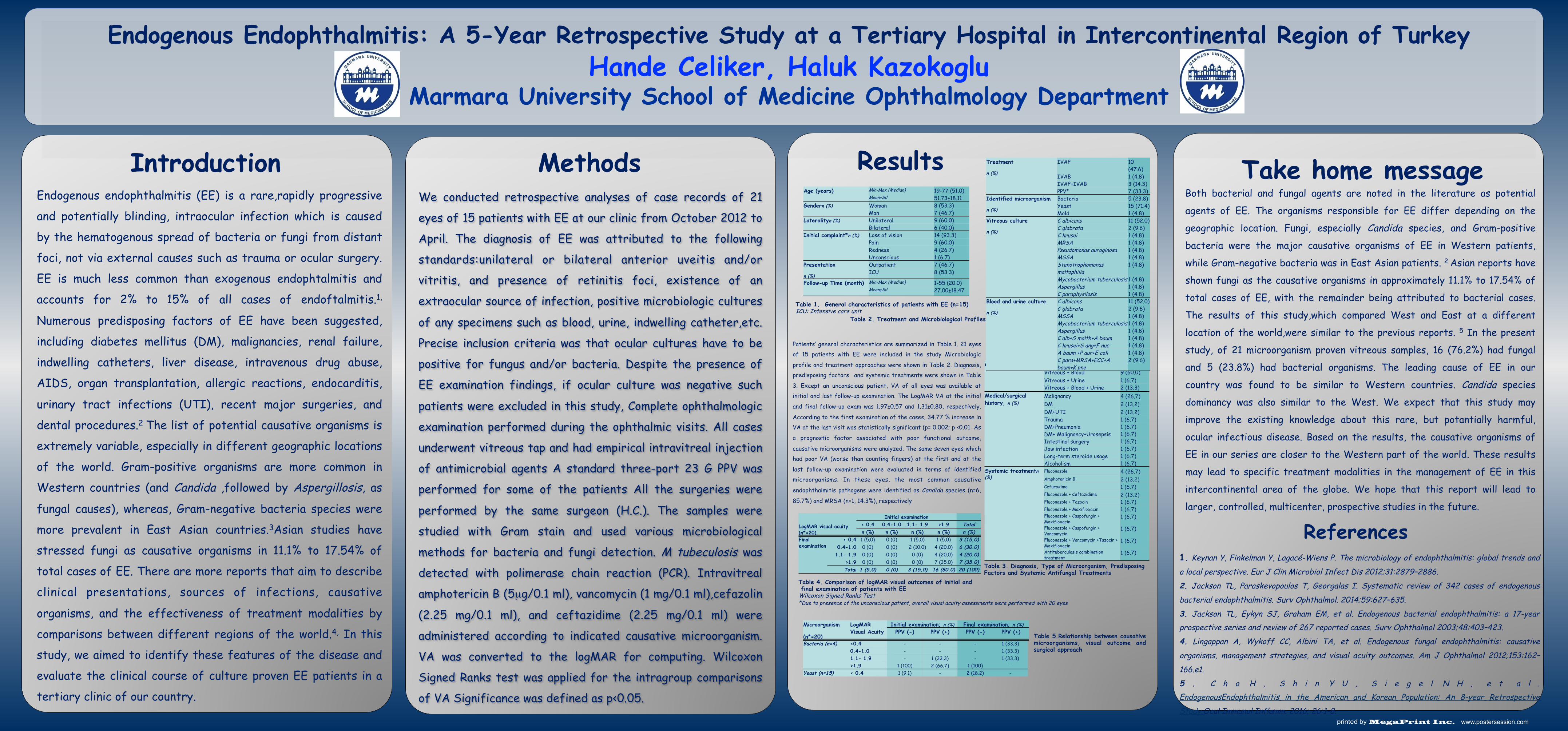
Patients’ general characteristics are summarized in Table 1. 21 eyes of 15 patients with EE were included in the study Microbiologic
profile and treatment approaches were shown in Table 2. Diagnosis, predisposing factors and systemic treatments were shown in Table
3. Except an unconscious patient, VA of all eyes was available at initial and last follow-up examination. The LogMAR VA at the initial
and final follow-up exam was 1.97±0.57 and 1.31±0.80, respectively.
According to the first examination of the cases, 34.77 % increase in VA at the last visit was statistically significant (p= 0.002; p <0.01 As
a prognostic factor associated with poor functional outcome,
causative microorganisms were analyzed. The same seven eyes which had poor VA (worse than counting fingers) at the first and at the
last follow-up examination were evaluated in terms of identified microorganisms. In these eyes, the most common causative
endophthalmitis pathogens were identified as Candida species (n=6,
85.7%) and MRSA (n=1, 14.3%), respectively
Both bacterial and fungal agents are noted in the literature as potential
agents of EE. The organisms responsible for EE differ depending on the
geographic location. Fungi, especially Candida species, and Gram-positive
bacteria were the major causative organisms of EE in Western patients,
while Gram-negative bacteria was in East Asian patients. 2 Asian reports have
shown fungi as the causative organisms in approximately 11.1% to 17.54% of
total cases of EE, with the remainder being attributed to bacterial cases.
The results of this study,which compared West and East at a different
location of the world,were similar to the previous reports. 5 In the present
study, of 21 microorganism proven vitreous samples, 16 (76.2%) had fungal
and 5 (23.8%) had bacterial organisms. The leading cause of EE in our
country was found to be similar to Western countries. Candida species
dominancy was also similar to the West. We expect that this study may
improve the existing knowledge about this rare, but potantially harmful,
ocular infectious disease. Based on the results, the causative organisms of
EE in our series are closer to the Western part of the world. These results
may lead to specific treatment modalities in the management of EE in this
intercontinental area of the globe. We hope that this report will lead to
larger, controlled, multicenter, prospective studies in the future.
Introduction Results
We conducted retrospective analyses of case records of 21
eyes of 15 patients with EE at our clinic from October 2012 to
April. The diagnosis of EE was attributed to the following
standards:unilateral or bilateral anterior uveitis and/or
vitritis, and presence of retinitis foci, existence of an
extraocular source of infection, positive microbiologic cultures
of any specimens such as blood, urine, indwelling catheter,etc.
Precise inclusion criteria was that ocular cultures have to be
positive for fungus and/or bacteria. Despite the presence of
EE examination findings, if ocular culture was negative such
patients were excluded in this study, Complete ophthalmologic
examination performed during the ophthalmic visits. All cases
underwent vitreous tap and had empirical intravitreal injection
of antimicrobial agents A standard three-port 23 G PPV was
performed for some of the patients All the surgeries were
performed by the same surgeon (H.C.). The samples were
studied with Gram stain and used various microbiological
methods for bacteria and fungi detection. M tubeculosis was
detected with polimerase chain reaction (PCR). Intravitreal
amphotericin B (5µg/0.1 ml), vancomycin (1 mg/0.1 ml),cefazolin
(2.25 mg/0.1 ml), and ceftazidime (2.25 mg/0.1 ml) were
administered according to indicated causative microorganism.
VA was converted to the logMAR for computing. Wilcoxon
Signed Ranks test was applied for the intragroup comparisons
of VA Significance was defined as p<0.05.
Table 1. General characteristics of patients with EE (n=15) ICU: Intensive care unit
Table 3. Diagnosis, Type of Microorganism, Predisposing Factors and Systemic Antifungal Treatments Table 4. Comparison of logMAR visual outcomes of initial and
final examination of patients with EE Wilcoxon Signed Ranks Test *Due to presence of the unconscious patient, overall visual acuity assessments were performed with 20 eyes
Age (years) Min-Max (Median) 19-77 (51.0) Mean±Sd 51.73±18.11
Gendern (%) Woman 8 (53.3) Man 7 (46.7)
Lateralityn (%) Unilateral 9 (60.0) Bilateral 6 (40.0)
Initial complaint*n (%) Loss of vision 14 (93.3) Pain 9 (60.0) Redness 4 (26.7) Unconscious 1 (6.7)
Presentation
n (%)
Outpatient 7 (46.7) ICU 8 (53.3)
Follow-up Time (month) Min-Max (Median) 1-55 (20.0) Mean±Sd 27.00±18.47
Culture source,n (%) Vitreous 3 (20.0) Vitreous + Blood 9 (60.0) Vitreous + Urine 1 (6.7) Vitreous + Blood + Urine 2 (13.3)
Medical/surgical history, n (%)
Malignancy 4 (26.7) DM 2 (13.2) DM+UTI 2 (13.2) Trauma 1 (6.7) DM+Pneumonia 1 (6.7) DM+ Malignancy+Urosepsis 1 (6.7) Intestinal surgery 1 (6.7) Jaw infection 1 (6.7) Long-term steroide usage 1 (6.7) Alcoholism 1 (6.7)
Systemic treatmentn (%)
Fluconazole 4 (26.7) Amphotericin B 2 (13.2) Cefuroxime 1 (6.7) Fluconazole + Ceftazidime 2 (13.2) Fluconazole + Tazocin 1 (6.7) Fluconazole + Moxifloxacin 1 (6.7) Fluconazole + Caspofungin + Moxifloxacin
1 (6.7)
Fluconazole + Caspofungin + Vancomycin
1 (6.7)
Fluconazole + Vancomycin +Tazocin + Moxifloxacin
1 (6.7)
Antituberculosis combination treatment
1 (6.7)
LogMAR visual acuity (n*=20)
Initial examination < 0.4 0.4-1.0 1.1- 1.9 >1.9 Total n (%) n (%) n (%) n (%) n (%)
Final examination
< 0.4 1 (5.0) 0 (0) 1 (5.0) 1 (5.0) 3 (15.0) 0.4-1.0 0 (0) 0 (0) 2 (10.0) 4 (20.0) 6 (30.0) 1.1- 1.9 0 (0) 0 (0) 0 (0) 4 (20.0) 4 (20.0)
>1.9 0 (0) 0 (0) 0 (0) 7 (35.0) 7 (35.0) Total 1 (5.0) 0 (0) 3 (15.0) 16 (80.0) 20 (100)
Microorganism
(n*=20)
LogMAR Visual Acuity
Initial examination; n (%) Final examination; n (%) PPV (-) PPV (+) PPV (-) PPV (+)
Bacteria (n=4) <0.4 - - - 1 (33.3) 0.4-1.0 - - - 1 (33.3) 1.1- 1.9 - 1 (33.3) - 1 (33.3) >1.9 1 (100) 2 (66.7) 1 (100) -
Yeast (n=15) < 0.4 1 (9.1) - 2 (18.2) -
Table 5.Relationship between causative microorganisms, visual outcome and surgical approach
Treatment
n (%)
IVAF 10 (47.6)
IVAB 1 (4.8) IVAF+IVAB 3 (14.3) PPV* 7 (33.3)
Identified microorganism
n (%)
Bacteria 5 (23.8) Yeast 15 (71.4) Mold 1 (4.8)
Vitreous culture
n (%)
C albicans 11 (52.0) C glabrata 2 (9.6) C krusei 1 (4.8) MRSA 1 (4.8) Pseudomonas auroginosa 1 (4.8) MSSA 1 (4.8) Stenotrophomonas maltophilia
1 (4.8)
Mycobacterium tuberculosis 1 (4.8) Aspergillus 1 (4.8) C paraphysilosis 1 (4.8)
Blood and urine culture
n (%)
C albicans 11 (52.0) C glabrata 2 (9.6) MSSA 1 (4.8) Mycobacterium tuberculosis 1 (4.8) Aspergillus 1 (4.8) C alb+S malth+A baum 1 (4.8) C krusei+S ang+F nuc 1 (4.8) A baum +P aur+E coli 1 (4.8) C para+MRSA+ECC+A baum+K pne
2 (9.6)
Table 2. Treatment and Microbiological Profiles

![Scanned Document - Prof. Dr. Alpay Celiker · TOF, tetrology of fallot; VSD, ventricular septa] defect. during follow up. 'Partial relief' meant no evidence of arrhythmia symptoms](https://img.pdfslide.us/doc/110x75/5f314ec70852e8379b0c9851/scanned-document-prof-dr-alpay-tof-tetrology-of-fallot-vsd-ventricular-septa.jpg)


![[PPT]Presentación de PowerPoint - Carpe Diem – Cogito ergo … · Web viewEn caso de endocarditis la sustitución valvular es el tratamiento de elección, mientras que en la endoftalmitis](https://img.pdfslide.us/doc/110x75/5b3765937f8b9a600a8c2f55/pptpresentacion-de-powerpoint-carpe-diem-cogito-ergo-web-viewen-caso.jpg)