Embed Size (px)
Citation preview

19Photoactive Yellow Protein, the Xanthopsin
Michael A. van der Horst, Johnny Hendriks, Jocelyne Vreede, Sergei Yeremenko,Wim Crielaard and Klaas J. Hellingwerf
19.1Introduction
19.1.1Discovery of the Photoactive Yellow Protein
In 1985 T.E. Meyer reported the isolation of a series of colored proteins from thehalophilic phototrophic bacterium Ectothiorhodospira (Halorhodospira) halophila(Meyer 1985). One of these proteins was yellow and was named ‘Photoactive YellowProtein’ (PYP) in a subsequent study (McRee et al. 1986), because of its observed pho-toactivity. H. halophila is a unicellular phototrophic purple sulfur bacterium that de-posits sulfur extracellularly. It was first isolated from the shores of Summer Lake,Lake County, Oregon (Raymond and Sistrom 1967; Raymond and Sistrom 1969), andsubsequently also from the extremely saline lakes of the Wadi el Natrun in Egypt(Imhoff et al. 1978). Both locations are salt lakes and indeed H. halophila is ahalophilic organism.
As a phototroph, H. halophila is able to exploit the free energy available from sun-light. However, like most organisms, H. halophila is not immune to the effects of UV-radiation, and by consequence protective mechanisms have evolved. Like most pho-totrophs H. halophila can perceive the quality and the quantity of the ambient radia-tion: It is attracted by red light that can be absorbed through its photosynthetic ma-chinery, but repelled by high intensities of white and in particular blue light. Thislatter response has a wavelength dependence that fits the absorption spectrum of thephotoactive yellow protein (Sprenger et al. 1993). This fit indicates that PYP may bethe light-sensor in this blue-light response of H. halophila. Accordingly, the functionof PYP may be similar to that of sensory rhodopsins, particularly to sensoryrhodopsin II from Halobacterium salinarum, an archaebacterium that is also abun-dant in (solar) salt lakes. Rhodopsins (Spudich et al. 2000; Hoff et al., 1997) form alarge family with members in organisms in all kingdoms of life, from unicellular bac-teria to Homo sapiens sapiens. It is the most extensively studied family of photorecep-
Handbook of Photosensory Receptors. Edited by W. R. Briggs, J. L. SpudichCopyright © 2005 WILEY-VCH Verlag GmbH & Co. KGaA, WeinheimISBN 3-527-31019-3

392
tor proteins, especially via some of the visual rhodopsins, and bacteriorhodopsin, thelatter being a light-activated proton pump found in the cytoplasmic membrane of H.salinarum. Sensory rhodopsins are structurally and photochemically closely related tobacteriorhodopsin and are present in the same organism. The similarity betweenPYP and the sensory rhodopsins was already noted after the first characterization ofPYP (McRee et al. 1986). A major difference between PYP and sensory rhodopsins isthat PYP is highly water soluble, whereas the rhodopsins, being membrane proteins,are not. The two proteins belong to structurally completely different families of pho-toreceptor proteins (which, however, may be similar in function; see above).
19.1.2A Family of Photoactive Yellow Proteins: the Xanthopsins
After the initial biochemical characterization of PYP, reverse genetics led to the iden-tification of the pyp operon that encodes this photoreceptor protein In H. halophila(Kort et al. 1996). Meanwhile, Southern hybridizations and PCR led to the discoveryof a photoactive yellow protein in six other organisms – all purple bacteria – and asecond one has been discovered in H. halophila itself (unpublished observation). Thisfamily of photoactive yellow proteins has been christened the Xanthopsin family (Ko-rt et al. 1996). Presently, the eight known xanthopsins can be divided into three sub-groups, based on mutual sequence homology. The first sub-group is formed by theproteins found in H. halophila [pyp(A)] (Meyer 1985), Rhodothalassium salexigens(Meyer et al. 1990), and Halochromatium salexigens (Koh et al. 1996). The second isformed by proteins found in Rhodobacter sphaeroides (Kort et al. 1996), and Rhodobac-ter capsulatus (Jiang and Bauer 1998), and the third sub-group consists of proteinsfound in Rhodospirillum centenum (Jiang et al. 1999) and Thermochromatium tepidum(Cusanovich and Meyer 2003). In these latter two xanthopsins the homology to PYPis limited to their amino-terminal domain; they both have a larger bacteriophy-tochrome domain as well. Recently a second pyp gene (i.e. pyp(B)) was discovered inH. halophila (M.A. van der Horst et al., unpublished observation). Based on mutualhomologies this xanthopsin can also be classified in sub-group III.
19.1.3Differentiation of Function among the Xanthopsins
Though all these xanthopsins presumably absorb blue light, their role in the variousorganisms differs. In H. halophila PYP is reported to mediate intiation of a photo-phobic tactile response (Sprenger et al. 1993). It is relevant to note that this does nothold for the xanthopsin from Rb. sphaeroides, provided that no genetic redundancy ex-ists in this organism (Kort et al. 2000). The results of the sequence comparisonshown in Figure 19.1, revealing three sub-groups, are consistent with this conclusion.The xanthopsin from Rs. centenum transcriptionally regulates chalcone synthesis(Jiang et al. 1999). These different functions of the xanthopsin members coincidewith the sub-group assignments. Though the function of only a few xanthopsins hasbeen elucidated, it may well be that its members within the different sub-groups have
19 Photoactive Yellow Protein, the Xanthopsin

393
a similar biological function, while these functions may differ between the sub-groups.
19.1.4PYP: The Prototype PAS Domain
The xanthopsins thus far have only been identified in proteobacteria. Its members,however, also show a striking similarity with the much larger family of the PAS do-mains. These PAS domains have been identified in proteins from all three kingdomsof life, i.e. in the Bacteria, the Archaea, and the Eucarya. PAS is an acronym formedfrom the names of the proteins in which the PAS motive was first recognized (Nam-bu et al. 1991): the Drosophila period clock protein (PER), the vertebrate aryl hydro-
19.1 Introduction
Figure 19.1 Multiple sequence alignment ofall currently known xanthopsins. Alignmentswere made using the program ClustalW athttp://www.ebi.ac.uk/clustalw/ (Thompson etal. 1994). Residues displayed with black back-ground are conserved in more than 50% ofthe sequences; residues with a grey back-
ground are similar in more than 50% of thesequences. The Roman numbers indicate thesubgroups to which the particular xanthopsinbelongs. The location of the elements of sec-ondary structure is shown via cartoons belowthe sequence alignment, together with thenames of the sub-domains.

394
carbon receptor nuclear translocator (ARNT), and the Drosophila single-minded pro-tein (SIM).
Proteins containing PAS domains are predominantly involved in signal transduc-tion. Over 2000 proteins have been identified that contain one or more PAS domains(Taylor and Zhulin 1999). Most of these proteins are receptors, signal transducers, ortranscriptional regulators. PAS domains are usually present in proteins with a mul-ti-domain architecture, such that a single protein can have up to six of these domains.Although most are present in the intracellular compartment, examples of periplas-mic sensing domains with a PAS structure have also been described (Reinelt et al.2003). The entire Photoactive Yellow Protein from H. halophila can be considered asingle PAS domain with a ∼30 amino acid N-terminal extension. As it is the first pro-tein from the PAS domain family for which the 3-D structure has been elucidated,PYP has been proclaimed to be the structural prototype of the PAS domain fold(Pellequer et al. 1998).
19.2Structure
The backbone of PYP is folded into an α/β-fold with a six-stranded anti-parallel β-sheet as a scaffold, f lanked by several helices (Borgstahl et al. 1995). The loops con-taining helices α3 and α4, as well as the one containing helix α5, fold on top of it toform the major hydrophobic core of PYP and a pocket in which the chromophore re-sides. Helices α1 and α2, which comprise the majority of the N-terminal domain foldbehind it to form a second, smaller hydrophobic core. A spatial model of this arrange-ment of elements of secondary structure is shown in Figure 19.2.
In photoreceptor proteins the light-absorbing chromophore triggers functional ac-tivity. Its chemical structure typically correlates with the wavelength range in whichthe holo-protein is biologically active (Hellingwerf et al. 1996). Accordingly, the xan-thopsins were shown to use an aromatic chromophore. After a long period of confu-sion this chromophore was identified in 1994 in H. halophila PYP as 4-hydroxycin-namic acid, covalently bound to the apo-protein via a thiol ester linkage with Cys69(Baca et al. 1994; Hoff et al. 1994a).
19.2.1Primary, Secondary, and Tertiary Structure
The amino acids that line the chromophore-binding pocket are (in the order in whichthey appear in the sequence): Ile31, Tyr42, Glu46, Thr50, Arg52, Phe62, Val66, Ala67,Cys69, Thr70, Phe96, Asp97, Tyr98, Met100, Val120 and Val122. When residing in thispocket the chromophore is completely buried in the major hydrophobic core of PYPand has no direct contact with solvent.
In the ground state of PYP, the chromophore is present as a phenolate anion (Ba-ca et al. 1994; Kim et al. 1995). The resulting negative charge is delocalized over thechromophore via an extensive π-orbital system. It is additionally stabilized via a hy-
19 Photoactive Yellow Protein, the Xanthopsin

395
drogen-bonding network and, presumably, by the positive charge of Arg52 (Borgstahlet al. 1995; Yoda et al. 2001; Groenhof et al. 2002b). The hydrogen-bonding networkincludes, besides the chromophore, residues Tyr42, Glu46, and Thr50, such that Oη
from Tyr42 and Oε,2 from Glu46 directly hydrogen-bond with the phenolate oxygen ofthe chromophore. Oγ,1 from Thr50 forms a hydrogen bond with Oη from Tyr42, yetdoes not line the chromophore-binding pocket.
19.2.2Solution Structure vs. Crystal Structure
Up till now we have described the structure of PYP as determined by X-ray diffrac-tion analyses, i.e. in the confines of a crystal lattice. In vivo, the protein is located inthe cytoplasm, on the intracellular side of the cytoplasmic membrane (Hoff et al.1994b), where it may have more freedom for dynamical alterations in its structurethan in a crystal lattice. It is therefore relevant to know whether there are differencesbetween its structure in a crystal and in aqueous solution [note that in the intact cell
19.2 Structure
Figure 19.2 Tertiary structure of the Photoac-tive Yellow Protein. The backbone is represent-ed as a ribbon with α helices, loops and βstrands. The chromophore is shown in yellow.
The transparent surface indicates the molecu-lar surface of PYP. This picture was createdusing PyMOL (DeLano 2002).

396
PYP may also be confined or partly constrained in its movements by e.g. (a) trans-ducer protein(s)].
The solution structure of PYP has been determined via multi-dimensional NMRspectroscopy (Dux et al. 1998) and is very similar to the structure determined with X-ray crystallography. Most of the elements of secondary structure are present in bothstructures, though they may start/end 1 to 2 residues earlier or later. However, helixα2 and the π-helix are not detectable in the solution structure. Additionally, there arethree poorly defined regions in the solution structure comprising residues 1–5, 17–23, and 113–117. This is caused by lack of structural constraints in the NMR dataset,which might be caused by high side-chain and/or backbone mobility (Dux et al. 1998).In agreement with this suggestion, the crystal structure indicates that the correspon-ding regions have higher B-factors.
From an ensemble of structures, such as obtained with NMR and using EssentialDynamics, it is possible to determine eigenvectors that describe the path along whichthe atoms of the protein may move (van Aalten et al. 1998; Van Aalten et al. 2000).Strikingly, by using these eigenvectors, it is possible to transform the solution struc-ture into the crystal structure and visa versa, indicating that the observed differencesare within the confines of the intrinsic f lexibility of the protein.
The solution structure, as determined with NMR, confirms the presence of the hy-drogen-bonding network that stabilizes the anionic chromophore in the chro-mophore-binding pocket. However, there is one notable difference with the crystalstructure: The side chain of Arg52 is present in two conformations, one in whichArg52 is clustered about 4 Å above the aromatic ring of the chromophore, and onewith the guanidinium group of Arg52 positioned about 4 Å above the aromatic ringof Tyr98 (Dux et al. 1998). This latter position is in line with the observation thatcations preferentially position themselves within 3.4 to 6 Å of the centroid of an aro-matic ring [a phenomenon called π-stacking (Scrutton and Raine 1996)]. The confor-mation of Arg52 and Tyr98 as observed in the crystal is different from the two con-formations for Arg52 and from the conformation of Tyr98 as detected in solution.
19.2.3The Xanthopsins Compared
The xanthopsins can be divided into three sub-groups, based on their primary struc-ture (see Section 19.1.2). Primary structure within the sub-groups is very similar,with identities around 75% (or: 87% similarity, i.e. including conserved substitu-tions) in pair-wise alignments. In a comparison of xanthopsins from sub-group I withxanthopsins from other groups, the similarity decreases with identities around 45%(67% similarity). Comparison of xanthopsins from sub-group II with those from sub-group III provides even poorer results.
Via comparison of the functional sub-domains of PYP in these sequence align-ments, insight is obtained on which domains are most typical for a xanthopsin, andwhich domains are important for its function according to its sub-family classifica-tion. As alluded to above, PYP was proposed to be the prototype PAS domain. ThePAS domain family is very large, spanning all three kingdoms of life. PAS domains
19 Photoactive Yellow Protein, the Xanthopsin

397
are not so much defined by their primary structure, but rather by their secondary andtertiary structural elements. They can be divided into four sub-domains: the N-ter-minal cap, the PAS core, the helical connector, and the β-scaffold (Figure 19.1). InPYP from H. halophila these sub-domains comprise residues 1–28, 29–69, 70–87,and 88–125, respectively (Pellequer et al. 1998). With respect to secondary structurethis implies that the N-terminal cap contains helices α1 and α2, the PAS core β-strands β1 to β3 and helices α3 and α4, the helical connector helix α5 and the β-scaf-fold β-strands β4 to β6. The residues that line the chromophore-binding pocket areall contained within the PAS core plus β-scaffold, which are sandwiched together.
Within the xanthopsin sub-families, no significant differences are detectable, withrespect to the degree of homology, between the different PAS sub-domains. This in-dicates that mutations are spread evenly over the entire protein. However, in a com-parison of all xanthopsins, a clear distinction can be made between the PAS sub-do-mains: the PAS-core and β-scaffold have mutual similarities of ∼60 %, whereas thissimilarity amongst the N-terminal caps and the helical connectors is in the order of30%. This difference suggests that the PAS-core and β-scaffold are the most typicaldomains of a xanthopsin and that the N-terminal cap and the helical connector de-termine the specific function of a particular xanthopsin (or: of one of its sub-fami-lies).
The N-terminal cap shows considerable similarity only between sub-families II andI. This similarity may be explainable by the fact that members of these sub-groups arecomplete proteins, whereas those from sub-group III are domains of a larger protein.This difference adds to the evidence that the N-terminal cap plays an important partin signal transduction (see further below).
19.3Photoactivity of the Xanthopsins
Upon photoactivation, the xanthopsins enter a cyclic series of dark reactions called aphotocycle. Chromophore trans–cis isomerization is the chemical trigger of thisprocess. Conformational changes in the surrounding protein follow, which lead toformation of a signaling state. This self-regenerative cycle only requires the holo-pro-tein to be sufficiently hydrated and does not require the presence of a membrane, ad-ditional proteins, or co-factors, etc. Most xanthopsin research is focused on charac-terization of the structural changes that underlie this photocycle, or a part of it. Thebest-studied xanthopsin by far is PYP from H. halophila.
19.3.1The Basic Photocycle
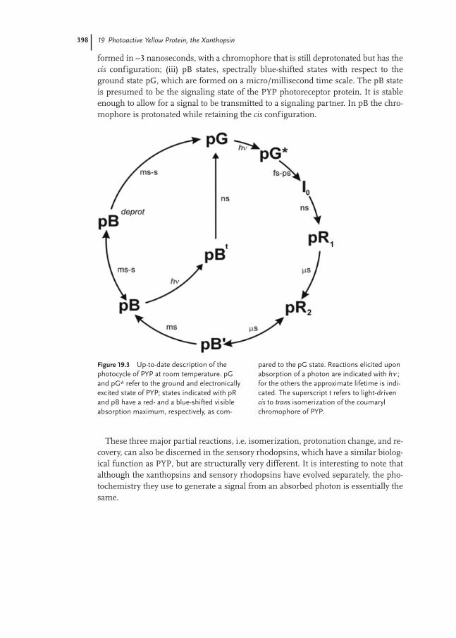
Models of the photocycle of Photoactive Yellow Protein have become more and morecomplex over the years (see Figure 19.3 for a recent version). Key states in this cycleare: (i) The ground- or dark-adapted state, pG, with a deprotonated chromophore intrans configuration; (ii) pR states, spectrally red-shifted compared to pG; the first is
19.3 Photoactivity of the Xanthopsins
398
formed in ∼3 nanoseconds, with a chromophore that is still deprotonated but has thecis configuration; (iii) pB states, spectrally blue-shifted states with respect to theground state pG, which are formed on a micro/millisecond time scale. The pB stateis presumed to be the signaling state of the PYP photoreceptor protein. It is stableenough to allow for a signal to be transmitted to a signaling partner. In pB the chro-mophore is protonated while retaining the cis configuration.
These three major partial reactions, i.e. isomerization, protonation change, and re-covery, can also be discerned in the sensory rhodopsins, which have a similar biolog-ical function as PYP, but are structurally very different. It is interesting to note thatalthough the xanthopsins and sensory rhodopsins have evolved separately, the pho-tochemistry they use to generate a signal from an absorbed photon is essentially thesame.
19 Photoactive Yellow Protein, the Xanthopsin
Figure 19.3 Up-to-date description of thephotocycle of PYP at room temperature. pGand pG* refer to the ground and electronicallyexcited state of PYP; states indicated with pRand pB have a red- and a blue-shifted visibleabsorption maximum, respectively, as com-
pared to the pG state. Reactions elicited uponabsorption of a photon are indicated with hν ;for the others the approximate lifetime is indi-cated. The superscript t refers to light-drivencis to trans isomerization of the coumarylchromophore of PYP.

399
19.3.2Photocycle Nomenclature
Over the years, multiple nomenclatures for the photocycle intermediates of PYP havebeen introduced. They all make reference to the above-mentioned three basic photo-cycle intermediates and can therefore be compared using these three species as a ref-erence. Initially, the ground-, red-shifted, and blue-shifted state were called P, I1, andI2 (Meyer et al. 1989). In 1994 Hoff et al. (Hoff et al. 1995) introduced the names pG,pR, and pB, thus stressing the wavelength shift in their UV/Vis absorption maxi-mum. Yet another nomenclature (Imamoto et al. 1996) was introduced in 1996, inwhich the corresponding species are referred to as PYP, PYPL, and PYPM, i.e. anomenclature based upon the similarities with bacterial rhodopsins. These nomen-clatures have been made even more complicated by the use of sub- and/or super-scripts that refer to a specific property of the species (e.g. the absorption maximum).As such properties may depend on the measurement conditions used, different sub-scripts are used for the same species.
More and more spectroscopic techniques are used to analyze the photocycle ofPYP. This has led to the discovery of several new photocycle species that were unde-tectable previously. To provide these new intermediates with a new name that fits log-ically within one of the existing nomenclatures is close to impossible.
19.3.3Experimental Observation: Context Dependence
It is of crucial importance to take the experimental context of the PYP protein intoconsideration when comparing different experiments. Four different experimentalparameters are of basic importance: Temperature, nature and pH of the solvent,mesoscopic context (or phase), and illumination conditions. The first one, tempera-ture, is obviously important when comparing kinetic experiments. But temperaturemay also allow one to trap certain photocycle species (Hoff et al. 1992; Imamoto et al.1996) and prevent others from being formed. The second one, the nature of the sol-vent, is also very important, and is usually different between different experiments.Though pH is probably the most important solvent feature that has an effect on e.g.the kinetics of the photocycle (Genick et al. 1997b; Hendriks et al. 2003), other solventfeatures such as its viscosity, hydrophobicity (Meyer et al. 1989), type and concentra-tion of solutes present (Meyer et al. 1987; Meyer et al. 1989), and the nature of the sol-vent itself (e.g. water vs. deuterium oxide (Hendriks et al. 2003)) can also have theireffect on the performance of a dissolved photoreceptor protein. The third importantparameter, the mesoscopic context or phase, dictates whether or not gross structuralchange is observed in the protein upon formation of the signaling state. This struc-tural change is limited in a crystalline lattice, whereas when the protein in solutiondoes show such a significant overall structural change (Xie et al. 2001). Recently,(single-crystal) absorption spectroscopy and X-ray diffraction experiments, as well asmeasurements in (partially) dehydrated protein films, have confirmed that the pho-tocycle can proceed through a short-cut version under these conditions (Kort et al.
19.3 Photoactivity of the Xanthopsins

400
2003; Mano et al. 2003; Moffat et al. 2003; van der Horst et al. 2004). Also illumina-tion conditions can have pronounced effects on the photocycle characteristics. Par-ticularly light color and intensity are important. The choice of wavelength can alreadyhave an effect on which photocycle species are formed (Hoff et al. 1992; Imamoto etal. 1996). Also the duration of the excitation pulse can have an effect, because ex-tended illumination allows photoactivation of photocycle species other than theground state (Gensch et al. 1998). This may lead to hysteresis effects in ground staterecovery (T. Gensch et al., unpublished). However, the intensity of the probe beamcan also inf luence the data, especially in protein variants with a high quantum yieldof photochemistry or with a slowly recovering photocycle.
19.3.4Mutants and Hybrids
PYP can be engineered genetically, via site-directed mutagenesis, and chemically, e.g.through the use of chromophore derivatives. The type of protein obtained in thesetwo approaches is referred to as mutant and hybrid, respectively. Many of the residuesthat interact with the chromophore (e.g. Tyr42, Glu46, Thr50 and Cys69) have beenmutated, resulting in (dramatically) altered photocycle characteristics. Various chro-mophore analogues, either with one or more ring substituents or “locked” in a spe-cific configuration, have been used for holo-protein reconstitution.
19.3.5Photo-activation in the Different Xanthopsins Compared
Besides the features of the photocycle of PYP from H. halophila described above, ad-ditional members of the xanthopsins have been characterized, albeit in much less de-tail. Both members of sub-group II (i.e. the PYP’s from Rb. sphaeroides and Rb. cap-sulatus) show comparable spectroscopic features, which are significantly differentfrom those of H. halophila PYP (Haker et al. 2000; Kyndt et al. 2003). Both show a sec-ond absorption maximum at 360 nm; this form is in a temperature- and pH-depend-ent equilibrium with the pG form. For PYP from Rb. sphaeroides it has been shownthat, upon excitation, the species at 360 can also enter a photocycle, independent fromthe photocycle of the 446 nm species (Haker et al. 2003). Furthermore, in both pro-teins, the ground state is recovered after photoexcitation ∼100-fold faster than in PYPfrom H. halophila.
As opposed to the faster photocycle recovery in these proteins, two xanthopsinshave been found that show considerably slower ground state recovery after photoex-citation: Ppr from Rhodospirillum centenum (Jiang et al. 1999) and a recently discov-ered second PYP protein from H. halophila (PYP(B); Van der Horst et al., unpublishedresults); in these, the recovery is ∼300-fold and ∼100-fold slower, respectively. Sincethe mechanism of the dark recovery of the ground state is poorly understood, an ex-planation for these differences cannot be given at this point.
19 Photoactive Yellow Protein, the Xanthopsin

401
19.4The Photocycle of Photoactive Yellow Protein
19.4.1Initial Events
The initial events in the photocycle of PYP are usually considered as transformationspreceding the formation of the most stable red-shifted intermediate state (pR). Thus,they comprise the initiation and the first, (ultra) fast, phase of the photocycle. It isgenerally accepted that the first basic step of the photocycle is the trans–cis isomer-ization of the chromophore. However, the exact spatial description of this process stillremains to be completed.
Femtosecond and picosecond transient absorption (Devanathan et al. 1999b; Bal-tuska et al. 1997), f luorescence (Mataga et al. 2003; Chosrowjan et al. 2004), and IRstudies (Groot et al. 2003) have revealed that initiation of the photocycle occurs on thepicosecond timescale. Femtosecond infrared spectroscopy – a method providing site-and bond-specific information – allowed the conclusion that breaking of the hydro-gen bond of the chromophore’s C=O group with the N–H group of Cys69 and the for-mation of a stable cis ground state occur in ∼2 ps (Groot et al. 2003). This observationis consistent with the timescale of the main component in the f luorescence decaysignal, which is usually associated with f luorescence quenching due to chromophoreisomerization (Changenet et al. 1998; Mataga et al. 2003; Chosrowjan et al. 2004).
Several models for isomerization have been suggested, but it is not clear which (ifany) of these models is correct. Some features have nevertheless been resolved: atleast two different f luorescent states are formed after photo-excitation of PYP. In oneof these states, which presents itself as an anisotropy difference with respect to theground state, the chromophore configuration is clearly different from that in theground state (Gensch et al. 2002). It has not been revealed whether these two f luo-rescent states are true photocycle intermediates (i.e. upon formation they return tothe ground state via the regular photocycle intermediates) or whether they are photo-cycle dead ends. Femtosecond time-resolved f luorescence studies of wild type PYPplus several site-directed mutants and hybrids with chromophore analogues showedthat the protein environment plays an important role in the kinetics and efficiency ofphotocycle initiation. Molecular-dynamics simulations and time-dependent densityfunctional theory calculations also corroborate this conclusion. The simulations sug-gest that the protein regulates the isomerization of the chromophore via stabilizationof the transition state in the isomerization pathway (Groenhof et al. 2002b; Groenhofet al. 2004).
Transient absorption measurements have revealed that, at room temperature, atleast one photocycle intermediate is formed between the excited (f luorescent) state(s)and pR. This so called I0 intermediate, is formed on a picosecond timescale and trans-forms at the nanosecond timescale into pR. In addition, the existence of yet anotherintermediate, between I0 and pR, has been reported. This intermediate, referred to asI0
‡, has absorption characteristics very similar to those of I0 (Ujj et al. 1998; Gensch
19.4 The Photocycle of Photoactive Yellow Protein

402
et al. 2002). An alternative interpretation, however, holds that a long living (o2 ps) ex-cited state causes these signals.
At cryogenic temperatures a branched photocycle pathway exists. Each of the twobranches contains two intermediates. One branch proceeds via an intermediate thatis similar to I0 (PYPB, converted into PYPBL). The two branches merge upon forma-tion of pR. Selective use of either of these two branches can be accomplished via tun-ing of the excitation wavelength. Shorter wavelengths make the branch with the I0-like intermediate the preferred one (Hoff et al. 1992; Masciangioli et al. 2000). Thismight be a true PYP characteristic but it is also possible that PYPB is photo-convert-ed back to pG, which occurs with optimal efficiency at longer wavelengths. Similarlythe PYPH intermediate (converted in the dark into PYPHL), at shorter wavelengths,can be photo-converted back to pG (Imamoto et al. 1996). This is one of the drawbackswhen cryotrapping is used to characterize photocycle intermediates. Photoactivity,however, is not exclusive for the ground state; transient intermediates may be pho-toactive as well. For accumulation of a cryotrapped intermediate in the sample, ex-tended illumination is usually needed. Consequently, the cryotrapped intermediatemust also be significantly illuminated. As a result it can be photo-converted, e.g. tothe ground state. It is also possible, however, that non-physiological intermediates,are formed as a result of these secondary photoreactions. A case in point may be thef luorescent species F430, which may well be formed from the PYPH intermediate(Hoff et al. 1992).
Of course it is important to identify spectroscopic intermediates correctly, but it iseven more interesting to understand their spatial structure. It is evident that the pro-tein itself displays only very modest structural changes during the first step of thephotocycle. Time-resolved X-ray diffraction revealed that the aromatic ring of thechromophore of PYP stays approximately at the same position during its pho-toisiomerization. (Genick et al. 1998; Imamoto et al. 2001b; Ren et al. 2001). The on-ly way to facilitate isomerization of the chromophore under these conditions is by ro-tation of the thiol-ester carbonyl. This carbonyl f lip can be interpreted as a double iso-merization around the C7=C8 double bond and the C9–Sγ single bond. The chro-mophore configuration accordingly changes from C7=C8-trans, C9–Sγ-cis to C7=C8-cis,C9–Sγ-trans. This model was first introduced on the basis of low temperature FTIRspectroscopy (Xie et al. 1996), and later confirmed (Brudler et al. 2001; Imamoto et al.2001b; Xie et al. 2001).
The structure of two intermediates from the first steps in the photocycle, PYPBL
and pR, have also been determined with X-ray crystallography. In both intermediates,structural changes are largely limited to the immediate surroundings of the chro-mophore. The structure of the PYPBL intermediate was obtained via cryotrapping andresolved down to a resolution of 0.85 Å. Based on the temperature and illuminationconditions used, and the absorption spectrum of the sample, it was concluded thatPYPBL was the cryotrapped intermediate (Genick et al. 1998). It can not be excluded,however, that PYPHL was also formed and thus that a mixture of intermediates waspresent. In the resolved structure the chromophore is in a distorted cis configuration;it has barely crossed the trans to cis transition point and by consequence isomeriza-tion is only completed in pR. An important conclusion is that the chromophore iso-
19 Photoactive Yellow Protein, the Xanthopsin

403
merizes in a way that minimizes movement of the chromophore within the chro-mophore pocket. Isomerization is achieved by rotating the carbonyl function 166.5°,with respect to the aromatic ring, breaking the hydrogen bond between the carbonylfunction and the backbone amide group of Cys69. Recent low-temperature analyses,however, show that complete trans to cis isomerization of the chromophore is possi-ble under these conditions (Kort et al. 2004). A key element in this latter study is theextensive correction for X-ray radiation damage.
The 1.9 Å resolution structure of the pR intermediate was obtained via the 1 nstime slice of a dataset encompassing time slices up to 1 ms, in a time-resolved X-raycrystallography experiment (at near room temperature) (Perman et al. 1998). The de-posited pR structure has a completely cis isomerized chromophore for the C7=C8 dou-ble bond. In this structure the carbonyl function has rotated an additional 108.5° withrespect to the cryo-trapped intermediate PYPBL. This may facilitate the formation ofa hydrogen bond between the carbonyl oxygen and the backbone amide of Tyr98.
The (free) energy content of PYP increases 120–160 kJ mol–1 upon formation of pR(van Brederode et al. 1995; Takeshita et al. 2000). Half of the energy of an absorbedphoton is thus stored in the holo-protein at this point (a photon with a wavelength of446 nm has an energy content of 268 kJ mol–1). This energy should be (and is) enoughto drive the remainder of the photocycle and make PYP return to the pG state. Theother half of the energy of the absorbed photon is lost in e.g. thermal relaxations.FTIR analysis of the cryotrapped intermediates in the first step of the photocycle, sug-gests that there is very little structural difference between these intermediates(Imamoto et al. 2001b). Thus small movements induced by thermal relaxations maydictate the exact isomerization route of the chromophore.
19.4.2Signaling State Formation and Ground State Recovery
Signaling of a blue photon in the cell occurs via interaction of the signaling state ofPYP with a (putative) transducer protein. To allow protein–protein interactions thesignaling state needs to have a relatively long lifetime. The PYP-transducer proteinhas not been identified yet; all conclusions so far are based on experiments with on-ly purified PYP and therefore it is possible that certain characteristics of the signal-ing state will change when transducer protein is added. Such differences indeed canbe observed in sensory rhodopsin I: Without the transducer protein sensoryrhodopsin I acts as a proton pump. In the presence of the transducer protein this ac-tivity is lost and also the pH dependence of the photocycle kinetics differs (Spudichand Spudich 1993). Formation of the signaling state of PYP from the red-shifted pho-tocycle intermediate pR is accomplished via two key events: protonation of the chro-mophore (neutralizing its negative charge) and a structural change of the protein.Ground state recovery, through reversal of these events, completes the photocycle. In-sight in the formation of the signaling state has increased significantly over the pastfew years, but little still is known on the specifics of the recovery of the ground state.
Significant new insights in the mechanism of signaling-state formation haveemerged recently. Early photocycle models considered only three states, the ground
19.4 The Photocycle of Photoactive Yellow Protein

404
state pG, pR and the signaling state pB. Formation of pB (from pR) was initially de-scribed as a mono-exponential process but, when higher resolution was achievable,as a bi-exponential process. Because of this bi-exponential character of pB formation,a detailed photocycle model for PYP (Meyer et al. 1987) postulated an additional in-termediate with similar spectral properties as pB. This model was abandoned in asubsequent paper (Meyer et al. 1989). Based on insights obtained with FTIR spec-troscopy, recently, the intermediate pB’ was introduced as an intermediate linking thepR and pB states (Xie et al. 2001). The role of this intermediate was subsequently con-firmed in a detailed kinetic UV/Vis analysis of the photocycle. pB’ was shown to bespectroscopically very similar to pB (Hendriks et al. 2003) and in equilibrium withpR, which explains the bi-exponential character of the pR to pB photocycle step (Mey-er et al. 1987; Hoff et al. 1994c). Existence of the pB’ intermediate was recently alsoconfirmed with time-resolved resonance Raman measurements (Pan et al. 2004).
Besides the thermal recovery of the ground state (i.e. spontaneously in the dark), alight-induced recovery pathway also exists. This path is initiated after photon absorp-tion by pB (and pB’). Via the light-induced cis to trans re-isomerization of the chro-mophore a thousand-fold increase in the rate of recovery, as compared to the ther-mally activated process, is observed (Hendriks et al. 1999b). For the thermal recoveryit has been suggested that before isomerization can take place the chromophore mustfirst be deprotonated. By consequence an intermediate must exist between pB andpG (Demchuk et al. 2000). This intermediate, pBdeprot, was recently detected in astudy of the kinetic deuterium isotope effect on photocycle transitions in PYP (Hen-driks et al. 2003).
19.4.3Structural Relaxation of pR
Recently it was demonstrated that after pR has formed, significant relaxation in PYPstructure takes place (Takeshita et al. 2002a; Takeshita et al. 2002b). Via transient grat-ing and pulsed-laser photoacoustic methods, a µs dynamic component was shown toexist during the lifetime of pR. This indicates that after completion of structuralchanges in and immediately around the chromophore, additional structural changestake place further away from the chromophore. Accordingly, pR actually representstwo intermediates, pR1 and pR2, and in hindsight evidence for the existence for thesetwo pR states was already available in early UV/Vis data (Hoff et al. 1994c). In a re-cent UV/Vis study (Hendriks et al. 2003), kinetics were obtained that exactly fit thepR1-to-pR2 transition observed via transient grating. The spectra for both pR inter-mediates are very similar, pR1 seems to have a slightly higher extinction coefficientthan pR2, but the λmax values are indistinguishable.
19.4.4Protonation Change upon pB’ Formation
After formation of the pR intermediates, the next event is protonation of the chro-mophore. This results in the formation of pB’. The first experimental evidence for the
19 Photoactive Yellow Protein, the Xanthopsin
405
existence of this intermediate was obtained in FTIR measurements (Xie et al. 2001).It was shown that deprotonation of Glu46 and protonation of the chromophore are si-multaneous events that are followed by a structural change of the protein. Absorptionchanges, registered using UV/Vis spectroscopy, mainly display changes of the chro-mophore and its immediate surroundings. Since all the details of the structure of thesurrounding protein cannot be probed with this technique, it is difficult to distin-guish between pB’ and pB with UV/Vis spectroscopy. Based on the FTIR evidence apB’ intermediate was, however, incorporated in a photocycle model, which was thenused to analyze UV/Vis data in a study on the kinetic deuterium-isotope effect in thephotocycle of PYP (Hendriks et al. 2003). From this approach it became clear that pRand pB’ exist in an equilibrium that shifts towards pB’ upon going to the extremes(both low and high) of pH. The observed kinetic deuterium isotope effect, for thewhole pH range that was investigated (pH 5 to 11), is in line with proton transfer fromGlu46 to the chromophore. For the reverse reaction the situation is more complex. Asa function of pH, the mechanism of formation of pR from pB’ may vary. The pH-de-pendence of the pR/pB’ equilibrium can also explain the shift towards mono-expo-nential behavior of pB formation at very high or low pH.
Direct proton transfer from Glu46 to the chromophore is therefore considered asthe most likely mechanism of the chromophore protonation upon the formation ofthe pB state. This conclusion also follows from the results of molecular dynamicssimulations and semi-empirical calculations, which suggest that proton transferfrom Glu46 to the phenolate oxygen of the chromophore is facilitated by the photo-induced isomerization of the chromophore (Sergi et al. 2001). It is relevant to notethat, by necessity, the residue at position 46 cannot protonate the chromophore in theE46Q mutant.
19.4.5Structural Change upon pB Formation
The non-linear Arrhenius kinetic behavior of the recovery reaction of pG (vanBrederode et al. 1996) was the first evidence that the signaling state of PYP is at leastpartly unfolded. A mutant form of PYP, which lacks the first 25 N-terminal aminoacids (van der Horst et al. 2001) displays close to normal Arrhenius behavior, indicat-ing that its N-terminal region is largely responsible for the large structural change up-on formation of the signaling state. Several additional methods, e.g. circular dichro-ism spectroscopy (CD) (Harigai et al. 2003), NMR (Rubinstenn et al. 1998; Craven etal. 2000; Derix et al. 2003), f luorescent-probe binding (Lee et al. 2001b; Hendriks etal. 2002), and FTIR spectroscopy (Hoff et al. 1999; Xie et al. 2001; Harigai et al. 2003)have confirmed this partial unfolding of PYP in its signaling state pB.
H/D exchange measurements have added to our current insight into the structur-al change that underlies signaling-state formation in PYP. An exposed hydrogenatom may be exchanged within seconds, whereas for a buried hydrogen atom this cantake days. PYP contains 235 potentially exchangeable hydrogen atoms, 42 of whichare from (de)protonatable groups. It was demonstrated, using electrospray ionizationmass spectrometry (Hoff et al. 1999), that more hydrogen atoms were exchanged for
19.4 The Photocycle of Photoactive Yellow Protein
406
deuterium atoms in the presence of light compared to PYP in the dark. The light-in-duced H/D exchange was independently confirmed by FTIR difference spectroscopy(Hoff et al. 1999). These experiments showed that there is a difference between theground and signaling state of PYP with respect to H/D exchange protection. NMRspectroscopy (Craven et al. 2000) revealed some of the (exposed) sites involved: therate of exchange of the backbone amide was recorded for 51 residues: 14 of whichshowed a significant change in exchange rate upon pB formation, only 2 less than thenumber predicted by mass spectrometry, a method which is not limited to the back-bone amide hydrogen atoms. Most significant loss in protection was observed forresidues Phe28, Glu46, and Thr70. The latter two are in the vicinity of the chro-mophore, Phe28 is close to Glu46.
A solution-NMR study of pB showed that, with respect to the ground state, this in-termediate exhibits more structural and dynamic disorder (Rubinstenn et al. 1998).In a follow-up study on pBdark formation (Craven et al. 2000) it was revealed that thisintermediate is very similar to pB. These NMR data suggest that the pB intermediateis in fact a mixture of structurally perturbed forms and a form structurally similar topB in crystalline PYP (Genick et al. 1997a).
The trigger for the large structural change upon formation of pB is the formationof a buried negative charge (i.e. on Glu46) upon proton transfer to the chromophore(Xie et al. 2001). While residing on the chromophore, this negative charge is stabi-lized by delocalization, by the hydrogen-bonding network, and possibly by the posi-tive charge on Arg52. A comparable stabilization of this negative charge is not possi-ble when it resides on Glu46. This situation then generates a free-energy stress with-in the protein, which can be relayed via several routes. One is return to the pR state,thus explaining the reversibility of pB’ formation (see above). Other routes lead to pBformation; the extent of structural change upon formation of the pB intermediatethen depends on the details of this route. One option is to relieve the buried negativecharge via exposure to the solvent. This requires a large structural change of the pro-tein. Another route is to protonate Glu46 via a donor different from the chro-mophore. Once Glu46 is protonated (large) structural changes are no longer a neces-sity.
As in these processes protonation changes play a key role, it is to be expected thatthey will display pronounced pH dependency. Also, the pH may inf luence the preva-lence of one route over another. Indeed a pH dependence of the extent of structuralchanges has been observed to coincide with the protonation state of Glu46 (J. Hen-driks and A. Xie et al., unpublished results). Significantly, both routes to pB implythat pB’ is formed as an intermediate. An alternative possibility is that a direct routefrom pR to pB exists in which Glu46 stays protonated, i.e. it would not donate a pro-ton to the chromophore in such a route. The chromophore then becomes protonatedonly after exposure to the solvent, or via another residue (e.g. Tyr42). A mechanismlike this presumably would require less structural change, and might be preferred ina crystalline environment.
All of these routes may be possible and depending on the conditions one particu-lar route may dominate. A key factor is what happens to the protonation state of bothGlu46 and the chromophore. Consistently, in the Glu46Gln mutant of PYP the struc-
19 Photoactive Yellow Protein, the Xanthopsin

407
tural change upon formation of pB is significantly less than in wild type PYP (Xie etal. 2001); this has also been observed for the His108Phe mutant (Kandori et al. 2000).
The pH dependence of structural change has also been demonstrated via transientprobe binding (Hendriks et al. 2002). Since in experiments monitoring the pH-de-pendent net proton uptake/release of PYP during pB formation (Hendriks et al.1999a), and in FTIR spectra, a similar pH dependence was observed (Hendriks 2002),it is likely that the pKa of Glu46 in pB is 5.5.
19.4.6Recovery of the Ground State
For recovery of the ground state of PYP the chromophore has to re-isomerize, the pro-tonation state of several functional groups needs to be restored and the protein needsto refold to its ground state conformation. Until recently, these partial reactions werethought to occur concertedly. The kinetic deuterium isotope effects (Hendriks et al.2003), however, have shown that deprotonation of the chromophore occurs before re-isomerization. By consequence the intermediate pBdeprot is formed in the recovery ofpG from pB. Deprotonation of the chromophore accelerates this re-isomerization(Sergi et al. 2001), although it remains a rate-controlling step, in which the exact pro-tein conformation likely plays a crucial role. The pBdeprot intermediate therefore willhave a deprotonated chromophore and a folding state that allows chromophore re-iso-merization. By consequence, the absorption spectrum of this intermediate may besimilar to that of pG, rather than to the pB intermediate with a deprotonated chro-mophore. In agreement with this possibility, its absorption maximum has been esti-mated to be around 430 nm (Hendriks et al. 1999a; Hendriks et al. 2003). Deproto-nation of the chromophore upon formation of pBdeprot is most likely facilitated by ahydroxyl ion (Hendriks et al. 2003).
As discussed above, the recovery rate of the ground state increases a thousand-foldwhen re-isomerization of the chromophore is facilitated by blue light (Hendriks et al.1999b). In the ensuing branching pathway an intermediate, pBt (t for trans) is formedon a nanosecond time scale. The slight blue shift of pBt, with respect to pB, can be ex-plained by the altered chromophore configuration. UV/Vis spectroscopy did not re-veal any additional intermediates between pBt to pG. This lack indicates that re-fold-ing and reformation of proper protonation states can proceed quickly once the chro-mophore is re-isomerized. The fact that this branching reaction exists can inf luencethe interpretation of data collected in the presence of light. Activation of the pB in-termediates, may allow recovery kinetics to appear faster than they really are in theabsence of this (probe) light (Miller et al. 1993). Light-accelerated recovery can be ex-ploited in the study of the very slow recovery variants, like M100A. It may have bio-logical relevance in PYP variants with strongly decreased recovery rates, as in the sec-ond PYP discovered in H. halophila (i.e. PYP(B); M.A. van der Horst et al., unpub-lished observations).
Several studies of the refolding of PYP have been performed. Often, these studiesemploy a denaturant to assist the initial unfolding of the protein. The exception is anNMR study (Rubinstenn et al. 1998), in which recovery of the ground state was meas-
19.4 The Photocycle of Photoactive Yellow Protein

408
ured on the basis of light-induced unfolding of the protein only. A dynamic processof refolding was observed in which the central β-sheet and parts of the α-helical struc-ture refold slightly faster, after which the region around the chromophore returns tothe ground state fold. The differences between the refolding rates are small, howev-er, and it is unclear if these rates differ significantly. The observed trend is in line withthe idea that for efficient re-isomerization of the chromophore, it not only needs tobe deprotonated, but that also the protein needs to be in a relatively folded state.
In a study utilizing urea and guanidinium·HCl, refolding was studied with the un-folded ground and signaling state of PYP (Lee et al. 2001c). The major difference be-tween these two denatured forms of the protein is the isomerization state of the chro-mophore. Refolding from the denatured ground state is a mono-exponential event,whereas refolding from the denatured signaling state is bi-exponential. The fast com-ponent in the latter process is identical to the refolding component from the dena-tured ground state. The slow exponent has a rate similar to the photocycle ground-state recovery rate under similar conditions. These observations indicate that afterthe signaling state renatures, it recovers to the ground state through regular photo-cycle states. Interestingly, extrapolation of the refolding kinetics to the absence of de-naturant shows an approximate thousand fold faster rate for refolding with the chro-mophore in the trans state, compared to the cis state. This is similar to the differencesobserved between the thermal- and the photo-activated ground state recovery (Hen-driks et al. 1999b). Similar results were obtained in experiments with the acid-dena-tured state of PYP (Lee et al. 2001a). Refolding from the acid denatured state with atrans chromophore, i.e. pBdark, is 3 to 5 orders of magnitude faster than from the aciddenatured state with the chromophore in the cis configuration.
For several PYP mutants, a severe decrease in the recovery rate has been reported(Glu46Asp (Devanathan et al. 1999a), Met100Ala (Devanathan et al. 1998), andMet100Leu (Sasaki et al. 2002)). These results indicate that Glu46 and Met100 are im-portant for recovery. It was argued that the electron-donating character of the residueat position 100 inf luences the rate of recovery, presumably through interaction withArg52 (Kumauchi et al. 2002).
19.5Spectral Tuning of Photoactive Yellow Protein
Tuning of the UV/Vis absorption bands of PYP, away from the values of neutral and/or anionic coumaric acid to longer wavelengths, is of interest for several reasons.First, there is the tuning of the absorption band of the ground state structure. Here,the contribution of specific structural characteristics must be analyzed to explain thedestabilization of the ground state and/or destabilization of the electronically excitedstate. However, during the photocycle of PYP, the maxima in the absorption spec-trum change as well. These changes contain valuable information regarding the chro-mophore and its surroundings in the respective photocycle states. A proper under-standing of the tuning of PYP will therefore aid to understand the events that occurduring the photocycle.
19 Photoactive Yellow Protein, the Xanthopsin

409
19.5.1Ground State Tuning
The interaction of the chromophore and apo-protein parts of PYP produces an ab-sorption band with 446 nm as its UV/Vis absorption maximum. As the free chro-mophore, trans-4-hydroxycinamic acid, has an absorption maximum at 284 nm (inaqueous solvent around neutral pH) (Aulin-Erdtman and Sandén 1968), this interac-tion of the chromophore with the protein induces a large red shift. Several specificcontributions to this large shift can be distinguished. For one, the thiol ester link ofthe chromophore with Cys69, causes a red shift of ∼5713 cm–1 (from 284 to 339 nm).This conclusion follows from a comparison of the absorption maximum of 4-hy-droxycinamic acid and the denatured form of PYP in aqueous solution at pH 7 (Kroonet al. 1996). An additional red-shift of 4310 cm–1 (from 339 to 397 nm) occurs whenthe chromophore becomes deprotonated, which follows from a comparison of the ab-sorption maximum of the denatured form of PYP in aqueous solution at pH 7 and 11(Kroon et al. 1996). This leaves a red shift of ∼2767 cm–1 (from 397 to 446 nm) that iscaused by interactions with the protein. Though this description is very illustrative, itdoes not provide specific insight regarding the nature of the interactions between thechromophore and the protein.
In a recent study, a closer look was taken at the mechanism(s) that lead(s) to thespectroscopic tuning of PYP in its ground state (Yoda et al. 2001). Two model com-pounds were used for comparison, i.e. a propyl ester and a propyl-thiol ester of 4-hy-droxycinnamic acid. The thiol ester model compound was consistently red-shifted by∼1000 cm–1, with respect to the ester model compound, irrespective of the protona-tion state of the chromophore. With regard to tuning in PYP, three tuning contribu-tions were considered, i.e. a medium effect of the protein matrix (700 cm–1), a count-er ion effect (5300 cm–1), and a hydrogen bonding effect (–1600 cm–1). The mediumeffect of the protein matrix takes into account non-specific solvent effects. For themodel compounds the absorption maximum shifted with the solvent that was used,e.g. for the thiol-ester model compound the absorption maximum ranged from34800 cm–1 (287 nm) in pentane to 31500 cm–1 (317 nm) in pyridine. This differenceis mainly caused by differences in the dielectric constant and refractive index be-tween the solvents. To determine the medium effect in PYP the values for the dielec-tric constant and the refractive index were estimated for the protein. Absorption max-ima of the thiol ester model compound in hexane and in a protein environment werecalculated. Note, that only the solvent properties of the protein are considered hereand not counter-ion and hydrogen-bonding effects. This leads to a contribution of∼700 cm–1 to the tuning of the chromophore in the protein.
The counter ion effect considers the difference in position of the counter ion of thethiol ester model compound in solution (sodium ion at 2.5 Å in a straight line fromthe phenolate oxygen bond) and in the protein (position of Arg52 in the crystal struc-ture PDB ID: 2PHY (Borgstahl et al. 1995)). Here protein solvent conditions wereused in the calculation of the absorption maxima. This leads to a contribution of∼5300 cm–1 to the tuning of the chromophore. The hydrogen-bonding effect was de-termined by placing methanol at the positions of the hydroxy groups of residues
19.5 Spectral Tuning of Photoactive Yellow Protein

410
Tyr42, Glu46, and Thr50, which are involved in the hydrogen-bonding network withthe chromophore, in the calculations. By comparing the situation incorporating themedium effect of the protein matrix and the counter-ion effect, with the situation thatalso takes into account the hydrogen-bonding effect, the contribution of the latter wascalculated as –1600 cm–1, i.e. a blue shift is caused by formation of a hydrogen bondto the phenolate oxygen of the chromophore! Furthermore, also with respect to thecounter ion effect, an interesting observation was made. The position of Arg52 issuch that it appears that the counter ion is infinitely far apart from the chromophore,i.e. it does not contribute to the tuning. Movement of the counter ion towards the thi-ol ester linkage would result in a red shift, whereas movement toward the phenolateoxygen would result in a blue shift. From NMR measurements (Dux et al. 1998) andmolecular dynamics studies (Groenhof et al. 2002b) it is evident that Arg52, may havetwo distinct positions in the ground state. The two distinct positions obtained withthe molecular dynamics studies would result in a difference of 20 nm between the ab-sorption maxima of the two conformers (Groenhof et al. 2002b).
When the hydrogen-bonding network in PYP is weakened (by replacing the brid-ging hydrogen atoms with deuterium atoms) indeed a small red shift is observed,which would be expected when the hydrogen bonding effect contributes a smallerblue shift to the total tuning. Furthermore, the residues that are involved in the hy-drogen-bonding network that stabilizes the anionic chromophore (i.e. Tyr42, Glu46,and Thr50) have been altered through mutagenesis. Mutants in these residues indeedresult in a red shift of the UV/Vis absorbance maximum of the chromophore (e.g.Tyr42Ala, Tyr42Phe, Glu46Gln, Glu46Ala, Thr50Val, and Thr50Ala (Genick et al.1997b; Mihara et al. 1997; Devanathan et al. 1999a; Brudler et al. 2000; Imamoto et al.2001a)) ref lecting the weaker hydrogen bonding effect in these mutants. In somemutants though, protein stability has been severely affected as indicated by a major,additional, blue-shifted absorption band (Tyr42Phe, Tyr42Ala, and Glu46Ala). Here,the weakened hydrogen-bonding network causes a shift in pKa of the chromophore,leading to a protonated state (Meyer et al. 2003; El-Mashtoly et al. 2004).
Several mutants altered at the position of Arg52 have also been prepared. TheArg52Ala mutant is slightly red-shifted (Genick et al. 1997b), while the Arg52Gln mu-tant shows no shift of the absorption maximum (Mihara et al. 1997). As describedabove, removal of the counter ion would not lead to a change in absorption maxi-mum, which is exactly what is observed in the Arg52Gln mutant. A small red shiftthat is observed in the Arg52Ala mutant may be explained by a more open structureof the chromophore-binding pocket, possibly allowing a solvent cation to act as acounter ion.
19.5.2Spectral Tuning in Photocycle Intermediates
The negative charge on the chromophore is most effectively delocalized if the chro-mophore is planar. During isomerization planarity is lost, and the negative charge isnot as efficiently delocalized, which results in a red shift of the absorption spectrum.This would explain why the intermediate I0 is more red-shifted than pR, as in the for-
19 Photoactive Yellow Protein, the Xanthopsin

411
mer intermediate the chromophore may still be in a twisted form. In pR the chro-mophore is still not quite planar due to steric hindrance between the carboxylic oxy-gen and atoms of the phenolate ring (Groenhof et al. 2002a), which explains the per-sistent red shift. Additionally, in pR the chromophore has contracted ∼0.5 Å (Groen-hof et al. 2002a), while the structure of the protein is very similar to that of the groundstate. Such a contraction could lead the counter ion Arg52 to become located closerto the phenolate oxygen of the chromophore, which would also lead to a red shift. Theresidues lining the binding pocket of the chromophore restrict its isomerization tothe least volume-demanding mechanism (Imamoto et al. 2002).
Protonation of the chromophore leads to a large blue shift, which is exactly what isobserved. In pB’ the structure of the protein is still very similar to that of pR, and thusto the structure of the ground state. The transfer of the proton leads to a more relaxedconformation of the chromophore (Pan et al. 2004), but interactions with the proteinlikely persist (Genick et al. 1997a). When pB is formed in solution, the structure ofthe protein has dramatically changed, so that the interaction of the protein with thechromophore most likely diminishes, which is also indicated by the slight additionalblue-shift of the pB intermediate with respect to pB’. However, even though the chro-mophore has become more exposed in pB, its absorption maximum is still slightlyred-shifted compared to the situation in denatured protein (Lee et al. 2001c). Thechromophore is therefore still tuned in pB, through interactions with the protein. Thepresence of these interactions is further demonstrated by the pKa of the chromophorein pB which is ∼10 and not 8.7 as is the case in fully denatured protein (Hendriks etal. 2003).
19.6Summary and Future Perspective
After excitation of the ground state of Photoactive Yellow Protein, its chromophorewill form a twisted excited state, which subsequently results in formation of the I0
photocycle intermediate with the chromophore in cis configuration, be it still twisted.Further relaxation leads to formation of pR. Isomerization proceeds with a minimalamount of movement of the chromophore, i.e. through a concerted rotation aroundseveral bonds that can be described as a C7=C8-trans C9-Sγ-cis to C7=C8-cis C9-Sγ-transmultiple-bond isomerization, i.e. a rotary movement of the carbonyl oxygen. Thisisomerization increases the probability of proton transfer from Glu46 to the chro-mophore, which results in formation of pB’. As a result, the negative charge – whichwas stabilized via delocalization when it resided on the chromophore, via a hydrogen-bonding network, and via a counter ion – now resides on Glu46 where it is very lo-calized. This is an energetically unfavorable situation that drives subsequent struc-tural changes, both in the N-terminal domain of the protein and around the chro-mophore. Particularly the involvement of the independently folded N terminus inthis partial unfolding is remarkable. Together with the sequence comparisons with-in the PAS domains, this suggests that indeed the N terminus is important for sig-naling. The extent of these structural changes depends on experimental conditions
19.6 Summary and Future Perspective

412
and may vary from very little structural change in crystals, to large structural changesin aqueous solution. This structural change will then have to elicit detectable struc-tural change in a downstream signal transduction partner, be it in a domain of thesame full-length protein like in Ppr, or via intermolecular interactions.
After formation of the signaling state, the ground state in the photocycle recovers,which requires chromophore re-isomerization and protein re-folding. Before re-iso-merization can take place, the chromophore presumably needs to be deprotonated(leading to formation of the pBdeprot intermediate), and the protein needs to adopt aspecific fold that allows for the re-isomerization to take place. Chromophore re-iso-merization can also take place photochemically, which results in a rate of recoverythat is three orders of magnitude faster than dark recovery. It is still unresolvedwhether this two-photon photocycle is biologically relevant.
During the past few years PYP has become popular as a model system. Three ar-eas of research are relevant in this respect: (i) signaling state formation in PAS-do-mains, (ii) functional protein (un)folding and (iii) the primary photochemistry ofphotoreceptors. This popularity is based on favorable handling characteristics, avail-ability of high-resolution structures, and the relatively simple structure of the chro-mophore of PYP. Significantly, several additional fields may benefit from the effortsto understand the details of signaling in PYP. An example is molecular-dynamicsmodeling, because this approach may benefit from the availability of transient refer-ence structures from time-resolved X-ray diffraction. The field of signal transductionin photobiology could also benefit enormously from the detailed insight in the mech-anism that leads to signal generation within PYP. However, for this to bear fruit thesignal transduction component(s) that link PYP to the f lagellar apparatus will have tobe identified.
References
19 Photoactive Yellow Protein, the Xanthopsin
G. Aulin-Erdtman, R. Sandén. Acta Chem.Scand. 1968, 22, 1187–1209.
M. Baca, G.E. Borgstahl, M. Boissinot, P.M.Burke, D.R. Williams, K.A. Slater, E.D. Get-zoff. Biochemistry 1994, 33, 14369–14377.
A. Baltuska, I.H.M. van Stokkum, A. Kroon, R.Monshouwer, K.J. Hellingwerf, R. van Gron-delle. Chem. Phys. Lett. 1997, 270, 263–266.
H.M. Berman, J. Westbrook, Z. Feng, G. Gilli-land, T.N. Bhat, H. Weissig, I.N. Shindyalov,P.E. Bourne. Nucleic Acids Res. 2000, 28,235–242.
G.E. Borgstahl, D.R. Williams, E.D. Getzoff.Biochemistry 1995, 34, 6278–6287.
R. Brudler, T.E. Meyer, U.K. Genick, S. De-vanathan, T.T. Woo, D.P. Millar, K. Gerwert,M.A. Cusanovich, G. Tollin, E.D. Getzoff.Biochemistry 2000, 39, 13478–13486.
R. Brudler, R. Rammelsberg, T.T. Woo, E.D.Getzoff, K. Gerwert. Nat. Struct. Biol. 2001,8, 265–270.
P. Changenet, H. Zhang, M.J. van der Meer,K.J. Hellingwerf, M. Glasbeek. Chem. Phys.Lett. 1998, 282, 276–282.
H. Chosrowjan, S. Taniguchi, N. Mataga,M. Unno, S. Yamauchi, N. Hamada, M. Ku-mauchi, F. Tokunago. J. Phys. Chem. B 2004,108, 2686–2698.
C.J. Craven, N.M. Derix, J. Hendriks, R. Boe-lens, K.J. Hellingwerf, R. Kaptein. Biochem-istry 2000, 39, 14392–14399.
M.A. Cusanovich, T.E. Meyer. Biochemistry2003, 42, 4759–4770.
W.L. DeLano, The PyMOL User’s Manual, De-Lano Scientific
E. Demchuk, U.K. Genick, T.T. Woo, E.D. Get-zoff, D. Bashford. Biochemistry 2000, 39,1100–1113.

413
N.M. Derix, R.W. Wechselberger, M.A. van derHorst, K.J. Hellingwerf, R. Boelens, R. Kap-tein, N.A.J. van Nuland. Biochemistry 2003,42, 14501–14506.
S. Devanathan, R. Brudler, B. Hessling, T.T.Woo, K. Gerwert, E.D. Getzoff, M.A. Cu-sanovich, G. Tollin. Biochemistry 1999a, 38,13766–13772.
S. Devanathan, U.K. Genick, I.L. Canestrelli,T.E. Meyer, M.A. Cusanovich, E.D. Getzoff,G. Tollin. Biochemistry 1998, 37, 11563–11568.
S. Devanathan, A. Pacheco, L. Ujj, M. Cu-sanovich, G. Tollin, S. Lin, N. Woodbury.Biophys. J. 1999b, 77, 1017–1023.
P. Dux, G. Rubinstenn, G.W. Vuister, R. Boe-lens, F.A. Mulder, K. Hard, W.D. Hoff, A.R.Kroon, W. Crielaard, K.J. Hellingwerf,R. Kaptein. Biochemistry 1998, 37, 12689–12699.
S.F. El-Mashtoly, M. Unno, M. Kumauchi,N. Hamada, K. Fujiwara, J. Sasaki, Y.Imamoto, M. Kataoka, F. Tokunaga, S. Ya-mauchi. Biochemistry 2004, 43, 2279–2287.
U.K. Genick, G.E. Borgstahl, K. Ng, Z. Ren,C. Pradervand, P.M. Burke, V. Srajer,T.Y. Teng, W. Schildkamp, D.E. McRee,K. Moffat, E.D. Getzoff. Science 1997a, 275,1471–1475.
U.K. Genick, S. Devanathan, T.E. Meyer, I.L.Canestrelli, E. Williams, M.A. Cusanovich,G. Tollin, E.D. Getzoff. Biochemistry 1997b,36, 8–14.
U.K. Genick, S.M. Soltis, P. Kuhn, I.L.Canestrelli, E.D. Getzoff. Nature 1998, 392,206–209.
T. Gensch, C.C. Gradinaru, I.H.M. van Stok-kum, J. Hendriks, K.J. Hellingwerf, R. vanGrondelle. Chem. Phys. Lett. 2002, 356, 347–354.
T. Gensch, K.J. Hellingwerf, S.E. Braslavsky,K. Schaffner. J. Phys. Chem. A 1998, 102,5398–5405.
G. Groenhof, M. Bouxin-Cademartory,B. Hess, S.P. De Visser, H.J. Berendsen,M. Olivucci, A.E. Mark, M.A. Robb. J. Am.Chem. Soc. 2004, 126, 4228–4233.
G. Groenhof, M.F. Lensink, H.J. Berendsen,A.E. Mark. Proteins 2002a, 48, 212–219.
G. Groenhof, M.F. Lensink, H.J. Berendsen,J.G. Snijders, A.E. Mark. Proteins 2002b, 48,202–211.
M.L. Groot, L. van Wilderen, D.S. Larsen, M.A.van der Horst, I.H.M. van Stokkum, K.J.
Hellingwerf, R. van Grondelle. Biochemistry2003, 42, 10054–10059.
A. Haker, J. Hendriks, T. Gensch, K. Hellingw-erf, W. Crielaard. FEBS Lett. 2000, 486, 52–56.
A. Haker, J. Hendriks, I.H.M. van Stokkum,J. Heberle, K.J. Hellingwerf, W. Crielaard,T. Gensch. J. Biol. Chem. 2003, 278, 8442–8451.
M. Harigai, Y. Imamoto, H. Kamikubo, Y. Ya-mazaki, M. Kataoka. Biochemistry 2003, 42,13893–13900.
K.J. Hellingwerf, W.D. Hoff, W. Crielaard. Mol.Microbiol. 1996, 21, 683–693.
J. Hendriks 2002, Shining light on the Pho-toactive yellow Protein from Halorhodospirahalophila, University of Amsterdam
J. Hendriks, T. Gensch, L. Hviid, M.A. van DerHorst, K.J. Hellingwerf, J.J. van Thor. Bio-phys. J. 2002, 82, 1632–1643.
J. Hendriks, W.D. Hoff, W. Crielaard, K.J.Hellingwerf. J. Biol. Chem. 1999a, 274,17655–17660.
J. Hendriks, I.H. van Stokkum, W. Crielaard,K.J. Hellingwerf. FEBS Lett. 1999b, 458,252–256.
J. Hendriks, I.H.M. van Stokkum, K.J. Helling-werf. Biophys. J. 2003, 84, 1180–1191.
W.D. Hoff, P. Dux, K. Hard, B. Devreese,I.M. Nugteren-Roodzant, W. Crielaard,R. Boelens, R. Kaptein, J. van Beeumen,K.J. Hellingwerf. Biochemistry 1994a, 33,13959–13962.
W.D. Hoff, S.L.S. Kwa, R. van Grondelle,K.J. Hellingwerf. Photochem. Photobiol. 1992,56, 529–539.
W.D. Hoff, H.C.P. Matthijs, H. Schubert,W. Crielaard, K.J. Hellingwerf. Biophys.Chem. 1995, 56, 193–199.
W.D. Hoff, W.W. Sprenger, P.W. Postma, T.E.Meyer, M. Veenhuis, T. Leguijt, K.J. Helling-werf. J. Bacteriol. 1994b, 176, 3920–3927.
W.D. Hoff, I.H. van Stokkum, H.J. van Rames-donk, M.E. van Brederode, A.M. Brouwer,J.C. Fitch, T.E. Meyer, R. van Grondelle,K.J. Hellingwerf. Biophys. J. 1994c, 67, 1691–1705.
W.D. Hoff, K. H. Jung and J. L. Spudich. AnnuRev Biophys Biomol Struct 1997, 26: 223–58
W.D. Hoff, A. Xie, I.H. Van Stokkum, X.J.Tang, J. Gural, A.R. Kroon, K.J. Hellingwerf.Biochemistry 1999, 38, 1009–1017.
Y. Imamoto, M. Kataoka, R.S.H. Liu. Photo-chem. Photobiol. 2002, 76, 584–589.
References

414
Y. Imamoto, M. Kataoka, F. Tokunaga.Biochemistry 1996, 35, 14047–14053.
Y. Imamoto, H. Koshimizu, K. Mihara,O. Hisatomi, T. Mizukami, K. Tsujimoto,M. Kataoka, F. Tokunaga. Biochemistry2001a, 40, 4679–4685.
Y. Imamoto, Y. Shirahige, F. Tokunaga, T. Ki-noshita, K. Yoshihara, M. Kataoka. Biochem-istry 2001b, 40, 8997–9004.
J.F. Imhoff, F. Hashwa, H.G. Trüper. Arch. Hy-drobiol. 1978, 84, 381–388.
Z. Jiang, E.C. Bauer. unpublished 1998.Z. Jiang, L.R. Swem, B.G. Rushing, S. De-
vanathan, G. Tollin, C.E. Bauer. Science 1999,285, 406–409.
H. Kandori, T. Iwata, J. Hendriks, A. Maeda,K.J. Hellingwerf. Biochemistry 2000, 39,7902–7909.
M. Kim, R.A. Mathies, W.D. Hoff, K.J. Helling-werf. Biochemistry 1995, 34, 12669–12672.
M. Koh, G. Van Driessche, B. Samyn, W.D.Hoff, T.E. Meyer, M.A. Cusanovich, J.J. VanBeeumen. Biochemistry 1996, 35, 2526–2534.
R. Koradi, M. Billeter, K. Wuthrich. J. Mol.Graph. 1996, 14, 51–55.
R. Kort, W. Crielaard, J.L. Spudich, K.J.Hellingwerf. J. Bacteriol. 2000, 182, 3017–3021.
R. Kort, K.J. Hellingwerf, R.B.G. Ravelli. J. Bi-ol. Chem. 2004, in press.
R. Kort, W.D. Hoff, M. Van West, A.R. Kroon,S.M. Hoffer, K.H. Vlieg, W. Crielaand, J.J.Van Beeumen, K.J. Hellingwerf. EMBO J.1996, 15, 3209–3218.
R. Kort, R.B. Ravelli, F. Schotte, D. Bourgeois,W. Crielaard, K.J. Hellingwerf, M. Wulff.Photochem. Photobiol. 2003, 78, 131–137.
A.R. Kroon, W.D. Hoff, H.P. Fennema, J. Gi-jzen, G.J. Koomen, J.W. Verhoeven, W.Crielaard, K.J. Hellingwerf. J. Biol. Chem.1996, 271, 31949–31956.
M. Kumauchi, N. Hamada, J. Sasaki, F. Toku-naga. J. Biochem. (Tokyo) 2002, 132, 205–210.
J.A. Kyndt, F. Vanrobaeys, J.C. Fitch, B.V. De-vreese, T.E. Meyer, M.A. Cusanovich, J.J. VanBeeumen. Biochemistry 2003, 42, 965–970.
B.C. Lee, P.A. Croonquist, W.D. Hoff. J. Biol.Chem. 2001a, 276, 44481–44487.
B.C. Lee, P.A. Croonquist, T.R. Sosnick, W.D.Hoff. J. Biol. Chem. 2001b, 276, 20821–20823.
B.C. Lee, A. Pandit, P.A. Croonquist, W.D.Hoff. Proc. Natl. Acad. Sci. USA 2001c, 98,9062–9067.
E. Mano, H. Kamikubo, Y. Imamoto, M. Katao-ka. Spectr.-Int. J. 2003, 17, 345–353.
T. Masciangioli, S. Devanathan, M.A. Cu-sanovich, G. Tollin, M.A. el-Sayed. Pho-tochem. Photobiol. 2000, 72, 639–644.
N. Mataga, H. Chosrowjan, S. Taniguchi,N. Hamada, F. Tokunaga, Y. Imamoto,M. Kataoka. Phys. Chem. Chem. Phys. 2003,5, 2454–2460.
D.E. McRee, T.E. Meyer, M.A. Cusanovich,H.E. Parge, E.D. Getzoff. J. Biol. Chem. 1986,261, 13850–13851.
T.E. Meyer. Biochim. Biophys. Acta 1985, 806,175–183.
T.E. Meyer, S. Devanathan, T. Woo, E.D. Get-zoff, G. Tollin, M.A. Cusanovich. Biochem-istry 2003, 42, 3319–3325.
T.E. Meyer, J.C. Fitch, R.G. Bartsch, G. Tollin,M.A. Cusanovich. Biochim. Biophys. Acta1990, 1016, 364–370.
T.E. Meyer, G. Tollin, J.H. Hazzard, M.A. Cu-sanovich. Biophys. J. 1989, 56, 559–564.
T.E. Meyer, E. Yakali, M.A. Cusanovich,G. Tollin. Biochemistry 1987, 26, 418–423.
K. Mihara, O. Hisatomi, Y. Imamoto, M. Katao-ka, F. Tokunaga. J. Biochem. (Tokyo) 1997,121, 876–880.
A. Miller, H. Leigeber, W.D. Hoff, K.J. Helling-werf. Biochim. Biophys. Acta 1993, 1141, 190–196.
K. Moffat, S. Crosson, S. Anderson. Abstr. Pap.Am. Chem. Soc. 2003, 226, U339–U339.
J.R. Nambu, J.O. Lewis, K.A. Wharton, Jr., S.T.Crews. Cell 1991, 67, 1157–1167.
D. Pan, A. Philip, W.D. Hoff, R.A. Mathies.Biophys. J. 2004, 86, 2374–2382.
J.L. Pellequer, K.A. Wager-Smith, S.A. Kay,E.D. Getzoff. Proc. Natl. Acad. Sci. USA1998, 95, 5884–5890.
B. Perman, V. Srajer, Z. Ren, T. Teng,C. Pradervand, T. Ursby, D. Bourgeois,F. Schotte, M. Wulff, R. Kort, K. Helling-werf, K. Moffat. Science 1998, 279, 1946–1950.
J.C. Raymond, W.R. Sistrom. Arch. Mikrobiol.1967, 59, 255–268.
J.C. Raymond, W.R. Sistrom. Arch. Mikrobiol.1969, 69, 121–126.
S. Reinelt, E. Hofmann, T. Gerharz, M. Bott,D.R. Madden. J. Biol. Chem. 2003, 278,39189–39196.
Z. Ren, B. Perman, V. Srajer, T.Y. Teng,C. Pradervand, D. Bourgeois, F. Schotte,
19 Photoactive Yellow Protein, the Xanthopsin

415
T. Ursby, R. Kort, M. Wulff, K. Moffat. Bio-chemistry 2001, 40, 13788–13801.
G. Rubinstenn, G.W. Vuister, F.A. Mulder, P.E.Dux, R. Boelens, K.J. Hellingwerf, R.Kaptein. Nat. Struct. Biol. 1998, 5, 568–570.
J. Sasaki, M. Kumauchi, N. Hamada, T. Oka,F. Tokunaga. Biochemistry 2002, 41, 1915–1922.
N.S. Scrutton, A.R. Raine. Biochem. J. 1996,319, 1–8.
A. Sergi, M. Gruning, M. Ferrario, F. Buda.J. Phys. Chem. B 2001, 105, 4386–4391.
W.W. Sprenger, W.D. Hoff, J.P. Armitage,K.J. Hellingwerf. J. Bacteriol. 1993, 175,3096–3104.
E.N. Spudich, J.L. Spudich. J. Biol. Chem. 1993,268, 16095–16097.
J.L. Spudich, C. S. Yang, K. H. Jung and E. N.Spudich. Annu Rev Cell Dev Biol 2000, 16,365–92
K. Takeshita, N. Hirota, Y. Imamoto, M. Katao-ka, F. Tokunaga, M. Terazima. J. Am. Chem.Soc. 2000, 122, 8524–8528.
K. Takeshita, Y. Imamoto, M. Kataoka, K. Mi-hara, F. Tokunaga, M. Terazima. Biophys. J.2002a, 83, 1567–1577.
K. Takeshita, Y. Imamoto, M. Kataoka, F. Toku-naga, M. Terazima. Biochemistry 2002b, 41,3037–3048.
B.L. Taylor, I.B. Zhulin. Microbiol. Mol. Biol.Rev. 1999, 63, 479–506.
J.D. Thompson, D.G. Higgins, T.J. Gibson.Nucleic Acids Res. 1994, 22, 4673–4680.
L. Ujj, S. Devanathan, T.E. Meyer, M.A. Cu-sanovich, G. Tollin, G.H. Atkinson. Biophys.J. 1998, 75, 406–12.
D.M. van Aalten, W.D. Hoff, J.B. Findlay,W. Crielaard, K.J. Hellingwerf. Protein Eng.1998, 11, 873–879.
D.M.F. Van Aalten, W. Crielaard, K.J. Helling-werf, L. Joshua-Tor. Protein Sci. 2000, 9, 64–72.
M.E. van Brederode, T. Gensch, W.D. Hoff,K.J. Hellingwerf, S.E. Braslavsky. Biophys.J. 1995, 68, 1101–1109.
M.E. van Brederode, W.D. Hoff, I.H. VanStokkum, M.L. Groot, K.J. Hellingwerf. Bio-phys. J. 1996, 71, 365–380.
M.A. van der Horst, I.H. van Stokkum,W. Crielaard, K.J. Hellingwerf. FEBS Lett.2001, 497, 26–30.
M.A. van der Horst, I.H. Van Stokkum,N. Dencher, K.J. Hellingwerf. 2004, underreview.
M.J. van der Meer 2000, Femtosecond f luores-cence studies of intramolecular reorienta-tional motions, University of Amsterdam
A. Xie, W.D. Hoff, A.R. Kroon, K.J. Helling-werf. Biochemistry 1996, 35, 14671–14678.
A. Xie, L. Kelemen, J. Hendriks, B.J. White,K.J. Hellingwerf, W.D. Hoff. Biochemistry2001, 40, 1510–1517.
M. Yoda, H. Houjou, Y. Inoue, M. Sakurai.J. Phys. Chem. B 2001, 105, 9887–9895.
References