Embed Size (px)
Citation preview
C18
The Five Common Neuro-ophthalmic Symptoms You Should
Not Disregard
12 June 2017
08:00 – 09:30hrs
Room 118/119
HAND-OUTS
Valerie Purvin, MD
Barcelona, 2017
1
Intermittent Diplopia
Patients whose symptoms occur intermittently often pose a particular diagnostic challenge.
We will start by looking at the various mechanisms that can produce such intermittent visual
symptoms and then, based on this list, will offer a diagnostic approach to the problem.
MECHANISM
A. Misalignment present only in a particular field of gaze
Some patients with incomitant misalignment are aware that their diplopia occurs only on
gaze in a certain direction but others are not, reporting instead that their double vision comes and
goes. Examples of this mechanism include the following:
1. Internuclear ophthalmoplegia (INO)
Most patients with an INO are orthophoric in primary position, even in the face of a marked
adduction deficit. The key to the diagnosis of an INO is the characteristic slowing of medial
rectus saccades, the most sensitive sign of the condition. Abduction nystagmus, while
characteristic, is actually non-specific.
2. Restrictive orbitopathy
Examples include Graves disease and orbital fracture. Similar to INO, such eyes are often
aligned in primary, the misalignment only becoming manifest on upgaze (for a floor fracture) or
on abduction (for medial wall entrapment). In contrast to INO, saccades in the direction of gaze
that is limited are of normal velocity.
3. Mild 6th nerve palsy (NP)
In cases of partial 6th NP, the misalignment may be sufficiently small in positions of gaze
away from the action of the muscle that it can be kept in check by fusion. Diplopia is thus
present only on lateral gaze or on far distance viewing.
4. Vergence disorders
Convergence or divergence insufficiency may be similarly present just in certain fields of
gaze. Divergence insufficiency is characterized by horizontal diplopia just at distance, most
typically when driving and especially at night. Convergence insufficiency produces horizontal
diplopia at near, often accompanied by blur due to associated accommodative insufficiency.
B. Underlying heterophoria that intermittently escapes fusion
Having some degree of latent misalignment (heterophoria) is quite common in normal
individuals but usually asymptomatic because it is kept in check by fusion. Fusion however can
be impaired for a number of reasons, thus producing diplopia. It is important to be familiar with
these precipitating factors.
Valerie Purvin, MD
Barcelona, 2017
2
1. Fusional amplitudes may be reduced with normal aging, often exacerbated by
intercurrent illness.
2. Loss of fusion is sometimes brought on by a change in optical status such as first
bifocals, or after refractive surgery.
3. Fusional capacity is particular sensitive to the effects of medications that have central
nervous system depressant action. Medications in this category include: sedative hypnotics, anti-
convulsants, opiates and muscle relaxants.
4. Certain neurologic degenerative diseases, particularly Parkinsonian syndromes, cause
early and prominent loss of fusional capacity. One form, Progressive Supranuclear Palsy (PSP),
also causes early loss of vergence (both divergence and convergence), adding to difficulty with
fusion.
5. In addition to the above ocular motor factors, it is important to realize that afferent visual
loss can also diminish fusion. The basis for fusion is the brain receiving a slightly different
version of the same object in each eye. The temporal fields are especially important in this
regard. The optical challenge for someone with a dense bi-temporal hemianopia is to line up the
two nasal hemi-fields to provide a coherent and complete image. Failure to do so produces
intermittent diplopia, which can be horizontal or vertical, depending on the direction of an
underlying phoria.
C. Truly intermittent disorders
After the above mechanisms are ruled out, we then move on to those ocular motor disorders
that are genuinely episodic. In most cases the clinical features enable diagnosis; sometimes there
are clues from the examination, even when the patients is between episodes. In some cases
additional ancillary testing is needed.
1. Myasthenia
The hallmark of myasthenia is actually fatigability rather than weakness. Improvement
upon awakening is more helpful than worsening later in the day.
2. Vertebro-basilar transient ischemic attacks (TIA’s)
Most V-B TIA’s have other focal deficits besides diplopia.
3. Spasm of the near reflex (convergence spasm)
Look for associated miosis and myopia.
4. Increased intracranial pressure (ICP)
When increased ICP causes diplopia, it is usually due to 6th nerve palsy, which is often
intermittent. Occasional cases are due to 4th nerve weakness.
5. EOM ischemia
There are not many conditions that cause ischemia of the eye muscles. In older individuals,
think about giant cell arteritis. In those with vascular risk factors, consider ocular ischemic
syndrome. EOM ischemia is usually accompanied by pain, which should help distinguish it from
myasthenia.
Valerie Purvin, MD
Barcelona, 2017
3
6. Irritable EOM disorders
These are disorders in which the eye muscle, or the nerve innervating it, becomes
intermittently overactive rather than weak. The most common of these disorders is superior
oblique myokymia (SOM) and ocular neuromyotonia (ONM). SOM is characterized by brief
episodes of monocular oscillopsia and vertical diplopia, lasting just a few seconds but often
recurring multiple times per day. Episodes are sometimes precipitated by looking in the field of
action of the superior oblique muscle.
ONM occurs most often after radiation for skull base tumor. The diplopia in this disorder is
due to delayed relaxation of the involved eye muscle. For example, if the left medial rectus is
affected, the eyes may be orthophoric in primary position initially but, following conjugate right
gaze then returning to primary, continued contraction of the MR produces esotropia. This
situation thus mimics a left 6th NP.
7. Periodic or paroxysmal skew deviation
This rare disorder is associated with other midbrain signs.
8. Cyclic esotropia
This rare condition is more properly termed congenital 3rd nerve palsy with cyclic spasm.
This condition has also been reported in acquired 3rd NP.
B. DIAGNOSTIC APPROACH
1. Establish that the diplopia is in fact binocular
Monocular diplopia is due to aberrations in the ocular media, never neurologic. When due to
refractive error or lenticular change, diplopia is usually constant; when intermittent, more likely
due to keratopathy. Accommodative insufficiency may cause intermittent monocular diplopia at
near. If the patient is certain about this question, it is sometimes appropriate to give them a
‘homework assignment” – to check with each eye covered the next time they have a spell.
2. Is the diplopia gaze-evoked?
Convergence insufficiency/paresis produces horizontal diplopia at near. Divergence paresis
or 6th nerve paresis causes horizontal diplopia at distance. Fourth nerve palsy causes vertical
diplopia on down gaze and with head tilt. Ocular neuromyotonia and superior oblique myotonia
are often precipitated by activation of the involved EOM (e.g. gaze down-in for SOM).
3. Is there an underlying phoria that is intermittently decompensating?
The examination should include cross-cover testing and Maddox rod for small vertical
deviations. Fusional amplitudes may be reduced by medication effect so ask about recent
additions or changes in dose.
If a small phoria is present, consider measuring fusional amplitudes (using a prism bar) and
then comparing the phoria with the amplitude to judge its significance. Normally we wouldn’t
expect a 1 diopter vertical heterophoria to be symptomatic, but if the fusional amplitude is <1
diopter then it may be.
Valerie Purvin, MD
Barcelona, 2017
4
Assess the visual field, at least by confrontation, and consider perimetry if intermittent
diplopia is still unexplained.
4. Are there signs/symptoms of increased intracranial pressure?
Ask about headaches, pulsatile tinnitus, look for papilledema.
5. Are there brainstem symptoms?
Most vertebro-basilar TIA’s are not monosymptomatic. Ask about other brainstem
symptoms such as vertigo (most common), peri-oral numbness, dysphagia, dysarthria, and
weakness of face or limbs. TIAs are sometimes precipitated by head/neck movements.
6. Are there signs or symptoms of myasthenia?
Is the diplopia present upon awakening? Is it worse with prolonged effort?
Look for myasthenic features on examination. These include: ptosis/lid fatigability/lid
twitches, Cogan’s sign, EOM fatigability/variability/quiver movements, and orbicularis
weakness or fatigability. A modified sleep test (eyes closed for about 20 minutes) is very useful,
especially for ptosis: look for brief improvement immediately upon re-opening the eyes.
7. Are there signs or symptoms of giant cell arteritis?
Intermittent diplopia is a common symptom of GCA, probably due to eye muscle ischemia
rather than cranial nerve weakness. As such, it can have any pattern. In any elderly patient with
diplopia, persistent or intermittent, ask about scalp tenderness, jaw pain, weight loss, fever and
other systemic manifestations.
C. ANCILLARY TESTING
Additional diagnostic testing depends on results of the historical and exam findings as noted
above. If there are provocative maneuvers that bring on symptoms, efforts should be made to
reproduce this in the office (e.g. prolonged reading, physical exertion). In cases with a
completely normal exam it may be appropriate to obtain an MRI scan, acetylcholine receptor
antibodies and (in older individuals) an ESR and CRP for giant cell arteritis. It should be noted
that antibody tests are not as sensitive for ocular myasthenia as for the generalized form of the
disease. Negative antibody testing does not rule out the disease.
REFERENCES
1. Frohman L, Kupersmith M. Reversible vertical ocular deviations associated with raised
intracranial pressure. J Clin Neuro-ophthalmol 1985;5:158-163.
2. Jacobson D. Divergence insufficiency revisited. Arch Ophthalmol 2000;118:1237-1241.
3. Sarkies NJC, Sanders MD. Convergence spasm. Trans Ophthalmol Soc 1985;104:782-786.
Valerie Purvin, MD
Barcelona, 2017
5
4. Purvin V, Kawasaki A. Giant cell arteritis with spontaneous remission. Clin Exp
Ophthalmol 2007;35:59-61.
5. Brazis, P, Miller N, Henderer J, Lee A. The natural history and results of treatment of
superior oblique myokymia. Arch Ophthalmol 1994;112:1063-1067.
6. Williams PE, Purvin VA, Kawasaki A. Superior oblique myokymia: Efficacy of medical
treatment. J AAPOS 2007; 11:254-257.
7. Yee R, Purvin V. Ocular neuromyotonia: three case reports with eye movement recordings.
J Neuro-ophthalmol 1998;18:1-8.
8. Plant GT. Putting ocular neuromyotonia in context. J Neuro-ophthalmol 2006;26:241-3.
9. Tapiero B, Pedespan JM, Rougier MB, et al. Cyclic strabismus. Presentation of two new
cases and critical review of the literature. J Fr Ophthalmol 1995;18:411-420.
10. Miller NR, Lee AG. Adult-onset acquired oculomotor nerve paresis with cyclic spasms:
relationship to ocular neuromyotonia. Am J Ophthalmol 2004;137:70-76.
Transient Visual Loss
Jonathan D. Trobe, MD
Professor of Ophthalmology and Neurology
University of Michigan
Ann Arbor, Michigan, USA
Transient visual loss (TVL) lasts from seconds to minutes in one eye or both. By convention, the
definition includes abrupt onset. The visual loss does not clear with blinking (as would an abnormal tear
film). TVL is a challenging clinical problem because the symptom is gone by the time you examine the
patient, and most often there are no helpful traces. But you have to start somewhere.
Begin by dividing TVL into transient MONOCULAR visual loss (TMVL) and transient BINOCULAR
visual loss (TBVL) because they have different causes and demand different investigations. But here
again you run into a problem: it is often hard to determine whether the visual loss was MONOCULAR or
BINOCULAR! Most patients will attribute homonymous binocular visual loss to the eye that suffered
temporal visual field loss. Four historical clues tell you that the TVL was probably binocular and
homonymous: 1) It interfered with visual clarity. (If the patient has intact vision in both eyes,
monocular TVL does not disturb visual clarity.) 2) It occurred in a temporal field. (Monocular TVL may
be nasal, superior, or inferior, but rarely temporal.) 3) It included sparkles or bright spots. (The retina
and optic rarely do not often produce positive visual phenomenon when ischemic.) 4) The scotoma
migrated across the visual field. (Retinal or optic nerve ischemia never produces a migrating scotoma,
whereas migrainous occipital events often do.)
Let us consider TMVL first. It is always caused by a failure of blood flow (perfusion) in the
affected eye. There are 7 main causes: 1) low blood pressure; 2) high blood pressure; 3) ipsilateral
cervical carotid occlusive disease; 4) distal (ophthalmic, ciliary, retinal artery) occlusive disease; 5)
cardioembolic or carotid embolic disease; 6) hypercoagulable states and 7) papilledema. (Low blood
pressure is an underestimated cause of TMVL, especially if it is provoked by assuming the upright
posture, by an increase in blood pressure-lowering medication, or by dehydration.)
To evaluate TMVL, start by asking if it is provoked, in which case systemic hypotension is a
common cause. Measure systemic blood pressure! (To be fancy, measure it in lying and standing
positions, looking for orthostatic hypotension if TMVL is provoked by those circumstances.) Perform
ophthalmoscopy to exclude papilledema, hypertensive retinopathy, and venous stasis retinopathy.
Stethoscope auscultation of the neck is a quaint but insensitive exercise. You will have to rely on neck
ultrasound or CT/MR/digital angiography to rule out carotid or aortic arch stenosis. You would
investigate cardioembolic disease, a rare cause of TMVL, by echocardiography and heart rhythm
monitoring, which has low yield unless the patient has known heart disease or reports palpitations.
Vascular occlusive disease in the ophthalmic or ciliary circulations is undetectable and can only be
surmised. Hypercoagulable states usually occur within known systemic disorders, which you can
sometimes elicit by history, but otherwise you may have to order a complete blood count and protein
electrophoresis. In elderly adults, TMVL can be a warning sign of occlusive disease in giant cell arteritis,
so ask about appropriate systemic symptoms and order a sedimentation rate and C-reactive protein.
Because TMVL is a transient ischemic attack (TIA), your evaluation must occur quickly if the
event has been recent. Some writers suggest that the patient be sent to an emergency room for
expedited evaluation. If critical ipsilateral carotid stenosis is found, the patient will be considered for
carotid endarterectomy or carotid stenting. There is little evidence to support the efficacy of that
procedure for TMVL alone—but welcome to controversy!
We all know that the evaluation of TMVL is often completely negative. In that case, you can
presume the cause to be occult vascular occlusive disease in older adults and perhaps vasospasm in
younger adults. Treat older adults with low-dose aspirin. No one knows what to do with younger
adults.
Transient binocular visual loss (TBVL) has 4 main causes: 1) migraine; 2) low perfusion of visual
cortex (TIA); 3) visual cortex seizure, or 4) papilledema.
Migraine, the most common cause, usually signals itself with a sparkling visual disturbance
(“scintillating scotoma”) that migrates across the hemifield in about 20 minutes. Headache may develop
afterwards. The first episode usually occurs before age 40. But there are exceptions to this classic
pattern: no scintillations, no migration, no confinement to a hemifield, onset after age 40. The less
classic the pattern, the more you must consider TIA or seizure.
TIA typically lasts less than 1 minute, may be homonymous or total, and usually occurs without
scintillations. If there are scintillations, the patient had a stroke or a seizure. TBVL from visual cortex
TIA may be accompanied by other brain symptoms of low perfusion, such as presyncope, disequilibrium,
diplopia, dysphagia, limb weakness, or limb numbness, but do not except those symptoms. Visual los
often occurs alone.
Visual cortex seizures, an uncommon but important cause of TBVL, typically have sparkling—
often colored-- scintillations that do not migrate and last from minutes to hours. The scintillations may
be formed if they involve adjacent temporal cortex. Most visual cortex seizures have isolated visual
manifestations, but rarely they spread to adjacent parietal or frontal cortex to cause head and gaze
deviation, jerking limb movements, and even temporary unconsciousness.
To evaluate TBVL, begin by trying to elicit the classic symptoms of migraine. If you find them,
you can probably stop the evaluation there. Anything less than a classic pattern of migraine, however,
demands that you do more. Ophthalmoscopy will rule out ocular causes such as papilledema (and rarely
binocular low-flow states). If that is negative, undertake formal visual field testing to rule out
homonymous hemianopia, which would indicate a lesion in the posterior cerebral hemisphere.
(Caution: a normal visual field does not entirely exclude such a lesion, which may be just outside the
visual pathway!) If you cannot blame migraine or the eyes, you must proceed with brain imaging,
especially if visual fields suggest a homonymous hemianopia. MRI will detect all but the most unusual
causes: status migrainosus, focal status epilepticus, Creutzfeldt-Jacob disease, Alzheimer disease, and
nonketotic hyperglycemia. If MRI is negative, the patient must undergo electroencephalography.
If this evaluation of TBVL is negative, and you believe the symptoms to be organic, presume TIA
and treat with aspirin.
4/24/17
1
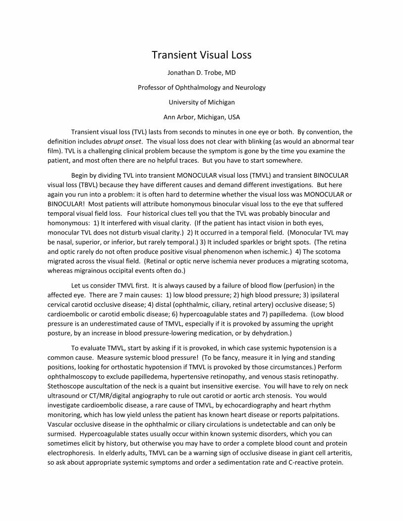
Photopsias
Prem S. Subramanian, MD, PhDProfessor of Ophthalmology, Neurology, Neurosurgery
Vice Chair for Academic AffairsUniversity of Colorado School of Medicine
Aurora, Colorado, USA4/24/17 2
Migraine Aura• Homonymous• Colored or black and white• Expands from near fixation• 5 min ≥ aura ≥ 60 min• Subsequent headache
Schott. Brain 2007;130:1690-1703.
4/24/17 3
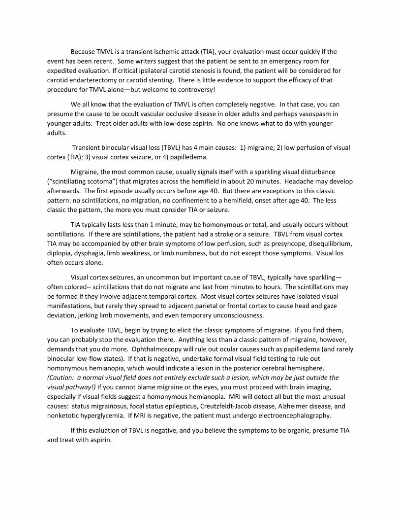
Case
• 17 year old woman• No past medical history• Intermittent right sided flashes, sparks• Lasting 30 seconds to 10 minutes• Sometimes associated with headache• No persistent visual complaints
4/24/17 4
“Migraine” Aura• Symptoms less than 5 min• Photopsias without form• Highly stereotyped symptoms• Constant laterality• Absence of headache
– No history of migraine– Older age of onset
Shams & Plant. Surv Ophthalmol 2011;56:135-161.
4/24/17 5
Visual Snow• Continuous dots in vision• Variant of migraine aura?
– Constant– Unformed– Throughout the visual field
• Young patients– May be present lifelong– Avg age at onset: 23 yrs old
Schankin et al. Brain 2014;137:1419-1428.4/24/17 6
Case• 63 year old man• Trouble driving at night• Bumps into objects• Sees flashes with eyes closed
• Va 6/6 in both eyes• Slow pupil reactions
4/24/17
2
4/24/17 7
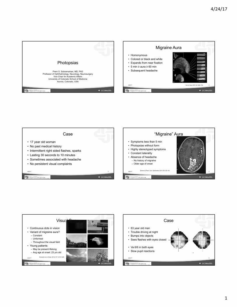
Cancer-Associated Retinopathy• Paraneoplastic syndrome• Symptoms before cancer is
found?• Autoimmune
– Anti-recoverin– Anti-enolase
• Treat the malignancy!
Mohamed & Harper. Arch Ophthalmol. 2007;125:1132-1133. 4/24/17 8
Case• 43 year old woman• Past history of Lyme disease
(treated)• Several days of photopsia OS• Missing objects in OS
• Va 6/6 OD, 6/12 OS, RAPD OS
4/24/17 9
AZOOR/AIBSE• Spectrum of disease with
photoreceptor dysfunction• Unilateral or bilateral• Cause unknown• ?Systemic associations• Other white dot syndromes
• Treatment unclear
Volpe et al. Arch Ophthalmol. 2001;119:59-63.
4/30/2017
1
Neuro-ophthalmic symptoms you
should not disregard: Vision Blur
with a “Normal Examination”
Nicholas J. Volpe, MDGeorge and Edwina Tarry Professor of Ophthalmology
Chairman, Department of Ophthalmology
Feinberg School of Medicine
Northwestern University, Chicago, USA
Possibilities
• Symptom is transient
• Condition or exam findings are transient
• Exam is not really normal
• Too soon for the exam to be abnormal
• Pathogen is too “small” and condition insidious
• Exam is normal but “imaging” is not
– OCT
– MRI
• Non Organic overlay confuses situation
• Subtle Motility problem is causing symptom
• Higher cortical function is causing symptom
Symptom is transient
Condition or Exam findings are transient
• Transient monocular blindness
• Giant cell arteritis
Exam is not really normal
• “occult” maculopathy
• You misses history or setting of amblyopia
• You did not test visual fields
• You missed cornea issue or subtle cataract
• You missed APD
• Choroidal ischemia (giant cell arteritis)
Exam is normal but “imaging” is not
• Neuro-ophthalmic exam is not complete until VFs, OCT,
(sometimes FA) and MRI are done
• OCT
• Occult maculopathies
• VR interface disorders
• MRI
• Tumor
• Stroke
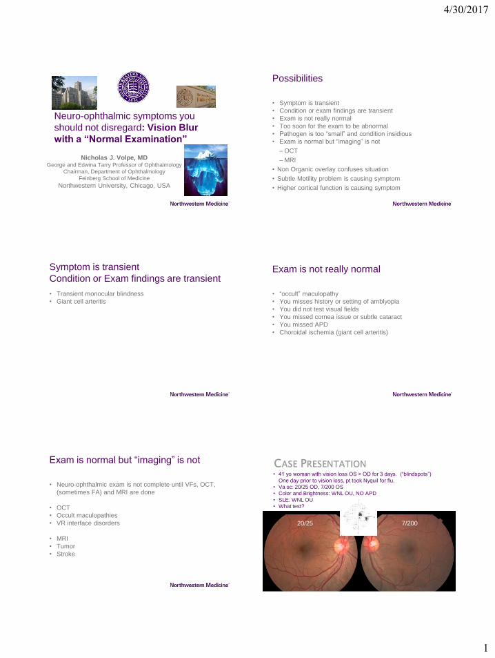
• 41 yo woman with vision loss OS > OD for 3 days. (“blindspots”)
One day prior to vision loss, pt took Nyquil for flu.
• Va sc: 20/25 OD, 7/200 OS
• Color and Brightness: WNL OU, NO APD
• SLE: WNL OU
• What test?
20/25 7/200

4/30/2017
2
ACUTE MACULAR NEURORETINOPATHY15 year old boy failed school vision screen
20/100 OD, 20/80 OS
Pupils: normal
Color: normal
Case
• 58 year old man• Routine Exam• Abnl Screening VF• Unaware of the defect
• 20/20 OU
• Color: nl
• Pupils: nl
Maculopathies in NO
• Should be easy based on History and
Exam, OCT:
• cystoid macula edema
• Macula hole
• Some V-R interface cases
• central serous chorioretinopathy
• Stargardt’s Disease
• ischemic maculopathy in diabetes
4/30/2017
3
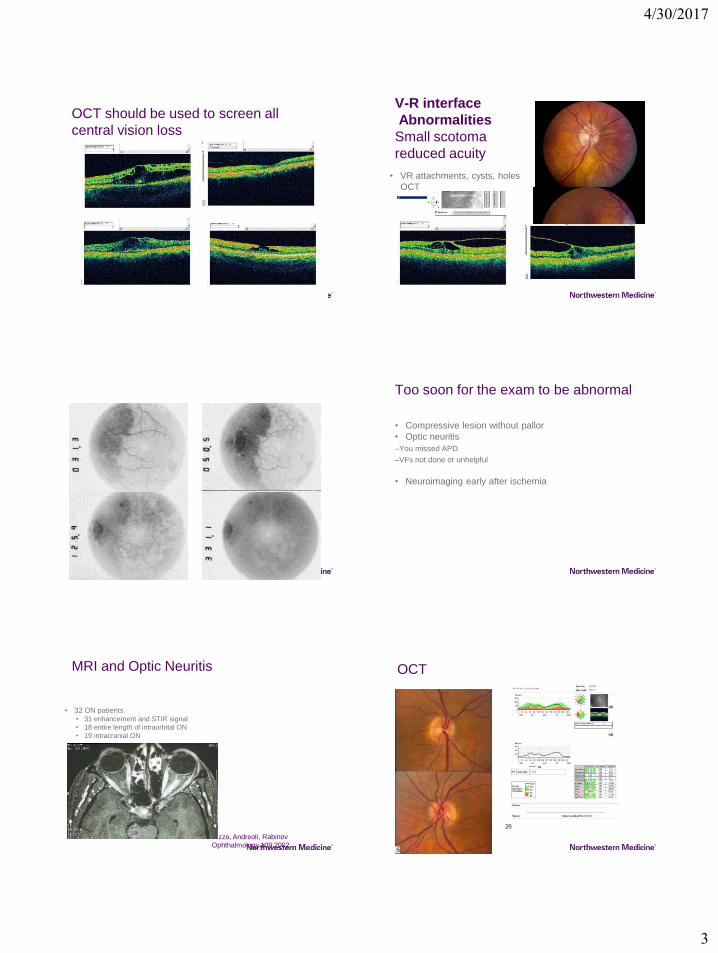
OCT should be used to screen all
central vision loss
V-R interface
Abnormalities
Small scotoma
reduced acuity
• VR attachments, cysts, holes
OCT
Too soon for the exam to be abnormal
• Compressive lesion without pallor
• Optic neuritis
–You missed APD
–VFs not done or unhelpful
• Neuroimaging early after ischemia
MRI and Optic Neuritis
• 32 ON patients• 31 enhancement and STIR signal
• 18 entire length of intraorbital ON
• 19 intracranial ON
Rizzo, Andreoli, Rabinov
Ophthalmology 109:2002
OCT
4/30/2017
4
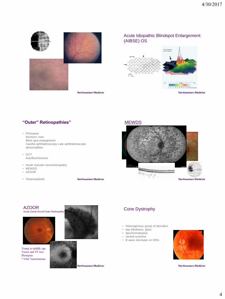
Acute Idiopathic Blindspot Enlargement:
(AIBSE) OS
“Outer” Retinopathies”
• Photopsia
Women> men
Blind spot enlargement
Careful ophthalmoscopy Late ophthalmoscopic
abnormalities
• OCT
Autofluorescence
• Acute macular neuroretinopathy
• MEWDS
• AZOOR
• Paraneoplastic
MEWDS
AZOORAcute Zonal Occult Outer Retinopathy
Young to middle age
Vision and VF loss
Photopsia
? Viral ?autoimmune
Cone Dystrophy
• Heterogenous group of disroders
• day blindness, glare
• dyschromatopsia
• central scotoma
• B wave decrease on ERG
4/30/2017
5
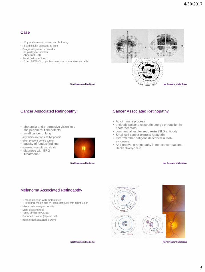
Case
• 58 y.o. decreased vision and flickering
• First difficulty adjusting to light
• Progressing over six weeks• 60 pack year smoker• Abnormal CXR
• Small cell ca of lung• Exam 20/80 OU, dyschromatopsia, some vitreous cells
Cancer Associated Retinopathy
• photopsia and progressive vision loss• mid peripheral field defects• small cancer of lung• any tumor-uterine and lymphoma
• often present before tumor
• paucity of fundus findings• narrowed vessels and vitritis
• diagnose with ERG• Treatment?
Cancer Associated Retinopathy
• Autoimmune process• antibody poisons recoverin energy production in
photoreceptors• commercial test for recoverin 23kD antibody• Small cell cancer express recoverin• Over 20 other antigens described in CAR
syndrome• Anti-recoverin retinopathy in non cancer patients-
Heckenlively-1998
Melanoma Associated Retinoapthy
• Late in disease with metastases• Flickering, vision and VF loss, difficulty with night vision
• Many maintain good acuity
• Male predominace• ERG similar to CSNB
• Reduced b wave (bipolar cell)
• normal dark adapted a wave
4/30/2017
6
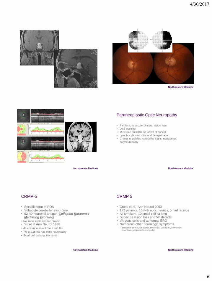
Paraneoplastic Optic Neuropathy
• Painless, subacute bilateral vision loss
• Disc swelling
• Must rule out DIRECT affect of cancer
• Lymphocyte vasculitis and demyelination
• Cranial n. palsies, cerebellar signs, nystagmus,
polyneuropathy
CRMP-5
• Specific form of PON• Subacute cerebellar syndrome• 62 kD neuronal antigen-Collapsin Response
Mediating Protein-5• Neuronal cystoplasmic protein
• Yu et al Ann Neurol 1998• As common as anti Yo < anti Hu
• 7% of 116 pts had optic neuropathy
• Small cell ca lung, thymoma
CRMP 5
• Cross et al, Ann Neurol 2003• 172 patients, 15 with optic neuritis, 5 had retinitis• All smokers, 10 small cell ca lung• Subacute vision loss and VF defects• Vitreous cells and abnormal ERG• Numerous other neurologic symptoms
– Subacute cerebellar ataxia, dementia, cranial n., movement disorders, peripheral neuropathy
4/30/2017
7
Pathogen is too “small” and/or condition
insidious
• Toxic optic neuropathies
• Nutritional optic neuropathies
• LHON
• Prion Disease
• Dosing according to ideal body weight• SD-OCT• Fundus Autofluorescence• Psychophysical
• Amsler grid (Threshold, Red)
• HVF-10-2 (white or Red target)
• Ishihara plates• Multifocal ERG
Plaquenil Screening: AAO guidelines 2009
Toxic Retinopathies
• Digoxin
• yellow vision
• canthaxanthine
• (hydroxy)chloroquine
• Bull’s eye maculopathy
• niacin
• CME
• tamoxifen
• crystals deposit in macula
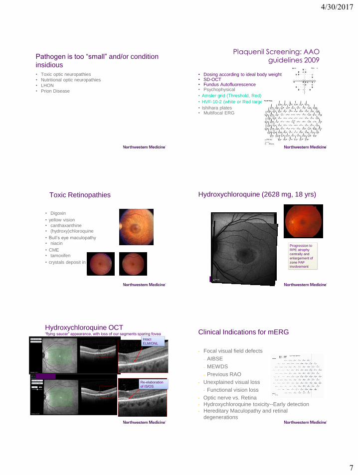
Hydroxychloroquine (2628 mg, 18 yrs)
Progression to
RPE atrophy
centrally and
enlargement of
zone FAF
involvement
Hydroxychloroquine OCT“flying saucer” appearance, with loss of our segments sparing fovea
Intact
ELM/ONL
Re-elaboration
of IS/OS
Clinical Indications for mERG
• Focal visual field defects
– AIBSE
– MEWDS
– Previous RAO
• Unexplained visual loss
• Functional vision loss
• Optic nerve vs. Retina
• Hydroxychloroquine toxicity--Early detection
• Hereditary Maculopathy and retinal
degenerations
4/30/2017
8
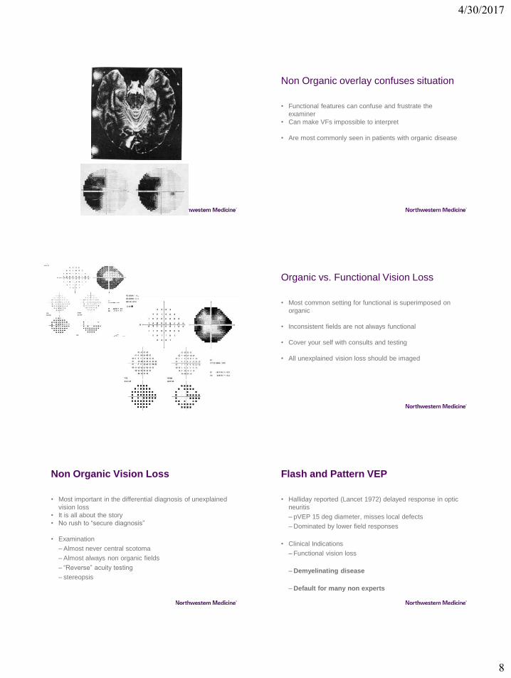
Non Organic overlay confuses situation
• Functional features can confuse and frustrate the
examiner
• Can make VFs impossible to interpret
• Are most commonly seen in patients with organic disease
Organic vs. Functional Vision Loss
• Most common setting for functional is superimposed on
organic
• Inconsistent fields are not always functional
• Cover your self with consults and testing
• All unexplained vision loss should be imaged
Non Organic Vision Loss
• Most important in the differential diagnosis of unexplained
vision loss
• It is all about the story
• No rush to “secure diagnosis”
• Examination
– Almost never central scotoma
– Almost always non organic fields
– “Reverse” acuity testing
– stereopsis
Flash and Pattern VEP
• Halliday reported (Lancet 1972) delayed response in optic
neuritis
– pVEP 15 deg diameter, misses local defects
– Dominated by lower field responses
• Clinical Indications
– Functional vision loss
– Demyelinating disease
– Default for many non experts
4/30/2017
9
Subtle Motility problem is causing
symptom
• If subtle misalignment, symptom will improve with
monocular occlusion
• Occasional nystagmoid movement with fixation instability
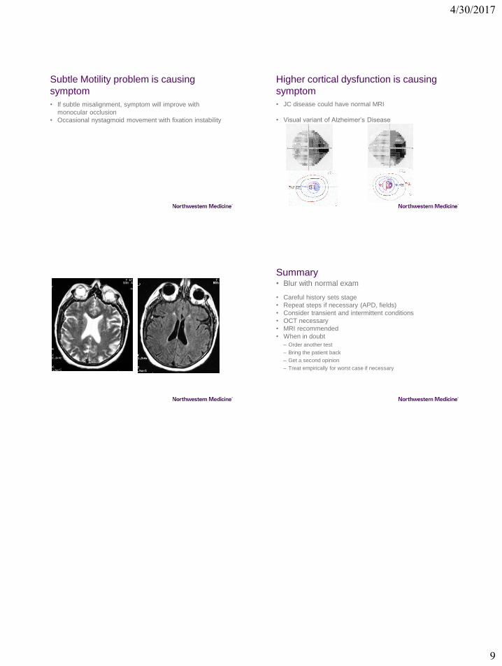
Higher cortical dysfunction is causing
symptom
• JC disease could have normal MRI
• Visual variant of Alzheimer’s Disease
Summary
• Careful history sets stage
• Repeat steps if necessary (APD, fields)
• Consider transient and intermittent conditions
• OCT necessary
• MRI recommended
• When in doubt
– Order another test
– Bring the patient back
– Get a second opinion
– Treat empirically for worst case if necessary
• Blur with normal exam