Embed Size (px)
Citation preview
CHAPTER 4
Hammerhead Ribozyme CrystalStructures and Catalysis
WILLIAM SCOTT
Center for the Molecular Biology of RNA, University of California, SantaCruz, CA 95064, USA
4.1 Introduction
After the discovery of RNase P1 (Chapter 9) and the Group I2 (Chapter 10)intron ribozymes, both of which are comparatively large and complex catalyticRNAs, the identification of the hammerhead ribozyme offered hope that thephenomenon of RNA catalysis might be best understood within the frameworkof a smaller, more tractable RNA that catalyzed a simple phosphodiesterisomerization reaction. Indeed, the first ribozyme crystal structures were in factthose of hammerhead ribozymes, but they seemed to create more questionsthan compelling explanations for RNA catalysis. The twelve years subsequentto the publication of these structures saw only increasing discord; the crystalstructure analyses seemed hopelessly irreconcilable with a growing corpus ofbiochemical evidence. Meanwhile, crystal structures for many of the otherribozymes, including the Group I intron and RNase P, started to appear.Finally, a new crystal structure of the hammerhead ribozyme emerged, 20 yearsafter the hammerhead’s discovery. This structure included a set of distaltertiary contacts whose importance was largely unrecognized until 2003, butwhose incorporation increased catalytic prowess by a factor ofB1000. The newcrystal structure reveals that this remarkable rate enhancement is a directconsequence of localized yet dramatic active site conformational changes thatare stabilized by a comparatively distant set of tertiary interactions. The newstructure appears to reconcile twenty years of discord while offering some newinsights into RNA structure and catalysis, as well as the foibles of experimentalinterpretation. The hammerhead ribozyme has indeed taught us much aboutRNA catalysis, despite (or, more likely, because of) major deviations from ourseemingly best-devised lesson plans.
48
4.2 A Catalytic RNA Prototype
The discovery that RNA can be an enzyme1,2 created the fundamental questionof how RNA enzymes work. Before this discovery, it was generally assumedthat proteins were the only biopolymers that had sufficient complexity andchemical heterogeneity to catalyze biochemical reactions. Clearly, RNA canadopt sufficiently complex tertiary structures that make catalysis possible. Buthow does the three-dimensional (3D) structure of an RNA endow it withcatalytic activity? What structural and functional principles are unique to RNAenzymes (or ribozymes), and what principles are so fundamental that they areshared with protein enzymes?
By understanding how ribozymes work, we might also learn more about howlife originated. RNA may have been the original self-replicating pre-bioticmolecule, according to the ‘‘RNA World’’ hypothesis,3 potentially catalyzingits own replication. Understanding the fundamental principles of ribozymecatalysis therefore may also give us new insights into the origin of life itself. Theanswer to the question of how ribozymes work also has practical consequences,as RNA enzymes are particularly well-suited for design as targeted therapeuticsfor various diseases (for a recent review, see ref. 4).
The hammerhead ribozyme has been thought of as a ‘‘prototype’’ ribozymein the same sense that lysozyme and serine proteases have been thought of as‘‘prototype’’ conventional protein enzymes, and for that reason the hammer-head ribozyme has attracted intense experimental scrutiny. The hammerheadribozyme is a comparatively simple and well-studied ribozyme that in principleshould be capable of revealing the secrets of its catalytic potential, if we are ableto pose the right questions and carry out useful and informative experiments.Much attention has been focused upon this particular ribozyme with the hopethat if its catalytic properties become well-understood our grasp of the phe-nomenon of RNA catalysis in general will become more comprehensive so thatgeneralizations may appear that are applicable to the larger ribozymes, to RNAsplicing and peptidyl transfer, and perhaps even beyond to a unified under-standing of RNA and protein enzymology.
4.3 A Small Ribozyme
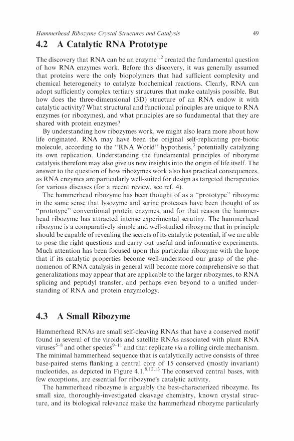
Hammerhead RNAs are small self-cleaving RNAs that have a conserved motiffound in several of the viroids and satellite RNAs associated with plant RNAviruses5–8 and other species9–11 and that replicate via a rolling circle mechanism.The minimal hammerhead sequence that is catalytically active consists of threebase-paired stems flanking a central core of 15 conserved (mostly invariant)nucleotides, as depicted in Figure 4.1.8,12,13 The conserved central bases, withfew exceptions, are essential for ribozyme’s catalytic activity.
The hammerhead ribozyme is arguably the best-characterized ribozyme. Itssmall size, thoroughly-investigated cleavage chemistry, known crystal struc-ture, and its biological relevance make the hammerhead ribozyme particularly
49Hammerhead Ribozyme Crystal Structures and Catalysis
well-suited for biochemical and biophysical investigations into the fundamentalnature of RNA catalysis. Despite the extensive structural and biochemicalcharacterization of the hammerhead ribozyme (reviewed in refs. 14–16) manyimportant questions have remained concerning how this RNA molecule’sstructure enables it to have catalytic activity. Our understanding of the rela-tionship between the structure of the hammerhead RNA and its catalyticactivity has enjoyed a particularly tumultuous history.
4.4 Chemistry of Phosphodiester Bond Isomerization
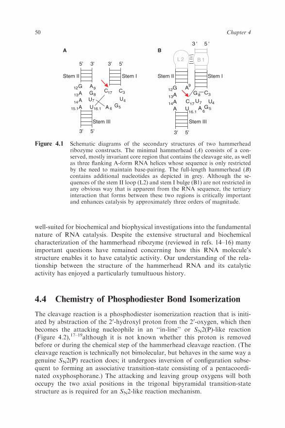
The cleavage reaction is a phosphodiester isomerization reaction that is initi-ated by abstraction of the 20-hydroxyl proton from the 20-oxygen, which thenbecomes the attacking nucleophile in an ‘‘in-line’’ or SN2(P)-like reaction(Figure 4.2),17–19although it is not known whether this proton is removedbefore or during the chemical step of the hammerhead cleavage reaction. (Thecleavage reaction is technically not bimolecular, but behaves in the same way agenuine SN2(P) reaction does; it undergoes inversion of configuration subse-quent to forming an associative transition-state consisting of a pentacoordi-nated oxyphosphorane.) The attacking and leaving group oxygens will bothoccupy the two axial positions in the trigonal bipyramidal transition-statestructure as is required for an SN2-like reaction mechanism.
3'5' 5'
3' 5'3' 5'
3'
12G
14A13A
15.1A A 6G5U16.1
C317CA9
U4U7
G812G
14A13A
A A6G5U
16.1
C3
17C
A9
U4U7
G8
3 ' 5 '
L 2 B 1
A B
Stem II Stem I Stem IStem II
Stem III Stem III
Figure 4.1 Schematic diagrams of the secondary structures of two hammerheadribozyme constructs. The minimal hammerhead (A) consists of a con-served, mostly invariant core region that contains the cleavage site, as wellas three flanking A-form RNA helices whose sequence is only restrictedby the need to maintain base-pairing. The full-length hammerhead (B)contains additional nucleotides as depicted in grey. Although the se-quences of the stem II loop (L2) and stem I bulge (B1) are not restricted inany obvious way that is apparent from the RNA sequence, the tertiaryinteraction that forms between these two regions is critically importantand enhances catalysis by approximately three orders of magnitude.
50 Chapter 4
The 50-product, as a result of this cleavage reaction mechanism, possesses a20,30-cyclic phosphate terminus, and the 30-product possesses a 50-OH termi-nus,20–21 as with non-enzymatic alkaline cleavage of RNA. The reaction istherefore, in principle, reversible, as the scissile phosphate remains a phos-phodiester, and may thus act as a substrate for hammerhead RNA-mediatedligation without a requirement for ATP or a similar exogenous energy source.The hammerhead ribozyme-catalyzed reaction, unlike the formally identicalnon-enzymatic alkaline cleavage of RNA, is a highly sequence-specific cleavagereaction with a typical turnover rate of approximately one molecule of substrate per molecule of enzyme per minute at pH 7.5 in 10mMMg21 (so-called‘‘standard reaction conditions’’ for the minimal hammerhead RNA sequence),depending upon the sequence of the particular hammerhead ribozyme con-struct measured. This represents an approximately 10 000-fold rate enhance-ment over the non-enzymatic cleavage of RNA.
4.5 Hammerhead Ribozyme Structure Nailed Down
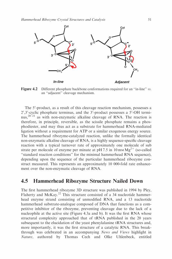
The first hammerhead ribozyme 3D structure was published in 1994 by Pley,Flaherty and McKay.22 This structure consisted of a 34 nucleotide hammer-head enzyme strand consisting of unmodified RNA, and a 13 nucleotidehammerhead substrate-analogue composed of DNA that functions as a com-petitive inhibitor of the ribozyme, preventing cleavage due to the lack of anucleophile at the active site (Figure 4.3a and b). It was the first RNA whosestructural complexity approached that of tRNA published in the 20 yearssubsequent to the elucidation of the yeast phenylalanine tRNA structures and,more importantly, it was the first structure of a catalytic RNA. This break-through was celebrated in an accompanying News and Views highlight inNature, authored by Thomas Cech and Olke Uhlenbeck, entitled
O
O
O
O
O
CH2
O
Cyt
O
O
O
O
O
O
CH2
O
Cyt
O
AdjacentIn-line
2' 2'
5'5'
5' 5'
3'3'
--
P P
Figure 4.2 Different phosphate backbone conformations required for an ‘‘in-line’’ vs.an ‘‘adjacent’’ cleavage mechanism.
51Hammerhead Ribozyme Crystal Structures and Catalysis
‘‘Hammerhead nailed down’’.23 Subsequent publication of two all-RNA ham-merhead ribozyme crystal structures, the first with a 20-OMe replacing thenucleophile at the active site,24 and the second with an unmodified nucleotide atthe active site,25 revealed the same 3D structure of the invariant core nucleo-tides in the ribozyme well within experimental error (Figure 4.3c and d), despitesignificant differences in the sequence within the nonessential regions, DNA vs.RNA substrates, presence vs. absence of divalent metal ions and active
Figure 4.3 Three crystallographically independent hammerhead ribozymes (A) occu-pied the asymmetric unit in the first hammerhead crystal structure; one ofthese is displayed in what has become the conventional orientation (B)such that the RNA enzyme strand (green) and DNA substrate-analogue(cyan) are clearly visible. Two crystallographically independent all-RNAhammerheads occupied the asymmetric unit (C) of the second hammer-head ribozyme crystal structure. One of these (D) is chosen and displayedto facilitate comparison with (b). Here, the shorter strand is (nominally)the enzyme strand and the longer strand (cyan) is the substrate, where a 20-OMe nucleotide occupies the cleavage site. A subsequent crystal formhaving but one molecule per asymmetric unit (not shown) is otherwiseidentical, despite the absence of the 20-OMe modification.
52 Chapter 4
nucleophile, and crystal packing schemes. In essence, the spatial positioning ofthe invariant region of the hammerhead ribozyme appeared to be immune to aseries of potential perturbations in at least six crystallographically independentmolecules, further corroborating, it seemed, the initial assessment that thestructure had indeed been ‘‘nailed down’’ in 1994.
4.6 Catalysis in the Crystal
The hammerhead ribozyme sequence that contained all unmodified RNA,including the active-site nucleophile, was catalytically active both in solutionand in the crystal.25,26 The crystals of the active hammerhead ribozyme thusenabled us to test for cleavage activity in the crystalline state. We initiated thecleavage reaction by flooding the crystal with divalent cations while raising thepH above the apparent kinetic pKa using a soaking solution buffered atpH8.5.25–27 In conditions with a Mg21 concentration of 50mM and pH8.5,the self-cleavage rate in the crystal is approximately 0.4moleculesmin�1. Undersimilar conditions in solution, this same hammerhead ribozyme construct, asequence that was optimized for purposes of growing crystals rather than forcatalytic prowess, cleaves at a rate of approximately 0.08moleculesmin�1,permitting us to suggest that the crystal lattice did more to aid in the properfolding of the ribozyme than it did to inhibit the hammerhead ribozyme’scleavage activity. Moreover, the extent of cleavage of the substrate in thecrystal was almost complete.26
Although this collection of minimal hammerhead ribozyme crystal structuresprovided rationalizations for several of the previously reported experimentalobservations, many important problems remained unresolved (see, for exam-ple, ref. 14). One of the most important and immediately recognized22 problemswas that the scissile phosphate in all of the structures just described wasobserved to be in a conformation that is completely incompatible28 with an ‘‘in-line’’ mechanism. Hence the need to bring the scissile phosphate into aconformation amenable to an in-line attack can be (and in fact was) taken asprima facie evidence for a required conformational change prior to bondcleavage.
4.7 Making Movies from Crystallographic Snapshots
The fact that the minimal hammerhead RNA sequence can cleave after beingcrystallized25 presented the opportunity to capture various states, includingpre-catalytic conformational changes, along the reaction pathway, using cry-stallographic freeze-trapping techniques.
Although conventional X-ray crystallography is essentially static and bynecessity involves both a spatial and a temporal averaging of a macroscopicnumber of molecules and a time period of hours (the duration of data collec-tion), time-resolved crystallographic experiments may be possible under the
53Hammerhead Ribozyme Crystal Structures and Catalysis
right conditions. Two options for time-resolved experiments exist that permitone to obtain crystallographic snapshots during catalysis.
The Laue method uses polychromatic X-rays to enable collection of a fairlycomplete X-ray dataset in a matter of milliseconds or less, and has been usedwith success in the case of many protein enzymes that catalyze rapid reac-tions.29,30Laue experiments require ideal crystals that possess very low mosai-city, as well as a way of very rapidly initiating a reaction simultaneouslythroughout the crystal lattice. Once the reaction has been initiated, eachenzyme–substrate complex evolves stochastically with respect to time. Inter-mediates can be observed only when the majority of molecules in a crystaloccupy the same intermediate state at the same time. Lower-occupancy inter-mediates remain essentially unobservable.
When combined with flash-freezing (e.g., immersion in liquid nitrogen) as aphysical trapping method, conventional monochromatic X-ray data collectionalso offers the opportunity to conduct time-resolved experiments using asecond approach. Instead of recording live snapshots, as with the Laue method,the reaction is simply initiated in the crystal, allowed to evolve, and themolecular contents of the crystal are immobilized by freeze-trapping prior tostandard data collection.31 Since most conventional X-ray data collection isalready performed using crystals frozen at 100K, this procedure in practiceinvolves very little additional experimental modification, and possesses theadded advantage of being much more tolerant of mosaicity and the other smallcrystal imperfections that often accompany reactivity in the crystal. The utilityof this approach is confined to comparatively slowly reactive enzymes. Fortu-nately, the cleavage rate of the minimal hammerhead ribozyme not only is onthe order of 1 min�1, it can be further modulated as a function of pH andpresence of divalent cations, making it an ideal candidate for the freeze-trapping approach.
To better understand the structural basis of the proposed conformationalchange required to activate the hammerhead ribozyme for catalysis, we per-formed monochromatic time-resolved crystallographic freeze-trapping studieswith the aim of observing conformational intermediates preceding cataly-sis.25,27,32,33 To trap the structure of a precatalytic structural intermediate, ahammerhead ribozyme having a ‘‘kinetic bottleneck’’ at the final or bond-breaking point on the reaction pathway was synthesized using a modifiedleaving-group, (Figure 4.4a). This modified hammerhead RNA was used tocapture a conformational intermediate that began to approach an in-lineconformation27 that evolved subsequent to triggering the reaction (Figure4.4b). Prior to triggering the reaction, the modified RNA possessed a structureindistinguishable from that of the initial-state structure of the unmodifiedRNA, an important control that was conducted and reported,27 contrary tosome claims that have been made.34 Two other pre-catalytic conformationalchanges were also captured using complementary approaches that did notinvolve modification of the leaving group, and appeared consistent in that theymade up a trajectory toward an in-line structure.25,33 (In retrospect, this
54 Chapter 4
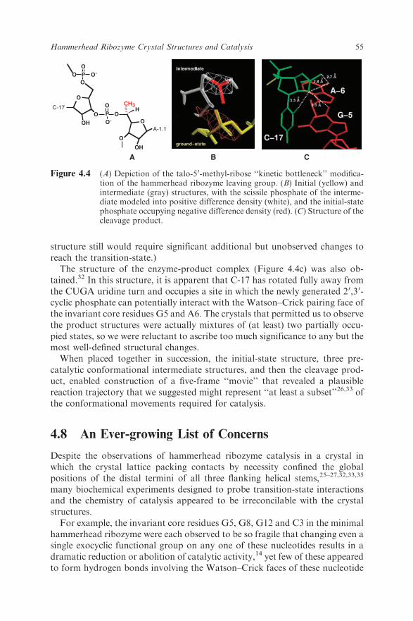
structure still would require significant additional but unobserved changes toreach the transition-state.)
The structure of the enzyme-product complex (Figure 4.4c) was also ob-tained.32 In this structure, it is apparent that C-17 has rotated fully away fromthe CUGA uridine turn and occupies a site in which the newly generated 20,30-cyclic phosphate can potentially interact with the Watson–Crick pairing face ofthe invariant core residues G5 and A6. The crystals that permitted us to observethe product structures were actually mixtures of (at least) two partially occu-pied states, so we were reluctant to ascribe too much significance to any but themost well-defined structural changes.
When placed together in succession, the initial-state structure, three pre-catalytic conformational intermediate structures, and then the cleavage prod-uct, enabled construction of a five-frame ‘‘movie’’ that revealed a plausiblereaction trajectory that we suggested might represent ‘‘at least a subset’’26,33 ofthe conformational movements required for catalysis.
4.8 An Ever-growing List of Concerns
Despite the observations of hammerhead ribozyme catalysis in a crystal inwhich the crystal lattice packing contacts by necessity confined the globalpositions of the distal termini of all three flanking helical stems,25–27,32,33,35
many biochemical experiments designed to probe transition-state interactionsand the chemistry of catalysis appeared to be irreconcilable with the crystalstructures.
For example, the invariant core residues G5, G8, G12 and C3 in the minimalhammerhead ribozyme were each observed to be so fragile that changing even asingle exocyclic functional group on any one of these nucleotides results in adramatic reduction or abolition of catalytic activity,14 yet few of these appearedto form hydrogen bonds involving the Watson–Crick faces of these nucleotide
OO
CH3H
O
OH
O
O
P
O
O
O
OH
O
OP O
A-1.1
C-17
-
-
A B C
Figure 4.4 (A) Depiction of the talo-50-methyl-ribose ‘‘kinetic bottleneck’’ modifica-tion of the hammerhead ribozyme leaving group. (B) Initial (yellow) andintermediate (gray) structures, with the scissile phosphate of the interme-diate modeled into positive difference density (white), and the initial-statephosphate occupying negative difference density (red). (C) Structure of thecleavage product.
55Hammerhead Ribozyme Crystal Structures and Catalysis
bases in any of the minimal hammerhead structures, apart from a G-5 inter-action in the product structure.
A particularly striking and only recently observed example consisted of G8and G12, which were identified36 as possible participants in acid–base catalysis(Chapter 3). Once it was demonstrated that the hammerhead RNA does notrequire divalent metal ions for catalysis,37,38 it gradually became apparent thatthat the RNA itself, rather than passively bound divalent metal ions, must play adirect chemical role in any acid–base chemistry in the hammerhead ribozymeactive site. It was, however, completely unclear how G12 and G8 could accom-plish this, given the original structures of the minimal hammerhead ribozyme.
Other concerns included an NOE between U4 and U7 of the cleavedhammerhead ribozyme39 that had also been observed during NMR character-ization, which suggested that these nucleotide bases must approach one anothercloser than about 6 A, although close approach of U7 to U4 did not appear tobe possible from the crystal structure. Finally, as previously discussed, theattacking nucleophile in the original structures, the 20-OH of C17, was not in aposition amenable to in-line attack upon the adjacent scissile phosphate.22
Perhaps most worrisome were experiments that suggested the A-9 and scissilephosphates must come within about 4 A of one anther in the transition-state,based upon double phosphorothioate substitution and soft metal ion rescueexperiments;40 the distance between these phosphates in the crystal structurewas about 18 A, with no clear mechanism for close approach if the stem II andstem I A-form helices were treated as rigid bodies. Taken together, these resultsappeared to suggest that a fairly large-scale conformational change must havetake place to reach the transition-state within the minimal hammerheadribozyme structure.
For these reasons, the two sets of experiments (biochemical vs. crystallo-graphic) appeared not only to be at odds, but to be completely and hopelesslyirreconcilable, generating a substantial amount of discord in the field.34 Nocompelling evidence for dismissing either set of experimental results was evermade successfully, although many claims to the contrary34,40,41 were made infavor of each.
4.9 Occam’s Razor Can Slit Your Throat
The principle of parsimony, attributed to William of Occam, states that oneshould favor simple scientific explanations and hypotheses that make a mini-mum number of assumptions over more complicated alternatives. The ‘‘Oc-cam’s Razor’’ principle, in which one shaves away extraneous and irrelevantdecorations to obtain the most parsimonious hypothesis, has also been ex-tended to the physical investigation of nucleic acids and proteins: study thesimplest macromolecular assemblies that possess the biological function ofinterest, and cut everything else off. This ‘‘reductionist paradigm’’ has beenemployed with such success in molecular biology that one can easily lose sightof the larger, physiological context.
56 Chapter 4

When the hammerhead RNA was first discovered, it was observed in aB370nucleotide single-stranded genomic satellite RNA, most of which could bedeleted while preserving the RNA’s catalytic properties.6–8 Eventually, it wasfound that about 13 core nucleotides and a minimal number of flanking helicalnucleotides were all that was required for a respectable catalytic turnover rateof 1 to 10min�1, and this ‘‘minimal’’ hammerhead construct became the focusof almost all of the biochemical, biophysical and crystallographic investiga-tions, as described earlier.
It thus came as a great surprise to most in the field when, in 2003, it wasfinally pointed out that for optimal activity, the hammerhead ribozyme requiresthe presence of sequences in stems I and II that interact to form tertiarycontacts42,43 (Figure 4.1b) that were removed in the process of shaving seem-ingly superfluous structures from the hammerhead ribozyme. Once the fullramifications of this revelation became apparent, i.e., that the entire field hadbeen studying the residual catalytic activity of an over-zealously truncatedversion of the full-length ribozyme, attention shifted away from the minimalconstructs.44 It also quickly became clear to us that a crystal structure of thefull-length hammerhead ribozyme, in which these distal tertiary contacts werepresent, might be of considerable interest.
4.10 Structure of a Full-length Hammerhead Ribozyme
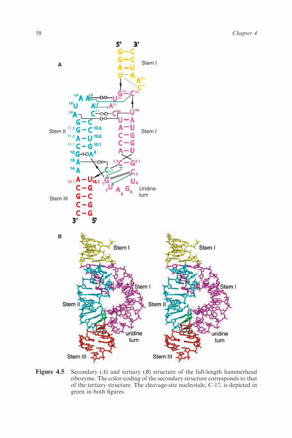
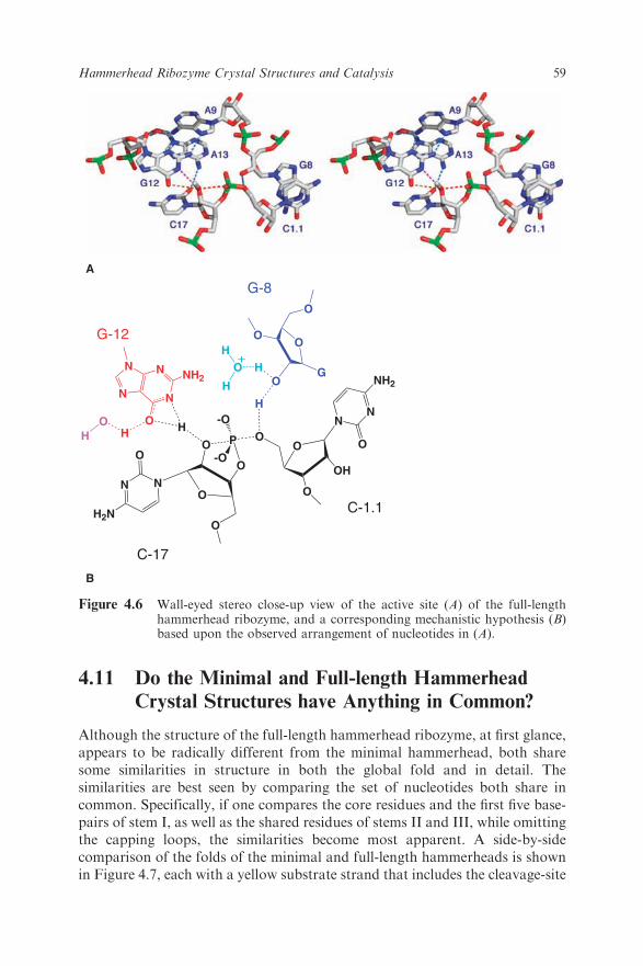
After several years of struggle, in 2006 we finally obtained a 2.2 A resolutioncrystal structure of the full-length hammerhead ribozyme.45 This new structure(Figure 4.5) appears to resolve the most worrisome of the previous discrepan-cies. In particular, C17 is now positioned for in-line attack, and the invariantresidues C3, G5, G8 and G12 all appear involved in vital interactions relevantto catalysis. Moreover, the A9 and scissile phosphates are observed to be 4.3 Aapart, which is consistent with the idea that, when modified, these phosphatescould bind a single thiophilic metal ion. The structure also reveals how twoinvariant residues, G-12 and G-8, are positioned within the active site –consistent with their previously proposed36 role in acid–base catalysis. G12 iswithin hydrogen bonding distance to the 20-O of C17, the nucleophile in thecleavage reaction, and the ribose of G8 hydrogen bonds to the leaving group50-O (Figure 4.6), while the nucleotide base of G8 forms a Watson–Crick pairwith the invariant C3. This arrangement permits us to suggest that G12 is thegeneral base in the cleavage reaction, and that G8 may function as the generalacid, which is consistent with previous biochemical observations.36 G5 hydro-gen bonds to the furanose oxygen of C17, helping to position it for in-lineattack. U4 and U7, as a consequence of the base-pair formation between G8and C3, are now positioned such that an NOE between their bases is easilyexplained.
The crystal structure of the full-length hammerhead ribozyme thus clearlyaddresses all of the major concerns that appeared irreconcilable with theprevious crystal structure.46
57Hammerhead Ribozyme Crystal Structures and Catalysis
333
666 555
8
999
777
444
17
10.110.110.111.1
121212
131313
141414
15.1 16.116.116.1
Stem II
Stem III
Stem I
Stem I
Uridine turn
2.12.12.11.11.11.1
L1
L3L3L3
L2L2L2
L6L6L6
L5L5L5
L4L4L4
B8
B5B5B5
B2
B6
B4B4B4
B3
B1
B7
11.2
11.3
10.210.210.2
10.310.310.3
A
B
Figure 4.5 Secondary (A) and tertiary (B) structure of the full-length hammerheadribozyme. The color-coding of the secondary structure corresponds to thatof the tertiary structure. The cleavage-site nucleotide, C-17, is depicted ingreen in both figures.
58 Chapter 4
4.11 Do the Minimal and Full-length Hammerhead
Crystal Structures have Anything in Common?
Although the structure of the full-length hammerhead ribozyme, at first glance,appears to be radically different from the minimal hammerhead, both sharesome similarities in structure in both the global fold and in detail. Thesimilarities are best seen by comparing the set of nucleotides both share incommon. Specifically, if one compares the core residues and the first five base-pairs of stem I, as well as the shared residues of stems II and III, while omittingthe capping loops, the similarities become most apparent. A side-by-sidecomparison of the folds of the minimal and full-length hammerheads is shownin Figure 4.7, each with a yellow substrate strand that includes the cleavage-site
N
H2N
O
NO
O
O
P
-OO
O
-OO
OH
O
N
NH2
O
N
G
O
O
O
O
N
NN
N
O
NH2
H
H
HO
H
O HH
H
G-12
G-8
C-17
C-1.1
B
A
+
Figure 4.6 Wall-eyed stereo close-up view of the active site (A) of the full-lengthhammerhead ribozyme, and a corresponding mechanistic hypothesis (B)based upon the observed arrangement of nucleotides in (A).
59Hammerhead Ribozyme Crystal Structures and Catalysis
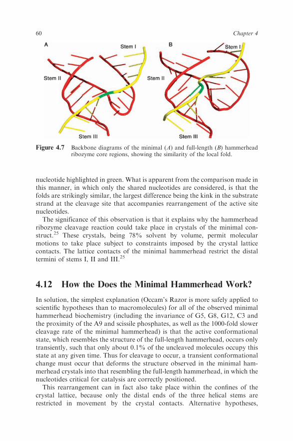
nucleotide highlighted in green. What is apparent from the comparison made inthis manner, in which only the shared nucleotides are considered, is that thefolds are strikingly similar, the largest difference being the kink in the substratestrand at the cleavage site that accompanies rearrangement of the active sitenucleotides.
The significance of this observation is that it explains why the hammerheadribozyme cleavage reaction could take place in crystals of the minimal con-struct.25 These crystals, being 78% solvent by volume, permit molecularmotions to take place subject to constraints imposed by the crystal latticecontacts. The lattice contacts of the minimal hammerhead restrict the distaltermini of stems I, II and III.25
4.12 How the Does the Minimal Hammerhead Work?
In solution, the simplest explanation (Occam’s Razor is more safely applied toscientific hypotheses than to macromolecules) for all of the observed minimalhammerhead biochemistry (including the invariance of G5, G8, G12, C3 andthe proximity of the A9 and scissile phosphates, as well as the 1000-fold slowercleavage rate of the minimal hammerhead) is that the active conformationalstate, which resembles the structure of the full-length hammerhead, occurs onlytransiently, such that only about 0.1% of the uncleaved molecules occupy thisstate at any given time. Thus for cleavage to occur, a transient conformationalchange must occur that deforms the structure observed in the minimal ham-merhead crystals into that resembling the full-length hammerhead, in which thenucleotides critical for catalysis are correctly positioned.
This rearrangement can in fact also take place within the confines of thecrystal lattice, because only the distal ends of the three helical stems arerestricted in movement by the crystal contacts. Alternative hypotheses,
Figure 4.7 Backbone diagrams of the minimal (A) and full-length (B) hammerheadribozyme core regions, showing the similarity of the local fold.
60 Chapter 4
including the suggestion that the minimal hammerhead cleaves via a differentpathway than that of the full-length hammerhead, and that the minimalhammerhead structure in solution is identical to the full-length hammerheadconformation observed in the crystal structure, have considerably less explan-atory power. The first hypothesis cannot explain the requirement for theinvariant residues, and the second hypothesis cannot account for the observed1000-fold rate enhancement. Hence it seems most likely that in solution, theminimal hammerhead has nearly the same structure as it does in the crystal, andthat in both cases the minimal ribozyme only occasionally visits the confor-mation that is stabilized and therefore dominates in the full-length hammer-head construct.
4.13 A Movie Sequel with a Happy Ending
In silico adiabatic morphing47 of the minimal into the full-length hammerheadribozyme structure is possible when the nucleotides shared in common by bothhammerhead constructs are interpolated. The structure observed in the mini-mal hammerhead ribozyme can be continuously deformed via low energy-barrier torsion angle conformational changes into the structure observed in thefull-length hammerhead. This process is best represented as a series of consec-utive structures viewed as a movie (link to Quicktime movie: http://tinyurl.com/2vly4d). It is likely that the first (1994 and 1995), initial-state hammerheadribozyme crystal structures, represent more or less accurately the dominantstructure of the minimal hammerhead ribozyme in the crystal – consistent withthe minimal hammerhead being 1000-fold less active in solution than the full-length hammerhead. The product, or cleaved state, of the minimal hammer-head in some ways resembles the full-length hammerhead to a greater extentthan does the uncleaved minimal hammerhead structure. Specifically, in thecleaved structure,32 the cleavage-site base, C-17, is observed to make contactswith G5 and A6 that are similar to those observed in the full-length structure,and the interactions with C3 are completely absent in both cases.
The more developed cleavage intermediates, in retrospect, appear to resem-ble a torsion angle conformational change of only about 1/3 of what is requiredto morph the structure from the minimal hammerhead to the full-lengthhammerhead active-site conformation. The crystallographic observations ofvarious states along the cleavage reaction pathway thus appear in retrospect tobe more incomplete than erroneous, in that they appear consistent with the firstB1/3 of the conformational change ultimately required to reach a structuresimilar to that of the full-length hammerhead. Missing by necessity from the setof snapshots was the low-occupancy transient conformation that is stabilizedby the distal tertiary contacts in the full-length hammerhead ribozyme. Incrystallographic experiments, one can only hope to resolve the dominant, high-occupancy, species in the crystal, so it is likely that the true pre-catalyticintermediate would never be observed crystallographically in the context of theminimal hammerhead construct.
61Hammerhead Ribozyme Crystal Structures and Catalysis
4.14 Concluding Remarks
In summary, it appears that the actual experimental data obtained from thecrystallographic analyses and the biochemical characterizations, which wereperformed on high-occupancy, near-ground-state and transient near-transition-state structures, respectively, were sound within the confines imposedby the minimal hammerhead structure. The mutually held interpretation thatacceptance of one set of experimental results precluded acceptance of the other,however, was based on the flawed assumption that the two sets of observationswere incommensurate and irreconcilable. In our case, the flawed assumptionmanifested itself most explicitly as the claim that unwinding and unpairing ofhelical elements was unlikely to take place.41 In the other case, the flawedassumption manifested itself with the claim that any cleavage observed in thecrystal must be due to an off-pathway artifact or experimental incompe-tence.34,40 In retrospect, neither dismissal was justified, nor compelling. Theresolution of the apparent paradox came with the structure of the full-lengthhammerhead, which reconciles and permits explanations of both sets ofexperimental results.
Acknowledgements
Sung-Hou Kim, Aaron Klug, Olke Uhlenbeck, David McKay, Barry Stoddard,David Lilley, Fritz Eckstein, Harry Noller, George Bruening, Monika Martick,Christine Dunham, James Murray, Michael Gait, Jane Grasby, Dan Hersch-lag, Jay Pandit, Ann Caviani Pease, Rosalind Kim, Elizabeth Holbrook,Young-In Chi and many others have been enormously helpful as collaborators,advisors, friends, and scientific sparring partners on many aspects of hammer-head ribozyme structure and catalysis from 1987 to the present. The AmericanCancer Society, the MRC Laboratory for Molecular Biology, The RNA Centerat the University of California at Santa Cruz, The William Keck Foundation,the National Science Foundation and the National Institutes of Health have allprovided crucial funding for hammerhead ribozyme structural studies in oneway or another at some point during that 20 year period.
References
1. C. Guerrier-Takada, K. Gardiner, T. Marsh, N. Pace and S. Altman, TheRNA moiety of ribonuclease P is the catalytic subunit of the enzyme, Cell,1983, 35, 849–857.
2. A.J. Zaug and T.R. Cech, The intervening sequence RNA of Tetrahymenais an enzyme, Science, 1986, 231, 470–475.
3. R.F. Gesteland and J.F. Atkins (eds.), The RNA World, Cold SpringHarbor Laboratory Press, Plainview, NY, 1993.
4. N.K. Vaish, A.R. Kore and F. Eckstein, Recent developments in thehammerhead ribozyme field, Nucleic Acids Res., 1998, 26, 5237–5242.
62 Chapter 4
5. G.A. Prody, J.T. Bakos, J.M. Buzayan, I.R. Schneider and G. Breuning,Autolytic processing of dimeric plant virus satellite RNA, Science, 1986,231, 1577–1580.
6. A.C. Forster and R.H. Symons, Self-cleavage of virusoid RNA is per-formed by the proposed 55-nucleotide active site, Cell, 1987, 50, 9–16.
7. J. Haseloff and W.L. Gerlach, Sequences required for self-catalysed cleav-age of the satellite RNA of tobacco ringspot virus, Gene, 1989, 82, 43–52.
8. O.C. Uhlenbeck, A small catalytic oligoribonucleotide, Nature, 1987, 328,596–600.
9. G. Ferbeyre, J.M. Smith and R. Cedergren, Schistosome satellite DNAencodes active hammerhead ribozymes, Mol. Cell Biol., 1998, 18, 3880–3888.
10. V. Bourdeau, G. Ferbeyre, M. Pageau, B. Paquin and R. Cedergren, Thedistribution of RNA motifs in natural sequences, Nucleic Acids Res., 1999,27, 4457–4467.
11. A.C. Forster, C. Davies, C.C. Sheldon, A.C. Jeffries and R.H. Symons,Self-cleaving viroid and newt RNAs may only be active as dimers, Nature,1988, 334, 265–267.
12. D.E. Ruffner, G.D. Stormo and O.C. Uhlenbeck, Sequence requirementsof the hammerhead RNA self-cleavage reaction, Biochemistry, 1990, 29,10695–10702.
13. C.C. Sheldon and R.H. Symons, Mutagenesis analysis of a self-cleavingRNA, Nucleic Acids Res., 1989, 17, 5679–5685.
14. D.B. McKay, Structure and function of the hammerhead ribozyme: Anunfinished story, RNA, 1996, 2, 395–403.
15. J.E. Wedekind and D.B. McKay, Crystallographic structures of the ham-merhead ribozyme: Relationship to ribozyme folding and catalysis, Annu.Rev. Biophys. Biomol. Struct., 1998, 27, 475–502.
16. W.G. Scott, Biophysical and biochemical investigations of RNA catalysisin the hammerhead ribozyme, Q. Rev. Biophys., 1999, 32, 241–284.
17. H. van Tol, J.M. Buzayan, P.A. Feldstein, F. Eckstein and G. Bruening,Two autolytic processing reactions of a satellite RNA proceed with inver-sion of configuration, Nucleic Acids Res., 1990, 18, 1971–1975.
18. G. Slim and M.J. Gait, Configurationally defined phosphorothioate-con-taining oligoribonucleotides in the study of the mechanism of cleavage ofhammerhead ribozymes, Nucleic Acids Res., 1991, 19, 1183–1188.
19. M. Koizumi and E. Ohtsuka, Effects of phosphorothioate and 2-aminogroups in hammerhead ribozymes on cleavage rates and Mg2+ binding,Biochemistry, 1991, 30, 5145–5150.
20. J.M. Buzayan, A. Hampel and G. Bruening, Nucleotide sequence andnewly formed phosphodiester bond of spontaneously ligated satellite to-bacco ringspot virus RNA, Nucleic Acids Res., 1986, 14, 9729–9743.
21. C.J. Hutchins, P.D. Rathjen, A.C. Forster and R.H. Symons, Self-cleavageof plus and minus RNA transcripts of avocado sunblotch viroid, NucleicAcids Res., 1986, 14, 3627–3640.
63Hammerhead Ribozyme Crystal Structures and Catalysis
22. H.W. Pley, K.M. Flaherty and D.B. McKay, Three-dimensional structureof a hammerhead ribozyme, Nature, 1994, 372, 68–74.
23. T.R. Cech and O.C. Uhlenbeck, Ribozymes. Hammerhead nailed down,Nature, 1994, 372, 39–40.
24. W.G. Scott, J.T. Finch and A. Klug, The crystal structure of an all-RNAhammerhead ribozyme: A proposed mechanism for RNA catalytic cleav-age, Cell, 1995, 81, 991–1002.
25. W.G. Scott, J.B. Murray, J.R. Arnold, B.L. Stoddard and A. Klug,Capturing the structure of a catalytic RNA intermediate: The hammerheadribozyme, Science, 1996, 274, 2065–2069.
26. J.B. Murray, C.M. Dunham and W.G. Scott, A pH-dependent conformat-ional change,rather than the chemical step, appears to be rate-limiting inthe hammerhead ribozyme cleavage reaction, J. Mol. Biol., 2002, 315,121–130.
27. J.B. Murray, D.P. Terwey, L. Maloney, A. Karpeisky, N. Usman, L.Beigelman and W.G. Scott, The structural basis of hammerhead ribozymeself-cleavage, Cell, 1998, 92, 665–673.
28. W.G. Scott, Ribozyme catalysis via orbital steering, J. Mol. Biol., 2001,311, 989–999.
29. K. Moffat, D. Bilderback, W. Schildkamp, D. Szebenyi and T.Y. Teng,Laue photography from protein crystals, Basic Life Sci., 1989, 51, 325–330.
30. L.N. Johnson, Time-resolved protein crystallography, Protein Sci., 1992, 1,1237–1243.
31. B.L. Stoddard, Intermediate trapping and laue X-ray diffraction: Potentialfor enzyme mechanism, dynamics, and inhibitor screening, Pharmacol.Ther., 1996, 70, 215–256.
32. J.B. Murray, H. Szoke, A. Szoke and W.G. Scott, Capture and visualiza-tion of a catalytic RNA enzyme-product complex using crystal latticetrapping and X-ray holographic reconstruction, Mol. Cell, 2000, 5,279–287.
33. C.M. Dunham, J.B. Murray and W.G. Scott, A helical twist-inducedconformational switch activates cleavage in the hammerhead ribozyme,J. Mol. Biol., 2003, 332, 327–336.
34. K.F. Blount and O.C. Uhlenbeck, The structure-function dilemma of thehammerhead ribozyme, Annu. Rev. Biophys. Biomol. Struct., 2005, 34,415–440.
35. W.G. Scott, Visualizing the structure and mechanism of a small nucleolyticribozyme, Methods, 2002, 28, 302–306AQ1 .
36. J. Han and J.M. Burke, Model for general acid-base catalysis by thehammerhead ribozyme: pH–activity relationships of G8 and G12 variantsat the putative active site, Biochemistry, 2005, 44, 7864–7870.
37. J.B. Murray, A.A. Seyhan, N.G. Walter, J.M. Burke and W.G. Scott, Thehammerhead, hairpin and VS ribozymes are catalytically proficient inmonovalent cations alone,, Chem. Biol., 1998, 5, 587–595.
38. W.G. Scott, RNA structure metal ions, and catalysis, Curr. Opin. Chem.Biol., 1999, 3, 705–709.
64 Chapter 4
39. J.P. Simorre, P. Legault, A.B. Hangar, P. Michiels and A. Pardi, Aconformational change in the catalytic core of the hammerhead ribozymeupon cleavage of an RNA substrate, Biochemistry, 1997, 36, 518–525.
40. S. Wang, K. Karbstein, A. Peracchi, L. Beigelman and D. Herschlag,Identification of the hammerhead ribozyme metal ion binding site respon-sible for rescue of the deleterious effect of a cleavage site phosphorothioate,Biochemistry, 1999, 38, 14363–14378.
41. J.B. Murray and W.G. Scott, Does a single metal ion bridge the A-9 andscissile phosphate groups in the catalytically active hammerhead ribozymestructure? J. Mol. Biol., 2000, 296, 33–41.
42. M. De la Pena, S. Gago and R. Flores, Peripheral regions of naturalhammerhead ribozymes greatly increase their self-cleavage activity, EMBOJ., 2003, 22, 5561–5570.
43. A. Khvorova, A. Lescoute, E. Westhof and S.D. Jayasena, Sequenceelements outside the hammerhead ribozyme catalytic core enable intracel-lular activity, Nat. Struct. Biol., 2003, 10, 708–712.
44. D.M. Lilley, Ribozymes–a snip too far? Nat. Struct. Biol., 2003, 10, 672–673.
45. M. Martick and W.G. Scott, Tertiary contacts distant from the active siteprime a ribozyme for catalysis, Cell., 2006, 126, 309–320.
46. J.A. Nelson and O.C. Uhlenbeck, When to believe what you see, Mol. Cell,2006, 23, 447–450.
47. W.G. Krebs and M. Gerstein, The morph server: A standardized systemfor analyzing and visualizing macromolecular motions in a databaseframework, Nucleic Acids Res., 2000, 28, 1665–1675.
65Hammerhead Ribozyme Crystal Structures and Catalysis
Ribozymes and RNA Catalysis edited by D.M.J. Lilley and F. Eckstein Chapter 4 Author queries (1) Ref. 35: Please complete the journal title (Methods…).


![Attenuation of Telomerase Activity by a Hammerhead ......(CANCER RESEARCH 58. 5406-5410, December I. 1998] Attenuation of Telomerase Activity by a Hammerhead Ribozyme Targeting the](https://img.pdfslide.us/doc/110x75/603d4e0fb422b843a43f3d6c/attenuation-of-telomerase-activity-by-a-hammerhead-cancer-research-58.jpg)


![540 RIBONUCLEOLYTIC NUCLEIC ACIDS [341 et al... · holds a M.Sc. scholarship from the Fonds pour la Formation de Chercheurs et l'Aide a la Recherche du Quebec. [34] Hammerhead Ribozyme](https://img.pdfslide.us/doc/110x75/5fc5df8697690d0d117a8d3d/540-ribonucleolytic-nucleic-acids-341-et-al-holds-a-msc-scholarship-from.jpg)