Embed Size (px)
Citation preview
1500
M. A. ABDEL-WAHAB, E. B. G. JONES AND L. L. P. VRIJMOED
Department of Biology and Chemistry, City University of Hong Kong, Tat Chee Avenue, Kowloon, Hong Kong Special Administrative Region,
People’s Republic of China
Halosarpheia kandeliae sp. nov. collected on intertidal bark of Kandelia candel is described and its unique ascus morphology illustrated.
It is compared with other Halosarpheia spp. and Aniptodera salsuginosa.
During a study of fungi growing on Kandelia candel in Hong
Kong, a Halosarpheia species was found on 5% of the 861
wood samples examined from four mangrove stands. It could
not be assigned to any of the described species, and is
described herein as new. Halosarpheia species are common in
aquatic habitats with 15 and 5 found in marine and freshwater
habitats respectively (Kohlmeyer & Volkmann-Kohlmeyer,
1991 ; Leong et al., 1991 ; Hsieh et al., 1995 ; Poon & Hyde,
1998 ; Hyde, Ho & Tsui, 1998). In marine habitats Halosarpheia
is the genus with the most species after Corollospora
(Kohlmeyer & Volkmann-Kohlmeyer, 1997). Halosarpheia
species are well adapted for the aquatic environment with
polar appendages that uncoil in water and aid in entrapment
and adhesion to suitable substrata (Shearer & Crane 1980 ;
Hyde, Greenwood & Jones, 1993).
MATERIAL AND METHODS
Dead attached intertidal branches of Kandelia candel were
collected from four mangrove stands in Hong Kong (Three
Fathoms Cove, Ting Kok, Ho Chung and Discovery Bay). The
samples were returned to the laboratory in plastic bags,
examined, incubated in plastic boxes, and re-examined after
4–6 wk. Ascospore appendage morphology was investigated
at different salinity levels (0, 3, 5, 10, 20, 33^) (Nakagiri &
Ito, 1994). Photographs of sections and squash mounts were
taken with a BH-2 Olympus differential interference contrast
microscope. Materials for SEM were prepared as described by
Moss & Jones (1977). Single-spore isolates were established
on cornmeal seawater agar (CMSA) with added antibiotics :
streptomycin and penicillin at 0±5 g l−" each.
Halosarpheia kandeliae Abdel-Wahab & E. B. G. Jones, sp.nov.
Etymology : From Kandelia candel, the host.
Ascomata200–310 µmalta,120–180 µmlata, ellipsoidea, axeprincipali
horizontali, nigra in partem superam, dilute brunnea in partem
inferam, immersa, membranacea, ostiolata, papillata. Peridiis 25–42 µm
crassis, bistratis, texturam angularem formantibus ; stratum externo
10–15 µm crasso, 3–5 stratis cellularum poligonalis, atrobrunneis,
luminibus grandibus, strato interno 15–28 µm crasso, 5–7 stratis
cellularum elongatis, poligonalis, dilute brunneis, luminibus angustis,
in pseudoparenchyma venteris transientis. Collis 110–137 µm longis,
40–77 µm latis, cylindricis, dilute brunneis, periphysatis ; periphysibus
7±5–12±5¬0±5−" µm. Catenophyses praesentes. Ascis 50–110¬12–
22 µm, octosporis, clavatis, longistipitatis, 10–44¬1–3 µm, lepto-
dermis, persistentibus, in parte inferiore loculorum insertorum.
Ascosporis 12–21¬4–7±5 µm (appendicibus exclusis), ellipsoideis,
uniseptatis, non constrictis, hyalinis, leptodermis, appendicibus
apicalibus, bipolaribus, initio galericulatis et rigidis, 5–10¬2–3±5 µm,
postea in filamenta longa tenuia in aqua resolventes.
Substratum : in cortice e Kandelia candel (L.) Druce.
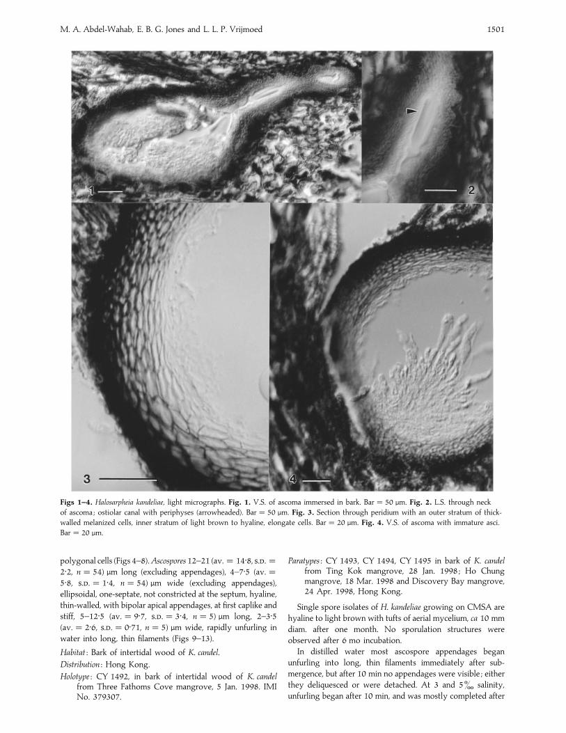
Ascomata 200–310 (av.¯ 234±6, ..¯ 55±4, n¯ 12) µmhigh,
120–180 (av.¯ 156±7, ..¯ 22±5, n¯ 12) µm wide, el-
lipsoidal, horizontal, black upper side, light brown bottom,
immersed under blackened bark, membranous, ostiolate and
papillate (Fig. 1). Peridium 25–42 (av.¯ 32±7, ..¯ 4±7, n¯9) µm thick, two layered, forming textura angularis, outer
stratum 10–15 (av.¯ 11±7, ..¯ 1±8, n¯ 9) µm thick, 3–5
layers of polygonal dark brown melanized cells ; inner stratum
15–28 (av.¯ 20±8, ..¯ 4±8, n¯ 9) µm thick, 5–7 layers of
elongated polygonal, light brown cells, merging with the
pseudoparenchyma of the venter (Fig. 3). Necks lateral bending
upward, 110–137 (av.¯ 129±5, ..¯ 47±5, n¯ 7) µm long,
40–77 (av.¯ 63±4, ..¯ 12±9, n¯ 7) µm wide, cylindrical,
light brown, ostiolar canal periphysate, periphyses 7±5–12±5(av.¯ 11±5, ..¯ 1±7, n¯ 10) µm long, 0±5–1 (av.¯ 0±9,..¯ 0±19, n¯ 10) µm wide (Fig. 2). Catenophyses present.
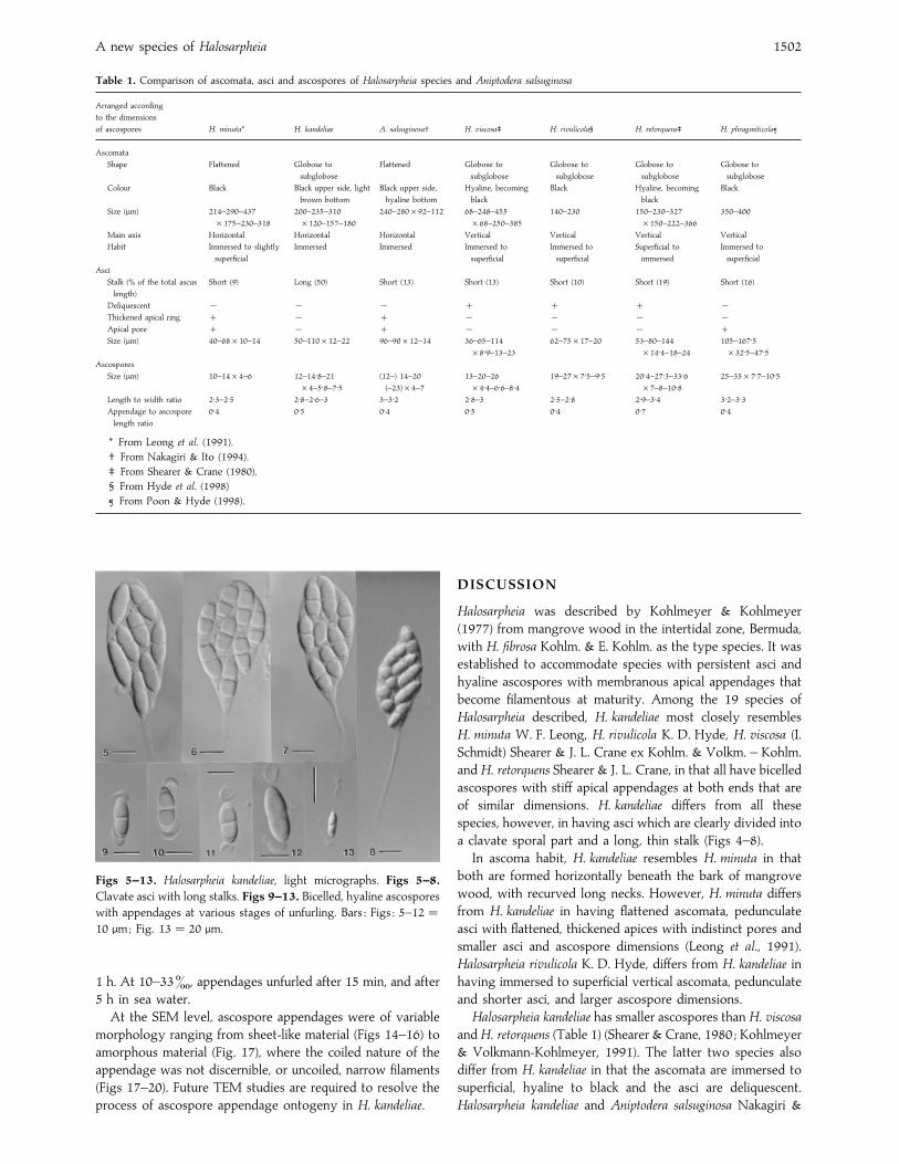
Asci 50–110 (av.¯ 79±0, ..¯ 12±4, n¯ 30) µm long, 12–22
(av.¯ 19±1, ..¯ 2±1, n¯ 30) µm wide, eight-spored, clav-
ate, with narrow long stalks 10–44 (av.¯ 27±5, ..¯ 9±4,n¯ 28) µm long, 1–3 (av.¯ 2±3, ..¯ 0±5, n¯ 28) µm wide,
thin-walled, persistent, without an apical apparatus, developing
at the base of the ascoma venter on small-celled ascogenous
tissue which is separated from the peridium by thin-walled
Mycol. Res. 103 (11) : 1500–1504 (1999) Printed in the United Kingdom
Halosarpheia kandeliae sp. nov. on intertidal bark of themangrove tree Kandelia candel in Hong Kong
M. A. Abdel-Wahab, E. B. G. Jones and L. L. P. Vrijmoed 1501
Figs 1–4. Halosarpheia kandeliae, light micrographs. Fig. 1. V.S. of ascoma immersed in bark. Bar¯ 50 µm. Fig. 2. L.S. through neck
of ascoma ; ostiolar canal with periphyses (arrowheaded). Bar¯ 50 µm. Fig. 3. Section through peridium with an outer stratum of thick-
walled melanized cells, inner stratum of light brown to hyaline, elongate cells. Bar¯ 20 µm. Fig. 4. V.S. of ascoma with immature asci.
Bar¯ 20 µm.
polygonal cells (Figs 4–8).Ascospores 12–21 (av.¯ 14±8, ..¯2±2, n¯ 54) µm long (excluding appendages), 4–7±5 (av.¯5±8, ..¯ 1±4, n¯ 54) µm wide (excluding appendages),
ellipsoidal, one-septate, not constricted at the septum, hyaline,
thin-walled, with bipolar apical appendages, at first caplike and
stiff, 5–12±5 (av.¯ 9±7, ..¯ 3±4, n¯ 5) µm long, 2–3±5(av.¯ 2±6, ..¯ 0±71, n¯ 5) µm wide, rapidly unfurling in
water into long, thin filaments (Figs 9–13).
Habitat : Bark of intertidal wood of K. candel.
Distribution : Hong Kong.
Holotype : CY 1492, in bark of intertidal wood of K. candelfrom Three Fathoms Cove mangrove, 5 Jan. 1998. IMINo. 379307.
Paratypes : CY 1493, CY 1494, CY 1495 in bark of K. candelfrom Ting Kok mangrove, 28 Jan. 1998 ; Ho Chungmangrove, 18 Mar. 1998 and Discovery Bay mangrove,24 Apr. 1998, Hong Kong.
Single spore isolates of H. kandeliae growing on CMSA are
hyaline to light brown with tufts of aerial mycelium, ca 10 mm
diam. after one month. No sporulation structures were
observed after 6 mo incubation.
In distilled water most ascospore appendages began
unfurling into long, thin filaments immediately after sub-
mergence, but after 10 min no appendages were visible ; either
they deliquesced or were detached. At 3 and 5^ salinity,
unfurling began after 10 min, and was mostly completed after
A new species of Halosarpheia 1502
Table 1. Comparison of ascomata, asci and ascospores of Halosarpheia species and Aniptodera salsuginosa
Arranged according
to the dimensions
of ascospores H. minuta* H. kandeliae A. salsuginosa† H. viscosa‡ H. rivulicola§ H. retorquens‡ H. phragmiticola¶
Ascomata
Shape Flattened Globose to
subglobose
Flattened Globose to
subglobose
Globose to
subglobose
Globose to
subglobose
Globose to
subglobose
Colour Black Black upper side, light
brown bottom
Black upper side,
hyaline bottom
Hyaline, becoming
black
Black Hyaline, becoming
black
Black
Size (µm) 214–290–437
¬175–230–318
200–235–310
¬120–157–180
240–280¬92–112 68–248–455
¬68–250–385
140–230 150–230–327
¬150–222–366
350–400
Main axis Horizontal Horizontal Horizontal Vertical Vertical Vertical Vertical
Habit Immersed to slightly
superficial
Immersed Immersed Immersed to
superficial
Immersed to
superficial
Superficial to
immersed
Immersed to
superficial
Asci
Stalk (% of the total ascus
length)
Short (9) Long (50) Short (13) Short (13) Short (10) Short (19) Short (16)
Deliquescent ® ® ® ®Thickened apical ring ® ® ® ® ®Apical pore ® ® ® ® Size (µm) 40–68¬10–14 50–110¬12–22 96–90¬12–14 36–65–114
¬8±9–13–23
62–75¬17–20 53–80–144
¬14±4–18–24
105–167±5¬32±5–47±5
Ascospores
Size (µm) 10–14¬4–6 12–14±8–21
¬4–5±8–7±5(12–) 14–20
(–23)¬4–7
13–20–26
¬4±4–6±6–8±419–27¬7±5–9±5 20±4–27±3–33±6
¬7–8–10±825–35¬7±7–10±5
Length to width ratio 2±3–2±5 2±8–2±6–3 3–3±2 2±8–3 2±5–2±8 2±9–3±4 3±2–3±3Appendage to ascospore
length ratio
0±4 0±5 0±4 0±5 0±4 0±7 0±4
* From Leong et al. (1991).
† From Nakagiri & Ito (1994).
‡ From Shearer & Crane (1980).
§ From Hyde et al. (1998)
¶ From Poon & Hyde (1998).
Figs 5–13. Halosarpheia kandeliae, light micrographs. Figs 5–8.
Clavate asci with long stalks. Figs 9–13. Bicelled, hyaline ascospores
with appendages at various stages of unfurling. Bars : Figs : 5–12¯10 µm; Fig. 13¯ 20 µm.
1 h. At 10–33^, appendages unfurled after 15 min, and after
5 h in sea water.
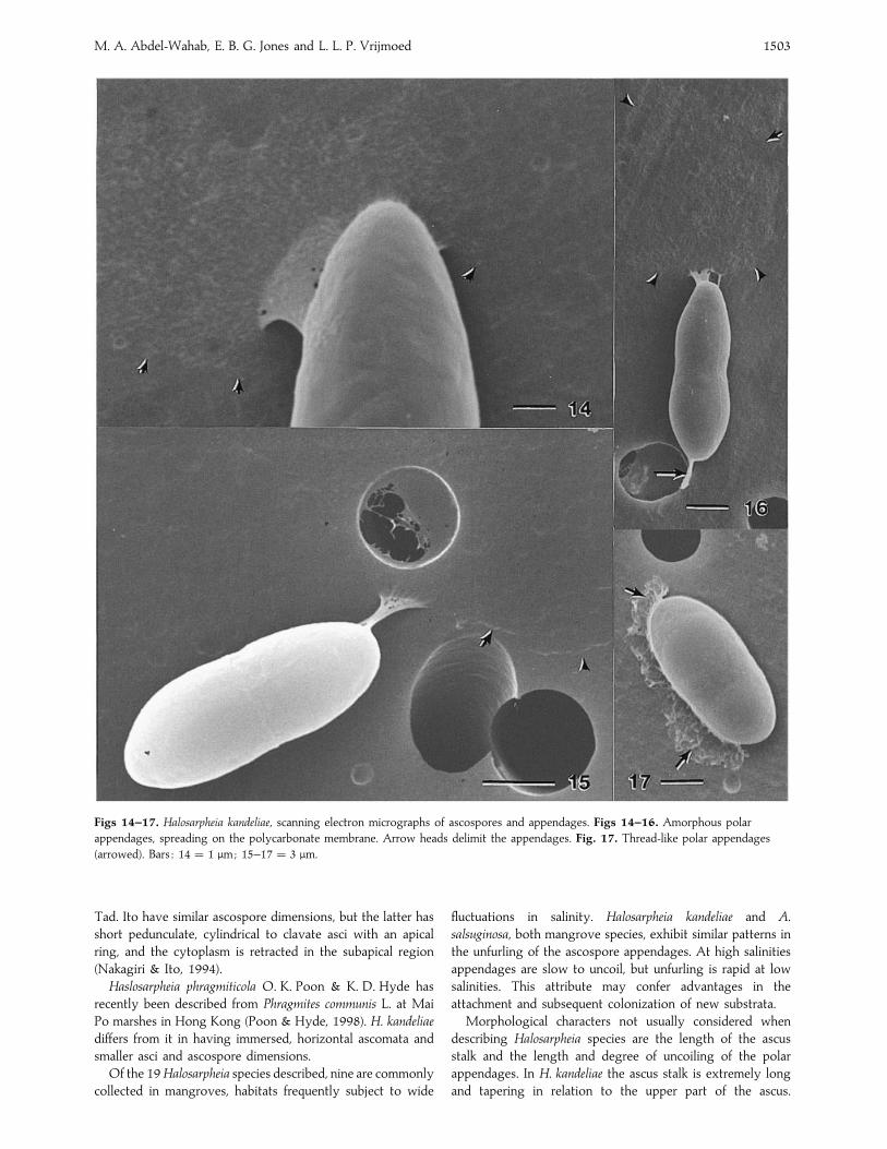
At the SEM level, ascospore appendages were of variable
morphology ranging from sheet-like material (Figs 14–16) to
amorphous material (Fig. 17), where the coiled nature of the
appendage was not discernible, or uncoiled, narrow filaments
(Figs 17–20). Future TEM studies are required to resolve the
process of ascospore appendage ontogeny in H. kandeliae.
DISCUSSION
Halosarpheia was described by Kohlmeyer & Kohlmeyer
(1977) from mangrove wood in the intertidal zone, Bermuda,
with H. fibrosa Kohlm. & E. Kohlm. as the type species. It was
established to accommodate species with persistent asci and
hyaline ascospores with membranous apical appendages that
become filamentous at maturity. Among the 19 species of
Halosarpheia described, H. kandeliae most closely resembles
H. minuta W. F. Leong, H. rivulicola K. D. Hyde, H. viscosa (I.
Schmidt) Shearer & J. L. Crane ex Kohlm. & Volkm. – Kohlm.
and H. retorquens Shearer & J. L. Crane, in that all have bicelled
ascospores with stiff apical appendages at both ends that are
of similar dimensions. H. kandeliae differs from all these
species, however, in having asci which are clearly divided into
a clavate sporal part and a long, thin stalk (Figs 4–8).
In ascoma habit, H. kandeliae resembles H. minuta in that
both are formed horizontally beneath the bark of mangrove
wood, with recurved long necks. However, H. minuta differs
from H. kandeliae in having flattened ascomata, pedunculate
asci with flattened, thickened apices with indistinct pores and
smaller asci and ascospore dimensions (Leong et al., 1991).
Halosarpheia rivulicola K. D. Hyde, differs from H. kandeliae in
having immersed to superficial vertical ascomata, pedunculate
and shorter asci, and larger ascospore dimensions.
Halosarpheia kandeliae has smaller ascospores than H. viscosa
and H. retorquens (Table 1) (Shearer & Crane, 1980 ; Kohlmeyer
& Volkmann-Kohlmeyer, 1991). The latter two species also
differ from H. kandeliae in that the ascomata are immersed to
superficial, hyaline to black and the asci are deliquescent.
Halosarpheia kandeliae and Aniptodera salsuginosa Nakagiri &
M. A. Abdel-Wahab, E. B. G. Jones and L. L. P. Vrijmoed 1503
Figs 14–17. Halosarpheia kandeliae, scanning electron micrographs of ascospores and appendages. Figs 14–16. Amorphous polar
appendages, spreading on the polycarbonate membrane. Arrow heads delimit the appendages. Fig. 17. Thread-like polar appendages
(arrowed). Bars : 14¯ 1 µm; 15–17¯ 3 µm.
Tad. Ito have similar ascospore dimensions, but the latter has
short pedunculate, cylindrical to clavate asci with an apical
ring, and the cytoplasm is retracted in the subapical region
(Nakagiri & Ito, 1994).
Haslosarpheia phragmiticola O. K. Poon & K. D. Hyde has
recently been described from Phragmites communis L. at Mai
Po marshes in Hong Kong (Poon & Hyde, 1998). H. kandeliae
differs from it in having immersed, horizontal ascomata and
smaller asci and ascospore dimensions.
Of the 19 Halosarpheia species described, nine are commonly
collected in mangroves, habitats frequently subject to wide
fluctuations in salinity. Halosarpheia kandeliae and A.
salsuginosa, both mangrove species, exhibit similar patterns in
the unfurling of the ascospore appendages. At high salinities
appendages are slow to uncoil, but unfurling is rapid at low
salinities. This attribute may confer advantages in the
attachment and subsequent colonization of new substrata.
Morphological characters not usually considered when
describing Halosarpheia species are the length of the ascus
stalk and the length and degree of uncoiling of the polar
appendages. In H. kandeliae the ascus stalk is extremely long
and tapering in relation to the upper part of the ascus.
A new species of Halosarpheia 1504
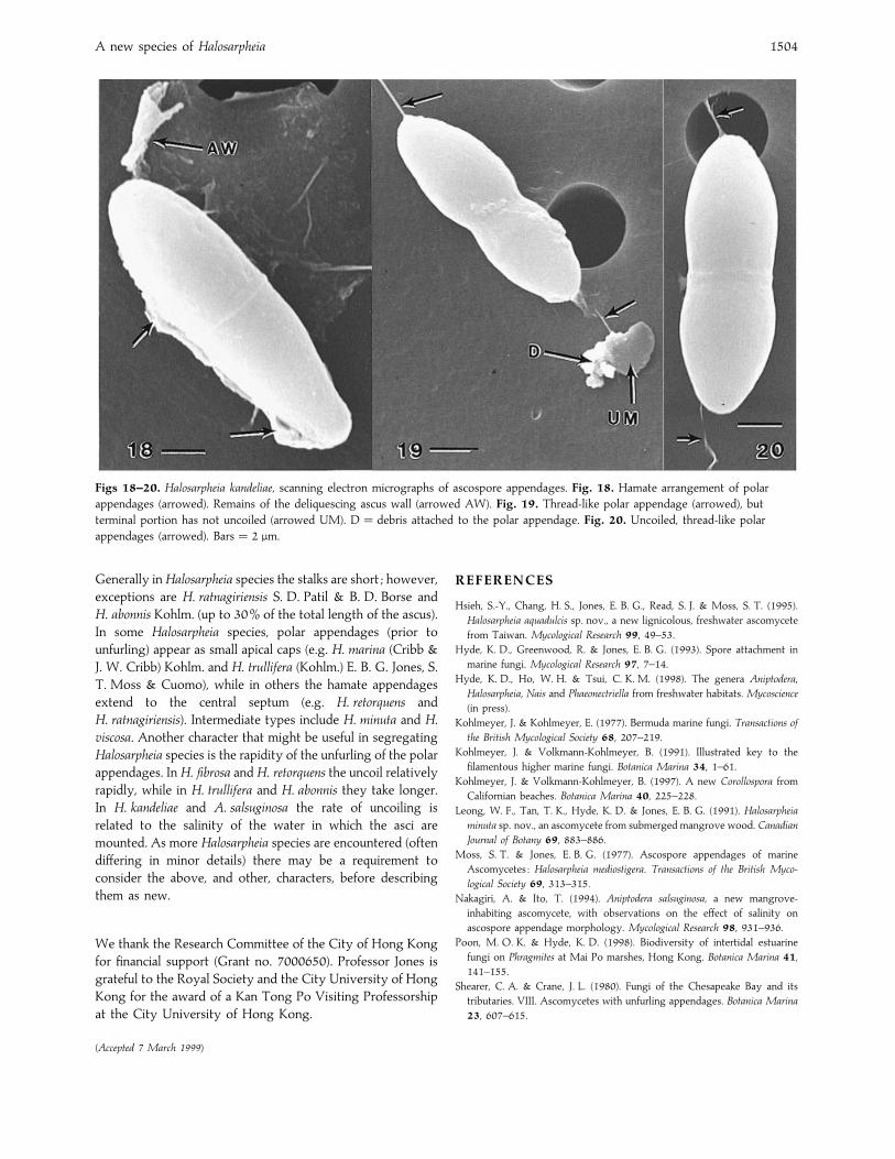
Figs 18–20. Halosarpheia kandeliae, scanning electron micrographs of ascospore appendages. Fig. 18. Hamate arrangement of polar
appendages (arrowed). Remains of the deliquescing ascus wall (arrowed AW). Fig. 19. Thread-like polar appendage (arrowed), but
terminal portion has not uncoiled (arrowed UM). D¯ debris attached to the polar appendage. Fig. 20. Uncoiled, thread-like polar
appendages (arrowed). Bars¯ 2 µm.
Generally in Halosarpheia species the stalks are short ; however,
exceptions are H. ratnagiriensis S. D. Patil & B. D. Borse and
H. abonnis Kohlm. (up to 30% of the total length of the ascus).
In some Halosarpheia species, polar appendages (prior to
unfurling) appear as small apical caps (e.g. H. marina (Cribb &
J. W. Cribb) Kohlm. and H. trullifera (Kohlm.) E. B. G. Jones, S.
T. Moss & Cuomo), while in others the hamate appendages
extend to the central septum (e.g. H. retorquens and
H. ratnagiriensis). Intermediate types include H. minuta and H.
viscosa. Another character that might be useful in segregating
Halosarpheia species is the rapidity of the unfurling of the polar
appendages. In H. fibrosa and H. retorquens the uncoil relatively
rapidly, while in H. trullifera and H. abonnis they take longer.
In H. kandeliae and A. salsuginosa the rate of uncoiling is
related to the salinity of the water in which the asci are
mounted. As more Halosarpheia species are encountered (often
differing in minor details) there may be a requirement to
consider the above, and other, characters, before describing
them as new.
We thank the Research Committee of the City of Hong Kong
for financial support (Grant no. 7000650). Professor Jones is
grateful to the Royal Society and the City University of Hong
Kong for the award of a Kan Tong Po Visiting Professorship
at the City University of Hong Kong.
(Accepted 7 March 1999)
REFERENCES
Hsieh, S.-Y., Chang, H. S., Jones, E. B. G., Read, S. J. & Moss, S. T. (1995).
Halosarpheia aquadulcis sp. nov., a new lignicolous, freshwater ascomycete
from Taiwan. Mycological Research 99, 49–53.
Hyde, K. D., Greenwood, R. & Jones, E. B. G. (1993). Spore attachment in
marine fungi. Mycological Research 97, 7–14.
Hyde, K. D., Ho, W. H. & Tsui, C. K. M. (1998). The genera Aniptodera,
Halosarpheia, Nais and Phaeonectriella from freshwater habitats. Mycoscience
(in press).
Kohlmeyer, J. & Kohlmeyer, E. (1977). Bermuda marine fungi. Transactions of
the British Mycological Society 68, 207–219.
Kohlmeyer, J. & Volkmann-Kohlmeyer, B. (1991). Illustrated key to the
filamentous higher marine fungi. Botanica Marina 34, 1–61.
Kohlmeyer, J. & Volkmann-Kohlmeyer, B. (1997). A new Corollospora from
Californian beaches. Botanica Marina 40, 225–228.
Leong, W. F., Tan, T. K., Hyde, K. D. & Jones, E. B. G. (1991). Halosarpheia
minuta sp. nov., an ascomycete from submerged mangrove wood. Canadian
Journal of Botany 69, 883–886.
Moss, S. T. & Jones, E. B. G. (1977). Ascospore appendages of marine
Ascomycetes : Halosarpheia mediostigera. Transactions of the British Myco-
logical Society 69, 313–315.
Nakagiri, A. & Ito, T. (1994). Aniptodera salsuginosa, a new mangrove-
inhabiting ascomycete, with observations on the effect of salinity on
ascospore appendage morphology. Mycological Research 98, 931–936.
Poon, M. O. K. & Hyde, K. D. (1998). Biodiversity of intertidal estuarine
fungi on Phragmites at Mai Po marshes, Hong Kong. Botanica Marina 41,
141–155.
Shearer, C. A. & Crane, J. L. (1980). Fungi of the Chesapeake Bay and its
tributaries. VIII. Ascomycetes with unfurling appendages. Botanica Marina
23, 607–615.