Embed Size (px)
Citation preview
Halophilic Microorganisms Are Responsible for the RosyDiscolouration of Saline Environments in Three HistoricalBuildings with Mural PaintingsJorg D. Ettenauer1*, Valme Jurado2, Guadalupe Pinar1, Ana Z. Miller2,3, Markus Santner4,
Cesareo Saiz-Jimenez2, Katja Sterflinger1
1 VIBT-BOKU, University of Natural Resources and Life Sciences, Department of Biotechnology, Vienna, Austria, 2 Instituto de Recursos Naturales y Agrobiologia, IRNAS-
CSIC, Sevilla, Spain, 3 CEPGIST/CERENA, Instituto Superior Tecnico, Universidade de Lisboa, Lisboa, Portugal, 4 Bundesdenkmalamt, Abteilung fur Konservierung und
Restaurierung, Vienna, Austria
Abstract
A number of mural paintings and building materials from monuments located in central and south Europe are characterizedby the presence of an intriguing rosy discolouration phenomenon. Although some similarities were observed among thebacterial and archaeal microbiota detected in these monuments, their origin and nature is still unknown. In order to get acomplete overview of this biodeterioration process, we investigated the microbial communities in saline environmentscausing the rosy discolouration of mural paintings in three Austrian historical buildings using a combination of culture-dependent and -independent techniques as well as microscopic techniques. The bacterial communities were dominated byhalophilic members of Actinobacteria, mainly of the genus Rubrobacter. Representatives of the Archaea were also detectedwith the predominating genera Halobacterium, Halococcus and Halalkalicoccus. Furthermore, halophilic bacterial strains,mainly of the phylum Firmicutes, could be retrieved from two monuments using special culture media. Inoculation ofbuilding materials (limestone and gypsum plaster) with selected isolates reproduced the unaesthetic rosy effect andbiodeterioration in the laboratory.
Citation: Ettenauer JD, Jurado V, Pinar G, Miller AZ, Santner M, et al. (2014) Halophilic Microorganisms Are Responsible for the Rosy Discolouration of SalineEnvironments in Three Historical Buildings with Mural Paintings. PLoS ONE 9(8): e103844. doi:10.1371/journal.pone.0103844
Editor: Ali Al-Ahmad, University Hospital of the Albert-Ludwigs-University Freiburg, Germany
Received April 12, 2014; Accepted July 2, 2014; Published August 1, 2014
Copyright: � 2014 Ettenauer et al. This is an open-access article distributed under the terms of the Creative Commons Attribution License, which permitsunrestricted use, distribution, and reproduction in any medium, provided the original author and source are credited.
Data Availability: The authors confirm that all data underlying the findings are fully available without restriction. All relevant data are within the paper and itsSupporting Information files. The ribosomal sequences of the bacterial- and archaeal clones and the bacterial isolates have been deposited at the NCBI GenBankdatabase under the accession numbers (KF692550–KF692709 for the cloned sequences and HG515390–HG515401 for the bacterial isolates).
Funding: JE, GP and KS were partly financially supported with funding provided from the Ministry of Care of Monuments in Austria (Bundesdenkmalamt). Furtherfunding was provided by the University of Natural Resources and Life Sciences (Vienna). CSJ, VJ and AZM acknowledge support from project Consolider projectfrom the Spanish Ministry (CSD) 2007-00058. The funders had no role in study design, data collection and analysis, decision to publish, or preparation of themanuscript.
Competing Interests: The authors have declared that no competing interests exist.
* Email: [email protected]
Introduction
It is well-known that microorganisms play a crucial role in the
degradation and deterioration of mural paintings and building
materials. Stone materials and wall paintings provide a great
variety of ecological niches for all types of microorganisms that can
induce biodeterioration. Biodegradation is caused by biochemical
processes, through bio-corrosion, dissolution and solubilization of
material components, however aesthetical effects are often more
evident in some biodeterioration processes. The aesthetical
changes are triggered by the deterioration of painting pigments
on walls and/or by the formation of coloured biofilms or excretion
of extracellular pigments. Fungi, algae, different bacteria and
archaea produce a wide variety of biogenic pigments such as
chlorophyll, carotenes, phenols, anthraquinones and melanin with
colours ranging from light yellow, orange, pink, purple, violet,
green, grey, dark brown to black [1–5]. The formation of orange
to red pigments is due to the production of carotenes as a means of
protecting the cells against high UV-radiation, chemical- and/or
salt stress [6]. On salty walls the inhabiting halophilic bacteria and
haloarchaea usually form pink to purple or violet stains. Orange
pigmentations on sandstone or marble often resemble iron oxide
and therefore it has to be clarified if the discolouration is due to
microbial growth [7,8]. The biogenic pigments are usually very
stable on the materials even if the causative microorganisms are
already dead.
In this study we investigated the pink to rosy discolouration
phenomenon presented by two historical chapels and a medieval
castle located in Austria (Figure S1). The Johannes Chapel in
Purgg (Styria) was built in Romanesque style and dates back to the
12th century. The frescos inside with the famous and mysterious
motive of the ‘‘cats-mice-war’’ was one of the most prominent and
well-preserved Romanesque paintings in Europe, dated to 1160
(Figure 1A). After constructional changes on the whole west wall a
rosy biofilm established which further spread across the whole
chapel (Figure 1B).
The castle of Rappottenstein (Lower Austria) is a medieval
castle, which was never conquered and is, therefore, one of the
most well-preserved castles in Austria. It dates back to 12th
century and combines three constructional ages with correspon-
PLOS ONE | www.plosone.org 1 August 2014 | Volume 9 | Issue 8 | e103844
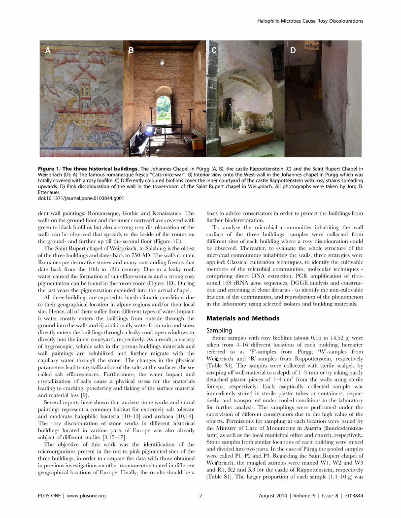
dent wall paintings: Romanesque, Gothic and Renaissance. The
walls on the ground floor and the inner courtyard are covered with
green to black biofilms but also a strong rosy discolouration of the
walls can be observed that spreads to the inside of the rooms on
the ground- and further up till the second floor (Figure 1C).
The Saint Rupert chapel of Wei priach, in Salzburg is the oldest
of the three buildings and dates back to 750 AD. The walls contain
Romanesque decorative stones and many outstanding frescos that
date back from the 10th to 13th century. Due to a leaky roof,
water caused the formation of salt efflorescences and a strong rosy
pigmentation can be found in the tower room (Figure 1D). During
the last years the pigmentation extended into the actual chapel.
All three buildings are exposed to harsh climatic conditions due
to their geographical location in alpine regions and/or their local
site. Hence, all of them suffer from different types of water impact:
i) water mostly enters the buildings from outside through the
ground into the walls and ii) additionally water from rain and snow
directly enters the buildings through a leaky roof, open windows or
directly into the inner courtyard, respectively. As a result, a variety
of hygroscopic, soluble salts in the porous buildings materials and
wall paintings are solubilized and further migrate with the
capillary water through the stone. The changes in the physical
parameters lead to crystallization of the salts at the surfaces, the so-
called salt efflorescences. Furthermore, the water impact and
crystallization of salts cause a physical stress for the materials
leading to cracking, powdering and flaking of the surface material
and material loss [9].
Several reports have shown that ancient stone works and mural
paintings represent a common habitat for extremely salt tolerant
and moderate halophilic bacteria [10–13] and archaea [10,14].
The rosy discolouration of stone works in different historical
buildings located in various parts of Europe was also already
subject of different studies [3,15–17].
The objective of this work was the identification of the
microorganisms present in the red to pink pigmented sites of the
three buildings, in order to compare the data with those obtained
in previous investigations on other monuments situated in different
geographical locations of Europe. Finally, the results should be a
basis to advice conservators in order to protect the buildings from
further biodeterioration.
To analyse the microbial communities inhabiting the wall
surface of the three buildings, samples were collected from
different sites of each building where a rosy discolouration could
be observed. Thereafter, to evaluate the whole structure of the
microbial communities inhabiting the walls, three strategies were
applied: Classical cultivation techniques, to identify the cultivable
members of the microbial communities, molecular techniques -
comprising direct DNA extraction, PCR amplification of ribo-
somal 16S rRNA gene sequences, DGGE analysis and construc-
tion and screening of clone libraries - to identify the non-cultivable
fraction of the communities, and reproduction of the phenomenon
in the laboratory using selected isolates and building materials.
Materials and Methods
SamplingStone samples with rosy biofilms (about 0.16 to 14.52 g) were
taken from 4–16 different locations of each building, hereafter
referred to as ‘P’-samples from Purgg, ‘W’-samples from
Wei priach and ‘R’-samples from Rappottenstein, respectively
(Table S1). The samples were collected with sterile scalpels by
scraping off wall material to a depth of 1–3 mm or by taking partly
detached plaster pieces of 1–4 cm2 from the walls using sterile
forceps, respectively. Each aseptically collected sample was
immediately stored in sterile plastic tubes or containers, respec-
tively, and transported under cooled conditions to the laboratory
for further analysis. The samplings were performed under the
supervision of different conservators due to the high value of the
objects. Permissions for sampling at each location were issued by
the Ministry of Care of Monuments in Austria (Bundesdenkma-
lamt) as well as the local municipal office and church, respectively.
Stone samples from similar locations of each building were mixed
and divided into two parts. In the case of Purgg the pooled samples
were called P1, P2 and P3. Regarding the Saint Rupert chapel of
Wei priach, the mingled samples were named W1, W2 and W3
and R1, R2 and R3 for the castle of Rappottenstein, respectively
(Table S1). The larger proportion of each sample (1.4–10 g) was
Figure 1. The three historical buildings. The Johannes Chapel in Purgg (A, B), the castle Rappottenstein (C) and the Saint Rupert Chapel inWei priach (D): A) The famous romanesque fresco ’’Cats-mice-war‘‘. B) Interior view onto the West-wall in the Johannes chapel in Purgg which wastotally covered with a rosy biofilm. C) Differently coloured biofilms cover the inner courtyard of the castle Rappottenstein with rosy strains spreadingupwards. D) Pink discolouration of the wall in the tower-room of the Saint Rupert chapel in Wei priach. All photographs were taken by Jorg D.Ettenauer.doi:10.1371/journal.pone.0103844.g001
Halophilic Microbes Cause Rosy Discolourations
PLOS ONE | www.plosone.org 2 August 2014 | Volume 9 | Issue 8 | e103844
sent under cooled conditions to Spain for cultivation analyses and
the smaller part was used for molecular analyses.
Cultivation analysisAll samples were inoculated at 28uC for 30 days on different
culture media: trypticase soy agar supplemented with Na and Mg
(TSA Na-Mg [18], TSA Na-Mg supplemented with 15% NaCl
(w/v) instead of 3% NaCl (w/v), nutrient agar (Difco, Becton
Dickinson, Sparks MD, USA) diluted 1:100, nutrient agar
supplemented with NaCl (3%, w/v), marine agar 2216 (Difco,
Becton Dickinson, Sparks MD, USA), DSMZ media 372 (http://
www.dsmz.de/microorganisms/medium/pdf/DSMZ_Medium372.
pdf) and 1018 (http://www.dsmz.de/microorganisms/medium/
pdf/DSMZ_Medium1018.pdf). The last two culture media were
specific for Archaea. All orange or pink pigmented colonies that
might be responsible for the aesthetical damage of the objects were
picked up and transferred to fresh medium.
Molecular characterization of the isolated strainsBacterial DNA was extracted following the method described by
Marmur [19]. The 16S rRNA gene was amplified by PCR using
the conserved primers 27F [20] and 1522R [21] with the following
PCR thermal conditions: 95uC for 1 min; 35 cycles of 95uC for
15 s, 55uC for 15 s, 72uC for 2 min; and a final extension cycle at
72uC for 10 min. Forward and reverse strands of the amplified
DNA fragment were sequenced in an ABI 3700 sequencer
(Applied Biosystems). The identification of phylogenetic neigh-
bours was carried out by the BLAST program [22] against the
NCBI database and the database of type strains EZtaxon [23] with
validly published prokaryotic names.
Molecular analysis - DNA extraction from the wallsThe mingled samples of each building were ground for 2
minutes in liquid nitrogen using a sterile mortar and pestle. From
the homogenized material each 100 mg were weighed in a
Sartorius precision scale for DNA extraction. The complete
microbial DNA was directly isolated using the FastDNA SPIN Kit
for soil from MP Biomedicals (Illkrich, France). The DNA
concentration, -quality and -purity, respectively, was measured
using a NanoDrop ND-1000 spectrophotometer (peqLabBiotech-
nologie GmbH, Linz, Austria). Additionally, the extracted DNA
was visualized on 1.5 (w/v) agarose gels at 110V for 40 minutes,
stained in an ethidium bromide solution [1 mg ml21; stock: 10 mg
ml21] for 20 minutes and documented using an UVP documen-
tation system (BioRad Transilluminator, Universal Hood; Mitsu-
bishi P93D-printer).
PCR amplification of bacterial 16S rDNA fragmentsAll PCR reactions were executed in a BioRad C1000 Thermal
Cycler. The 26 PCR Master Mix (Promega, Mannheim,
Germany) [50 units ml21 of TaqDNA Polymerase in a supplied
reaction buffer (pH 8.5), 400 mM dATP, 400 mM dGTP, 400 mM
dCTP, 400 mM dTTP, 3 mM MgCl2] was diluted to 16,
12.5 pmol of each primer and 2.5 ml of template DNA were
added to 25 ml total reaction volumes.
For genetic fingerprinting of the eubacterial 16S rDNA
fragments by DGGE two different PCR reactions were performed.
For the first round the universal primers 341f [24] and 907r [25]
were used. The second round, a semi-nested PCR for genetic
fingerprints, was done using the primers 341GC and 518r [26].
The forward primer possesses a 40-base Guanine-Cytosine (GC)
clamp at its 59 end that stabilizes the melting behaviour of the
DNA fragments in DGGE analysis [24]. The semi-nested PCR
was executed in 100 ml volumes, separated into two tubes to which
each 50 ml mastermix, 25 pmol of each primer and 3.5 ml of
template were added. The thermocycling conditions described by
Schabereiter-Gurtner et al. [27] were used for genetic fingerprint-
ing. Seven microliter of each PCR product was electrophoresed on
a 2% (w/v) agarose gel as described above. In each PCR reaction
a negative control was included, where no DNA template was
added, to exclude the possibility of cross-contamination.
PCR amplification of archaeal 16S rDNA fragmentsThe amplification of archaeal 16S rDNA fragments was carried
out similar to the bacterial PCR analysis with the addition of BSA
(25 pmol in 25 ml reaction volume) to the mastermix. For the first
round the primers ARC344 and ARC915 [28] were applied, using
the thermocycling program described by Pinar et al. [29]. To
obtain genetic fingerprints a semi-nested PCR was performed with
primers 518r carrying a GC clamp at its 59 end [24] and the
archaea specific primer ARC344. The same cycling conditions
were used as described for the amplification of the bacterial 16S
rDNA.
Fingerprint analysis by DGGE – Denaturing Gradient GelElectrophoresis
For DGGE fingerprinting 100 ml PCR products from the semi-
nested PCR were pooled, precipitated overnight with 96% ethanol
at 220uC and re-suspended in 20 ml ultra-pure water (Qiagen
GmbH, Hilden, Germany). The concentrated PCR products
supplemented with 5 ml 66 Loading Dye Solution (Thermo
Scientific) were separated on gels in 0.56 TAE buffer [20 mM
Tris, 10 mM acetate, 0.5 mM Na2EDTA; pH 8.0] for 3.5 hours
at 200 V and 60uC in a Bio-Rad-DCode – Universal Mutation
Detection System [24]. A linear chemical gradient ranging from
35 to 55% of urea and formamide in an 8% (w/v) polyacrylamide
gel (Bio-Rad, Munich, Germany) for screening of bacterial
communities and from 35 to 50% for separation of bands of the
archaeal population was used. After completion of electrophoresis
staining of the gels was done in an ethidium bromide solution for
20 minutes and afterwards visualized by a UVP documentation
system.
Construction of 16S rDNA clone libraries and screeningby DGGE
In order to obtain phylogenetic identification data on the
inhabiting microorganisms, two clone libraries of each sample
containing the bacterial or the archaeal 16S rDNA fragments,
respectively, were created. Therefore, 263.5 ml DNA templates of
each sample were amplified in 2650 ml reaction volumes using the
primers for the first round as mentioned above. Aliquots of the
PCR products were electrophoresed, purified using the QIAquick
PCR Purification Kit (Qiagen GmbH, Hilden, Germany) and re-
suspended in 30 ml ultra-pure water. The purified DNA was again
analysed by gel electrophoresis and 5.5 ml were used as a ligation
template for the pGEM-T easy Vector system (Promega). The
ligation products were transformed into One Shot TOP10 cells
(Invitrogen, Carlsbad, USA) according to the manufacturer’s
instructions. Recombinant cells (white colonies) could be identified
on indicator LB medium containing ampicilline (100 mg ml21),
streptomycine (25 mg ml21) and X-Gal (5-bromo-4-chloro-3-
indolyl-b-D-galactopyranoside; 0.1 mM) [30].
About 50–150 white colonies from each clone library were
harvested and screened by DGGE as described by Schabereiter-
Gurtner et al. [27]. The band positions of the clones were
Halophilic Microbes Cause Rosy Discolourations
PLOS ONE | www.plosone.org 3 August 2014 | Volume 9 | Issue 8 | e103844
compared with the DGGE fingerprint of the original sample and
inserts of clones matching dominant- and faint bands of the
banding profile of the original sample were selected for
sequencing.
16S rDNA sequencing and sequence analysisIn 2650 ml reaction volumes with each 3 ml template DNA of
the clone inserts were amplified using the vector specific primers
SP6 and T7 (Promega, Mannheim, Germany) [27]. After
visualization on agarose gels and purification of the pooled PCR
products, 25 ml aliquots were sent to GATC Biotech sequencing
service (www.gatc-biotech.com). Comparative sequence analysis
was done by comparing pair-wise insert sequences with those
available in the online databases provided by the NCBI (National
Centre for Biotechnology Information), and RDP (Ribosomal
Database Project), respectively, using the search program BLAST
[22]. The ribosomal sequences of the bacterial- and archaeal
clones and the bacterial isolates have been deposited at the NCBI
GenBank database under the accession numbers (KF692550–
KF692709 for the cloned sequences and HG515390–HG515401
for the bacterial isolates) listed in Tables S2, S3 and S4, for each
16S rDNA sequence.
Laboratory-based colonization experiment of buildingmaterials
Strains of Halobacillus naozhouensis and Kocuria polaris were
used for inoculation and reproduction of biodeterioration
processes in the laboratory. Gypsum plasterand Hontoria lime-
stone [31] were sliced into squares of 36360.5 cm, sterilized in an
autoclave under fluent vapor and inoculated with suspensions of
each bacterium. Probes of gypsum plaster and limestone were
inoculated with three different suspensions of cells (A: Halobacillusnaozhouensis, B: Kocuria polaris and C: mixture of both strains) at
concentrations of 1.56109 cells ml21. All probes were inoculated
with 150 mL of suspension and incubated at 30uC for one month.
Field Emission Scanning Electron Microscopy (FESEM)FESEM was used to accurately assess surface topography,
microbial growth and biodeterioration phenomena on the
inoculated gypsum plaster and limestone probes. Bulk fragments
were directly mounted on a sample stub and sputter coated with a
thin gold/palladium film. Subsequently, samples were examined
on a Jeol JSM-7001F microscope equipped with an Oxford X-ray
energy dispersive spectroscopy (EDS) detector. FESEM examina-
tions were operated in secondary electron (SE) detection mode
with an acceleration potential of 15 kV.
Results
Phylogenetic identification of the cultivatedmicroorganisms
During the first 48 hours of incubation up to a maximum of 12
days, twenty-nine bacterial strains with yellow to orange or pink
appearance were isolated. Coloured bacterial strains isolated from
sample P2 of the Johannes Chapel in Purgg represented 41.4% of
all isolated strains, and, on average, from all three samples of the
castle Rappottenstein 19.5%. No cultivable bacteria were found
on samples from Wei priach, which might be due to the known
difficulties in culturing these pigmented, halophilic microorgan-
isms and perhaps also due to the low number of samples as well as
very small sample amounts that could be taken from this location
(Table S1). Similarly, no archaea could be isolated from none of
the samples. The molecular identification of the isolated strains is
shown in Table 1 and also in Table S2 where the detailed
information about each isolate, its closest related neighbour, the
isolation source as well as the accession number for the submitted
16S rDNA sequence are given. According to the NCBI database,
the bacterial strains showed similarity values ranging from 98 to
100% and could be grouped to cultured members of three
different bacterial phyla, namely the Firmicutes (89.7% of all
isolated strains), the Proteobacteria (6.9%) and the Actinobacteria
(3.5%).
Out of the twelve isolates obtained from sample P2, two isolates
from Purgg were closely affiliated with Planococcus salinarum(98%) and the other bacterial strains were highly related to
Halobacillus herbersteinensis (99%) - all belonging to the phylum
Firmicutes (Table S2).
From the castle of Rappottenstein seventeen isolates could be
grown on the used culture media: ten bacteria from sample R1, six
from sample R2 and one from sample R3. Five isolates from
sample R1 showed the best match in the NCBI database search
with Halobacillus herbersteinensis (99%). Further, three bacteria
were affiliated with Marinococcus luteus (99%) and one bacterium
with Paracoccus marcusii (100%), - being all representatives of the
phylum Firmicutes. Only one strain showed to be related to
members of the Proteobacteria, namely to Halomonas muralis(98%). Out of the six cultured bacteria from sample R2, five
isolates affiliated also with members of the Firmicutes. Two of
them were related to cultivable Planomicrobium flavidum (98%),
another one to Planococcus psychrotoleratus (99%), one to
Planococcus antarcticus (99%) and one to Planococcus donghaen-sis (99%). The last strain of sample R2 was the only member of the
Actinobacteria, which could be identified as Kocuria rosea (99%).
The only isolate obtained from sample R3 was similar to
Planococcus psychrotoleratus (99%) belonging to the Firmicutes
phylum (Table S2).
Alternatively, using EZtaxon (a 16S rRNA gene sequence
database of type strains), NCBI affiliations changed and
Halobacillus herbersteinensis was identified as Halobacillusnaozhouensis, Marinococcus luteus as Marinococcus tarijensis,Planococcus psychrotoleratus either as Planococcus okeanokoites or
Planococcus donghaensis, Planococcus antarcticus as Planococcusdonghaensis, and Kocuria rosea as Kocuria polaris (Table S2).
Phylogenetic identification of the microbial communitiesusing molecular techniques
The second part of each mixed sample was subjected to direct
DNA extraction to further elucidate the non-cultivable microbiota.
The bacterial 16S rDNA could be amplified by PCR using
universal primers from all investigated samples. However, the
amplification of archaeal ribosomal DNA was possible for nearly
all samples except from sample P1 of Purgg. DGGE fingerprint
analyses were conducted with the amplified 16S rDNA to obtain
information on the diversity present on the walls. The received
fingerprints of the inhabiting bacterial and archaeal communities
of all investigated samples from the three buildings are shown in
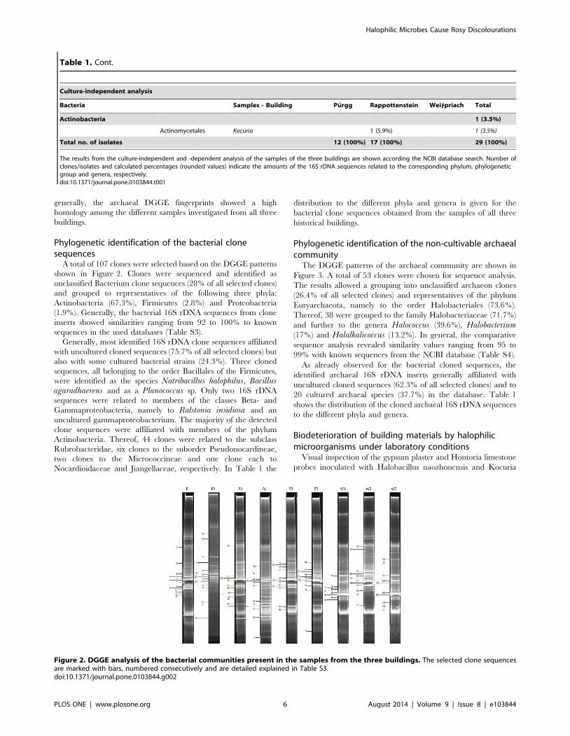
Figures 2 and 3, respectively. The band numbers of all identified
clones are indicated in Tables S3 and S4 and in Figures 2 and 3 to
allow an easy tracking of the corresponding band in the
community profile of each sample.
The bacterial fingerprints showed to be rather complex with
three to seven dominant bands and many faint bands (Figure 2).
The DGGE profiles from the different samples of each building
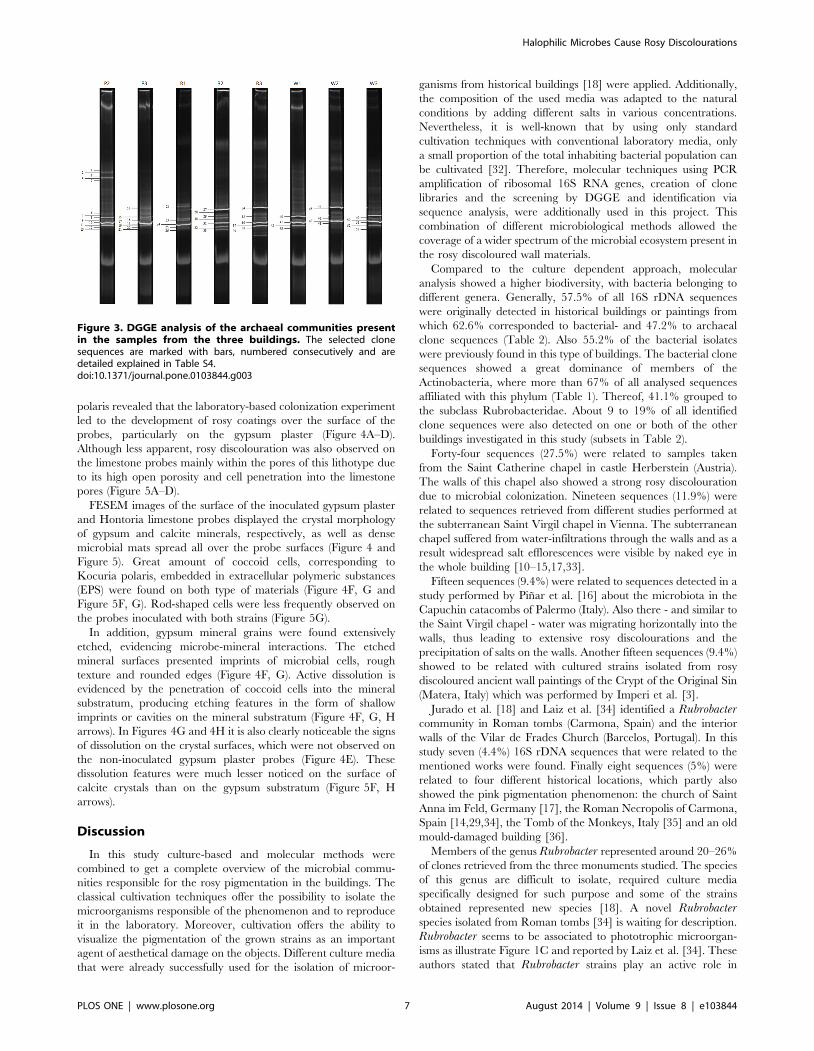
and among the different buildings were very similar. The archaeal
DGGE analysis showed rather simple banding profiles with two to
four dominant bands and only a few faint bands (Figure 3). As
already observed on the DGGE profiles derived from the bacteria,
Halophilic Microbes Cause Rosy Discolourations
PLOS ONE | www.plosone.org 4 August 2014 | Volume 9 | Issue 8 | e103844
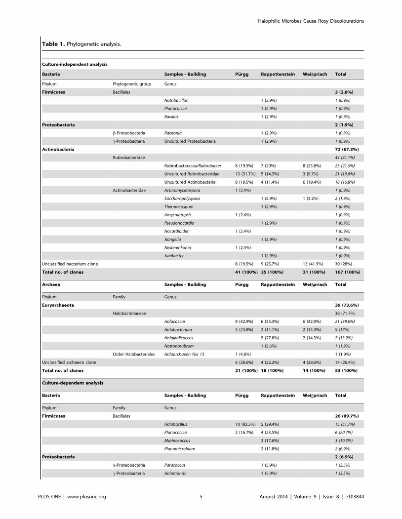
Table 1. Phylogenetic analysis.
Culture-independent analysis
Bacteria Samples - Building Purgg Rappottenstein Wei priach Total
Phylum Phylogenetic group Genus
Firmicutes Bacillales 3 (2.8%)
Natribacillus 1 (2.9%) 1 (0.9%)
Planococcus 1 (2.9%) 1 (0.9%)
Bacillus 1 (2.9%) 1 (0.9%)
Proteobacteria 2 (1.9%)
b-Proteobacteria Ralstonia 1 (2.9%) 1 (0.9%)
c-Proteobacteria Uncultured Proteobacteria 1 (2.9%) 1 (0.9%)
Actinobacteria 72 (67.3%)
Rubrobacteridae 44 (41.1%)
Rubrobacteracea/Rubrobacter 8 (19.5%) 7 (20%) 8 (25.8%) 23 (21.5%)
Uncultured Rubrobacteridae 13 (31.7%) 5 (14.3%) 3 (9.7%) 21 (19.6%)
Uncultured Actinobacteria 8 (19.5%) 4 (11.4%) 6 (19.4%) 18 (16.8%)
Actinobacteridae Actinomycetospora 1 (2.4%) 1 (0.9%)
Saccharopolyspora 1 (2.9%) 1 (3.2%) 2 (1.9%)
Thermocrispum 1 (2.9%) 1 (0.9%)
Amycolatopsis 1 (2.4%) 1 (0.9%)
Pseudonocardia 1 (2.9%) 1 (0.9%)
Nocardioides 1 (2.4%) 1 (0.9%)
Jiangella 1 (2.9%) 1 (0.9%)
Nesterenkonia 1 (2.4%) 1 (0.9%)
Janibacter 1 (2.9%) 1 (0.9%)
Unclassified bacterium clone 8 (19.5%) 9 (25.7%) 13 (41.9%) 30 (28%)
Total no. of clones 41 (100%) 35 (100%) 31 (100%) 107 (100%)
Archaea Samples - Building Purgg Rappottenstein Wei priach Total
Phylum Family Genus
Euryarchaeota 39 (73.6%)
Halobacteriaceae 38 (71.7%)
Halococcus 9 (42.9%) 6 (33.3%) 6 (42.9%) 21 (39.6%)
Halobacterium 5 (23.8%) 2 (11.1%) 2 (14.3%) 9 (17%)
Halalkalicoccus 5 (27.8%) 2 (14.3%) 7 (13.2%)
Natronorubrum 1 (5.6%) 1 (1.9%)
Order Halobacteriales Haloarchaeon Nie 13 1 (4.8%) 1 (1.9%)
Unclassified archaeon clone 6 (28.6%) 4 (22.2%) 4 (28.6%) 14 (26.4%)
Total no. of clones 21 (100%) 18 (100%) 14 (100%) 53 (100%)
Culture-dependent analysis
Bacteria Samples - Building Purgg Rappottenstein Wei priach Total
Phylum Family Genus
Firmicutes Bacillales 26 (89.7%)
Halobacillus 10 (83.3%) 5 (29.4%) 15 (51.7%)
Planococcus 2 (16.7%) 4 (23.5%) 6 (20.7%)
Marinococcus 3 (17.6%) 3 (10.3%)
Planomicrobium 2 (11.8%) 2 (6.9%)
Proteobacteria 2 (6.9%)
a-Proteobacteria Paracoccus 1 (5.9%) 1 (3.5%)
c-Proteobacteria Halomonas 1 (5.9%) 1 (3.5%)
Halophilic Microbes Cause Rosy Discolourations
PLOS ONE | www.plosone.org 5 August 2014 | Volume 9 | Issue 8 | e103844
generally, the archaeal DGGE fingerprints showed a high
homology among the different samples investigated from all three
buildings.
Phylogenetic identification of the bacterial clonesequences
A total of 107 clones were selected based on the DGGE patterns
shown in Figure 2. Clones were sequenced and identified as
unclassified Bacterium clone sequences (28% of all selected clones)
and grouped to representatives of the following three phyla:
Actinobacteria (67.3%), Firmicutes (2.8%) and Proteobacteria
(1.9%). Generally, the bacterial 16S rDNA sequences from clone
inserts showed similarities ranging from 92 to 100% to known
sequences in the used databases (Table S3).
Generally, most identified 16S rDNA clone sequences affiliated
with uncultured cloned sequences (75.7% of all selected clones) but
also with some cultured bacterial strains (24.3%). Three cloned
sequences, all belonging to the order Bacillales of the Firmicutes,
were identified as the species Natribacillus halophilus, Bacillusagaradhaerens and as a Planococcus sp. Only two 16S rDNA
sequences were related to members of the classes Beta- and
Gammaproteobacteria, namely to Ralstonia insidiosa and an
uncultured gammaproteobacterium. The majority of the detected
clone sequences were affiliated with members of the phylum
Actinobacteria. Thereof, 44 clones were related to the subclass
Rubrobacteridae, six clones to the suborder Pseudonocardineae,
two clones to the Micrococcineae and one clone each to
Nocardioidaceae and Jiangellaceae, respectively. In Table 1 the
distribution to the different phyla and genera is given for the
bacterial clone sequences obtained from the samples of all three
historical buildings.
Phylogenetic identification of the non-cultivable archaealcommunity
The DGGE patterns of the archaeal community are shown in
Figure 3. A total of 53 clones were chosen for sequence analysis.
The results allowed a grouping into unclassified archaeon clones
(26.4% of all selected clones) and representatives of the phylum
Euryarchaeota, namely to the order Halobacteriales (73.6%).
Thereof, 38 were grouped to the family Halobacteriaceae (71.7%)
and further to the genera Halococcus (39.6%), Halobacterium(17%) and Halalkalicoccus (13.2%). In general, the comparative
sequence analysis revealed similarity values ranging from 95 to
99% with known sequences from the NCBI database (Table S4).
As already observed for the bacterial cloned sequences, the
identified archaeal 16S rDNA inserts generally affiliated with
uncultured cloned sequences (62.3% of all selected clones) and to
20 cultured archaeal species (37.7%) in the database. Table 1
shows the distribution of the cloned archaeal 16S rDNA sequences
to the different phyla and genera.
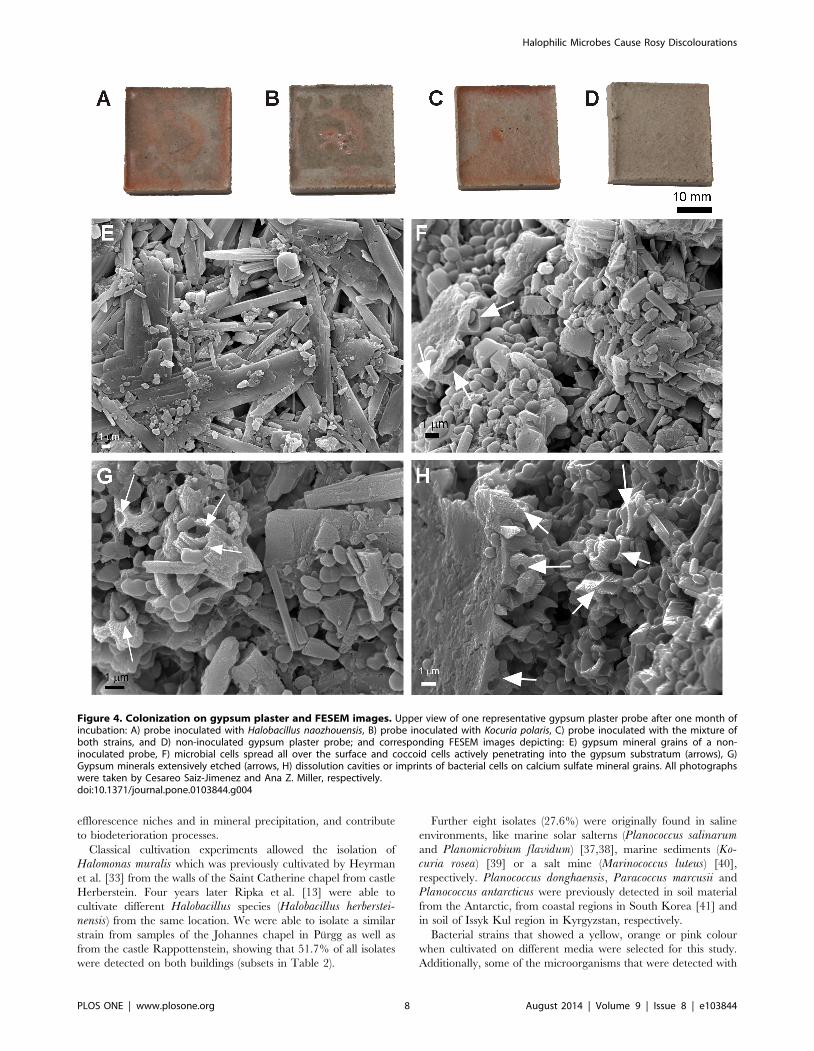
Biodeterioration of building materials by halophilicmicroorganisms under laboratory conditions
Visual inspection of the gypsum plaster and Hontoria limestone
probes inoculated with Halobacillus naozhouensis and Kocuria
Table 1. Cont.
Culture-independent analysis
Bacteria Samples - Building Purgg Rappottenstein Wei priach Total
Actinobacteria 1 (3.5%)
Actinomycetales Kocuria 1 (5.9%) 1 (3.5%)
Total no. of isolates 12 (100%) 17 (100%) 29 (100%)
The results from the culture-independent and -dependent analysis of the samples of the three buildings are shown according the NCBI database search. Number ofclones/isolates and calculated percentages (rounded values) indicate the amounts of the 16S rDNA sequences related to the corresponding phylum, phylogeneticgroup and genera, respectively.doi:10.1371/journal.pone.0103844.t001
Figure 2. DGGE analysis of the bacterial communities present in the samples from the three buildings. The selected clone sequencesare marked with bars, numbered consecutively and are detailed explained in Table S3.doi:10.1371/journal.pone.0103844.g002
Halophilic Microbes Cause Rosy Discolourations
PLOS ONE | www.plosone.org 6 August 2014 | Volume 9 | Issue 8 | e103844
polaris revealed that the laboratory-based colonization experiment
led to the development of rosy coatings over the surface of the
probes, particularly on the gypsum plaster (Figure 4A–D).
Although less apparent, rosy discolouration was also observed on
the limestone probes mainly within the pores of this lithotype due
to its high open porosity and cell penetration into the limestone
pores (Figure 5A–D).
FESEM images of the surface of the inoculated gypsum plaster
and Hontoria limestone probes displayed the crystal morphology
of gypsum and calcite minerals, respectively, as well as dense
microbial mats spread all over the probe surfaces (Figure 4 and
Figure 5). Great amount of coccoid cells, corresponding to
Kocuria polaris, embedded in extracellular polymeric substances
(EPS) were found on both type of materials (Figure 4F, G and
Figure 5F, G). Rod-shaped cells were less frequently observed on
the probes inoculated with both strains (Figure 5G).
In addition, gypsum mineral grains were found extensively
etched, evidencing microbe-mineral interactions. The etched
mineral surfaces presented imprints of microbial cells, rough
texture and rounded edges (Figure 4F, G). Active dissolution is
evidenced by the penetration of coccoid cells into the mineral
substratum, producing etching features in the form of shallow
imprints or cavities on the mineral substratum (Figure 4F, G, H
arrows). In Figures 4G and 4H it is also clearly noticeable the signs
of dissolution on the crystal surfaces, which were not observed on
the non-inoculated gypsum plaster probes (Figure 4E). These
dissolution features were much lesser noticed on the surface of
calcite crystals than on the gypsum substratum (Figure 5F, H
arrows).
Discussion
In this study culture-based and molecular methods were
combined to get a complete overview of the microbial commu-
nities responsible for the rosy pigmentation in the buildings. The
classical cultivation techniques offer the possibility to isolate the
microorganisms responsible of the phenomenon and to reproduce
it in the laboratory. Moreover, cultivation offers the ability to
visualize the pigmentation of the grown strains as an important
agent of aesthetical damage on the objects. Different culture media
that were already successfully used for the isolation of microor-
ganisms from historical buildings [18] were applied. Additionally,
the composition of the used media was adapted to the natural
conditions by adding different salts in various concentrations.
Nevertheless, it is well-known that by using only standard
cultivation techniques with conventional laboratory media, only
a small proportion of the total inhabiting bacterial population can
be cultivated [32]. Therefore, molecular techniques using PCR
amplification of ribosomal 16S RNA genes, creation of clone
libraries and the screening by DGGE and identification via
sequence analysis, were additionally used in this project. This
combination of different microbiological methods allowed the
coverage of a wider spectrum of the microbial ecosystem present in
the rosy discoloured wall materials.
Compared to the culture dependent approach, molecular
analysis showed a higher biodiversity, with bacteria belonging to
different genera. Generally, 57.5% of all 16S rDNA sequences
were originally detected in historical buildings or paintings from
which 62.6% corresponded to bacterial- and 47.2% to archaeal
clone sequences (Table 2). Also 55.2% of the bacterial isolates
were previously found in this type of buildings. The bacterial clone
sequences showed a great dominance of members of the
Actinobacteria, where more than 67% of all analysed sequences
affiliated with this phylum (Table 1). Thereof, 41.1% grouped to
the subclass Rubrobacteridae. About 9 to 19% of all identified
clone sequences were also detected on one or both of the other
buildings investigated in this study (subsets in Table 2).
Forty-four sequences (27.5%) were related to samples taken
from the Saint Catherine chapel in castle Herberstein (Austria).
The walls of this chapel also showed a strong rosy discolouration
due to microbial colonization. Nineteen sequences (11.9%) were
related to sequences retrieved from different studies performed at
the subterranean Saint Virgil chapel in Vienna. The subterranean
chapel suffered from water-infiltrations through the walls and as a
result widespread salt efflorescences were visible by naked eye in
the whole building [10–15,17,33].
Fifteen sequences (9.4%) were related to sequences detected in a
study performed by Pinar et al. [16] about the microbiota in the
Capuchin catacombs of Palermo (Italy). Also there - and similar to
the Saint Virgil chapel - water was migrating horizontally into the
walls, thus leading to extensive rosy discolourations and the
precipitation of salts on the walls. Another fifteen sequences (9.4%)
showed to be related with cultured strains isolated from rosy
discoloured ancient wall paintings of the Crypt of the Original Sin
(Matera, Italy) which was performed by Imperi et al. [3].
Jurado et al. [18] and Laiz et al. [34] identified a Rubrobactercommunity in Roman tombs (Carmona, Spain) and the interior
walls of the Vilar de Frades Church (Barcelos, Portugal). In this
study seven (4.4%) 16S rDNA sequences that were related to the
mentioned works were found. Finally eight sequences (5%) were
related to four different historical locations, which partly also
showed the pink pigmentation phenomenon: the church of Saint
Anna im Feld, Germany [17], the Roman Necropolis of Carmona,
Spain [14,29,34], the Tomb of the Monkeys, Italy [35] and an old
mould-damaged building [36].
Members of the genus Rubrobacter represented around 20–26%
of clones retrieved from the three monuments studied. The species
of this genus are difficult to isolate, required culture media
specifically designed for such purpose and some of the strains
obtained represented new species [18]. A novel Rubrobacterspecies isolated from Roman tombs [34] is waiting for description.
Rubrobacter seems to be associated to phototrophic microorgan-
isms as illustrate Figure 1C and reported by Laiz et al. [34]. These
authors stated that Rubrobacter strains play an active role in
Figure 3. DGGE analysis of the archaeal communities presentin the samples from the three buildings. The selected clonesequences are marked with bars, numbered consecutively and aredetailed explained in Table S4.doi:10.1371/journal.pone.0103844.g003
Halophilic Microbes Cause Rosy Discolourations
PLOS ONE | www.plosone.org 7 August 2014 | Volume 9 | Issue 8 | e103844
efflorescence niches and in mineral precipitation, and contribute
to biodeterioration processes.
Classical cultivation experiments allowed the isolation of
Halomonas muralis which was previously cultivated by Heyrman
et al. [33] from the walls of the Saint Catherine chapel from castle
Herberstein. Four years later Ripka et al. [13] were able to
cultivate different Halobacillus species (Halobacillus herberstei-nensis) from the same location. We were able to isolate a similar
strain from samples of the Johannes chapel in Purgg as well as
from the castle Rappottenstein, showing that 51.7% of all isolates
were detected on both buildings (subsets in Table 2).
Further eight isolates (27.6%) were originally found in saline
environments, like marine solar salterns (Planococcus salinarumand Planomicrobium flavidum) [37,38], marine sediments (Ko-curia rosea) [39] or a salt mine (Marinococcus luteus) [40],
respectively. Planococcus donghaensis, Paracoccus marcusii and
Planococcus antarcticus were previously detected in soil material
from the Antarctic, from coastal regions in South Korea [41] and
in soil of Issyk Kul region in Kyrgyzstan, respectively.
Bacterial strains that showed a yellow, orange or pink colour
when cultivated on different media were selected for this study.
Additionally, some of the microorganisms that were detected with
Figure 4. Colonization on gypsum plaster and FESEM images. Upper view of one representative gypsum plaster probe after one month ofincubation: A) probe inoculated with Halobacillus naozhouensis, B) probe inoculated with Kocuria polaris, C) probe inoculated with the mixture ofboth strains, and D) non-inoculated gypsum plaster probe; and corresponding FESEM images depicting: E) gypsum mineral grains of a non-inoculated probe, F) microbial cells spread all over the surface and coccoid cells actively penetrating into the gypsum substratum (arrows), G)Gypsum minerals extensively etched (arrows, H) dissolution cavities or imprints of bacterial cells on calcium sulfate mineral grains. All photographswere taken by Cesareo Saiz-Jimenez and Ana Z. Miller, respectively.doi:10.1371/journal.pone.0103844.g004
Halophilic Microbes Cause Rosy Discolourations
PLOS ONE | www.plosone.org 8 August 2014 | Volume 9 | Issue 8 | e103844
molecular methods are also known for their pigmented colonies
ranging from light yellow to light pink, orange, rosy to red or
brown due to the production of the characteristic carotenoids
bacterioruberin and monoanhydrobacterioruberin such as Nester-enkonia xinjiangensis, Janibacter corallicola, Natribacillus halo-philus, Saccharopolyspora salina and Rubrobacter sp.
[18,34,42,43]. All of those strains are moderately to extremely
halophilic bacteria that can grow up to a maximum NaCl
concentration of 7–23% (w/v).
By using culture dependent techniques members of the
Firmicutes phylum were predominantly found in the samples
(89.7%), whereas the actinobacterial fraction represented the
smallest part of the isolated strains (3.5%). Conversely, the
application of molecular methods for the identification of the
bacteria showed primarily representatives of the Actinobacteria
(67.3%) and the Firmicutes accounted only for 2.8% of the cloned
sequences.
Figure 5. Colonization on limestone and FESEM images. Upper view of one representative Hontoria limestone probe after one month ofincubation: A) probe inoculated with Halobacillus naozhouensis, B) probe inoculated with Kocuria polaris, C) probe inoculated with the mixture ofboth strains, and D) non-inoculated limestone probe; and FESEM images of the inoculated limestone probes depicting: E) calcium carbonate grains ofa non-inoculated stone probe, F) microbial cells spread all over the surface and within stone cavities embedded in EPS, G) coccoid and rod-shapedcells on a limestone probe inoculated with both strains, H) calcium carbonate mineral grain showing signs of dissolution (arrows). All photographswere taken by Cesareo Saiz-Jimenez and Ana Z. Miller, respectively.doi:10.1371/journal.pone.0103844.g005
Halophilic Microbes Cause Rosy Discolourations
PLOS ONE | www.plosone.org 9 August 2014 | Volume 9 | Issue 8 | e103844
Ta
ble
2.
Seq
ue
nce
affi
liati
on
s.
16
SrD
NA
clo
ne
s
Ba
cte
ria
Aff
ilia
tio
nw
ith
seq
ue
nce
fro
mC
lon
es
wit
ho
ut
am
atc
hC
lon
es
ma
tch
ing
ea
cho
the
rfr
om
dif
fere
nt
bu
ild
ing
s
his
tori
cal
bu
ild
ing
/p
ain
tin
gsa
lte
nv
iro
nm
en
tso
ilo
the
re
nv
iro
nm
en
tsP
RW
P#
RP
#W
R#
WP
#R
#W
P-c
lon
es
(41
)2
5(6
1%
)3
(7.3
%)
12
(29
.3%
)1
(2.4
%)
13
(31
.7%
)1
0(2
4.4
%)
9(2
2%
)9
(22
%)
R-c
lon
es
(35
)2
0(5
7.1
%)
6(1
7.1
%)
9(2
5.7
%)
20
(57
.1%
)5
(14
.3%
)5
(14
.3%
)5
(14
.3%
)
W-c
lon
es
(31
)2
2(7
1%
)2
(6.5
%)
6(1
9.4
%)
1(3
.2%
)9
(29
%)
11
(35
.5%
)5
(16
.1%
)6
(19
.4%
)
To
tal
no
.(1
07)
67(6
2.6%
)11
(10.
3%)
27(2
5.2%
)2
(1.9
%)
13
(12
.1%
)2
0(1
8.7
%)
9(8
.4%
)1
5(1
4%
)2
0(1
8.7
%)
10
(9.3
%)
20
(18
.7%
)
Arc
ha
ea
Aff
ilia
tio
nw
ith
seq
ue
nce
fro
mC
lon
es
wit
ho
ut
am
atc
hC
lon
es
ma
tch
ing
ea
cho
the
rfr
om
dif
fere
nt
bu
ild
ing
s
his
tori
cal
bu
ild
ing
/p
ain
tin
gsa
lte
nv
iro
nm
en
tso
ilo
the
re
nv
iro
nm
en
tP
RW
P#
RP
#W
R#
WP
#R
#W
P-c
lon
es
(21
)1
1(5
2.4
%)
5(2
3.8
%)
4(1
9%
)1
(4.8
%)
10
(47
.6%
)3
(14
.3%
)6
(28
.6%
)2
(9.5
%)
R-c
lon
es
(18
)6
(33
.3%
)9
(50
%)
1(5
.6%
)2
(11
.1%
)9
(50
%)
3(1
6.7
%)
4(2
2.2
%)
2(1
1.1
%)
W-c
lon
es
(14
)8
(57
.1%
)4
(28
.6%
)2
(14
.3%
)2
(14
.3%
)5
(35
.7%
)3
(21
.4%
)4
(28
.6%
)
To
tal
no
.(5
3)25
(47.
2%)
18(3
4%)
5(9
.4%
)5
(9.4
%)
10
(18
.9%
)9
(17
%)
2(3
.8%
)6
(11
.3%
)1
1(2
0.8
%)
7(1
3.2
%)
8(1
5.1
%)
Ba
cte
ria
lis
ola
tes
Aff
ilia
tio
nw
ith
seq
ue
nce
fro
mC
lon
es
wit
ho
ut
am
atc
hC
lon
es
ma
tch
ing
ea
cho
the
rfr
om
dif
fere
nt
bu
ild
ing
s
his
tori
cal
bu
ild
ing
/p
ain
tin
gsa
lte
nv
iro
nm
en
tso
ilo
the
re
nv
iro
nm
en
tsP
RP
#R
P-s
am
ple
s(1
2)
10
(83
.3%
)2
(16
.7%
)2
(16
.7%
)1
0(8
3.3
%)
R-s
am
ple
s(1
7)
6(3
5.3
%)
6(3
5.3
%)
3(1
7.6
%)
2(1
1.8
%)
12
(70
.6%
)5
(29
.4%
)
To
tal
no
.(2
9)16
(55.
2%)
8(2
7.6%
)3
(10.
3%)
2(6
.9%
)2
(6.9
%)
12
(41
.4%
)1
5(5
1.7
%)
Ove
rvie
wo
fth
e1
6S
rDN
Ase
qu
en
ces
fro
mth
eb
acte
rial
-an
dar
chae
alcl
on
es
seq
ue
nce
sas
we
llas
the
bac
teri
alis
ola
tes
fro
mth
isst
ud
yth
ataf
filia
ted
wit
hkn
ow
nse
qu
en
ces
fro
mth
eN
CB
Idat
abas
e,w
hic
hw
ere
pre
vio
usl
yd
ete
cte
dfr
om
dif
fere
nt
en
viro
nm
en
ts.
Nu
mb
er
of
clo
ne
san
dca
lcu
late
dp
erc
en
tag
es
ind
icat
eth
ere
late
dn
ess
toth
ese
hab
itat
sas
we
llas
the
sub
sets
of
the
seq
ue
nce
sd
eri
ved
fro
md
iffe
ren
tb
uild
ing
sto
eac
ho
the
r.d
oi:1
0.1
37
1/j
ou
rnal
.po
ne
.01
03
84
4.t
00
2
Halophilic Microbes Cause Rosy Discolourations
PLOS ONE | www.plosone.org 10 August 2014 | Volume 9 | Issue 8 | e103844
In a previous study 47.1% of the isolated bacteria from the Saint
Virgil Chapel belonged to the Halobacillus genus [10]. In this
study even a higher percentage of the cultured strains (55.2%)
were affiliated with this genus. However, it is worth noting that the
culture-independent analysis of the samples did not yield any
clones harbouring halobacilli sequences. A similar pitfall of
molecular analysis was already observed by Pinar et al. [12]
during the observation of the microbiota in the Saint Virgil chapel
and were discussed by several authors [24,44–49]. The disparities
in the results obtained by culture dependent and –independent
techniques in this study once more show the drawbacks of each
approach for an accurate description of the microbial community
in a certain habitat [50,51].
We could again proof the co-existence of moderately halophilic
bacteria and neutrophilic halophilic archaea on hypersaline
environments represented by historical stone works, which was
already shown before [10,12,52]. Similar to the bacterial clone
sequences, the majority of the archaeal 16S rDNA sequences
showed to be related to mural stonework or ancient paintings
(47.2%; Table 2). The identified archaeal sequences were previ-
ously detected either in the Saint Virgil chapel, the Saint
Catherine chapel or the Capuchin catacombs. Thirty-four percent
of all analyzed archaeal clones were originally found in saline
environments, e.g. in a solar saltern in Greece (Halococcus sp.)
[53] or in different saline sediments (Halalkalicoccus sp.,
Haloarchaeon Nie 13) [54,55]. A further 18.8% have diverse
origins (soil, groundwater, etc.).
Some of the identified archaeal species also produce colourful
pigments as means of protection against exposure to UV light and
chemicals. Halalkalicoccus sp. and Natronorubrum sp. show a
pink-pigmented colour appearance [56,57], whereas bright orange
to pink colonies are formed by Halococcus hamelinensis [58].
These haloarchaea are able to grow on even higher salt
concentrations than the detected halophilic bacteria – up to
30% NaCl (w/v) [59].
The interaction between microorganisms and mineral substrata
was studied by FESEM in order to address the real action of
Halobacillus naozhouensis and Kocuria polaris on gypsum plaster
and limestone probes, and certain biodeterioration phenomenon
on these building materials. The distribution pattern of the rosy
biofilms on the inoculated probes was different for both materials
due to their petrophysic characteristics, mainly, stone surface
roughness. The presence of larger pores on the Hontoria limestone
allowed the development of the inoculated microorganisms within
the stone probes, contrasting with the smoother surface of the
gypsum plaster. The development and activity of these microor-
ganisms on both substrata were responsible for the rosy
discolouration and might also cause the dissolution features
observed by FESEM, particularly on the gypsum mineral grains.
Biogeochemical deterioration is the direct action caused by the
metabolic processes of organisms on a substratum [60]. The
biogenic release of corrosive acids is probably the best well-known
and most commonly investigated biogeochemical damage mech-
anism in inorganic materials. The process known as biocorrosion,
involves the release of organic acids which can etch or solubilize
stone minerals [61]. The dissolution features observed on the
inoculated gypsum plaster probes is a clear evidence of the
microbial activity present on these mineral substrata inducing
biodeterioration. Microorganisms may also induce biodeteriora-
tion though actively dissolution of carbonates and other minerals
to enable penetration into the substratum enhancing stone
porosity [62,63]. These biogeochemical processes give rise to
changes on the lithic substratum, as particularly observed on the
inoculated gypsum plaster probes (Figure 4). The data shows that
the rosy discolouration phenomenon, in addition to an unaesthetic
effect, induces also biogeochemical deterioration.
Conclusions
In this work we could show that the rosy biofilms on the walls of
three different buildings harbour very similar bacterial and
archaeal communities. Similar climatic conditions with relatively
low UV irradiations and lowered annual temperatures, construc-
tional problems with water infiltrations into the walls, the
migration and further crystallisation of salts on the surface lead
to the formation of extreme saline environments that offer optimal
growth conditions for halophilic microorganisms. The inhabiting
members of the Firmicutes and Actinobacteria, mainly represen-
tatives of the subclass Rubrobacteridae, as well as Halobacteriales
members are the main cause for the rosy coloured biofilms on the
walls. These microorganisms were already detected in other
historical buildings from different locations in Europe. Further
investigations should address their goals in the design of special
cultivation media to isolate the so far unidentified members of the
Rubrobacter genus and the Halobacteriales order, which were also
involved in this phenomenon.
The results of this study show that halotolerant and halophilic
microbes with brilliant rosy to purple colourations are the most
important biodeteriogens of walls and wall paintings in salt-
burdened historical sites. The intensity of the stains often is a
serious aesthetical damage of wall paintings and in some cases it
might even lead to an illegibility of the painting. For this reason,
restorers often wish to carry out a treatment to remove this
microbiota from the surfaces. Since desalination – by use of
compresses – is a general tool in order to decrease the salt
crystallization and the mechanical damage related to this, this
method could also help to stop the growth of halophilic and
halotolerant microorganisms. However, without any accompany-
ing measures that decrease the humidity, the habitat would then
be open for a wide variety of less salt-tolerant microbes including
fungi [12]. Since especially fungi are very potent producers of
organic acids and also decomposers of organic binders in wall
paintings, such a microbiota might be even more harmful for the
object than the predominantly aesthetical damage caused by
pigmented bacteria and archaea. Therefore, a desalination of the
walls is only reasonable in combination with structural measures
that decrease the humidity down to a level that does not allow
microbial growth. Such measures could be drainage and repair of
constructional damages or better ventilation to avoid condensa-
tion. The same holds true for the application of biocides. None of
the biocide compounds that are currently used in restoration –
including quaternary ammonium compounds, ethanol or formal-
dehyde-releasers – have a preventive effect against re-colonization.
Thus, application of a biocide can only be recommended in
parallel to climate control measures. If climate control is
impossible, it should be considered to accept the coloured
microbiota rather than disturbing or changing the microbial
community by treatments like desalination or application of
biocides that, if not ineffective, can cause more fatal damage to the
paintings [5,64].
Supporting Information
Figure S1 Additional information about the buildings.The Johannes Chapel in Purgg (A, B), the castle Rappottenstein
Halophilic Microbes Cause Rosy Discolourations
PLOS ONE | www.plosone.org 11 August 2014 | Volume 9 | Issue 8 | e103844
(C, D) and the Saint Rupert Chapel in Wei priach (E, F). A) The
Johannes chapel in Purgg was first restored between 1889 and
1894 in the sense of historicism, which was again removed
between 1939 and 1949 where all original paintings were restored.
B) The chapel is built on top of a hill and due to the strong
exposure to the weather, in the 1960s a shingles wall was installed
outside of the west wall to protect it against rain, wind and snow.
Since 1996 a few additional actions to protect the chapel were
made: The whole chapel received an outside exterior rendering
with lime plaster, the entrance was moved from the west side to the
north with an additional small room as climatic sluice. These
constructional changes led to a cooling of the west wall and the
further establishment of a rosy colour on the whole wall. C) The
castle Rappottenstein was built on a hill and therefore under
strong atmospheric exposure. The walls in the inner courtyard are
highly exposed to rain and snow as well as water migrates through
the walls of the whole building leading to the formation of salt
efflorescences. The castle contains arcaded sidewalks with Sgraffiti
decoration over three floors and famous frescos with profane-
paintings from the 16th century. D) The famous ‘‘Green room’’,
also called ‘‘Brudermordzimmer’’ (brother-murderer-room), on
the 2nd floor shows medieval scenes on green background and
dates back to 1520-1480. E) Only 50 kilometres away from Purgg
is the Saint Rupert chapel of Wei priach that, due to its
geographical location in the Alps, is also exposed to alpine
climatic conditions. The chapel consists of a north orientated
tower and an inner hall with rectangular choir with a semicircle
apse. F) The most outstanding murals were discovered in 1949 and
also in 1977/78. The ‘‘Last Judgment’’ in two registers, the
‘‘Legend of Agidius’’, hunting scenes of Visigoth kings and martyr
were found underneath the plaster, laid open and subsequent
restored. All photographs were taken by Jorg D. Ettenauer.
(TIF)
Table S1 Description of the pooled and further ana-lysed samples from the three historical buildings. The
compositions of the mixed samples with the original samples
numbers, sample amounts (in gram) as well as the mixed sample
amounts (in gram) are given that were further used for cultivation-
and molecular analysis.
(DOCX)
Table S2 Phylogenetic affiliations of isolated strains.Phylogenetic affiliations of the 16S rRNA gene sequences obtained
from the cultivated bacteria in the samples from Purgg (P2) and
Rappottenstein (R1, R2 and R3). The number of isolates, the
colour appearance, the growth conditions and isolation time after
incubation start (in hours and days), the sequence length of the
16Sr DNA for database comparison, the similarity of the closest
relative from the NCBI- and EZtaxon- (marked with an asterisk*)
database and the accession numbers are given. Accession codes:
Sequences were deposited at the NCBI GenBank under the
accession numbers HG515390–HG515401.
(DOCX)
Table S3 Phylogenetic affiliations of the bacterialsequences. Phylogenetic affiliations of the partial 16S rRNA
gene sequences obtained from all bacterial clones of the samples
from the three buildings. Accession codes: Sequences were
deposited at the NCBI GenBank under the accession numbers
KF692550–KF692709 for the cloned sequences.
(DOCX)
Table S4 Phylogenetic affiliations of the archaealsequences. Phylogenetic affiliations of the partial 16S rRNA
gene sequences obtained from all archaeal clones in the samples
from the three buildings. Accession codes: Sequences were
deposited at the NCBI GenBank under the accession numbers
KF692550–KF692709 for the cloned sequences.
(DOCX)
Author Contributions
Conceived and designed the experiments: KS MS CSJ. Performed the
experiments: JE VJ AZM. Analyzed the data: JE VJ AZM CSJ.
Contributed reagents/materials/analysis tools: JE VJ GP AZM. Contrib-
uted to the writing of the manuscript: JE GP CSJ KS. Obtained permission
for sampling: MS.
References
1. Agarossi G (1994) Biodeterioramento in ambienti ipogei: esperienze e
considerazioni. In: Studi e ricerche sulla conservazione delle opere d’arte alla
memoria di Marcello Paribeni. CNR, Roma. pp. 1–18.
2. Bastian F, Jurado V, Novakova A, Alabouvette C, Saiz-Jimenez C (2010) The
microbiology of the Lascaux Cave. Microbiol 156: 644–652.
3. Imperi F, Caneva G, Cancellieri L, Ricci M, Sodo A, et al. (2007) The bacterial
aetiology of rosy discoloration of ancient wall paintings. Environ Microbiol 9:
2894–2902.
4. Saiz-Jimenez C, Haider K, Martin JP (1975) Anthraquinones and phenols as
intermediates in the formation of dark colored humic acid-like pigments by
Eurotium echinulatum. Soil Sci Soc Am Proc 39: 649–653.
5. Sterflinger K, Pinar G (2013) Microbial deterioration of cultural heritage and
works of art - tilting at windmills? Appl Microbiol Biotechnol 97: 9637–9646.
6. Agnanostidis K, Gehrmann M, Gross M, Krumbein WE, Lisi S, et al. (1992)
Biodeterioration of marbles of the Parthenon and Propylaea, Acropolis, Athens -
associated organisms, decay and treatment suggestions. In: Decrouez D,
Chamay J, Zezza F, editors. The Conservation of Monuments in the
Mediterranean Basin: Proceedings of the 2nd international symposium, Musee
d’art et d’histoire, Geneve.
7. Blazquez F, Garcia-Valles M, Krumbein WE, Sterflinger K, Vendrell-Saz M
(1997) Microstromatolitic deposits on granitic monuments: development and
decay. Europ J Mineral 9: 889–901.
8. Rullkotter J, Krumbein WE, Lellau T, Sterflinger K (1997) Patination of
monuments - Two cases of microbially produced stable pigments. In: American
Chemical Society, editor. 213th ACS National Meeting, San Francisco, CA,
paper No.10.
9. Saiz-Jimenez C, Laiz L (2000) Occurrence of halotolerant/halophilic bacterial
communities in deteriorated monuments. Int Biodeter Biodegr 46: 319–326.
10. Ettenauer J, Sterflinger K, Pinar G (2010) Cultivation and molecular monitoring
of halophilic microorganisms inhabiting an extreme environment presented by a
salt-attacked monument. Int J Astrobiol 9: 59–72.
11. Pinar G, Ramos C, Rolleke S, Schabereiter-Gurtner C, Vybiral D, et al. (2001)
Detection of indigenous Halobacillus populations in damaged ancient wall
paintings and building materials: Molecular monitoring and cultivation. Appl
Environ Microbiol 67: 4891–4895.
12. Pinar G, Ripka K, Weber J, Sterflinger K (2009) The micro-biota of a sub-
surface monument the medieval chapel of St. Virgil (Vienna, Austria). Int
Biodeter Biodegr 63: 851–859.
13. Ripka K, Denner E, Michaelsen A, Lubitz W, Pinar G (2006) Molecular
characterisation of Halobacillus strains isolated from different medieval wall
paintings and building materials in Austria. Int Biodeter Biodegr 58: 124–132.
14. Pinar G, Saiz-Jimenez C, Schabereiter-Gurtner C, Blanco-Varela M, Lubitz W,
et al. (2001) Archaeal communities in two disparate deteriorated ancient wall
paintings: detection, identification and temporal monitoring by denaturing
gradient gel electrophoresis. FEMS Microbiol Ecol 37: 45–54.
15. Gurtner C, Heyrman J, Pinar G, Lubitz W, Swings J, et al. (2000) Comparative
analyses of the bacterial diversity on two different biodeteriorated wall paintings
by DGGE and 16S rDNA sequence analysis. Int Biodeter Biodegr 46: 229–239.
16. Pinar G, Piombino-Mascali D, Maixner F, Zink A, Sterflinger K (2013)
Microbial survey of the mummies from the Capuchin Catacombs of Palermo,
Italy: biodeterioration risk and contamination of the indoor air. FEMS
Microbiol Ecol 82: 341–356.
17. Schabereiter-Gurtner C, Pinar G, Vybiral D, Lubitz W, Rolleke S (2001)
Rubrobacter-related bacteria associated with rosy discolouration of masonry and
lime wall paintings. Arch Microbiol 176: 347–354.
18. Jurado V, Miller A, Alias-Villegas C, Laiz L, Saiz-Jimenez C (2012) Rubrobacterbracarensis sp nov., a novel member of the genus Rubrobacter isolated from a
biodeteriorated monument. Syst Appl Microbiol 35: 306–309.
19. Marmur J (1961) A procedure for the isolation of deoxyribonucleic acid from
micro-organisms. J Mol Biol 3: 208–218.
Halophilic Microbes Cause Rosy Discolourations
PLOS ONE | www.plosone.org 12 August 2014 | Volume 9 | Issue 8 | e103844
20. Lane DJ, Pace B, Olsen GJ, Stahl DA, Sogin ML, et al. (1985) Rapid
determination of 16S ribosomal RNA sequences for phylogenetic analyses. ProcNat Acad Sci 82: 6955–6959.
21. Giovannoni JJ, Wing RA, Ganal MW, Tanksley SD (1991) Isolation of
molecular markers from specific chromosomal intervals using DNA pools fromexisting mapping populations. Nucleic Acids Res 19: 6553–6558.
22. Altschul S, Madden T, Schaffer A, Zhang J, Zhang Z, et al. (1997) GappedBLAST and PSI-BLAST: a new generation of protein database search
programs. Nucleic Acids Res 25: 3389–3402.
23. Chun J, Lee J, Jung Y, Kim M, Kim S, et al. (2007) EzTaxon: a web-based toolfor the identification of prokaryotes based on 16S ribosomal RNA gene
sequences. Int J Syst Evol Microb 57: 2259–2261.24. Muyzer G, de Waal EC, Uitterlinden AG (1993) Profiling of complex microbial
populations by denaturing gradient gel electrophoresis analysis of polymerasechain reaction-amplified genes coding for 16S rRNA. Appl Environ Microbiol
59: 695–700.
25. Teske A, Wawer C, Muyzer G, Ramsing N (1996) Distribution of sulfate-reducing bacteria in a stratified fjord (Mariager fjord, Denmark) as evaluated by
most-probable-number counts and denaturing gradient gel electrophoresis ofPCR-amplified ribosomal DNA fragments. Appl Environ Microbiol 62: 1405–
1415.
26. Neefs J, Vandepeer Y, Hendriks L, Dewachter R (1990) Compilation of smallribosomal-subunit RNA sequences. Nucleic Acids Res 18: 2237–2317.
27. Schabereiter-Gurtner C, Pinar G, Lubitz W, Rolleke S (2001) An advancedmolecular strategy to identify bacterial communities on art objects. J Microbiol
Meth 45: 77–87.28. Raskin L, Stromley J, Rittmann B, Stahl D (1994) Group-specific 16S ribosomal-
RNA hybridization probes to describe natural communities of methanogens.
Appl Environ Microbiol 60: 1232–1240.29. Pinar G, Gurtner C, Lubitz W, Rolleke S (2001) Identification of archaea in
objects of art by denaturing gradient gel electrophoresis analysis and shotguncloning. Meth Enzymol 336: 356–366.
30. Sambrook J, Russell DW (2001) Molecular Cloning: a laboratory manual (3rd
ed.), Cold Spring Harbor Laboratory Press, New York.31. Laiz L, Romanowska-Deskins A, Saiz-Jimenez C (2011) Survival of a bacterial/
archaeal consortium on building materials as revealed by molecular methods. IntBiodeter Biodegr 65: 1100–1103.
32. Amann RI, Ludwig W, Schleifer K (1995) Phylogenetic identification and in situdetection of individual microbial cells. Microbiol Rev 59: 143–169.
33. Heyrman J, Balcaen A, De Vos P, Swings J (2002) Halomonas muralis sp. nov.,
isolated from microbial biofilms colonizing the walls and murals of the Saint-Catherine chapel (Castle Herberstein, Austria). Int J Syst Evol Microbiol 52:
2049–2054.34. Laiz L, Miller A, Jurado V, Akatova E, Sanchez-Moral S, et al. (2009) Isolation
of five Rubrobacter strains from biodeteriorated monuments. Naturwissenschaf-
ten 96: 71–79.35. Diaz-Herraiz M, Jurado V, Cuezva S, Laiz L, Pallecchi P, et al. (2013) The
Actinobacterial colonization of Etruscan paintings. Sci Rep 3: doi:10.1038/srep01440.
36. Schafer J, Jackel U, Kampfer P (2010) Analysis of Actinobacteria from mould-colonized water damaged building material. Syst Appl Microbiol 33: 260–268.
37. Jung YT, Kang SJ, Oh TK, Yoon JH, Kim BH (2009) Planomicrobiumflavidum sp. nov., isolated from a marine solar saltern, and transfer ofPlanococcus stackebrandtii Mayilraj et al. 2005 to the genus Planomicrobium as
Planomicrobium stackebrandtii comb. nov. Int J Syst Evol Microbiol 59: 2929–2933.
38. Yoon JH, Kang SJ, Lee SY, Oh KH, Oh TK (2010) Planococcus salinarum sp.
nov., isolated from a marine solar saltern, and emended description of the genusPlanococcus. Int J Syst Evol Microbiol 60: 754–758.
39. Yu Y, Li HR, Zeng YX, Chen B (2005) Isolation and pyhlogenetic assignation ofactinomycetes in the marine sediments from the Artic Ocean. Acta
Oceanologica Sinica 24: 135–142.
40. Balderrama-Subieta A, Guzman D, Minegishi H, Echigo A, Shimane Y, et al.(2013) Marinococcus tarijensis sp. nov., a moderately halophilic bacterium
isolated from a salt mine. Int J Syst Evol Microb 63: 3319–23.41. Bhattarai H, Lee Y, Cho K, Lee H, Shin H (2006) The study of antagonistic
interactions among pelagic bacteria: a promising way to coin environmentalfriendly antifouling compounds. Hydrobiologia 568: 417–423.
42. Kageyama A, Takahashi Y, Yasumoto-Hirose M, Kasai H, Shizuri Y, et al.
(2007) Janibacter corallicola sp. nov., isolated from coral in Palau. J Gen Appl
Microbiol 53: 185–189.
43. Li WJ, Chen HH, Zhang YQ, Schumann P, Stackebrandt E, et al. (2004)
Nesterenkonia halotolerans sp. nov. and Nesterenkonia xinjiangensis sp. nov.,
actinobacteria from saline soils in the west of China. Int J Syst Evol Microbiol54: 837–841.
44. Ettenauer J, Pinar G, Lopandic K, Spangl B, Ellersdorfer G, et al. (2012)
Microbes on building materials - Evaluation of DNA extraction protocols ascommon basis for molecular analysis. Sci Total Environ 439: 44–53.
45. Farrelly V, Rainey FA, Stackebrandt E (1995) Effect of genome size and rrn
gene copy number on PCR amplification of 16S rRNA genes from a mixture ofbacterial species. Appl Environ Microbiol 61: 2798–801.
46. Head IM, Saunders JR, Pickup RW (1998) Microbial evolution, diversity, and
ecology: A decade of ribosomal RNA analysis of uncultivated microorganisms.Microb Ecol 35: 1–21.
47. Kuske CR, Banton KL, Adorada DL, Stark PC, Hill KK, et al. (1998) Small-
scale DNA sample preparation method for field PCR detection of microbial cellsand spores in soil. Appl Environ Microbiol 64: 2463–72.
48. Rappe MS, Giovannoni SJ (2003) The uncultured microbial majority. Ann Rev
Microbiol 57: 369–394.
49. Reysenbach AL, Giver LJ, Wickham GS, Pace NR (1992) Differential
amplification of rRNA genes by polymerase chain reaction. Appl Environ
Microbiol 58: 3417–3418.
50. Busse HJ, Denner EB, Lubitz W (1996) Classification and identification of
bacteria: current approaches to an old problem. Overview of methods used inbacterial systematics. J Biotechnol 47: 3–38.
51. Laiz L, Pinar G, Lubitz W, Saiz-Jimenez C (2003) Monitoring the colonization
of monuments by bacteria: cultivation versus molecular methods. EnvironMicrobiol 5: 72–74.
52. Pinar G, Sterflinger K (2009) Microbes and building materials. In: Cornejo DN,
Haro JL, editors. Building Materials: Properties, Performance and Applications,Nova Science Publishers Inc., New York. pp. 163–188.
53. Tsiamis G, Katsaveli K, Ntougias S, Kyrpides N, Andersen G, et al. (2008)
Prokaryotic community profiles at different operational stages of a Greek solarsaltern. Res Microbiol 159: 609–627.
54. Fukushima T, Usami R, Kamekura M (2007) A traditional Japanese-style salt
field is a niche for haloarchaeal strains that can survive in 0.5% salt solution.Saline Syst 3: 1–12.
55. Ozcan B, Cokmus C, Coleri A, Caliskan M (2006) Characterization of extremely
halophilic Archaea isolated from saline environment in different parts of Turkey.Microbiol 75: 739–746.
56. Gutierrez M, Castillo A, Corral P, Minegishi H, Ventosa A (2010)
Natronorubrum sediminis sp nov., an archaeon isolated from a saline lake.Int J Syst Evol Microbiol 60: 1802–1806.
57. Liu B, Tang S, Zhang Y, Lu X, Li L, et al. (2013) Halalkalicoccuspaucihalophilus sp nov., a halophilic archaeon from Lop Nur region inXinjiang, northwest of China. Antonie Van Leeuwenhoek 103: 1007–1014.
58. Goh F, Leuko S, Allen MA, Bowman JP, Kamekura M, et al. (2006) Halococcushamelinensis sp. nov., a novel halophilic archaeon isolated from stromatolites inShark Bay, Australia. Int J Syst Evol Microbiol 56: 1323–1329.
59. Roh SW, Nam YD, Nam SH, Choi SH, Park HS, et al. (2010) Complete
genome sequence of Halalkalicoccus jeotgali B3(T), an extremely halophilicarchaeon. J Bacteriol 192: 4528–4529.
60. Gorbushina AA (2007) Life on the rocks. Environ Microbiol 9: 1613–1631.
61. Krumbein WE (1988) Microbial interactions with mineral materials. In:
Hougthon DR, Eggins S, editors. Biodeterioration 7, Elsevier Applied Science.pp. 78–100.
62. Griffin PS, Indictor N, Kloestler RJ (1991) The biodeterioration of stone: a
review of deterioration mechanisms, conservation case histories, and treatment.Int Biodeter 28: 187–207.
63. Fernandes P (2006) Applied microbiology and biotechnology in the conservation
of stone cultural heritage materials. Appl Microbiol Biotechnol 73: 291–296.
64. Martin-Sanchez PM, Novakova A, Bastian F, Alabouvette C, Saiz-Jimenez C(2012) Use of biocides for the control of fungal outbreaks in subterranean
environments: The case of the Lascaux Cave in France. Environ Sci Technol 46:3762–3770.
Halophilic Microbes Cause Rosy Discolourations
PLOS ONE | www.plosone.org 13 August 2014 | Volume 9 | Issue 8 | e103844